Review

Recent Progress in Cellulose Nanofibril Hydrogels for Biomedical Applications

1
Department of Biofibers and Biomaterials Science, Kyungpook National University, Daegu 41566, Republic of Korea
2
Institute of Medical Imaging, Hengyang Medical School, University of South China, Hengyang 421009, China
3
Department of Chemistry, Kyungpook National University, Daegu 41566, Republic of Korea
4
KNU G-LAMP Project Group, KNU Institute of Basic Sciences, Kyungpook National University, Daegu 41566, Republic of Korea
5
Biomedical Research Institute, Kyungpook National University Hospital, Daegu 41940, Republic of Korea
6
Department of Corporate Support, Healthcare & Spa Industry Promotion Agency, Asan 31442, Republic of Korea
7
Medical Device Development Center, Osong Medical Innovation Foundation, 123 Osongsaengmyeong-ro, Cheongju 28160, Republic of Korea
8
Department of Chemical and Biomolecular Engineering, Yonsei University, Seoul 03722, Republic of Korea
*
Authors to whom correspondence should be addressed.
These authors contributed equally to this work.
Polymers 2025, 17(17), 2272; https://doi.org/10.3390/polym17172272
Submission received: 12 July 2025 / Revised: 14 August 2025 / Accepted: 18 August 2025 / Published: 22 August 2025

Abstract

Cellulose nanofibril (CNF)-based hydrogels, owing to their sustainability, biocompatibility, and versatile mechanical properties, are promising for biomedical applications. This review analyzes the recent advances and biomedical applications of CNF hydrogels. CNF hydrogels can be prepared via physical and chemical crosslinking. Physical crosslinking involves surface charge density control, pH manipulation, and flow-based processing to generate stable networks, whereas chemical crosslinking employs agents such as epichlorohydrin and citric acid to form permanent covalent bonds. These approaches enable precise control over hydrogel properties, including mechanical strength, porosity, and stimuli responsiveness. CNF hydrogels are particularly promising in drug delivery systems and tissue engineering. CNFs as drug delivery vehicles offer enhanced bioavailability and drug loading capacity owing to their open pore structure and large surface area. Recent developments in stimuli-responsive and injectable CNF hydrogels have enabled controlled drug release and improved targeting capabilities. Moreover, CNF hydrogels serve as effective scaffolds for cell growth and tissue regeneration, with applications in cartilage engineering and wound healing. Integrating CNF hydrogels with 3D bioprinting technology has generated complex tissue structures. However, several challenges remain, including the need for the standardization of toxicology assessments, optimization of large-scale production processes, and development of sophisticated control mechanisms for drug delivery. Future research should advance manufacturing technologies, improve long-term stability, and develop standardized testing protocols for regulatory compliance.

1. Introduction

In recent years, attempts to utilize natural polymers for biomedical applications, including drug delivery, wound dressings, and tissue engineering scaffolds, have notably increased [1,2]. Scientists and industrialists foresee that such attempts could yield environmentally friendly products that are renewable and sustainable. Among various natural polymers, cellulose has garnered significant attention in the biomedical field owing to its biocompatibility, structural tunability, and natural abundance, rendering it ideal for applications such as drug delivery, wound dressing, and tissue engineering (Figure 1A).
The advent of nanotechnology suggests new means for cellulose utilization, particularly in the form of nanocellulose. Nanocellulose, encompassing both cellulose nanocrystals (CNCs) and cellulose nanofibrils (CNFs), has garnered substantial attention from academic and industrial researchers owing to its versatile properties and extensive applications [3,4]. CNFs, occasionally designated as nanofibrillated cellulose or microfibrillated cellulose, are malleable, elongated fibrils with lengths exceeding 1 μm and a cross-section of approximately 5 nm. Typically, CNFs are produced by the high-energy mechanical homogenization of wood pulp, often in conjunction with enzymatic treatment [5], 2,2,6,6-tetramethyl-1-piperidinyloxy (TEMPO) oxidation [6], or other chemical modifications [7], to enhance colloidal stability and reduce the energy input required for fibrillation.
The distinctive structural and physicochemical characteristics of CNFs render them particularly well suited for biomedical applications. CNFs exhibit exceptional physical and chemical properties, including high tensile strength and modulus (in the range of 130–150 GPa), a large specific surface area (up to several hundred meters square per gram), low density (1.6 g/cm3), reactive surface chemistry, and intrinsic biodegradability and renewability [8]. These attributes, in conjunction with a high aspect ratio and partially crystalline nature, enable CNFs to form robust networks at low concentrations (<1 wt%) [9]. Interestingly, the usability of CNFs can be extended further when combined with a hydrogel system. Hydrogels represent a class of soft materials formed by three-dimensional networks of crosslinked polymer chains that can hold large volumes of water (up to 99.9%). The combination of CNFs with hydrogels yields an innovative biomaterial that synergizes the inherent properties of CNFs with the advantages of hydrogels.
CNF hydrogels have attracted considerable attention owing to their superior biocompatibility and biodegradability, rendering them suitable for safe application in biomedical and tissue engineering [10,11]. The versatility of CNF hydrogels permits their adaptation to specific biomedical applications, each of which has a unique set of requirements (Figure 1B). For example, cell and organoid cultures necessitate a malleable three-dimensional mechanical support coated with adhesion proteins that exhibit a sol–gel transition, thereby allowing the biological material to be manipulated at various stages [12]. In tissue engineering applications, such as engineered cartilage and skin regeneration, CNF hydrogels can be designed to control mechanical properties, including strength, rigidity, and elongation, and facilitate processing into complex shapes [13,14]. Moreover, in the field of drug delivery, CNF hydrogels have the potential to safeguard therapeutic agents until the target site is reached, where the agents are released in a controlled manner [15]. In diagnostic applications, these hydrogels can be designed to selectively retain a specific marker or biomolecule at a very low concentration and communicate the presence or concentration of this analyte [16].
CNF-based hydrogels offer notable advantages in terms of biocompatibility, renewability, and mechanical tunability. However, the widespread application of these hydrogels in the biomedical field has been constrained by several technical and chemical limitations. In particular, conventional fabrication methods often require the use of toxic polyfunctional crosslinkers that can leave harmful residues and require time-consuming purification. These drawbacks raise concerns regarding biosafety and hinder scalable production [17]. To address these limitations, novel crosslinking strategies that exploit the inherent functional groups of CNFs must be developed, enabling the design of safe, sustainable hydrogel systems.
This review provides comprehensive information regarding the fabrication and characterization methods used for CNF-based hydrogels and presents case studies of the use of these hydrogels in various biomedical applications, including drug delivery and tissue engineering. Furthermore, the current technical challenges and prospective research directions are examined. This review elucidates the potential of CNF hydrogels and clarifies their contribution to future biomedical research and industrial development.

2. Methods for Preparing CNF Hydrogels

2.1. Physical Crosslinking Methods

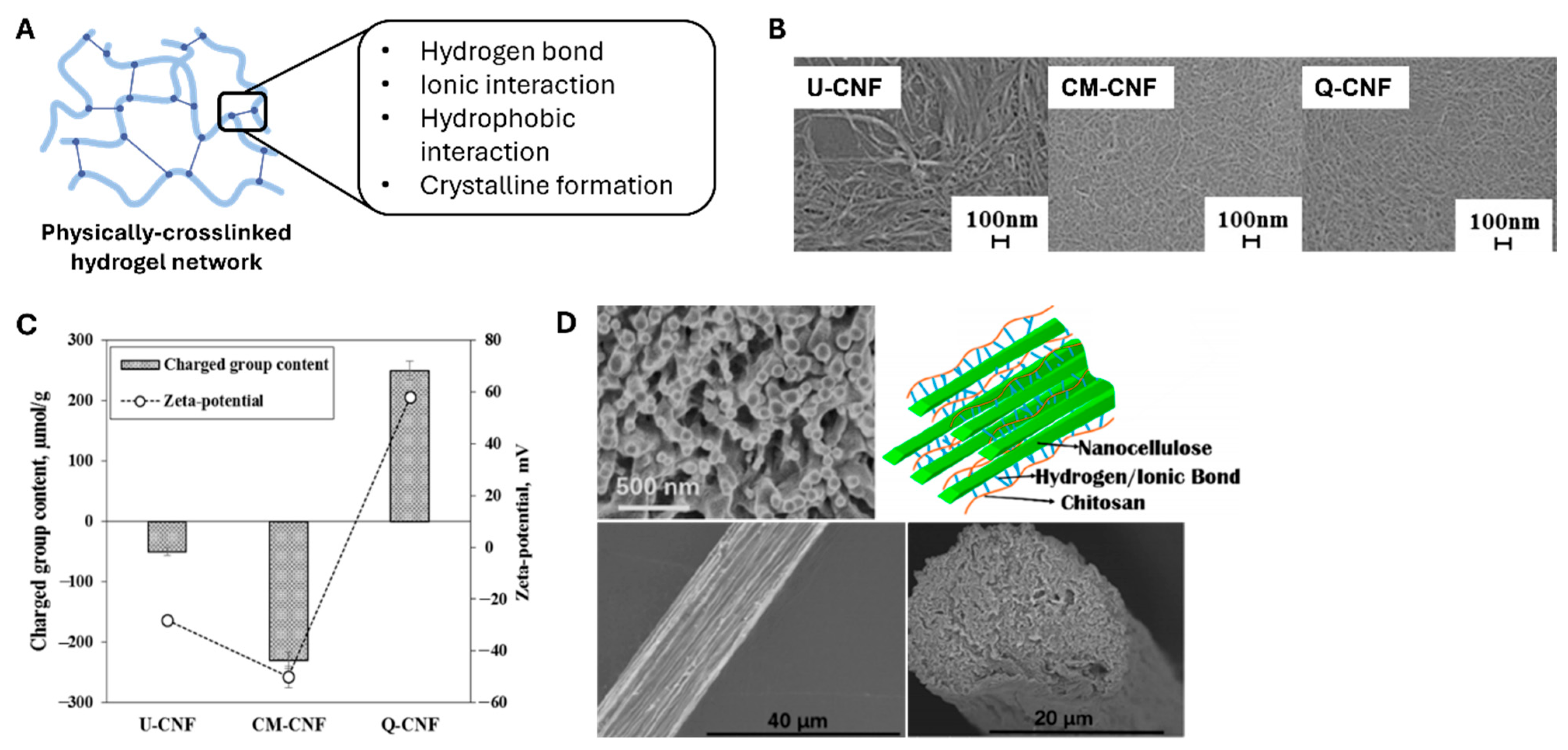
CNF hydrogels formed via physical crosslinking exhibit distinct structural and rheological characteristics, primarily owing to the anisotropic morphologies and high aspect ratios of nanocellulose fibrils. Unlike conventional polymer-based hydrogels that typically originate from molecular solutions, CNF dispersions exist as colloidal suspensions, critically influencing gelation mechanisms and viscoelastic behavior [18,19]. Fibrils, measuring between 5 and 50 nm in diameter and extending up to several microns in length, can entangle and form percolated networks via hydrogen bonding and van der Waals interactions, enabling stable hydrogel formation without the presence of chemical crosslinkers [18,20]. Figure 2A reveals that the physical crosslinking of such polymeric networks involves conjugation between polymeric chains via reversible interactions such as hydrogen bonding, ionic interactions, hydrophobic interactions, and crystalline formation.
CNF hydrogels can be crosslinked through several key approaches, and the most fundamental approach involves controlling surface charge density. At a given solid content, increasing the surface charge density of a CNF prevents agglomeration by electrostatic repulsion and promotes network entanglement. Im et al. [21] reported that three different composite hydrogels of CNFs and polyvinylpyrrolidone (PVP) were prepared with various surface charges of CNFs (Figure 2B,C). The group containing untreated CNFs (U-CNFs) possessed a zeta potential value of 0 mV. The hydrogels with carboxymethylated (CM-CNF) and quaternized (Q-CNF) CNFs possessed zeta potentials of −40 mV and +70 mV, respectively (Figure 2C). Interestingly, these three hydrogels demonstrated different transmittance values, crystallinities, distributions of nanofibrils, shear viscosities, and storage moduli.
Hydrogel gelation is highly dependent on controlling pH and ionic strength. Adding salt or reducing solution pH induces gelation by reducing surface charge via counterion-driven charge screening of surface carboxyl groups. The stability of these gels strongly depends on pH and salt concentration, which directly affect the network-bound water content and overall gel structure [23]. Fall et al. demonstrated that the degree of deprotonation and the number of charged carboxyl groups in hydrogels can be precisely controlled by varying salt concentration and pH [23]. Moreover, CNF concentration influences hydrogel formation, and studies have shown that stable gels can form at concentrations as low as 0.125 wt% when using enzymatically treated and homogenized CNFs [5]. This is particularly significant as it represents a concentration two orders of magnitude lower than that required for other nanocellulose materials [24].
Furthermore, structured CNF-based hydrogels are being developed by controlling the orientation of CNFs. Håkansson et al. [25] demonstrated that hydrodynamic forces and ionic interactions can be used to achieve fibril alignment and gelation by exposing CNFs to a focused flow. Similarly, Cai et al. [22] fabricated aligned filaments solely consisting of CNFs via flow-assisted assembling, as illustrated in Figure 2D. The aligned structure imparted better mechanical properties to the CNF-based hydrogel than the randomly oriented nanofibrils and improved applicability in the biomedical engineering field, such as muscle and neuronal engineering. Moreover, the crosslinking density strengthens the overall mechanical modulus of the hydrogel. However, it is also critical to consider the viscoelasticity of the CNF-based hydrogel resulting from the crosslinking density since the modulus exceeding a certain extent without degradability possibly hinders migration and proliferation of cells embedded within the network.

2.2. Chemical Crosslinking Methods

Chemically crosslinking CNFs creates strong and permanent network structures via various bonding mechanisms (Figure 3A). Chemically crosslinked networks can be achieved via covalent bonds formed through radical polymerization, chemical reactions, irradiation, or enzymatic reactions [26,27,28]. The preparation process begins with the challenging step of dissolving cellulose chains, governed by the balance between entropy and molecular interactions [29,30]. To commence the gelation procedure, different chemical procedures and modification protocols have been developed to dissolve cellulose in water or organic solvents [31,32]. For instance, aqueous solutions of NaOH/urea and LiOH/urea have been used to dissolve cellulose at temperatures as low as −10 °C [33,34].
Radical polymerization can also induce chemical crosslinking of CNF-based hydrogel, as demonstrated by Chen et al. The group demonstrated the fabrication of a composite hydrogel consisting of poly(vinyl alcohol) (PVA) and CNF through γ-irradiation, as shown in Figure 3B [35]. When CNF components are irradiated with γ-rays, free radicals are generated, which then form stable chemical bonds with hydrogen atoms in PVA components. Specifically, hydrogen atoms of the hydroxyl group and methylene group in both CNF and PVA form tertiary and secondary radicals, respectively, upon irradiation. These radicals eventually lead to formation of a 3D network to fabricate a hydrogel network. Basically, in this case, CNF acts as a crosslinker, and the composite form of natural polymer (CNF) and synthetic polymer (PVA) strengthen the mechanical properties of the final PVA/CNF hydrogel.
Composite hydrogel preparation offers another approach to enhance mechanical properties. CNFs can be used as reinforcing agents in polymer matrices; however, the loading levels of these CNFs must be carefully controlled to avoid entanglement. Such composites are more commonly prepared using synthetic polymers, such as PVA, poly (ethylene glycol) (PEG), or polyacrylamide, because such polymers are stable and malleable with excellent mechanical properties. Zhang et al. [36] and Takeno et al. [37] successfully fabricated a CNF/PVA hydrogel crosslinked with borax. They demonstrated that the mechanical properties of the PVA-based hydrogel improved owing to the addition of CNF and borax. In particular, Takeno et al. [37] emphasized the effect of CNF size on gel properties. The composite gel containing the CNF with a smaller length demonstrated better stretchability than the ones with longer fiber lengths. The self-healing capability of these two reported composite hydrogels was another significant feature. As shown in Figure 3C, the CNF/PVA hydrogel cut in half was healed in 5 min of physical contact.
The self-healing capacity of these composite CNF/PVA hydrogels reveals that chemical crosslinking can impart various interesting functionalities to the system. For example, ultraviolet radical polymerization has been used to prepare hydrogels from bacterial nanocellulose and poly(2-hydroxyethyl methacrylate). These composite hydrogels exhibit significantly improved tensile strengths and Young’s moduli with respect to single-component systems [38]. Moreover, temperature-responsive properties can be incorporated via specific chemical modifications. Specifically, poly(N-isopropylacrylamide) (PNIPAm)-based CNF hydrogels have demonstrated characteristic temperature-dependent swelling behavior (Figure 3D). Therefore, the mechanical properties of these thermo-responsive systems can be tuned by controlling the CNF content [37,39].

3. Biomedical Applications of CNF Hydrogels

3.1. Drug Delivery Systems

Drug delivery systems represent bioengineered technologies designed for the targeted transport of therapeutic agents to specific tissues and organs. These systems incorporate carrier vessels and coating treatments to control the release of medicines and biomolecules [40]. Such modifications enhance pharmacokinetics and optimize the biodistribution of substances in the human body. The development of effective drug delivery systems requires careful consideration of stability factors, including pH, ionic strength, and temperature variations before reaching the target site, to prevent premature release and ensure controlled release [41]. CNF-based hydrogels have emerged as promising carriers for bioactive molecules owing to their unique advantages involving nanostructures, biocompatibility, biodegradability, and tunable surface chemistry [42,43,44]. By incorporating functional groups or additional components, CNF hydrogels can be engineered to achieve stimuli-responsive release, sustained and sequential drug delivery, or injectable and localized therapeutic administration.

3.1.1. Stimuli-Responsive Hydrogels

The development of stimuli-responsive CNF hydrogels has garnered particular attention due to their ability to release drugs on demand in response to specific stimuli such as pH, temperature, mechanical force, light, and ionic strength. These systems offer precise spatiotemporal control over therapeutic delivery, making them attractive candidates for next-generation biomedical applications.
Masruchin et al. [45] synthesized dual-responsive composite hydrogels based on TEMPO-oxidized CNFs and thermally responsive PNIPAm for drug delivery. These hydrogels responded to both pH and temperature. The pH sensitivity of these hydrogels originates from the tunable ionization of the carboxyl groups on CNFs, whereas temperature responsiveness is imparted by PNIPAm, enabling control over swelling behavior via external thermal input. In another significant development, Zhang et al. [46] fabricated pH-responsive gel macrospheres using sodium alginate (SA) and TEMPO-oxidized CNFs for probiotic delivery. These macrospheres remained stable in simulated gastric fluid (SGF) due to reduced electrostatic repulsion and increased hydrogen bonding in acidic conditions and subsequently swelled in simulated intestinal fluid (SIF) to release their payload. The addition of CNFs significantly enhanced the mechanical strength and structural integrity of the macrospheres, improving resistance to shrinkage and disintegration under gastrointestinal conditions. Further advancing CNF-based intelligent hydrogels, Zhang et al. [47] developed a dual-stimuli responsive system composed of polyvinyl alcohol (PVA), polydopamine (PDA), and two-step oxidized CNFs (TOCN) for combined chemo-photothermal breast cancer therapy. TOCN introduced abundant carboxyl groups that enhanced doxorubicin (DOX) loading via electrostatic and hydrogen bonding interactions. The system exhibited controlled DOX release under mildly acidic (pH 6.8) and elevated temperature (42 °C) conditions. Additionally, PDA enabled near-infrared (NIR) photothermal responsiveness, allowing precise on-demand drug release and enhanced therapeutic efficacy (Figure 4A).
In addition to pH- and temperature-responsive designs, CNF-based hydrogels have also been explored for light-triggered controlled drug release. Notably, Lem et al. developed a far-red light-responsive platform using an anionic nanofibrillated cellulose (ANFC) hydrogel loaded with both a tetra-cationic zinc phthalocyanine photosensitizer (ZnPc(MePy)4) and cellulose-binding liposomes [48]. Upon irradiation at 730 nm, an optimal wavelength for deep tissue penetration, the photosensitizer generated singlet oxygen, which oxidatively disrupted the liposomal membranes and triggered the release of encapsulated hydrophilic drugs [48]. The ANFC hydrogel acted as a stable, biocompatible reservoir that confined ROS activity within the matrix, ensuring spatially controlled drug release.
Beyond chemical and photothermal responsiveness, mechanical stimuli have also been harnessed for smart release. Park et al. developed a mechanically responsive semi-interpenetrating polymer network (semi-IPN) hydrogel composed of TEMPO-oxidized bacterial cellulose nanofibers (BCNFs) and polyacrylamide (PAM) [49]. By tuning the aspect ratio of BCNFs, the hydrogel’s mechanical properties, including compressive strength and elasticity, were significantly enhanced. The incorporation of BCNFs reduced hydrogel permeability, slowing passive drug diffusion while enabling on-demand release upon mechanical compression. Notably, drug diffusion was selectively enhanced in hydrogels with higher BCNF content under compressive stimulation, without compromising structural integrity under repeated mechanical stress. This system offers a promising platform for wearable, pressure-sensitive drug delivery applications, such as artificial skin patches.
Together, these studies underscore the versatility of CNF-based hydrogels in engineering multi-responsive drug delivery platforms, capable of reacting to physiological and external cues for precision-controlled therapeutic outcomes.

3.1.2. Composite and Nanocomposite Hydrogels

A major limitation of conventional hydrogels in drug delivery applications is their tendency to exhibit an initial burst release and relatively low drug loading capacity [50]. To address these challenges, composite and nanocomposite hydrogels have been developed to enhance structural integrity, regulate release kinetics, and improve drug encapsulation efficiency. These improvements are typically achieved by either incorporating functional nanoparticles into CNF-based hydrogel matrices or embedding CNFs into other polymer systems to reinforce the hydrogel network. Such hybrid systems integrate the unique properties of both CNFs and functional additives, offering synergistic advantages that make them highly suitable for sustained and targeted drug delivery.
Liu et al. [51] constructed CNF-based composite hydrogels (MDPA@GO/CNF) by incorporating mesoporous polydopamine (MPDA) and graphene oxide (GO) to achieve sustained drug release and safe delivery of tetracycline hydrochloride (TH). MPDA loaded with TH was wrapped in GO sheets and embedded into CNF-based hydrogel (Figure 4B, left). In this system, CNFs serve as a hydrophilic fibrous matrix, offering abundant hydroxyl and carboxyl groups that promote structural integrity and crosslinking through non-covalent interactions with GO and calcium ions. The incorporation of MPDA@GO into the CNF matrix successfully enhanced the sustained and controlled release of TH compared to pure CNF hydrogel. Furthermore, drug release from the composite hydrogel could be regulated by environmental pH and near-infrared (NIR) irradiation (Figure 4B, right).
In another study, Yunnan Baiyao (YNBY) particles were incorporated into a CNF-based composite hydrogel to construct a wearable patch for sustained transdermal drug delivery [52]. In this formulation, CNFs form a highly entangled fibrous network that reinforces the mechanical strength of the hydrogel and reduces its swelling rate by increasing crosslinking density. This network acts as a diffusion barrier, indirectly modulating the release of embedded drug molecules. The incorporated YNBY serves as a particulate drug reservoir, gradually releasing its bioactive components, such as Panax notoginseng saponin R1, through a combination of diffusion and matrix swelling. The interaction between YNBY and the CNF hydrogel matrix further contributes to the sustained and controlled drug release behavior. Thus, this CNF-based nanocomposite hydrogel presents a promising strategy for long-lasting transdermal therapeutic applications.
Similarly, CNF/Zinc oxide nanohybrids (ZONHs) were incorporated into a hydrogel composed of starch, gelatin, and itaconic acid to create a pH-sensitive nanocomposite system for sustained drug delivery [53]. In this formulation, CNFs facilitate the stable dispersion of ZnO nanoparticles, ensuring uniform distribution and consistent functionality throughout the hydrogel matrix. The resulting ZONHs serve as reinforcing agents, enhancing the hydrogel’s water-swelling capacity and regulating drug release through strong interfacial interactions with the polymer network and encapsulated drug. Moreover, at higher pH levels, the hydrogel demonstrates increased swelling, promoting more effective diffusion of the encapsulated drug. Thus, this CNF-based nanocomposite system offers promising potential as a sustainable drug delivery platform in biomedical applications.
Composite and nanocomposite hydrogels incorporating cellulose nanofibers (CNFs) provide enhanced structural and functional properties for drug delivery systems. Through integration with various functional additives, these hydrogels offer improved mechanical strength, swelling control, and sustained drug release. Their versatility makes them suitable for a broad range of biomedical applications.
Figure 4. Application of CNF-based hydrogels in drug delivery systems. (A) SEM images for the TOCN/PVA hydrogel with changes in PVA content (left), and the NIR-responsive temperature changes observed in the hydrogel system (right). (Reproduced/Adapted with permission from [47], Elsevier, 2024). (B) SEM images of tetracycline hydrochloride (TH) drug-loaded mesoporous polydopamine (MPDA) particles wrapped in graphene oxide sheets (left), and release profile for TH from MPDA-encapsulated CNF hydrogel at different pH values (right). (Reproduced/Adapted with permission from [51], American Chemical Society, 2020). (C) Characterization of the tri-stimuli-responsive injectable hydrogel (CNF-TRIH@DOX): (i) Photographic image of the hydrogel and its injectability, the release profile of the loaded DOX depending on the hydrogel responsiveness to (ii) NIR, (iii) pH, and (iv) temperature. (Reproduced/Adapted with permission from [54], Elsevier, 2022.) (D) Cumulative release of drugs (IBU and DOX) from CNF/SA hydrogel in response to different pH values, and the swelling ratio of the hydrogel changing at different pH (5.0, 6.5, and 7.4). (Reproduced/Adapted with permission from [55], Elsevier, 2025).
Polymers 17 02272 g004

3.1.3. Injectable and Localized CNF Hydrogels

Injectable hydrogels have emerged as highly promising platforms for localized drug delivery due to their three-dimensional carrier capacity, biocompatibility, minimally invasive administration, and ability to conform to irregular tissue shapes. In recent years, they have been extensively studied for in vivo drug delivery, particularly in tumor therapy [56]. Cellulose nanofibrils (CNFs), owing to their inherent pseudoplastic and thixotropic behavior, can form shear-thinning hydrogels suitable for injection. These properties enable CNF hydrogels to be easily administered and to rapidly recover their structure post-injection, making them ideal candidates for localized therapeutic applications. For instance, Lauren et al. [57] developed technetium-99m-labeled CNF hydrogels to enable real-time tracking of hydrogel localization and drug release in vivo. The hydrogel exhibited sustained release of large molecules, making it particularly suitable for long-term or depot-based delivery, especially for biologics and protein therapeutics. These biocompatible and non-toxic CNF hydrogels were enzymatically degradable into glucose via localized cellulase activity, enabling an additional level of spatial and temporal control. Such systems demonstrate the potential of CNF-based injectable hydrogels for precision drug delivery in dynamic biological environments.
As shown in Figure 4C, Chen et al. developed a tri-stimuli-responsive injectable hydrogel (CNF-TRIH@DOX) based on TEMPO-oxidized cellulose nanofibrils (CNFs), modified with polyethyleneimine (PEI) and poly(N-isopropylacrylamide) (PNIPAm) to confer responsiveness to near-infrared (NIR) light (Figure 4C(ii)), pH (Figure 4C(iii)), and temperature (Figure 4C(iv)) for tumor therapy [54]. Combined with alginate and chitosan, CNF formed a 3D nanocage-like hydrogel structure with high loading capacity for doxorubicin (DOX) and Prussian blue nanoparticles (PBNPs), the latter acting as a contrast agent for imaging via electrostatic interactions. Thus, CNF plays a central role not only as a structural backbone and responsive switch for stimuli-triggered release, but also as a functional component that enables theranostic capabilities through intelligent drug delivery and imaging performance.
In summary, CNF-based injectable hydrogels represent a versatile and promising platform for localized and controlled drug delivery. Their unique rheological properties, biocompatibility, and ability to form responsive networks enable precise spatial and temporal control over therapeutic release. By integrating functional modifications and responsive components, CNF hydrogels can be tailored for advanced biomedical applications such as tumor-targeted therapy and real-time imaging. These systems exemplify the potential of smart, minimally invasive drug delivery technologies in addressing complex clinical challenges.

3.1.4. Sustained and Sequential Release Hydrogels

Despite these advances, several challenges remain in the development of CNF-based drug delivery systems. A key area for improvement is the design of hydrogel systems capable of simultaneously releasing multiple drugs at controlled, varying rates, an approach that is particularly advantageous for complex diseases like cancer [58].
For instance, a recent study demonstrated a sequential drug delivery system (SDS) using a cellulose nanofiber/sodium alginate (CNF/SA) hydrogel embedded with doxorubicin-loaded calcium carbonate hollow microspheres (CaCO3/DOX HMs) and ibuprofen (IBU) for colon cancer treatment [55]. In this system, CNF plays a crucial role in forming a pH-sensitive hydrogel matrix that enables stepwise drug release. At intestinal pH (~7.4), carboxyl group deprotonation in CNF and SA leads to electrostatic repulsion and matrix swelling, allowing the release of IBU to relieve pain. As the pH drops to ~6.5 in the colon, decomposition of the CaCO3 microspheres triggers the release of DOX for localized chemotherapy (Figure 4D). Thus, CNF contributes to both structural support and stimuli-responsive behavior, enabling targeted and temporally controlled release of multiple therapeutics.
CNF-based hydrogels offer significant promise as intelligent drug delivery platforms, particularly for localized and combination therapies. Their tunable chemical structure, responsiveness to physiological stimuli, and compatibility with other biomaterials make them ideal candidates for advanced delivery systems. As demonstrated by recent sequential delivery strategies, CNF enables precise spatial and temporal control over multi-drug release, addressing the complex needs of diseases like cancer. Moving forward, further research into multi-responsive and multifunctional CNF-based hydrogels will be key to unlocking their full clinical potential in personalized and effective treatment strategies.

3.2. Tissue Engineering

The fundamental requirements for tissue engineering scaffolds include the ability to promote nutritional transport, vascularization through their porous gel structure [57,59,60], and natural degradation in body fluids to eliminate the need for surgical removal [59]. In addition, scaffolds must exhibit sufficient mechanical strength to support cell adhesion, proliferation, and differentiation into specialized, functional tissues.

3.2.1. Scaffold Materials

In recent years, CNF-based hydrogels have garnered considerable attention in tissue engineering applications due to their highly hydrated three-dimensional porous structure that mimics biological tissue [42,61,62]. Moreover, CNFs serve as effective mechanical reinforcement agents, enhancing the structural integrity of hydrogel scaffolds without compromising biocompatibility [58,63]. CNF-based scaffolds have been explored for a variety of tissue engineering applications, including cartilage, meniscus, bone, intervertebral disc, cardiac, corneal, neural, and tracheal regeneration (Table 1).
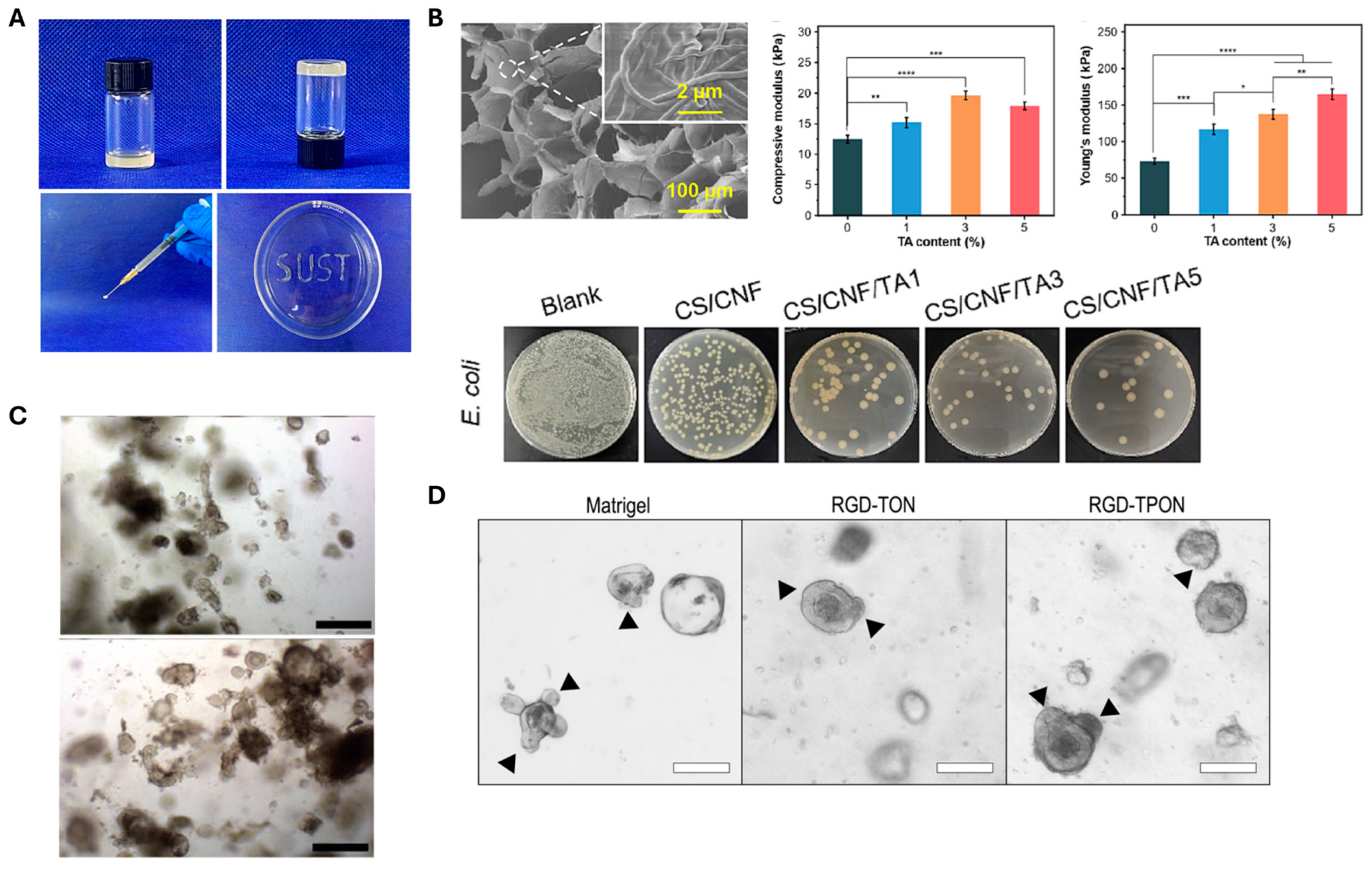
In the case of bone regeneration, one of the major limitations of natural polymer-based hydrogels is their insufficient mechanical strength, which hinders their effectiveness in load-bearing bone repair applications. To overcome this challenge, Cui et al. developed an injectable and self-healing semi-interpenetrating network (semi-IPN) hydrogel composed of oxidized alginate (OSA), gelatin (Gel), and CNFs, using a one-step synthesis method without the need for external crosslinking agents [65]. In this system, OSA served as a natural crosslinker by forming dynamic imine bonds with the amino groups of gelatin via Schiff base reactions. CNFs contributed to the formation of the hydrogel network through physical interactions, primarily hydrogen bonding, while also acting as mechanical reinforcement agents, thickeners, and rheological modifiers. The mechanical strength of the hydrogel increased with higher CNF content, and the formulation exhibited shear-thinning behavior, facilitating injectability (Figure 5A). Interestingly, while CNFs improved the structural integrity, they also reduced viscosity, which is advantageous for syringe-based administration. Functionally, the OSA/Gel/CNF hydrogel demonstrated excellent biomineralization capability, attributed to the abundant carboxyl and hydroxyl groups on the CNFs, which created strong negative dipoles for chelating free calcium ions. Furthermore, the hydrogel promoted MC3T3-E1 preosteoblast cell viability, proliferation, and osteogenic differentiation, highlighting its potential as an effective injectable scaffold for bone tissue engineering.
Injectable CNF-based hydrogels are highly attractive for minimally invasive applications due to their ability to conform to irregular defect geometries [76]. In addition to bone repair, they have been applied in intervertebral disc regeneration, where both mechanical reinforcement and injectability are essential. Doench et al. [67] developed an injectable CNF-filled chitosan hydrogel with tunable rheological and mechanical properties. CNFs were incorporated without compromising injectability, as their alignment under high shear minimized flow resistance. Rheological studies showed that CNFs formed hydrogen bonding and hydrophobic interactions with CHI, increasing viscosity at low shear rates and improving post-injection stability. The addition of CNFs also enhanced the elastic modulus, mimicking native disc stiffness. In vivo pig studies demonstrated effective implant localization, restoration of disc viscoelasticity, and increased disc height. These findings highlight the potential of CNF/CHI hydrogels as non-cellularized, bioactive materials for disc repair.
In cardiac tissue engineering, Hou et al. prepared a conductive CNF-based hydrogel system by polymerizing polypyrrole (PPy) within a TEMPO-oxidized CNF (TOCN) matrix [72]. The carboxyl groups introduced through TEMPO oxidation enabled hydrogel formation via ionic crosslinking with ferric ions (Fe3+), resulting in a stable and interconnected network. Subsequent in situ polymerization of PPy within the TOCN matrix not only imparted electrical conductivity but also mitigated aggregation issues, thereby preserving the mechanical integrity of the hydrogel. The resulting TOCN–PPy composite hydrogel exhibited both suitable mechanical strength and electrical conductivity, effectively mimicking the native properties of myocardial tissue. Notably, cardiomyocytes cultured on the scaffold showed enhanced expression of myocardial-specific proteins, including connexin 43 and cardiac troponin T, indicating favorable cell–material interactions and support for cardiac phenotype maintenance. These findings highlight the potential of TOCN–PPy composite hydrogels as promising scaffolds for myocardial tissue regeneration.
Overall, CNF-based hydrogels are versatile scaffold materials with high water content, ECM-like structure, and strong mechanical reinforcement capabilities. They enhance the strength, stability, and biofunctionality of hydrogels, supporting cell growth in diverse tissues. These properties have enabled their successful use in scaffolds for cartilage, bone, intervertebral disc, and cardiac regeneration, highlighting their potential as next-generation biomaterials for both load-bearing and electroactive applications.
Figure 5. Application of CNF-based hydrogels in tissue engineering. (A) Demonstration of sol–gel state and injectability of CNF-based hydrogel in composite with oxidized alginate and gelatin. (Reproduced/Adapted with permission from [65], Elsevier, 20203). (B) SEM images and modulus of TA-incorporated CNF hydrogel (top) and the antibacterial activity of the CNF/TA hydrogel (bottom). *: p < 0.05, **: p < 0.01, ***: p < 0.001, ****: p < 0.0001). (Reproduced/Adapted with permission from [77], Elsevier, 2024). (C) Maturation of liver organoids within TEMPO-oxidized CNF hydrogels at day 1 (top) and day 7 (bottom) of encapsulation. Scale bar = 500 μm. (Reproduced/Adapted with permission from [78], Wiley, 2020). (D) The morphology of intestinal organoids with buddings (indicated with black arrows) compared after 4 days of culture within Matrigel, RGD-TON, and RGD-TPON. Scale bar = 100 μm. (Reproduced/Adapted with permission from [79], American Chemical Society, 2021).
Polymers 17 02272 g005

3.2.2. Wound Dressing

Cutaneous wound healing is a highly dynamic and coordinated biological process involving four overlapping stages: hemostasis, inflammation, proliferation, and tissue remodeling [80]. However, bacterial infection remains one of the most significant challenges that can disrupt this healing cascade, leading to delayed recovery and increased risk of complications [81]. Therefore, effective wound management requires dressings that not only protect the wound but also actively support the healing process. An ideal wound dressing should meet several key criteria: it must be non-toxic, non-allergenic, and capable of maintaining a moist environment at the wound site. It should allow gas exchange, absorb excess exudate and toxins, prevent bacterial colonization, and ideally possess intrinsic antimicrobial properties. Additionally, the dressing should be easy to remove without causing further trauma to the wound bed [81,82,83].
In recent years, CNF-based hydrogels have emerged as promising candidates for advanced wound dressings due to their biocompatibility, high water-holding capacity, structural similarity to the extracellular matrix, and potential for functionalization with antimicrobial or therapeutic agents (Table 2). For instance, Li et al. developed a CNF-based hydrogel composed of chitosan (CS), CNF, and tannic acid (TA), demonstrating strong antibacterial activity and hemostatic performance for treating drug-resistant bacterial infections [77]. In this system, CNFs serve dual roles as both structural and functional elements. Structurally, they form a nanofibrous three-dimensional network that reinforces the hydrogel matrix, mimics the ECM, and supports cellular adhesion and proliferation (Figure 5B, top). Functionally, their abundant hydroxyl groups facilitate strong hydrogen bonding with TA, enabling stable incorporation and enhancing antibacterial efficacy through membrane disruption and protein denaturation (Figure 5B, bottom). Therefore, CNFs significantly contribute to the hydrogel’s mechanical robustness, bioactivity, and antimicrobial functionality, making it highly effective for the treatment of infected wounds. In another study, TA was also utilized as an antibacterial and antioxidant agent in a glucose-responsive hydrogel composed of CNFs, 3-acrylamidophenyl boronic acid (AAPBA), and acrylamide (AM) [84].
In a related development, Zhang et al. developed a hybrid hydrogel of CNF, polyvinyl alcohol (PVA), and curcumin-modified silver nanoparticles (cAg) through a one-step synthesis method based on hydrogen bonds, dynamic boronic ester bonds, and coordinate covalent bonds [87]. In this hydrogel system, CNFs play a vital dual role [87]. Firstly, CNFs act as a structural matrix for anchoring and uniformly dispersing cAg, which endow the hydrogel with broad-spectrum antibacterial activity against pathogens such as S. aureus, E. coli, and C. albicans. The high surface area and reactive hydroxyl groups on CNFs facilitate the formation of coordinate interactions with cAg, improving nanoparticle stability, preventing aggregation, and enabling sustained release. Secondly, the intrinsic self-assembly of CNF chains into nanoscale interpenetrating hierarchical networks enhances the hydrogel’s mechanical performance, flexibility, self-healing, and tissue adhesion. These effects are attributed to the synergistic interplay of physical interaction, such as hydrogen bonding and polymer chain entanglement, and dynamic covalent borate ester bonding between CNFs and PVA chains. Moreover, the reactive surface chemistry of CNFs further supports the hydrogel’s multifunctionality, including its biodegradability and biocompatibility, ultimately promoting re-epithelialization, collagen deposition, and accelerated wound healing.
Overall, CNF-based hydrogels are promising wound dressing materials, combining biocompatibility, high water retention, and ECM-like architecture to support tissue repair. Their surface chemistry allows functionalization with bioactive agents, enabling antibacterial, antioxidant, hemostatic, and self-healing properties. These features enhance mechanical stability, reduce infection risk, and promote faster, more effective healing.

3.2.3. Three-Dimensional Cell Culture Platforms

Three-dimensional (3D) cell culture systems better replicate the architecture, microenvironment, and cell–cell interactions of native tissues than conventional two-dimensional cultures, making them valuable for tissue engineering, disease modeling, and drug screening. Hydrogels are ideal 3D culture matrices due to their biocompatibility, tunable properties, and high water content [89]. CNF-based hydrogels offer additional advantages, including ECM-like nanofibrous architecture, adjustable mechanical strength and porosity, and the potential for biofunctionalization to support cell adhesion, proliferation, and differentiation [42].
CNF-based hydrogels formed either by self-assembly or ionic crosslinking have been have shown strong potential as 3D cell culture matrices for stem cells and organoids (Table 3). In the case of self-assembled CNF hydrogels without added crosslinkers, Sanandiya et al. demonstrated that injectable, surface-oxidized CNF thixogels supported high viability, long-term survival, and spherical morphology of human breast cancer cells and mouse embryonic stem cells [90]. These hydrogels exhibited shear-thinning and thixotropic behavior, remaining flowable under applied stress and rapidly recovering their structure within 60 s, while forming 3D networks through flexible nanofiber entanglement. The mechanical moduli could be tuned by adjusting CNF concentration or applying external stimuli. Similarly, TEMPO-oxidized CNF hydrogels with shear-healing and shear-thinning properties were shown to effectively support hepatic differentiation of human liver organoids into functional hepatocyte-like cells [78]. These organoids exhibited comparable expressions of hepatic genes, hepatocyte functions, and polarization to those cultured in Matrigel, suggesting that CNF-based hydrogels could serve as a promising, biocompatible alternative to Matrigel for tissue engineering and regenerative medicine applications (Figure 5C).
In ionic crosslinking systems, the introduction of carboxyl groups into CNFs via TEMPO oxidation enables the formation of metal–carboxylate complexes with multivalent cations, improving both biochemical and mechanical properties. Kim et al. reported the successful 3D encapsulation of osteoblasts in Ca2+-crosslinked CNF hydrogels, achieving high cell viability and long-term proliferation [91]. Notably, the cells remained uniformly distributed throughout the CNF hydrogel, avoiding the cell sinking observed in Matrigel. To further enhance biofunctionality, Curvello et al. functionalized TEMPO-oxidized (TON) and TEMPO/periodate-oxidized (TPON) CNFs with RGD peptides to promote cell attachment and differentiation [79]. Both TON and TPON hydrogels could be crosslinked with either Mg2+ or Ca2+ ions for intestinal organoid culture (Figure 5D). Due to their higher carboxyl content, TPON fibers allowed greater RGD grafting, providing stronger biochemical cues for cell adhesion. Organoids cultured in these hydrogels maintained viable regions for up to four days, and cell clusters recovered from Mg2+-crosslinked hydrogels could be passaged with intact RNA.
Table 3. CNF-based hydrogel as 3D cell culture platforms.
Hydrogel Composition/TypeFormation MethodFunctionalizationCell TypeReferences
TEMPO-oxidized CNF/injectable thixogelSelf-assembly (without added crosslinker)-Human breast cancer (MCF-7) and mouse embryonic stem cells
(mESC; E14TG2A)
[90]
TEMPO-oxidized CNF/Injectable thixogelSelf-assembly (without added crosslinker)-Human liver organoid[78]
TEMPO-oxidized nanofibrillated cellulose (NFC)/bulk gelSelf-assembly (without added crosslinker)-Mesenchymal stem cells (MSCs)[92]
TEMPO-oxidized CNF/bulk gelCalcium ion (Ca2+) crosslinking-Pre-osteoblast cells (MC3T3-E1)[91]
TEMPO-oxidized CNF/bulk gelCalcium ion (Ca2+) crosslinkingFibronectin-derived moieties (RGD peptides), laminin-1, insulin-like growth factor (IGF-1)Small intestinal organoids[93]
TEMPO-oxidized (TON) and TEMPO/periodate-oxidized (TPON) CNF/bulk gelMagnesium (Mg2+) and calcium ion (Ca2+) crosslinkingFibronectin-derived moieties (RGD peptides)Intestinal organoids[79]
TEMPO-oxidized CNF/microgelCalcium ion (Ca2+) crosslinkingHyaluronic acid (HA)Human adipose-derived stem cell (hADSC)[94]
Moving beyond bulk hydrogels, Goh et al. demonstrated the fabrication of TEMPO-oxidized CNF hydrogel microbeads for human adipose-derived stem cell (hADSC) culture by simply dropping cell-containing gel precursors into a CaCl2 solution [94]. To improve long-term culture performance, hyaluronic acid (HA) was incorporated into the CNF microbead matrix via physical interactions. Microbeads containing 0.2% high-molecular-weight HA (700 kDa) significantly enhanced cell proliferation, VEGF secretion, and stemness maintenance compared to bare CNF microbeads. This HA-incorporated CNF microbead system thus offers a low-cost, sustainable microgel platform suitable for stem cell 3D culture.
CNF-based hydrogels, whether self-assembled or ionically crosslinked, provide tunable, ECM-mimicking 3D culture environments that support high cell viability, proliferation, and differentiation. They perform comparably to Matrigel while avoiding its key drawbacks, namely its animal origin, variability, undefined composition, and high cost, offering a sustainable, reproducible, and customizable alternative for advanced stem cell and organoid culture systems.

4. Summary and Outlook

Over the past decades, biomedical materials have advanced substantially in terms of CNF hydrogel research and development. These advanced materials possess intricate three-dimensional networks that can retain substantial amounts of water while maintaining robust structural integrity. This unique combination of properties has established CNF hydrogels as promising candidates across multiple biomedical domains, including cellular research, regenerative medicine, and therapeutic delivery systems.
Current research has revealed several areas that require further investigation before the widespread implementation of these hydrogels becomes feasible. In cellular applications, a major concern centers on the methodology used for hydrogel dissolution post-culture. Recent studies rely heavily on enzymatic breakdown processes; however, insufficient research exists on the extended impact of these enzymes on cellular health and function. The scope of cellular research using CNF hydrogels has remained relatively narrow, focusing predominantly on specific cell lines while leaving vast areas of potential applications, such as complex human organ culture systems, largely unexplored. The field of regenerative medicine presents additional limitations, particularly in terms of optimizing bioprinting processes. Current evidence suggests that pure CNF systems rarely achieve optimal printing outcomes without requiring supplementary biomaterials to be incorporated. Additionally, applications in wound management typically require antimicrobial agents to be integrated to achieve the desired therapeutic outcomes.
Initial medical trials have generated encouraging results in terms of biocompatibility. Notable research involving burn treatment applications yielded favorable safety profiles with the minimal occurrence of adverse reactions, i.e., clinical trials [95] showed no allergic reaction or inflammatory response to CNF wound dressings. These preliminary findings, though promising, highlight the critical need for further comprehensive safety evaluations. The toxicology assessment and long-term evaluation of in vivo toxicity and biocompatibility of CNF-based hydrogels remain a crucial issue for real clinical applications [96]. The development of standardized assessment protocols remains essential for advancing medical implementation.
The transition to commercial-scale production introduces additional complexities. A critical challenge involves understanding and controlling material variability owing to different source materials and processing methods. Although some studies have investigated the effects of aspect ratio, surface charge, and fabrication conditions on CNF properties [97,98,99], maintaining consistent quality under high production volumes remains challenging. Advanced manufacturing technologies, particularly in the realm of precision printing, show promise in terms of addressing some of these challenges by forming precisely controlled structures. However, these processes need to be optimized for large-scale implementation, and therapeutic delivery systems could benefit significantly from the adaptable nature of CNF hydrogels, particularly for developing responsive release mechanisms. The potential for enhancing diagnostic capabilities through the precise control of surface chemistry represents another promising avenue for investigation. Additionally, the development of comprehensive testing standards will be crucial for regulatory compliance across various applications.
Moreover, the technological advancements in processing methods are promising. Recent innovations in manufacturing techniques have demonstrated potential for generating sophisticated structural arrangements while maintaining material integrity. These developments could yield efficient production methods while enabling excellent control over final product characteristics. Integration with existing medical technologies represents another critical area for development, requiring careful consideration of practical aspects such as sterilization procedures and storage requirements.
The environmental and economic advantages of using CNF hydrogels position them favorably for future development. The renewable nature and potential for cost-effective production of these hydrogels align well with current sustainability initiatives. However, realizing this potential will require continued advancement in processing efficiency and scale-up capabilities. The successful translation of laboratory findings to practical applications will depend on addressing the aforementioned challenges while maintaining the fundamental properties that render these materials attractive for biomedical applications.
Future research directions should focus on developing sophisticated controlled-release mechanisms, improving drug loading efficiencies, and addressing the challenges of large-scale production. Integrating multiple stimuli-responsive elements and developing precise targeting mechanisms represent promising areas for investigation. Research efforts should clarify the long-term stability and degradation behavior of CNF hydrogels under physiological conditions and the interactions with various therapeutic agents. The cost-effectiveness and sustainability of CNF enhance its attractiveness from the environmental and financial perspectives, rendering it a promising material for next-generation drug delivery systems.

Author Contributions

Conceptualization, T.W., M.G., H.J.H. and K.G.; methodology, H.J.H., Y.K. and M.G.; software, C.L., J.M., Y.K., J.P. and K.L.; validation, C.L., J.M., K.L. and K.C.; formal analysis, J.P. and K.C.; resources, K.G.; writing—original draft preparation, T.W., M.G., H.J.H., Y.K., K.L., J.P., K.C., S.L. and K.G.; writing—review and editing, H.J.H. and K.G.; visualization, C.L., J.M. and S.L.; supervision, K.G.; project administration, K.G.; funding acquisition, K.G. All authors have read and agreed to the published version of the manuscript.

Funding

This research was supported by Kyungpook National University Research Fund, 2024.

Data Availability Statement

No new data were created or analyzed in this study.

Acknowledgments

We would like to thank Keun Hyung YU for his valuable comments on this review.

Conflicts of Interest

The authors declare no conflicts of interest.

Abbreviations

The following abbreviations are used in this manuscript:
CNFCellulose nanofibril
CNCCellulose nanocrystal
TEMPO2,2,6,6-tetramethyl-1-piperidinyloxy
SEMScanning electron microscope
U-CNFUntreated CNF
CM-CNFCarboxymethylated CNF
Q-CNFQuaternized CNF
PVPPolyvinylpyrrolidone
PEGPoly (ethylene glycol)
PNIPAmPoly(N-isopropylacrylamide)
SASodium alginate
THTetracycline hydrochloride
MPDAMesoporous polydopamine
GOGraphene oxide
TOCNTEMPO-oxidized CNF
PPyPolypyrrole

References

  1. Khan, M.I.; An, X.; Dai, L.; Li, H.; Khan, A.; Ni, Y. Chitosan-based polymer matrix for pharmaceutical excipients and drug delivery. Curr. Med. Chem. 2019, 26, 2502–2513. [Google Scholar] [CrossRef] [PubMed]
  2. Lin, N.; Dufresne, A. Nanocellulose in biomedicine: Current status and future prospect European. Polym. J. 2014, 59, 302–325. [Google Scholar] [CrossRef]
  3. Rajinipriya, M.; Nagalakshmaiah, M.; Robert, M.; Elkoun, S. Importance of agricultural and industrial waste in the field of nanocellulose and recent industrial developments of wood based nanocellulose: A review. ACS Sustain. Chem. Eng. 2018, 6, 2807–2828. [Google Scholar] [CrossRef]
  4. Xie, H.; Du, H.; Yang, X.; Si, C. Recent strategies in preparation of cellulose nanocrystals and cellulose nanofibrils derived from raw cellulose materials. Int. J. Polym. Sci. 2018, 2018, 7923068. [Google Scholar] [CrossRef]
  5. Pääkkö, M.; Ankerfors, M.; Kosonen, H.; Nykänen, A.; Ahola, S.; Österberg, M.; Ruokolainen, J.; Laine, J.; Larsson, P.T.; Ikkala, O. Enzymatic hydrolysis combined with mechanical shearing and high-pressure homogenization for nanoscale cellulose fibrils and strong gels. Biomacromolecules 2007, 8, 1934–1941. [Google Scholar] [CrossRef]
  6. Saito, T.; Kimura, S.; Nishiyama, Y.; Isogai, A. Cellulose nanofibers prepared by TEMPO-mediated oxidation of native cellulose. Biomacromolecules 2007, 8, 2485–2491. [Google Scholar] [CrossRef]
  7. Wågberg, L.; Decher, G.; Norgren, M.; Lindström, T.; Ankerfors, M.; Axnäs, K. The build-up of polyelectrolyte multilayers of microfibrillated cellulose and cationic polyelectrolytes. Langmuir 2008, 24, 784–795. [Google Scholar] [CrossRef]
  8. Moon, R.J.; Martini, A.; Nairn, J.; Simonsen, J.; Youngblood, J. Cellulose nanomaterials review: Structure, properties and nanocomposites. Chem. Soc. Rev. 2011, 40, 3941–3994. [Google Scholar] [CrossRef]
  9. Aulin, C.; Ahola, S.; Josefsson, P.; Nishino, T.; Hirose, Y.; Osterberg, M.; Wagberg, L. Nanoscale Cellulose Films with Different Crystallinities and Mesostructures—Their Surface Properties and Interaction with Water. Langmuir 2009, 25, 7675–7685. [Google Scholar] [CrossRef]
  10. Lu, Y.; Aimetti, A.A.; Langer, R.; Gu, Z. Bioresponsive materials. Nat. Rev. Mater. 2016, 2, 16075. [Google Scholar] [CrossRef]
  11. Green, J.J.; Elisseeff, J.H. Mimicking biological functionality with polymers for biomedical applications. Nature 2016, 540, 386–394. [Google Scholar] [CrossRef]
  12. Haycock, J.W. 3D cell culture: A review of current approaches and techniques. 3D Cell Cult. Methods Protoc. 2010, 695, 1–15. [Google Scholar]
  13. Fu, N.; Zhang, X.; Sui, L.; Liu, M.; Lin, Y. Application of Scaffold Materials in Cartilage Tissue Engineering. In Cartilage Regeneration. Stem Cell Biology and Regenerative Medicine; Lin, Y., Ed.; Humana Press: Cham, Switzerland, 2017. [Google Scholar] [CrossRef]
  14. Jayakumar, R.; Prabaharan, M.; Kumar, P.S.; Nair, S.; Tamura, H. Biomaterials based on chitin and chitosan in wound dressing applications. Biotechnol. Adv. 2011, 29, 322–337. [Google Scholar] [CrossRef]
  15. Fenton, O.S.; Olafson, K.N.; Pillai, P.S.; Mitchell, M.J.; Langer, R. Advances in biomaterials for drug delivery. Adv. Mater. 2018, 30, 1705328. [Google Scholar] [CrossRef] [PubMed]
  16. Tavakoli, J.; Tang, Y. Hydrogel based sensors for biomedical applications: An updated review. Polymers 2017, 9, 364. [Google Scholar] [CrossRef]
  17. Caló, E.; Khutoryanskiy, V.V. Biomedical applications of hydrogels: A review of patents and commercial products. Eur. Polym. J. 2015, 65, 252–267. [Google Scholar] [CrossRef]
  18. De France, K.J.; Hoare, T.; Cranston, E.D. Review of hydrogels and aerogels containing nanocellulose. Chem. Mater. 2017, 29, 4609–4631. [Google Scholar] [CrossRef]
  19. Mendoza, L.; Batchelor, W.; Tabor, R.F.; Garnier, G. Gelation mechanism of cellulose nanofibre gels: A colloids and interfacial perspective. J. Colloid. Interface Sci. 2018, 509, 39–46. [Google Scholar] [CrossRef]
  20. Dufresne, A. Nanocellulose: A new ageless bionanomaterial. Mater. Today 2013, 16, 220–227. [Google Scholar] [CrossRef]
  21. Im, W.; Park, S.Y.; Goo, S.; Yook, S.; Lee, H.L.; Yang, G.; Youn, H.J. Incorporation of CNF with different charge property into PVP hydrogel and its characteristics. Nanomaterials 2021, 11, 426. [Google Scholar] [CrossRef] [PubMed]
  22. Cai, Y.; Geng, L.; Chen, S.; Shi, S.; Hsiao, B.S.; Peng, X. Hierarchical assembly of nanocellulose into filaments by flow-assisted alignment and interfacial complexation: Conquering the conflicts between strength and toughness. ACS Appl. Mater. Interfaces 2020, 12, 32090–32098. [Google Scholar] [CrossRef]
  23. Fall, A.B.; Lindstrom, S.B.; Sundman, O.; Odberg, L.; Wagberg, L. Colloidal stability of aqueous nanofibrillated cellulose dispersions. Langmuir 2011, 27, 11332–11338. [Google Scholar] [CrossRef]
  24. Ureña-Benavides, E.E.; Ao, G.; Davis, V.A.; Kitchens, C.L. Rheology and phase behavior of lyotropic cellulose nanocrystal suspensions. Macromolecules 2011, 44, 8990–8998. [Google Scholar] [CrossRef]
  25. Håkansson, K.M.; Fall, A.B.; Lundell, F.; Yu, S.; Krywka, C.; Roth, S.V.; Santoro, G.; Kvick, M.; Wittberg, L.P.; Wågberg, L. Hydrodynamic alignment and assembly of nanofibrils resulting in strong cellulose filaments. Nat. Commun. 2014, 5, 4018. [Google Scholar] [CrossRef]
  26. Shen, X.; Shamshina, J.L.; Berton, P.; Gurau, G.; Rogers, R.D. Hydrogels based on cellulose and chitin: Fabrication, properties, and applications. Green. Chem. 2016, 18, 53–75. [Google Scholar] [CrossRef]
  27. Demitri, C.; Del Sole, R.; Scalera, F.; Sannino, A.; Vasapollo, G.; Maffezzoli, A.; Ambrosio, L.; Nicolais, L. Novel superabsorbent cellulose-based hydrogels crosslinked with citric acid. J. Appl. Polym. Sci. 2008, 110, 2453–2460. [Google Scholar] [CrossRef]
  28. Navarra, M.A.; Bosco, C.D.; Moreno, J.S.; Vitucci, F.M.; Paolone, A.; Panero, S. Synthesis and characterization of cellulose-based hydrogels to be used as gel electrolytes. Membranes 2015, 5, 810–823. [Google Scholar] [CrossRef]
  29. Del Valle, L.J.; Díaz, A.; Puiggalí, J. Hydrogels for biomedical applications: Cellulose, chitosan, and protein/peptide derivatives. Gels 2017, 3, 27. [Google Scholar] [CrossRef] [PubMed]
  30. Lindman, B.; Medronho, B.; Alves, L.; Costa, C.; Edlund, H.; Norgren, M. The relevance of structural features of cellulose and its interactions to dissolution, regeneration, gelation and plasticization phenomena. Phys. Chem. Chem. Phys. 2017, 19, 23704–23718. [Google Scholar] [CrossRef]
  31. Raghuwanshi, V.S.; Cohen, Y.; Garnier, G.; Garvey, C.J.; Russell, R.A.; Darwish, T.; Garnier, G. Cellulose dissolution in ionic liquid: Ion binding revealed by neutron scattering. Macromolecules 2018, 51, 7649–7655. [Google Scholar] [CrossRef]
  32. Mohd, N.; Draman, S.; Salleh, M.; Yusof, N. Dissolution of cellulose in ionic liquid: A review. AIP Conf. Proc. 2017, 1809, 020035. [Google Scholar]
  33. Heinze, T.; Koschella, A. Solvents applied in the field of cellulose chemistry: A mini review. Polímeros 2005, 15, 84–90. [Google Scholar] [CrossRef]
  34. Cai, J.; Zhang, L. Rapid dissolution of cellulose in LiOH/urea and NaOH/urea aqueous solutions. Macromol. Biosci. 2005, 5, 539–548. [Google Scholar] [CrossRef]
  35. Chen, Y.; Yang, M.; Zhang, W.; Guo, W.; Zhang, X.; Zhang, B. Facile preparation of irradiated poly (vinyl alcohol)/cellulose nanofiber hydrogels with ultrahigh mechanical properties for artificial joint cartilage. Materials 2024, 17, 4125. [Google Scholar] [CrossRef]
  36. Zhang, H.; Fu, C.; Yong, L.C.; Sun, N.; Liu, F.G. Flexible and Transparent PVA/CNF Hydrogel with Ultrahigh Dielectric Constant. ACS Appl. Polym. Mater. 2024, 6, 5706–5713. [Google Scholar] [CrossRef]
  37. Takeno, H.; Inoguchi, H.; Hsieh, W.-C. Mechanical and structural properties of cellulose nanofiber/poly (vinyl alcohol) hydrogels cross-linked by a freezing/thawing method and borax. Cellulose 2020, 27, 4373–4387. [Google Scholar] [CrossRef]
  38. Hobzova, R.; Hrib, J.; Sirc, J.; Karpushkin, E.; Michalek, J.; Janouskova, O.; Gatenholm, P. Embedding of bacterial cellulose nanofibers within PHEMA hydrogel matrices: Tunable stiffness composites with potential for biomedical applications. J. Nanomater. 2018, 2018, 5217095. [Google Scholar] [CrossRef]
  39. Syverud, K.; Kirsebom, H.; Hajizadeh, S.; Chinga-Carrasco, G. Cross-linking cellulose nanofibrils for potential elastic cryo-structured gels. Nanoscale Res. Lett. 2011, 6, 626. [Google Scholar] [CrossRef]
  40. García-González, C.A.; Alnaief, M.; Smirnova, I. Polysaccharide-based aerogels—Promising biodegradable carriers for drug delivery systems. Carbohydr. Polym. 2011, 86, 1425–1438. [Google Scholar] [CrossRef]
  41. Hoare, T.R.; Kohane, D.S. Hydrogels in drug delivery: Progress and challenges. Polymer 2008, 49, 1993–2007. [Google Scholar] [CrossRef]
  42. Curvello, R.; Raghuwanshi, V.S.; Garnier, G. Engineering nanocellulose hydrogels for biomedical applications. Adv. Colloid Interface Sci. 2019, 267, 47–61. [Google Scholar] [CrossRef]
  43. Plackett, D.; Letchford, K.; Jackson, J.; Burt, H. A review of nanocellulose as a novel vehicle for drug delivery. Nord. Pulp Pap. Res. J. 2014, 29, 105–118. [Google Scholar] [CrossRef]
  44. Xue, Y.; Mou, Z.; Xiao, H. Nanocellulose as a sustainable biomass material: Structure, properties, present status and future prospects in biomedical applications. Nanoscale 2017, 9, 14758–14781. [Google Scholar] [CrossRef] [PubMed]
  45. Masruchin, N.; Park, B.-D.; Causin, V. Dual-responsive composite hydrogels based on TEMPO-oxidized cellulose nanofibril and poly (N-isopropylacrylamide) for model drug release. Cellulose 2018, 25, 485–502. [Google Scholar] [CrossRef]
  46. Zhang, H.; Yang, C.; Zhou, W.; Luan, Q.; Li, W.; Deng, Q.; Dong, X.; Tang, H.; Huang, F. A pH-responsive gel macrosphere based on sodium alginate and cellulose nanofiber for potential intestinal delivery of probiotics. ACS Sustain. Chem. Eng. 2018, 6, 13924–13931. [Google Scholar] [CrossRef]
  47. Zhang, Z.; Cui, H.; Wang, X.; Liu, J.; Liu, G.; Meng, X.; Lin, S. Oxidized cellulose-filled double thermo/pH-sensitive hydrogel for local chemo-photothermal therapy in breast cancer. Carbohydr. Polym. 2024, 332, 121931. [Google Scholar] [CrossRef] [PubMed]
  48. Lem, O.; Gangurde, P.; Koivuniemi, A.; Keskinen, A.; Efimov, A.; Durandin, N.; Laaksonen, T. Far-red light-triggered cargo release from liposomes b ound to a photosensitizer-cellulose nanofiber hydrogel. Carbohydr. Polym. 2024, 336, 122134. [Google Scholar] [CrossRef]
  49. Park, D.; Kim, J.W.; Shin, K.; Kim, J.W. Bacterial cellulose nanofibrils-reinforced composite hydrogels for mechanical compression-responsive on-demand drug release. Carbohydr. Polym. 2021, 272, 118459. [Google Scholar] [CrossRef]
  50. Osman, N.; Devnarain, N.; Omolo, C.A.; Fasiku, V.; Jaglal, Y.; Govender, T. Surface modification of nano-drug delivery systems for enhancing antibiotic delivery and activity. Wiley Interdiscip. Rev. Nanomed. Nanobiotechnology 2022, 14, e1758. [Google Scholar] [CrossRef]
  51. Liu, Y.; Fan, Q.; Huo, Y.; Liu, C.; Li, B.; Li, Y. Construction of a mesoporous polydopamine@ GO/cellulose nanofibril composite hydrogel with an encapsulation structure for controllable drug release and toxicity shielding. ACS Appl. Mater. Interfaces 2020, 12, 57410–57420. [Google Scholar] [CrossRef]
  52. Gao, X.; Zhang, H.; Yan, C.; Wu, J.; Wang, Y.; Jiang, M.; Wang, Y. Yunnan Baiyao-enhanced cellulose nanofiber composite hydrogel wearable patch for transdermal drug delivery and anti-freezing applications. Int. J. Biol. Macromol. 2025, 315, 144684. [Google Scholar] [CrossRef]
  53. Bora, A.; Sarmah, D.; Rather, M.A.; Mandal, M.; Karak, N. Nanocomposite of starch, gelatin and itaconic acid-based biodegradable hydrogel and ZnO/cellulose nanofiber: A pH-sensitive sustained drug delivery vehicle. Int. J. Biol. Macromol. 2024, 256, 128253. [Google Scholar] [CrossRef]
  54. Chen, Z.; Chen, R.; Zhao, C.; Quan, Z.; Zhu, H.; Wang, L.; Bu, Q.; He, Y.; He, H. A novel medically imageable intelligent cellulose nanofibril-based injectable hydrogel for the chemo-photothermal therapy of tumors. Chem. Eng. J. 2022, 431, 133255. [Google Scholar] [CrossRef]
  55. Bai, W.; Chen, H.; Li, J.; Cai, W.; Kong, Y.; Zuo, X. Calcium carbonate hollow microspheres encapsulated cellulose nanofiber/sodium alginate hydrogels as a sequential delivery system. Int. J. Biol. Macromol. 2025, 309, 142839. [Google Scholar] [CrossRef]
  56. Cheng, Y.; Zhang, H.; Wei, H.; Yu, C.-Y. Injectable hydrogels as emerging drug-delivery platforms for tumor therapy. Biomater. Sci. 2024, 12, 1151–1170. [Google Scholar] [CrossRef] [PubMed]
  57. Laurén, P.; Lou, Y.-R.; Raki, M.; Urtti, A.; Bergström, K.; Yliperttula, M. Technetium-99m-labeled nanofibrillar cellulose hydrogel for in vivo drug release. Eur. J. Pharm. Sci. 2014, 65, 79–88. [Google Scholar] [CrossRef] [PubMed]
  58. Zelikin, A.N.; Ehrhardt, C.; Healy, A.M. Materials and methods for delivery of biological drugs. Nat. Chem. 2016, 8, 997–1007. [Google Scholar] [CrossRef]
  59. Paukkonen, H.; Kunnari, M.; Laurén, P.; Hakkarainen, T.; Auvinen, V.-V.; Oksanen, T.; Koivuniemi, R.; Yliperttula, M.; Laaksonen, T. Nanofibrillar cellulose hydrogels and reconstructed hydrogels as matrices for controlled drug release. Int. J. Pharm. 2017, 532, 269–280. [Google Scholar] [CrossRef]
  60. Lin, N.; Gèze, A.; Wouessidjewe, D.; Huang, J.; Dufresne, A. Biocompatible double-membrane hydrogels from cationic cellulose nanocrystals and anionic alginate as complexing drugs codelivery. ACS Appl. Mater. Interfaces 2016, 8, 6880–6889. [Google Scholar] [CrossRef]
  61. Liu, H.; Liu, K.; Han, X.; Xie, H.; Si, C.; Liu, W.; Bae, Y. Cellulose nanofibrils-based hydrogels for biomedical applications: Progresses and challenges. Curr. Med. Chem. 2020, 27, 4622–4646. [Google Scholar] [CrossRef]
  62. Govindarasu, M.; Palanisamy, S.; Joy, J.G.; Sharma, G.; You, S.; Kim, J.-C. Advances of nanocellulose and cellulose-based derivatives for biomedical applications. Cellulose 2025, 32, 5735–5762. [Google Scholar] [CrossRef]
  63. Bazghaleh, A.A.; Dogolsar, M.A.; Salehi, R.; Barzin, J. Synthesis and characterization of an injectable, self-healing hydrogel based on succinyl chitosan, oxidized pectin, and cellulose nanofiber for biomedical applications. J. Polym. Res. 2025, 32, 63. [Google Scholar] [CrossRef]
  64. Shanto, P.C.; Park, S.; Park, M.; Lee, B.-T. Physico-biological evaluation of 3D printed dECM/TOCN/alginate hydrogel based scaffolds for cartilage tissue regeneration. Biomater. Adv. 2023, 145, 213239. [Google Scholar] [CrossRef]
  65. Cui, S.; Zhang, S.; Coseri, S. An injectable and self-healing cellulose nanofiber-reinforced alginate hydrogel for bone repair. Carbohydr. Polym. 2023, 300, 120243. [Google Scholar] [CrossRef]
  66. Liu, X.; Hu, H.; Ma, J.; Wang, B. Mineralized cellulose nanofibers reinforced bioactive hydrogel remodels the osteogenic and angiogenic microenvironment for enhancing bone regeneration. Carbohydr. Polym. 2025, 357, 123480. [Google Scholar] [CrossRef] [PubMed]
  67. Doench, I.; Torres-Ramos, M.E.; Montembault, A.; de Oliveira, P.N.; Halimi, C.; Viguier, E.; Heux, L.; Siadous, R.; Thiré, R.M.; Osorio-Madrazo, A. Injectable and gellable chitosan formulations filled with cellulose nanofibers for intervertebral disc tissue engineering. Polymers 2018, 10, 1202. [Google Scholar] [CrossRef] [PubMed]
  68. Doench, I.; Tran, T.A.; David, L.; Montembault, A.; Viguier, E.; Gorzelanny, C.; Sudre, G.; Cachon, T.; Louback-Mohamed, M.; Horbelt, N. Cellulose nanofiber-reinforced chitosan hydrogel composites for intervertebral disc tissue repair. Biomimetics 2019, 4, 19. [Google Scholar] [CrossRef]
  69. Jeencham, R.; Tawonsawatruk, T.; Numpaisal, P.-O.; Ruksakulpiwat, Y. Reinforcement of injectable hydrogel for meniscus tissue engineering by using cellulose nanofiber from cassava pulp. Polymers 2023, 15, 2092. [Google Scholar] [CrossRef] [PubMed]
  70. Sinna, J.; Jeencham, R.; Mueangkhot, P.; Sophon, S.; Noralak, P.; Raksapakdee, R.; Numpaisal, P.-O.; Ruksakulpiwat, Y. Development of Poly (vinyl alcohol) Grafted Glycidyl Methacrylate/Cellulose Nanofiber Injectable Hydrogels for Meniscus Tissue Engineering. Polymers 2023, 15, 4230. [Google Scholar] [CrossRef]
  71. Tohidi, H.; Maleki-Jirsaraei, N.; Simchi, A.; Mohandes, F.; Emami, Z.; Fassina, L.; Naro, F.; Conti, B.; Barbagallo, F. An electroconductive; thermosensitive, and injectable chitosan/pluronic/gold-decorated cellulose nanofiber hydrogel as an efficient carrier for regeneration of cardiac tissue. Materials 2022, 15, 5122. [Google Scholar] [CrossRef]
  72. Hou, R.; Xie, Y.; Song, R.; Bao, J.; Shi, Z.; Xiong, C.; Yang, Q. Nanocellulose/polypyrrole hydrogel scaffolds with mechanical strength and electrical activity matching native cardiac tissue for myocardial tissue engineering. Cellulose 2024, 31, 4247–4262. [Google Scholar] [CrossRef]
  73. Xeroudaki, M.; Rafat, M.; Moustardas, P.; Mukwaya, A.; Tabe, S.; Bellisario, M.; Peebo, B.; Lagali, N. A double-crosslinked nanocellulose-reinforced dexamethasone-loaded collagen hydrogel for corneal application and sustained anti-inflammatory activity. Acta Biomater. 2023, 172, 234–248. [Google Scholar] [CrossRef]
  74. Cheng, K.-C.; Huang, C.-F.; Wei, Y.; Hsu, S.-H. Novel chitosan–cellulose nanofiber self-healing hydrogels to correlate self-healing properties of hydrogels with neural regeneration effects. NPG Asia Mater. 2019, 11, 25. [Google Scholar] [CrossRef]
  75. Li, Y.; Xun, X.; Duan, L.; Gao, E.; Li, J.; Lin, L.; Li, X.; He, A.; Ao, H.; Xu, Y. Cartilage structure-inspired nanofiber-hydrogel composite with robust proliferation and stable chondral lineage-specific differentiation function to orchestrate cartilage regeneration for artificial tracheal construction. Bioact. Mater. 2025, 47, 136–151. [Google Scholar] [CrossRef]
  76. Liu, M.; Zeng, X.; Ma, C.; Yi, H.; Ali, Z.; Mou, X.; Li, S.; Deng, Y.; He, N. Injectable hydrogels for cartilage and bone tissue engineering. Bone Res. 2017, 5, 17014. [Google Scholar] [CrossRef] [PubMed]
  77. Li, D.; Dong, X.; Liu, X.; Lin, H.; Yang, D.; Shi, X.; Chen, C.; Tao, F.; Jiang, L.; Deng, H. Cellulose nanofibers embedded chitosan/tannin hydrogel with high antibacterial activity and hemostatic ability for drug-resistant bacterial infected wound healing. Carbohydr. Polym. 2024, 329, 121687. [Google Scholar] [CrossRef]
  78. Krüger, M.; Oosterhoff, L.A.; van Wolferen, M.E.; Schiele, S.A.; Walther, A.; Geijsen, N.; De Laporte, L.; van der Laan, L.J.; Kock, L.M.; Spee, B. Cellulose nanofibril hydrogel promotes hepatic differentiation of human liver organoids. Adv. Healthc. Mater. 2020, 9, 1901658. [Google Scholar] [CrossRef]
  79. Curvello, R.; Garnier, G. Cationic cross-linked nanocellulose-based matrices for the growth and recovery of intestinal organoids. Biomacromolecules 2020, 22, 701–709. [Google Scholar] [CrossRef]
  80. Goh, M.; Hwang, Y.; Tae, G. Epidermal growth factor loaded heparin-based hydrogel sheet for skin wound healing. Carbohydr. Polym. 2016, 147, 251–260. [Google Scholar] [CrossRef] [PubMed]
  81. Falcone, M.; De Angelis, B.; Pea, F.; Scalise, A.; Stefani, S.; Tasinato, R.; Zanetti, O.; Paola, L.D. Challenges in the management of chronic wound infections. J. Glob. Antimicrob. Resist. 2021, 26, 140–147. [Google Scholar] [CrossRef] [PubMed]
  82. Du, H.; Liu, W.; Zhang, M.; Si, C.; Zhang, X.; Li, B. Cellulose nanocrystals and cellulose nanofibrils based hydrogels for biomedical applications. Carbohydr. Polym. 2019, 209, 130–144. [Google Scholar] [CrossRef]
  83. Masoud, A.R.; Velisdeh, Z.J.; Bappy, M.J.P.; Pandey, G.; Saberian, E.; Mills, D.K. Cellulose-Based Nanofibers in Wound Dressing. Biomimetics 2025, 10, 344. [Google Scholar] [CrossRef] [PubMed]
  84. Ma, J.; Jin, L.; Xu, Q. Nanocellulose strengthened glucose-responsive, antioxidant and antibacterial hydrogels for wound dressings. Cellulose 2025, 32, 5575–5593. [Google Scholar] [CrossRef]
  85. Zhong, Y.; Seidi, F.; Li, C.; Wan, Z.; Jin, Y.; Song, J.; Xiao, H. Antimicrobial/biocompatible hydrogels dual-reinforced by cellulose as ultrastretchable and rapid self-healing wound dressing. Biomacromolecules 2021, 22, 1654–1663. [Google Scholar] [CrossRef]
  86. Yang, G.; Zhang, Z.; Liu, K.; Ji, X.; Fatehi, P.; Chen, J. A cellulose nanofibril-reinforced hydrogel with robust mechanical, self-healing, pH-responsive and antibacterial characteristics for wound dressing applications. J. Nanobiotechnol. 2022, 20, 312. [Google Scholar] [CrossRef] [PubMed]
  87. Zhang, S.; Gatsi, B.; Yao, X.; Jin, Y.; Amhal, H. Cellulose nanofiber-reinforced antimicrobial and antioxidant multifunctional hydrogel with self-healing, adhesion for enhanced wound healing. Carbohydr. Polym. 2025, 352, 123189. [Google Scholar] [CrossRef]
  88. Hong, G.; Li, J.; Wei, W.; Wu, Y.; Li, L.; Chen, Y.; Xie, D.; Qu, Q.; Rojas, O.J.; Hu, G. Starfish-Inspired Synergistic Reinforced Hydrogel Wound Dressing: Dual Responsiveness and Enhanced Bioactive Compound Delivery for Advanced Skin Regeneration and Management. ACS Nano 2025, 19, 10180–10198. [Google Scholar] [CrossRef]
  89. Zhang, B.; Duan, W.; Wang, Y.; Dai, L.; Cai, B.; Kong, L.; Fan, J.; Zhang, G.; Wang, L.; Wu, W. Recent advances of cellulose nanofiber-based materials in cell culture: From population to single-cell. TrAC Trends Anal. Chem. 2023, 166, 117159. [Google Scholar] [CrossRef]
  90. Sanandiya, N.D.; Vasudevan, J.; Das, R.; Lim, C.T.; Fernandez, J.G. Stimuli-responsive injectable cellulose thixogel for cell encapsulation. Int. J. Biol. Macromol. 2019, 130, 1009–1017. [Google Scholar] [CrossRef]
  91. Kim, H.J.; Oh, D.X.; Choy, S.; Nguyen, H.-L.; Cha, H.J.; Hwang, D.S. 3D cellulose nanofiber scaffold with homogeneous cell population and long-term proliferation. Cellulose 2018, 25, 7299–7314. [Google Scholar] [CrossRef]
  92. Nikolits, I.; Radwan, S.; Liebner, F.; Dietrich, W.; Egger, D.; Chariyev-Prinz, F.; Kasper, C. Hydrogels from TEMPO-oxidized nanofibrillated cellulose support in vitro cultivation of encapsulated human mesenchymal stem cells. ACS Appl. Bio Mater. 2023, 6, 543–551. [Google Scholar] [CrossRef]
  93. Curvello, R.; Kerr, G.; Micati, D.J.; Chan, W.H.; Raghuwanshi, V.S.; Rosenbluh, J.; Abud, H.E.; Garnier, G. Engineered plant-based nanocellulose hydrogel for small intestinal organoid growth. Adv. Sci. 2021, 8, 2002135. [Google Scholar] [CrossRef] [PubMed]
  94. Goh, M.; Tae, G. Mesenchymal stem cell-encapsulated cellulose nanofiber microbeads and enhanced biological activities by hyaluronic acid incorporation. Carbohydr. Polym. 2022, 280, 119026. [Google Scholar] [CrossRef]
  95. Hakkarainen, T.; Koivuniemi, R.; Kosonen, M.; Escobedo-Lucea, C.; Sanz-Garcia, A.; Vuola, J.; Valtonen, J.; Tammela, P.; Mäkitie, A.; Luukko, K. Nanofibrillar cellulose wound dressing in skin graft donor site treatment. J. Control. Release 2016, 244, 292–301. [Google Scholar] [CrossRef] [PubMed]
  96. Seabra, A.B.; Bernardes, J.S.; Fávaro, W.J.; Paula, A.J.; Durán, N. Cellulose nanocrystals as carriers in medicine and their toxicities: A review. Carbohydr. Polym. 2018, 181, 514–527. [Google Scholar] [CrossRef]
  97. Sacui, I.A.; Nieuwendaal, R.C.; Burnett, D.J.; Stranick, S.J.; Jorfi, M.; Weder, C.; Foster, E.J.; Olsson, R.T.; Gilman, J.W. Comparison of the properties of cellulose nanocrystals and cellulose nanofibrils isolated from bacteria, tunicate, and wood processed using acid, enzymatic, mechanical, and oxidative methods. ACS Appl. Mater. Interfaces 2014, 6, 6127–6138. [Google Scholar] [CrossRef]
  98. Yang, J.; Zhao, J.-J.; Han, C.-R.; Duan, J.-F.; Xu, F.; Sun, R.-C. Tough nanocomposite hydrogels from cellulose nanocrystals/poly (acrylamide) clusters: Influence of the charge density, aspect ratio and surface coating with PEG. Cellulose 2014, 21, 541–551. [Google Scholar] [CrossRef]
  99. Sun, B.; Hou, Q.; Liu, Z.; Ni, Y. Sodium periodate oxidation of cellulose nanocrystal and its application as a paper wet strength additive. Cellulose 2015, 22, 1135–1146. [Google Scholar] [CrossRef]
Figure 1. Schematic representation of the (A) source of cellulose nanofibrils and the (B) application of CNF-based hydrogels (created using Biorender.com).
Polymers 17 02272 g001
Figure 2. Preparation of CNF hydrogels via physical crosslinking methods. (A) Exemplary interactions that construct physically crosslinked hydrogel networks (created using Biorender.com). (B) Scanning electron microscope (SEM) images of untreated (U-CNF), carboxymethylated (CM-CNF), and quaternized (Q-CNF) CNFs. (Reproduced/Adapted with permission from [21], MDPI, 2021.) (C) Zeta potentials of U-CNF, CM-CNF, and Q-CNF. (Reproduced/Adapted with permission from [21], MDPI, 2021). (D) SEM images of the aligned nanocellulose-based filaments generated via flow-assisted assembly. (Reproduced/Adapted with permission from [22], American Chemical Society, 2020.)
Polymers 17 02272 g002
Figure 3. Preparation of CNF hydrogels via chemical crosslinking. (A) Exemplary interactions that yield chemically crosslinked hydrogel networks (created using Biorender.com). (B) Schematic representation of chemically conjugated network within PVA/CNF hydrogel upon γ-irradiation. (Reproduced/Adapted with permission from [35], MDPI, 2024). (C) Self-healing features of a CNF/PVA hydrogel demonstrated by observing full healing after being cut in half (Reproduced/Adapted with permission from [36], American Chemical Society, 2024). (D) Difference in deswelling (top) and swelling kinetics (bottom) of PNIPAm-CNF hydrogel due to changes in CNF content (0, 2.5, 5, 10%). (Reproduced/Adapted with permission from [37], Elsevier, 2016).
Polymers 17 02272 g003
Table 1. CNF-based hydrogel as scaffold materials in tissue engineering applications.
Hydrogel Composition/TypeFormation MethodTissue TypeRole of CNFKey Properties/ApplicationReferences
Poly(vinyl alcohol) (PVA), CNF γ-ray irradiation, annealing, and rehydrationCartilage
  • Reinforced mechanical properties via hydrogen bonding and molecular alignment with PVA matrix.
  • High tensile strength
  • Good lubrication
  • Mechanical mimicry of cartilage
  • Suitable for cartilage and load-bearing tissue regeneration
[35]
TEMPO-oxidized cellulose nanofiber (TOCN), Decellularized extracellular matrix (dECM), Sodium alginate (SA)/3D printableTwo-step calcium ion (Ca2+) crosslinking
  • Improved viscoelastic behavior, stability, mechanical properties as well as printability of the scaffolds.
  • Improved cell penetration in the scaffold.
  • Good printability
  • High cell proliferation, penetration, and chondrogenic differentiation
  • Suitable for cartilage tissue regeneration
[64]
Oxidized alginate (OSA), Gelatin (Gel), and CNF/Injectable and self-healingOne-step Schiff base reaction (aldehyde–amine)Bone
  • Reinforced mechanical properties via hydrogen bonding.
  • Rheological modifiers contributed to the shear-thinning behavior, facilitating injectability.
  • Induced biomineralization via Ca2+ chelation.
  • Osteoconductive
  • Sufficient mechanical strength
  • Suitable for bone tissue repair
[65]
Enzymatically
mineralized TEMPO-oxidized bacterial cellulose nanofibers (m-TOBC), Mesoporous silica nanoparticles
(MSNs) loaded with the angiogenic drug dimethyloxalylglycine (DMOG), Gelatin methacryloyl (GelMA)/3D printable
Visible light (405 nm) crosslinking after 3D printing
  • Improved the printability of GelMA ink.
  • Improved the mechanical properties of the hydrogel.
  • Improved the osteo-conduction of the hydrogel.
  • Osteoconductive and creates an angiogenic microenvironment for enhancing bone repair
  • Suitable for bone tissue repair
[66]
Viscous chitosan (CHI), CNF/InjectablePhysical mixing, no crosslinker (rheology-based structuring)Intervertebral Disc (IVD)
  • Improved the post-injection stability of the hydrogel via hydrogen bonding and hydrophobic interaction with CHI.
  • Improved hydrogel elastic modulus, mimicking native disc stiffness.
  • Restores disc height and biomechanics (in vivo)
  • Suitable as non-cellularized, bioactive materials for disc repair
[67]
Viscous chitosan (CHI), CNFPhysical mixing and further neutralized with sodium hydroxide (NaOH)
  • Improved the mechanical properties of CHI hydrogel.
  • Restores disc biomechanics (in vivo)
  • Serves as contention patches against nucleus protrusion
  • Supports cell growth in a 3D environment
  • Suitable for IVD tissue engineering
[68]
Poly (ethylene oxide)-poly(propylene
oxide)-poly(ethylene oxide) block copolymer-diacrylate (PEO-PPO-PEO-DA), CNF, Gelatin methacryloyl (GelMA)/Injectable
UV crosslinking (365 nm at 6 mW/cm2)Meniscus
  • Improved the mechanical characteristics (compression strength, compression modulus, ultimate tensile strength, tensile modulus, and elongation at break) of injectable hydrogels.
  • Good physicochemical properties
  • Promoted cell viability
  • Suitable and potential for meniscus tissue engineering
[69]
Poly (vinyl alcohol) (PVA), Glycidyl
Methacrylate (GMA), CNF/Injectable
UV crosslinking (365 nm at 6 mW/cm2)
  • Improved the mechanical characteristics (compression strength and modulus) of injectable hydrogels.
  • Improved human cartilage stem/progenitor cells (CSPCs) proliferation.
  • Good physicochemical properties
  • Promoted cell proliferation
  • Potential for meniscus tissue engineering
[70]
Pluronic® F-127 (PEO99–PPO65–PEO99), Chitosan, Gold nanoparticles attached on TEMPO-oxidized bacterial cellulose nanofibers (Au@OBC)Physical gelation via thermo-responsive behaviorCardiac
  • Acted as stabilizing and dispersing agents for gold nanoparticles (AuNPs), ensuring their uniform distribution within the hydrogel matrix. This homogeneous dispersion enhances both the mechanical strength and electroconductivity of the hydrogel.
  • Thermosensitive
  • Electroconductive
  • Good physicochemical properties
  • Potential to be employed as a platform for electroactive tissue repair
[71]
TEMPO-oxidized cellulose
nanofibers (TOCN), Pyrrole monomer (PPy)
Iron ion (Fe3+) crosslinking followed by in situ polymerization of PPy in the presence of Fe3+
  • Provided mechanical reinforcement via a nanofibrous network.
  • Served as a template for uniform dispersion of PPy monomers, enabled homogeneous in situ polymerization of PPy.
  • Good mechanical properties and conductive scaffold
  • Promotes cell growth and proliferation as well as myocardial-specific protein expression
  • Potential scaffold material for cardiac tissue engineering
[72]
Porcine skin collagen, TEMPO-oxidized cellulose
nanofibers (CNF), Dexamethasone
First linking collagen and CNFs via carbodiimide chemistry, followed by photo crosslinking Corneal
  • Supported the double-crosslinked hydrogel network.
  • Enhanced mechanical strength of the hydrogel through integration into both crosslinking phases.
  • Good mechanical strength with sufficient transparency
  • Sustained anti-inflammatory activity
  • Potential as corneal implant to treat corneal stromal disease
[73]
TEMPO-oxidized cellulose
nanofibers (CNF), Telechelic difunctional PEG (DF-PEG), Glycol chitosan (CS)/Injectable and self-healing
Schiff base reaction between aldehyde-functionalized telechelic difunctional PEG (DF-PEG-CHO) and amino groups on chitosan (CS)Neural
  • Enhanced self-healing of hydrogel via strain-sensitive reinforcement.
  • Improved thermal stability of the hydrogel.
  • Prolonged biodegradation by stabilizing the network.
  • Tunable self-healing
  • Enhanced neural stem cell oxygen metabolism and neural differentiation, and neuroregeneration (in vitro and in vivo)
  • Potential as scaffold in neural regeneration
[74]
Fragmented short-length TEMPO-oxidized bacterial cellulose
nanofibers (sOBC), Gelatin methacryloyl (GelMA), Transforming growth factor beta (TGF-β), and Fibroblast growth factor (FGF)
UV crosslinkingTracheal
  • Serves as the bioactive surface for TGF-β loading.
  • Enhanced structural stability of the hydrogel and mechanical strength via hydrogen bonding.
  • Controlled and sustained release of growth factors
  • Regeneration of mature neo-cartilage with typical lacunae, supports cartilage regeneration under low density cells
  • Formation of cartilage-ring analog
  • Potential candidate for tracheal defects repair
[75]
Table 2. CNF-based hydrogel as wound dressing.
Hydrogel Composition/TypeFormation MethodAntimicrobial or Therapeutic AgentsRole of CNFKey FindingsReferences
Chitosan (CS), CNF, Tannic acid (TA)Both chemical (amino group of the CS chain and the ester group of genipin) and physical crosslinking (hydrophobic and hydrogen bond formed between CS, CNF, and TA)Tannic acid (TA)
  • Reinforced the hydrogel matrix, mimics the ECM, and supports cellular adhesion and proliferation
  • Enabled stable incorporation of TA via hydrogen bonding to enhance antibacterial efficacy
  • Exhibited extraordinary hemostatic ability during the bleeding phase of the wound.
  • Accelerated cell proliferation and differentiation.
  • Suppressed bacterial growth, facilitating wound microenvironmental cleaning, promoting hair follicle and vessel regeneration, and expediting wound healing on full thickness rat skin wound.
[77]
CNFs, Tannin
(TA), 3-acrylamidophenyl boronic acid (AAPBA),
Acrylamide (AM)/Self-healing and glucose responsiveness
AAPBA was copolymerized with AM using APS and MBA as crosslinker, CNFs and TA were crosslinked with poly(AM–
AAPBA) through the formation of dynamic borate
ester bonds between the boronic acid groups in
AAPBA and the o-dihydroxy groups in TA and CNFs
Tannin (TA)
  • Improved the tensile stress of composite hydrogels
  • Endowed the hydrogel with self-healing property through the dynamic borate ester bonding with AAPBA and TA
  • Released more tannin in glucose solution.
  • Exhibited high antioxidant activity.
  • Exhibited antimicrobial effect against E. coli and S. aureus.
  • Able to adsorb proteins (bovine serum albumin (BSA)).
[84]
Poly(vinyl alcohol) (PVA), Borax, Dopamine-grafted
oxidized carboxymethyl cellulose (OCMC-DA), CNF, Neomycin (NEO)/Self-healing and pH responsive
Dynamic reversible borate ester
linkages and hydrogen bonds between OCMC-DA, PVA, and
CNF, along with dynamic crosslinking imine linkages between
NEO and OCMC-DA
Neomycin (NEO)
  • Reinforced the hydrogel matrix, enabling the integration of excellent ductility, self-adaptability, biodegradability, and biocompatibility
  • Exhibited excellent self-healing ability and stretchability (3300%).
  • Exhibited antibacterial effect against a broad spectrum of bacteria (E. coli and S. aureus).
[85]
Poly(vinyl alcohol) (PVA), Borax, Resveratrol-grafted cellulose nanofibrils (RPC)/Self-healing and pH-responsiveDynamic reversible borate ester
linkages and hydrogel bond between PVA, borax and RPC
Resveratrol
  • Enhanced the mechanical strength of the hydrogel by promoting extra hydrogen bonds and physical entanglement
  • Exhibited robust mechanical properties (fracture strength of 149.6 kPa), high self-healing efficiency (>90%), and excellent adhesion performance (tissue shear stress of 54.2 kPa).
  • Showed pH-responsive drug release behavior, the cumulative release amount of resveratrol in pH 5.4 was 2.33 times that of pH 7.4.
  • Exhibited excellent antioxidant effects and antibacterial effect against S. aureus.
  • Exhibited excellent antibacterial effect, skin tissue regeneration, and wound closure capabilities on the skin wounds of mice infected with S. aureus.
[86]
CNF, polyvinyl alcohol (PVA), and curcumin-modified silver nanoparticles (cAg), BoraxOne-step polymerization (hydrogen bonds between CNF and PVA, dynamic boronic ester bonds between borate ion, CNF and PVA, and coordinate covalent bonds between Ag and CNF)Curcumin-modified silver nanoparticles (cAg)
  • Acted as a structural matrix for anchoring and uniformly dispersing cAg, which endows the hydrogel with broad-spectrum antibacterial activity
  • Enhanced the hydrogel’s mechanical performance, flexibility, self-healing, and tissue adhesion via intrinsic self-assembly of CNF chains into nanoscale interpenetrating hierarchical networks
  • Exhibited combined superiorities of excellent mechanical performances (tensile stress of 231 kPa and compressive stress of 1.23 MPa), self-healing efficiency of 94.55%, and favorable adhesion strength of 48 kPa.
  • Exhibited robust antimicrobial activity against S. aureus, E. coli, and C. albicans and antioxidant activity.
  • Displayed outstanding drug release behavior of Ag+ and curcumin, great swelling ratio (>700%), enhanced cell viability (over 98%) and proliferation of L929 cells.
  • Showed good in vivo wound closure (wound closure rate up to 97%) collagen deposition and granulation thickness.
[87]
Dopamine-modified
Tempo-oxidized cellulose nanofibers (DA-TCNF), Chitosan, (3-aminobenzeneboronic acid)-
grafted oxidized dextran (POD), and Poly(vinyl alcohol) (PVA)/ROS and pH responsiveness
POD and DA-TCNF form dynamic Schiff base, boronic ester linkages and hydrogen bond with PVA and chitosanMangiferin and Vitamin C
  • Dopamine-modified TCNF introduced catechol groups into the hydrogel, mimicking the strong adhesive properties of mussel proteins and enabling firm skin attachment
  • Contributed to mechanical stability and dynamic responsiveness of the hydrogel
  • Exhibited self-healing, superior mechanical strength, self-adhesion, 3D printability, injectability, and hemostatic properties.
  • Exhibited excellent antioxidant and antibacterial activity against S. aureus and E. coli.
  • Exhibited dual pH- and ROS-responsive controlled release of mangiferin and vitamin C.
  • Improved wound healing of full thickness rat wound skin by facilitating angiogenesis, collagen deposition, and the inhibition of inflammation and bacterial infections.
[88]
Disclaimer/Publisher’s Note: The statements, opinions and data contained in all publications are solely those of the individual author(s) and contributor(s) and not of MDPI and/or the editor(s). MDPI and/or the editor(s) disclaim responsibility for any injury to people or property resulting from any ideas, methods, instructions or products referred to in the content.