3.1. Synthesized Materials Characterization
The phase formation and crystallinity of synthesized products, including Nano-Silica, Nano-SiO
2/C, PPy, and PPy@Nano-SiO
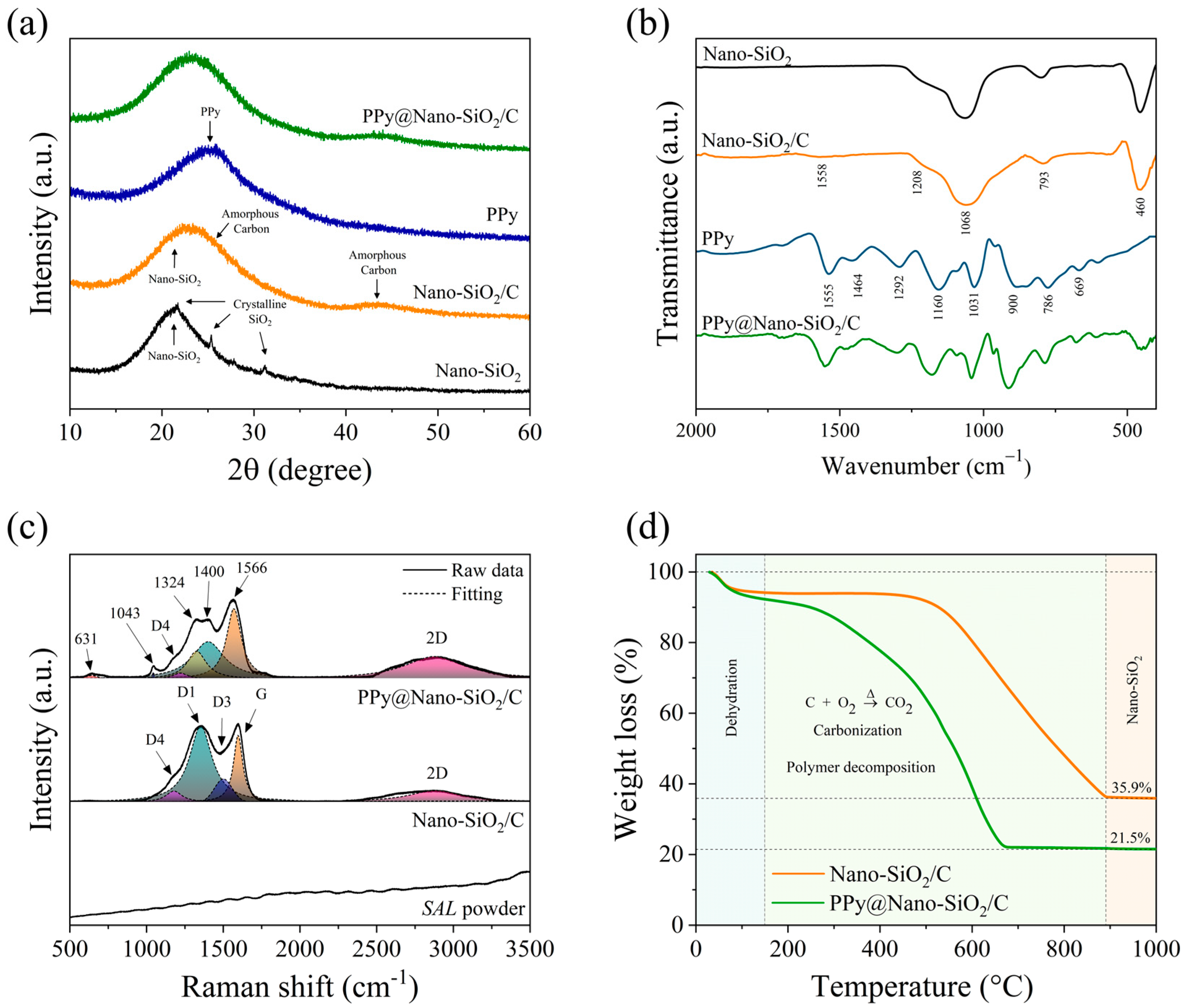
2/C nanocomposites, were investigated using the XRD technique as depicted in
Figure 1a. For the as-synthesized Nano-SiO
2 sample, the XRD patterns were observed to have a broad peak near 23° and small overlapped crystalline patterns at 21.83°, 28.33°, and 31.29°. These patterns corresponded to the characteristic patterns of amorphous SiO
2 combined with some low-crystalline SiO
2 (α-cristobalite, JCPDS No. 39-1425) [
36], respectively. After co-synthesis of SiO
2 and carbon, there were only two broad peaks centered at 26° and 43°, corresponding to the carbon planes (JCPDS No. 41-1487) of (002) and (100), respectively. These indicated the presence of amorphous carbon or low crystalline carbon. Thus, the precise carbon structures in all synthesized samples would be investigated in greater depth by HR-TEM and SAED techniques. Evidently, the amorphous and crystalline SiO
2 patterns disappeared completely. This could be due to the intensity-increasing overlapping of carbon patterns. On the other hand, the XRD patterns of pure PPy exhibited only a broad peak at 25° centered. This indicates that the nature of the as-synthesized PPy is amorphous. For the XRD patterns of PPy@Nano-SiO
2/C, there were also only two broad peaks, which are mainly influenced by carbon. However, because of the nanocomposite formation, the two broad peaks could occur due to overlapping patterns of SiO
2, carbon, and PPy. Compared to the two XRD patterns of Nano-SiO
2/C and PPy@Nano-SiO
2/C, there was no discernible peak displacement in the XRD patterns. More importantly, the center-highest intensity peak value of the prepared nanocomposite material (23.11°) is positioned between the two overlapped broadening peaks of Nano-SiO
2/C and PPy, which are centered at 22.22° and 25.88°, respectively. This indicates that the structure of SiO
2 and carbon was unaffected by the addition of PPy.
To evaluate the successful polymerization of pyrrole monomer and the existence of PPy following in situ polymerization, the characteristic patterns of PPy were examined by FTIR.
Figure 1b illustrates the FTIR spectra of all synthesized materials. For the chemical bonding characterization, the FTIR spectra of as-synthesized materials were obtained in the appropriate range of 2000–400 cm
−1. Recently, the characteristic FTIR peaks of PPy were reported by Sanches et al. [
37]. Therefore, the FTIR spectrum of synthesized pure PPy was also given for reference and compared in this work. Both the 786 cm
−1 and 900 cm
−1 bands were attributed to C–H stretching. Intriguingly, the low-intensity peak at 786 cm
−1 revealed a predominance of α-carbon atoms in the PPy ring, coinciding with the ordered planar configuration of PPy, whereas Py rings are linked sequentially via α–α connections, whereas alternative connections can lead to disorder in the polymer chain [
38]. This phenomenon is characteristic of conjugated polymers, whose electrical conductivity is primarily influenced by carrier concentration and mobility. A high α–α connection ratio signifies a high degree of order, which facilitates carrier migration [
39]. A band at 1031 cm
−1 can be attributed to the =C–H vibration in-plane deformation. C–H in-plane and out-of-plane deformations are responsible for the band at 1160 cm
−1. Bands at 1464 cm
−1 and 1555 cm
−1 were attributed, respectively, to the vibration of the pyrrole ring (C–N) and the ring stretching mode (C=C, C–C). The bands at 1206 cm
−1 and 1292 cm
−1 were designated as the N–C stretching band and =C–H in-plane vibration, respectively. Focusing on PPy@Nano-SiO
2/C, the spectra are unambiguous proof that the combination of Nano-SiO
2/C and the PPy spectra pattern actually occurred. Therefore, the bands that were detected in the PPy@Nano-SiO
2/C nanocomposite of this investigation recognize the polymerization of pyrrole in the nanocomposite and are compatible with those previously reported in the scientific literature [
37,
40].
As shown in
Figure 1c, room-temperature Raman spectroscopy of the produced materials, including SAL powder, Nano-SiO
2/C, and PPy@Nano-SiO
2/C, revealed diverse bands between 500 and 3500 cm
−1. The degree of graphitization and band vibrations were measured to identify structural changes from SAL powder, a raw material, to synthesized nanocomposites (Nano-SiO
2/C and PPy@Nano-SiO
2/C). The three major peaks—the D band, G band, and 2D band—in the Raman spectrum indicated the existence of graphitic carbon. According to the research of Eshun, J. et al. [
41], the D band at 1350 cm
−1 is due to defect structures in highly organized carbonaceous materials, which correspond not only to the condensed benzene rings in amorphous carbon, but are also associated with the vibrating modes of disordered graphite rings. In addition, the G band identified at roughly 1580 cm
−1 provides evidence of the vibrations of sp
2 carbon atoms, whereas double bonds are in graphitic materials. These are a result of the carbon aromatic ring systems present in pristine structures. In particular, Raman spectra determined that 2D bands detected at 2850 cm
−1 are indicative of graphitic structures’ unique multilayer characteristics. Interestingly, the spectra of Nano-SiO
2/C nanocomposite contained upwards of a single D band, which was consistent with the prior study [
42]. The D band was modeled using Lorentzian equations that resulted in three peaks (D1, D3, and D4). In contrast to the most prominent D1 band at 1350 cm
−1, the D3 band was identified at roughly 1500 cm
−1 and the D4 band was discovered as a shoulder peak at approximately 1200 cm
−1. It is hypothesized that the D3 band reflects sp
2-bonded carbon fragments or functional groups within the disordered structure, whereas the D4 band is devoted to the vibration modes of sp
3 carbon. Taking into account the SAL powder and Nano-SiO
2/C, the obtained carbon in the SAL raw materials, which was not detected in Raman spectra, was transformed into sp
2 and sp
3 carbons in the graphitic structure, despite the disordered nature of the material. Due to the influence of PPy addition, the D band shape of PPy@Nano-SiO
2 was altered and split into more than four peaks, which overlapped with the D band in Nano-SiO
2/C. This outcome assigned to predominant band vibrations in PPy is consistent with what has previously been reported in the scientific literature [
43]. C–C in-ring, antisymmetric C–N stretching, C–H bending, and N–H bending stretching were attributed to the peak at 1400 cm
−1. The peak at 1324 cm
−1 was assigned to antisymmetric C–N stretching and C–C in-ring stretching. The designation 1043 cm
−1 was a C–H in-plane distortion. The peak at 631 cm
−1 was finally classified as a C–C ring torsional. In accordance with the FTIR discovery, our analyses strongly corroborate the effective synthesis of PPy, whose molecular chains consist primarily of α–α links in Nano-SiO
2/C to achieve the PPy@Nano-SiO
2/C nanocomposite.
Employing TGA analysis, the weight content of as-prepared nanocomposites was investigated. TGA entails the measurement of a change in mass by subjecting samples to a predetermined temperature regimen. This measurement was conducted in an environment ranging from 25 to 1000 °C in an ambient atmosphere, as shown in
Figure 1d. Generally, when the temperature was up to 150 °C, the remaining moisture would be totally evaporated. Afterward, the sample predominantly lost weight due to thermal and oxidative degradation. After that, the steady weight loss proceeded exclusively in PPy@Nano-SiO
2/C at temperatures ranging from 280 °C to 800 °C, which can be attributed to PPy oxidation [
44]. This suggested that the residual PPy had been degraded, decreasing the amount of carbon bonded to PPy polymer chains and removing more permanent nitrogen-doped units from the PPy ring, both of which are evident from the FTIR study. Unfortunately, carbon in all its forms is eliminated from oxidizing when the temperature rises to 900 °C. The significant weight loss observed at temperatures above 500 °C appears to be primarily attributable to the pyrolysis and carbonization of the carbon backbone chain from PPy and carbon, resulting in the release of CO
2 [
40]. Eventually, the TGA curves demonstrated that the prepared nanocomposites disintegrated completely, although the weight loss of nanocomposite products remained rather steady beyond 900 °C. This suggests that the leftover white product of nanocomposites after high-temperature examination was only the incomplete combustion product of SiO
2, which corresponds to their superior thermal stability. For the Nano-SiO
2 sample, it is presumed that this sample consisted primarily of SiO
2. In addition, there is no silica loss throughout the entire production process because SiO
2 is chemically inert and stable in the ambient environment. The SiO
2 contents of Nano-SiO
2/C and PPy@Nano-SiO
2/C were determined to be 38.12 and 23.30 wt.%, whereas the carbon contents were 61.88 and 37.82 wt.%. Consequently, the PPy composition for PPy@Nano-SiO
2/C was 38.88 wt.%. Therefore, using these TGA results, the weight loss data of as-prepared nanocomposites are summarized, and the theoretical specific capacity is calculated in
Table 1.
Employing SEM investigation, the morphology of synthesized products was analyzed; the results are presented in
Figure 2a–c. Due to the natural structure of SAL, the morphology of Nano-SiO
2 was observed to be a coral-like structure composed of small individual SiO
2 nanoparticles, as illustrated in
Figure 2a. As depicted in
Figure 2b, the structure of Nano-SiO
2/C contains carbon layers because of the carbonization of hemicellulose in the SAL. This suggests that the open space obtained from the coral-like Nano-SiO
2 was filled and covered by carbon, which had a layered structure in the Nano-SiO
2/C nanocomposite.
Figure 2c demonstrates that, as a direct result, the morphology of the PPy@SiO
2/C sample was altered. After incorporating PPy into Nano-SiO
2/C, an SEM image of the PPy@SiO
2/C nanocomposite revealed that the material possessed a thicker sheet adorned with well-distributed particles, which may well be PPy formation during the in situ polymerization process. Considering the thick layer observation, it can be concluded that the PPy is successfully encapsulated on the Nano-SiO
2/C. This characteristic structure significantly improves the materials’ surface activity, and PPy greatly enhances the materials’ electrical conductivity. The PPy@SiO
2/C nanocomposite evident in the image enables the nanocomposite to offer double protection against the volume expansion of SiO
2.
This study addresses energy-dispersive X-ray spectroscopy mapping, where additional examination of element distribution in PPy@Nano-SiO
2/C can be conducted. The EDS mapping acquired the element signals from the captured area of the SEM image, as shown in
Figure 2d, demonstrating that the elements C, Si, O, and N, respectively (
Figure 2e–h), were uniformly distributed throughout the material. The EDS mapping identifies the presence of N, and it is highly probable that the predominance of nitrogen (N) sources from PPy. The formation of SiO
2 can be confirmed by the overlapping signals of Si and O. The overlap between C and Si signals is evidence that SiO
2 is being spread equally throughout the carbon matrix. In addition, the overlapping of N, C, and Si elements with each other unequivocally demonstrates that PPy is dispersed consistently throughout the Nano-SiO
2/C matrix. According to the findings, C, Si, O, and N elements are uniformly disseminated across the mixture. This confirms that the Nano-SiO
2 and PPy nanoparticles are scattered throughout the carbon layer. From the SEM and EDS mapping observations of PPy@Nano-SiO
2/C, it can be determined that the unique morphology of as-synthesized materials adheres to the proposed schematic diagram depicted in
Figure 2i.
The N
2 adsorption–desorption isotherms and associated pore-size distribution curves are shown in
Figure S1a–c and Figure S1d–f, respectively, for Nano-SiO
2, Nano-SiO
2/C, and PPy@Nano-SiO
2/C. The BET surface area of prepared materials is in the order of PPy@Nano-SiO
2/C (117.15 m
2/g) > Nano-SiO
2 (68.24 m
2/g) > Nano-SiO
2/C (59.31 m
2/g), whereas the corresponding pore volume is in the order of Nano-SiO
2 (0.2914 cm
3/g) > PPy@Nano-SiO
2/C (0.1691 cm
3/g) > Nano-SiO
2/C (0.1177 cm
3/g). Interestingly, similar type IV curves with hysteresis loops are observed for these three isotherms, as designated by the International Union of Pure and Applied Chemistry (IUPAC) [
45]. Depending on the pore width, the type-IV isotherm contains a single hysteresis due to the absorption of mesopores, indicating that the pore width is wider than 4 nm [
46]. In addition, a substantial hysteresis loop can indeed be noticed in the N
2 adsorption–desorption isotherm, confirming that the produced materials have a high mesopore proportion. The pore size distribution was then described in terms of differential pore volume against differential log diameter (dV
p/dlog(D)) in order to present pore size distributions, which are depicted in
Figure S1d–f. The BJH theory was used to examine the pore size distribution in this study. All samples contained peaks with a range between 2 and 50 nm. The increase in dV
p/dlog(D) peak intensity reflected a rise in porous density. After in situ polymerization of the pyrrole monomer, the number of pores in the PPy@Nano-SiO
2/C sample emerged as having increased significantly. Therefore, the increase in surface area and pore density may have been caused by the porous nature of PPy-coated layers and particles. In particular, all synthesized samples exhibited a significant intensity peak between 1.5 and 15 nm. The analysis revealed that mesopores (2–50 nm) predominated in the three synthesized materials. Notably, the presence of mesopores in all materials enhances the electrolyte’s penetration, which ultimately decreases the lithium-ion diffusion path length, resulting in superior electrochemical performance at high current densities [
47,
48]. All data on the surface area and the porosity analysis of the synthesized materials are summarized in
Table 2.
3.2. Electrochemical Performances
To assess the electrochemical performance, all prepared samples, Nano-SiO2, Nano-SiO2/C, and PPy@Nano-SiO2/C, were fabricated into working electrodes, for which SA brown algae was used as a binder, before being assembled into CR2016 half-coin battery cells with Li metal as a reference electrode. The fabricated cells were then aged for 12 h before being utilized for battery and electrochemical performance studies.
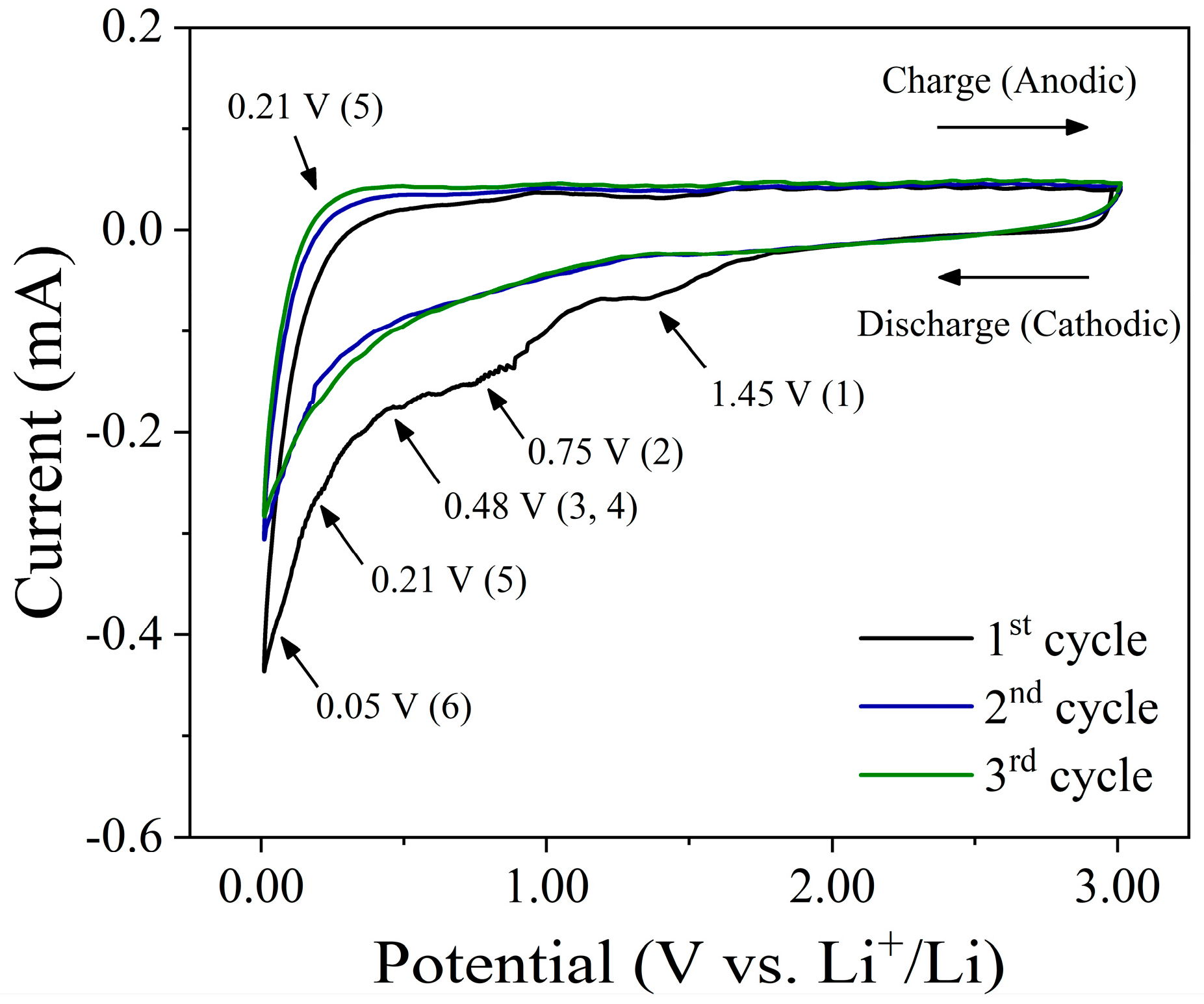
To evaluate the Li-storage mechanism, the cyclic voltammetry (CV) performance at a scan step of 0.1 mV s
−1 over a circuit operating voltage of 0.01–3.00 V for the first three cycles was examined at the SA-PPy@Nano-SiO
2/C electrode, as represented in
Figure 3. There seem to be three detectable peaks (approximately 1.45 V, 0.75 V, and 0.48 V) within the first cathodic scans of CV plots, which are also represented in the initial discharge curve. The peak around 1.45 V is largely attributable to carbonate decomposition in the electrolyte, which is an unanticipated side reaction between the electrolyte and the electrode material [
49]. In addition, the significant cathodic peak at 1.45 V, which reflects the irreversible reduction of SiO
2 to Si, is represented in Equation (1), and the peak at 0.75 V can be referred to as the formation of the solid electrolyte interface (SEI) layer on the electrode surface as a consequence of the consumption of oxygenic functional groups, as shown in Equation (2) [
50]. Upon the initial discharge of the fabricated electrode, amorphous SiO
2 was transformed into Si, and Li
2O was produced [
10,
51]. In addition, when the SiO
2-based material was first discharged, Li
2Si
2O
5 or Li
4SiO
4 would be created when the amorphous SiO
2 turned into Si, as shown in Equations (3) and (4) [
52,
53]. The emergence of irreversible phases during the reaction required considerable capacity. However, the aforementioned effects diminish completely in subsequent cycles. Therefore, this reaction contributes to the electrode’s lithium storage capacity. In the second and third cycles, the CV curves become stable over the following scan cycles, reflecting the equivalent reversible behavior. As shown in Equation (5), the typical peaks associated with the reversible alloy/dealloy reactions of Si with Li
+ involve cathodic peaks of approximately 0.21 V and anodic peaks of approximately 0.52 V, which correlate to the conversion between Si and Li
xSi [
12,
54]. Multiple Li
xSi phases coexisted during the lithiation (alloying) process, as shown by the appearance of a sharp reduction peak at 0.01–0.20 V in the cathodic scan [
55]. Accordingly, this process increases the lithium storage capacity of the electrode. On the other hand, during the charge–discharge process, the peak at around ~0.5–0.01 V corresponds to the Li-ion intercalate and the extraction from graphite-like nanosized domains within the carbon sheets. Since the initial discharge process, lithium has intercalated first into defect sites (1.00 V) and then into nanographite-like domains (0.20 V) [
56]. Thus, in subsequent cycles, the loss of capacity in the pseudo-graphitic nature of disordered hard carbons is mostly due to the decrease in lithium storage at defect sites, especially at carbon surfaces. The basic reaction mechanism of lithium-ion intercalation in the carbon material can be expressed in Equation (6). In the following scan, the second and third cycles almost overlap with each other, proving the high stability and reversibility of the lithiation and delithiation reactions. Obviously, the chemical reaction between PPy and Li was not observed, indicating that PPy may be an inactive material in this nanocomposite electrode. As aforementioned, all chemical reactions during charge–discharge processes could well be classified as follows:
Figure S3 illustrates the galvanostatic charge–discharge curves for the PPy@Nano-SiO
2/C electrode between the voltage cutoffs of 0.01 and 3.0 V at a current density of 0.3C (227.4 mA g
−1). The initial discharge capacity of the fabricated electrodes was discovered to be 1342.5 mAh g
−1. The first cycle’s charge capacity reduction is attributable to irreversible processes which is a side effect of electrolyte disintegration, the creation of a solid electrolyte interphase (SEI) layer, and unforeseen side reactions generated by the consumption of oxygenic functional groups corresponding to the CV result. The Coulombic efficiency (CE) of the second and third cycles improved to 86.0% and 97.7%, respectively, in subsequent cycles. These can be due to the decreasing effect of SEI film formation in the second cycle and their unique nanostructure’s ability to function as an effective anode, resulting in a nearly fully reversible Li
+ reaction.
To determine the morphology and structure of prepared PPy@Nano-SiO
2/C nanocomposite electrodes before and after cycles, a TEM investigation was performed.
Figure 4 displays TEM images of pre-cycled and post-cycled PPy@Nano-SiO
2/C nanocomposite electrodes, along with selected area electron diffraction (SAED) analysis and particle diameter histograms of SiO
2 nanoparticles (SiO
2 NPs) and Si nanoparticles (Si NPs) in the carbon matrix and polymeric network. For the pre-cycled PPy@Nano-SiO
2/C electrode, as depicted in
Figure 4a,b, SiO
2 NPs were principally observed in two configurations: agglomerated particles and dissociated SiO
2 NPs. The particles are highly aggregated due to their small size and Van der Waal’s forces. However, the size of the cluster is still as large as the nanoscale. Furthermore, it is evident that the spherical SiO
2 NPs developed predominantly within the carbon matrix.
Figure 4c represents the corresponding particle size distribution of SiO
2 NPs. It is evident that the diameter of the SiO
2 NPs varied from 2 to 10 nm, with an average value of 5.17 nm. According to the SAED pattern (inset of
Figure 4a), the amorphous structure was revealed. This can be identified by the amorphous nature of SiO
2 and low-crystalline carbon. To examine the in-depth structure of carbon, the area of the carbon sheet was specifically taken, as shown in
Figure S2. The TEM image (
Figure S2a) revealed a thin sheet structure, with the SAED pattern corresponding to low-crystalline carbon. Thus, in the HRTEM observation, the lattice view of the carbon sheet is shown in
Figure S2b, exposing the pseudo-graphitic nature of disordered hard carbon microstructures incorporating graphitic domains [
57]. For the post-cycled PPy@Nano-SiO
2/C electrode,
Figure 4d demonstrates that the small particles were evenly distributed and embedded inside the carbon matrix and self-healing polymeric network. The SAED pattern depicted in the inset of
Figure 4d exhibits three polycrystalline rings consistent with those of the Si (111), LiO
2 (110), and Si (220) planes. These could verify the lithiation–delithiation mechanism that converts SiO
2 NPs to Si quantum dots (Si QDs), whereas the SA-PPy polymer was transformed from spherical agglomerates to 3-dimensional networks.
Figure 4e depicts the formation of the irreversible LiO
2 reaction at this stage of the post-cycled electrode, which corresponds to the formation of the ~3 nm-thick SEI layer. A histogram analysis in
Figure 4f reveals that the Si QDs are monodispersed and have an average size of 2.40 nm. Due to the ultrafine size of Si QDs, the high surface area interacting with Li-ion enables rapid lithiation and delithiation rates, enhancing specific capacity during long-term battery cycling. The crystallinity of the Si-NPs in the post-cycled electrode was further investigated by high-resolution transmission electron microscopy (HR-TEM image), as shown in
Figure 4g. The HR-TEM displays Si-QDs distributed onto the carbon layer. We can observe the crystalline structure of QDs, which displays the (111) lattice sets with an interplanar spacing of ~3.10 Å, characteristic of Si.
Figure 4h shows the HAADF-STEM image of a single sheet of SA-PPy@Nano-SiO
2/C nanocomposite containing relatively heavy elements uniformly on the plate and lighter elements implied as the Si element of SiO
2 nanoparticles. The result clearly elucidates the SA-PPy@Nano-SiO
2/C nanocomposite structure, where the SiO
2 nanoparticles are denser than the carbon layer and conductive polymer network.
Figure 4i–l shows the results of the STEM-EDS elemental mapping analysis. The individual elemental maps show the relative positions of the synthesized materials, clearly identifying the plate structure visually decorated with carbon and SiO
2, which correspond to the C K edge (
Figure 4i), O K edge (
Figure 4j), and Si K edge (
Figure 4k), respectively. Moreover, this is unambiguous evidence of PPy network structure showing that N K edge (
Figure 4l) signals are localized in a good dispersion on the SiO
2/C nanocomposite area. From these advanced electron microscopy results, this work enriches the electrode engineering technology of nanocomposite materials using biomass waste recycling and opens up a new way to customize the self-healing anode for green and sustainable lithium-ion batteries.
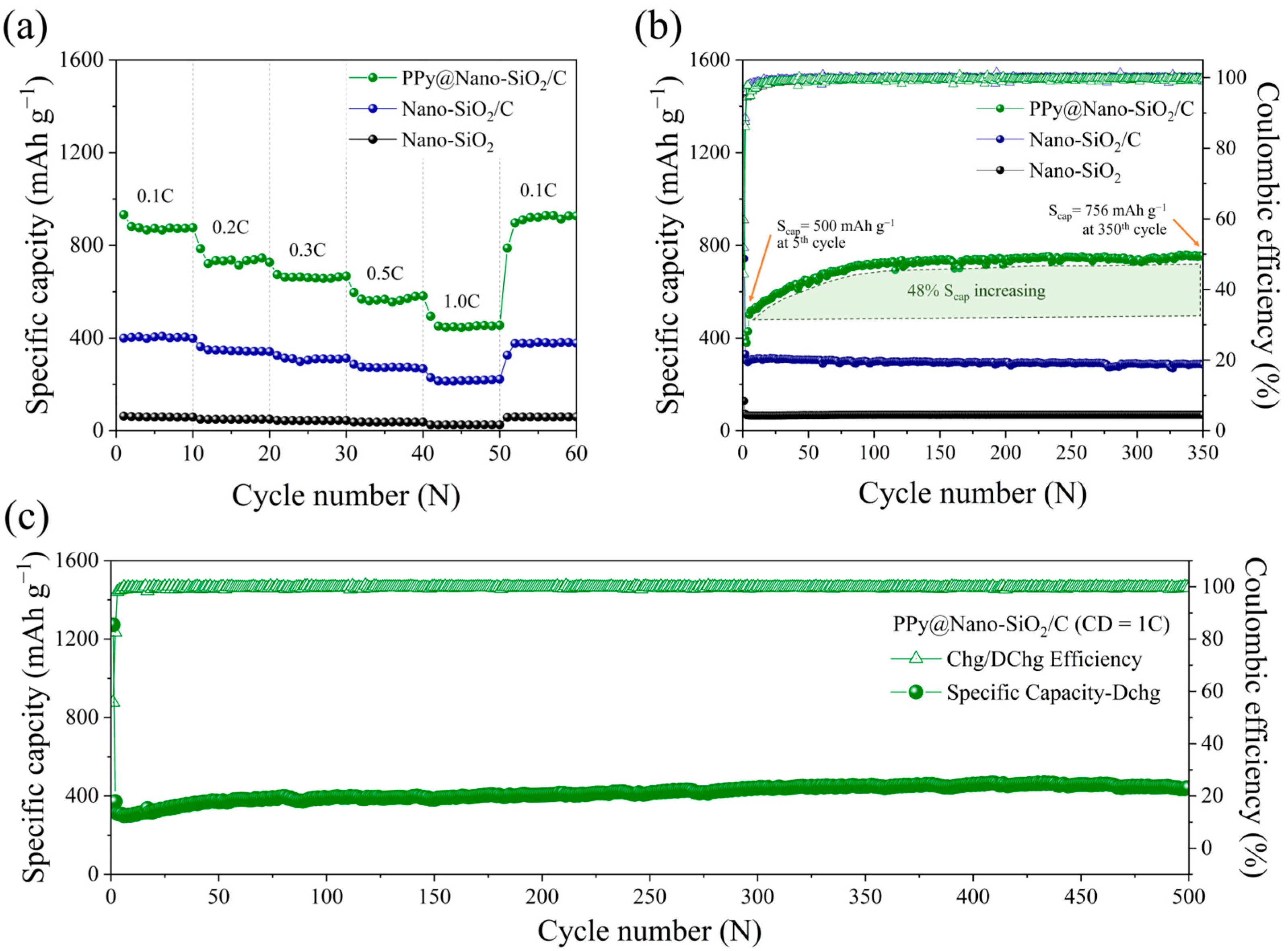
Figure 5a demonstrates the rate property on the lithiation (discharge) capacity of Nano-SiO
2, Nano-SiO
2/C, and PPy@Nano-SiO
2/C at current densities ranging from 0.1 C to 1 C, which was carried out to investigate the rate capability. Even after rapid cycling between 0.1 C and 1.0 C, the PPy@Nano-SiO
2/C electrodes offer the highest rate capability at 0.1 C. Evidently, PPy@Nano-SiO
2/C has improved rate capability, as its specific capacity is greater than that of the fabricated electrodes at each rate. PPy@Nano-SiO
2/C has reversible lithiation capacities of 879, 737, 663, 571, and 455 mAh g
−1 at 0.1 C, 0.2 C, 0.3 C, 0.5 C, and 1.0 C, respectively. In fact, as the charge–discharge rates increased, the lithiation capabilities declined. After measuring the rate capability, the charge–discharge rate rapidly reverted to 0.1C, and the specific capacity can be recovered to 906 mAh g
−1 with a 103% retention. This indicates that this higher percentage of retention may probably be due to the effect of SA-PPy’s self-healing character, which can improve not only the reversible capacity after many cycles but also the conductivity of composites during the charging–discharging process. The average specific capacity at various rates of current density and the percentage of retention at 0.1 C of prepared electrodes are recorded in
Table S1. Owing to the fact that in rate evaluations, fabricated half-cells were only cycled for 10 cycles at each current rate, the performance difference between prepared electrodes was not significant. In order to determine the cycle duration of the three synthesized materials, extensive cycling tests were conducted.
Figure 5b depicts the cycling stability profiles of prepared anodes measured over 350 cycles at a current density of 0.3 C. Importantly, considerable capacity fading typically emerges throughout the first cycle, most particularly due to SEI formation, the effect of irreversible capacity, and difficulty in extracting lithium from disordered materials during initial cycles, resulting in a loss of reversible capacity and a low CE. After 350 cycles, the specific capacities of Nano-SiO
2, Nano-SiO
2/C, and PPy@NanoSiO
2/C electrodes reached 756, 285, and 68 mAh g
−1, respectively, corresponding to CE values reinforced to nearly 100%. Surprisingly, the CE of all electrodes was immediate by the 5th cycle, and the overall cycling performance remained impressively consistent until the 350th cycle. In the scenario of PPy@Nano-SiO
2/C electrodes, the reversible capacity improved from 500 mAh g
−1 at the 5th cycle to 725 mAh g
−1 at the 100th cycle. Following this, the specific capacity climbed marginally to 756 mAh g
−1 compared to the fifth cycle, in which the specific capacity grew almost 48%. This remarkable occurrence may well be the result of three synergistic efforts. First, the reduction of SiO
2 nanoparticles to ultra-fine SiQDs in the subsequent cycle and their well-distribution into the carbon and SA-PPy networks agreed with the TEM images in
Figure 4d,g. This facilitated the electrochemical reaction with elevated and more stable active sites, resulting in a high specific capacity. Second, the design strategy of polymeric green nanocomposite manifests enhanced mechanical robustness that can inhibit SiQDs from being prematurely pulverized and electrical contract loss with that of the flexible backbone conductive network of dual SA-PPy polymer and restore the cracks from high-volume change of Si by filling as a result of its self-healing property. Together with the SA binder, the polymeric nanocomposite sustained the performance of PPy@Nano-SiO
2/C anodes for over 350 cycles. Importantly, the dual protection of carbon and SA-PPy on SiO
2 nanoparticles can significantly improve electrochemical performance, especially with respect to the excellent durability of PPy@Nano-SiO
2/C electrodes. Third, the electrolyte may not completely wet the electrode prior to cycling. Once charging and discharging commence, the movement of lithium ions can allow the electrolyte to more thoroughly wet the PPy layer and SiO
2/C. Consequently, in the initial few cycles, the capabilities rise with the number of cycles [
58].
One of the most important determinants of whether applications of lithium batteries might well be realized is their long-stable life cycling with high-rate features. Consequently, it is essential to concentrate on the rapid charge–discharge performance of the electrode over its multiple cycles. At 1.0C charge–discharge rates,
Figure 5c depicts the long-term cycling behavior of the PPy@Nano-SiO
2/C electrode for 500 cycles. After five cycles, the PPy@Nano-SiO
2/C anode exhibited an initial specific capacity of 306 mAh g
−1; after 500 cycles, its specific capacity increased gradually to 441 mAh g
−1. For the long-term test, this anode’s capacity retention at the 500th cycle was 46% higher than its initial capacity at the 5th cycle for the cycling assessment. These indicate that the PPy@Nano-SiO
2/C has excellent cycling stability in order to use the anode because no capacity fading was observed after the 500th cycle. Consequently, the superior cycle performance of PPy@Nano-SiO
2/C can be attributed to their SA-PPy network in the nanocomposite electrode together with a thin SEI layer, which has superior physical confinement on SiQDs, as evidenced in the microstructure of the cycled material in the TEM images in
Figure 4d,e. As a result, the SA-PPy network offers enhanced electronic conductivity and ion transport efficiency. This study indicates that this fabricated SA-PPy@Nano-SiO
2/C electrode has the ability to function well as an anode in sustainable LIBs for over 500 cycles.
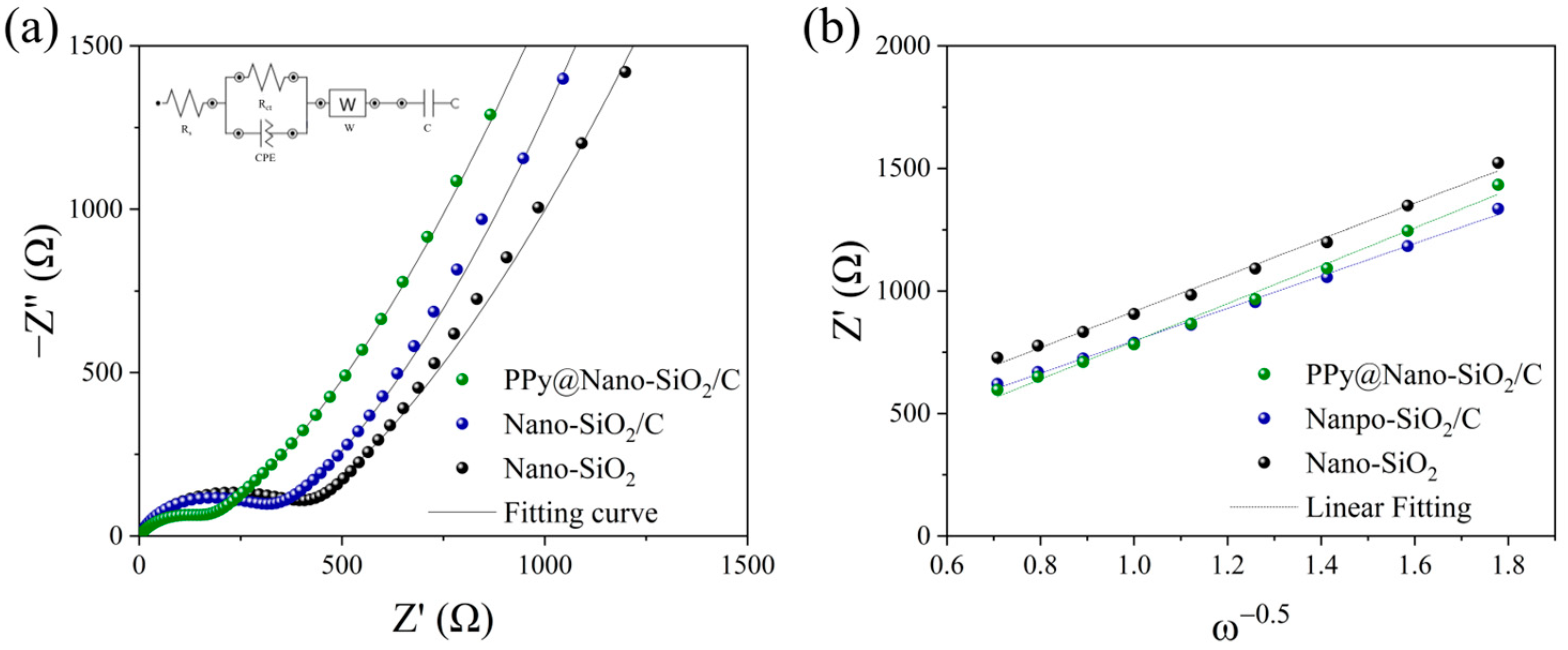
In order to comprehend the electrochemical kinetics of the fabricated electrodes, EIS measurements and equivalent impedance circuit fitting with the Nova 2.1 software have been conducted. The Nyquist plots of Nano-SiO
2, Nano-SiO
2/C, and PPy@Nano-SiO
2/C pre-cycled electrodes are exhibited in
Figure 6a, respectively. All measurements were performed on cells following a 12 h rest period. For all three electrodes, the plots can be considered to consist of a semicircle in the high-frequency region, followed by a sloping line in the middle to low-frequency region. The proposed circuit is an equivalent circuit (inset
Figure 6a) to fit the Nyquist plots represented by solid lines to the experimental data. It is made up of an R symbolizing a resistor, which is comprised of interphase electronic contact resistance (R
s) and charge transfer resistance (R
ct) through the electrode–electrolyte interface, as well as a constant phase element (CPE) in parallel, a Warburg diffusion element (Z
w), and a capacitor (C).
Table 3 outlines the different prepared electrodes analyzed during the interpretation of Nyquist plots, for which the calculation method was followed in our previous report [
49,
59]. Considering the interfacial impedance of the pre-cycle electrode, the R
ct of the PPy@Nano-SiO
2/C (156 Ω) shows the smallest resistance corresponding to the increment of lithium-ion diffusion, whereas the R
ct of the Nano-SiO
2/C and Nano-SiO
2 are 290 and 363 Ω, respectively, while the electrical conductivities (σ
ct) of the electrode are in the order of PPy@Nano-SiO
2/C > Nano-SiO
2/C > Nano-SiO
2. It means the PPy composite in materials has better effects on reducing interfacial impedance than carbon. Regarding the interfacial impedance of the pre-cycled electrode, the R
ct of the PPy@Nano-SiO
2/C (156 Ω) exhibits the lowest resistance correlating to the increase in lithium-ion diffusion, whereas the R
ct of Nano-SiO
2/C (290 Ω) and Nano-SiO
2 (363 Ω) are, respectively, 290 and 363 Ω. This indicates that the PPy network composite is more effective than carbon at reducing interfacial impedance. The angular frequency (
ω), which is related to
Z′, can be used to compute the Warburg factor (σ
w) throughout the low-frequency zone.
Figure 6b depicts the plot of the line relationship between
Zre and
ω−1/2. The Warburg factor (σ
w) was calculated using the slope of the line of prepared electrodes. The lithium-ion diffusion coefficient
, for which the proposed methodology and calculation were previously stated in our work [
49], can therefore be computed from the Warburg region using Equation (7).
where
R is the universal gas constant,
T is the absolute temperature,
F is the Faraday constant,
n is the number of electrons,
A is the electrode area of the electrode, and
C is the concentration of Li-ions in the electrolyte. According to Equation (8), σ
w is the Warburg factor and
ω is the angular frequency, which is related to
Z′ and can be determined from the slope of the fitting line of
Z′ vs.
ω−0.5 in the EIS data at low frequencies, as shown in
Figure 6b.
The Li
+ diffusion coefficients (
, cm
2 s
−1) of all cells are calculated using Equations (7) and (8), which
and
are shown in
Table 3. The PPy@Nano-SiO
2/C electrode has a higher
than that of the Nano-SiO
2 and Nano-SiO
2/C electrodes. It is evident that the
value is directly correlated to the rise in the components of carbon and PPy materials. This is due to the fact that SiO
2, a major component, performs slower conversion reactions (Equations (2) and (4)) in the initial stage and subsequent alloying and dealloying reactions (Equation (5)) with Li ions than the PPy reaction and carbon intercalation (Equation (1)). The unique architecture of SA-PPy contributes to the high electrochemical performances, where 3D networks with good structural stability give sufficient space to achieve maximal contact across electrode and electrolyte, shorten the lithium-ion diffusion length, and alleviate volume change. Thus, the nitrogen lone-pair electron in the PPy backbone chain facilitating electron delocalization further enhances electrical conductivity, accelerates electron transport, and avoids SiQD agglomeration following reduction. As a result, these advantages contribute to an increase in
value, which is beneficial for improving battery performance, notably in circulation stability and specific capacity, by enabling significantly faster Li-ion transfer. As a consequence, these positive impacts on the success verify the excellent rate capability and cycle stability of PPy@Nano-SiO
2/C electrode materials utilizing SA binder, which correlate to the previously discussed battery performances.
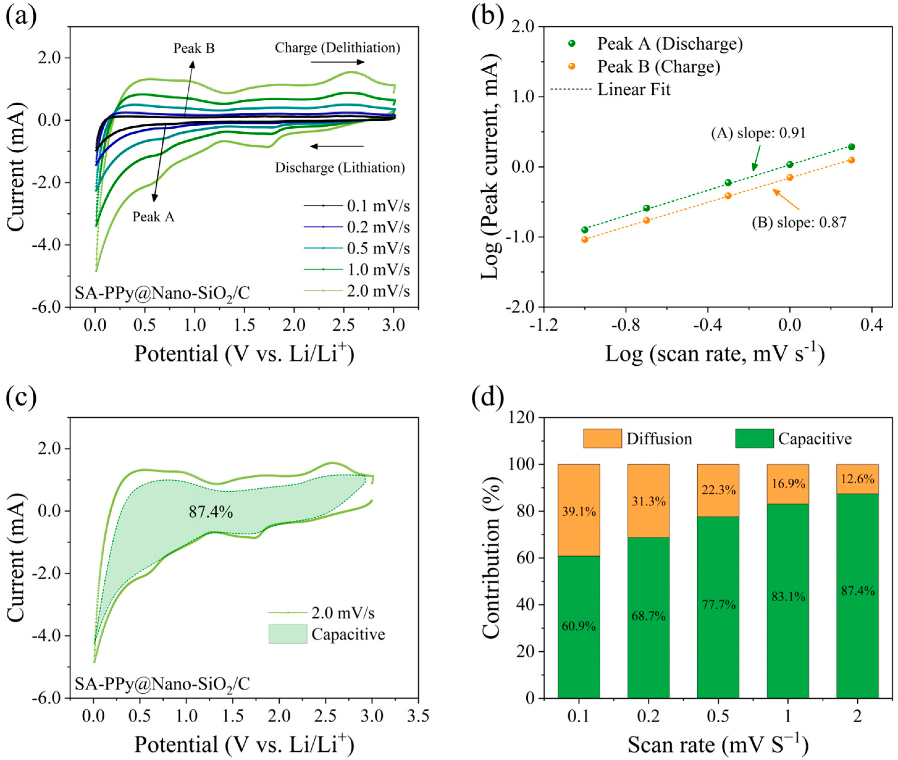
To illustrate the detailed kinetic analysis and to evaluate the likely explanations for the improved rate capability, the as-synthesized SA-PPy@SiO
2/C electrode was used as the anode for the suggested half-coin cells in order to separate the diffusion-controlled capacity and capacitive capacity.
Figure 7a shows the CV curve of this electrode at various scan rates of 0.1 to 2.0 mV s
−1 at room temperature, whose voltage window is 0.01–3.0 V. The capacitive and diffusive contributions can be used to evaluate the electrochemical kinetics of the electrode during high-rate performance. Comparable patterns have indeed been represented by the CV curves, and the peak intensities gradually rose as the scan rate increased. According to general knowledge, the measured current (
i) and scan rate (
v) satisfy the following power–law relationship in accordance with Equations [
60,
61]:
where
i represents the current’s magnitude,
v indicates the scan rate, and
a and
b are variables. The capacity provided by the capacitive effect could be determined by constructing log(
i)−log(
v) curves according to Equation (10) and calculating the slope of the line (
b value). According to prior research, as
b approaches 0.5, a process is governed by complete diffusion, and when
b approaches 1, the process is capacitive [
62,
63]. Therefore, by establishing the value of
b, the battery’s major contribution could well be quantified. The value of
b was 0.91 at peak A (discharge) and 0.87 at peak B (discharge).
Figure 7b displays the linear fitting. This value indicated that the capacitive and diffusion-controlled mechanisms control the peak current in approximately equal measure. The overall capacitive contribution at a specified scan rate can be estimated by separating the capacitive and diffusion-controlled contributions at a defined voltage using the following Equations [
24,
64]:
where
k1 and
k2 are constants for a particular potential that could be obtained by linearly fitting
i/
v1/2 versus
v1/2 under the defined potentials, and
v is the scan rate for certain potentials. By identifying
k1 as the slope and
k2 as the intercept, capacitive and diffusion contributions could be determined. Integrating the green shaded area (
k1v) with the measured currents (solid line) in
Figure 7c demonstrates that capacitive interactions account for roughly 87.4% of the total current at 2.0 mV s
−1 at the PPy@SiO
2/C electrode. As a result, contribution ratios between the two methods were calculated at various scan rates.
Figure 7d illustrates how the percentage of capacitive contribution grows with the scan rate. According to the current separation process, capacitive contributions rise significantly from 60.9% to 68.7%, 77.7%, 83.1%, and 87.4% of total capacitive contributions when the scan rate is increased by 0.1, 0.2, 0.5, 1.0, 1.5, and 2.0 mV s
−1, respectively. This is not surprising since the role of capacitive contribution is growing, and pseudocapacitive contribution is important for ultrafine-reduced SiQDs and SA-PPy networks with a large surface area and a fast scan rate. These studies revealed that the capacitive-controlled process compensated for a large amount of the overall electrochemical reaction of the prepared electrode. As a consequence, the high capacitive contribution to the rate performance of the prepared electrode proved that the material has efficient redox reactions and activity that is independent of the electrochemical reaction rate [
25]. Furthermore, this could also reflect that the conductive SA-PPy networks in the SA-PPy@SiO
2/C electrode are a fundamental requirement and ensure a sustainable structure during charge–discharge processes [
65,
66].
To influence the effect of the SA-PPy framework on the Li-ion attraction of the PPy@Nano-SiO
2/C electrode, the time-dependent transformation for 0, 1, 3, and 7 days during the dissolution experiments of the prepared anode in 1 M LiPF
6 EC/DMC electrolyte was monitored, as demonstrated in
Figure 8a. Determining their solubility by A snapshot revealed that even after seven days, the coloration of the solvent containing PPy@Nano-SiO
2/C electrodes had not significantly altered. The electrolyte was still in a colorless and transparent solution and was the same shade as the electrolyte without an immersed electrode. Similarly, no noticeable evidence of electrode material detachment was discovered. As is evident from these, together with PPy, the high-binding SA binder efficiently prevented the surface delamination and dissolution of the prepared polymer-based materials.
Figure 8b,c illustrate the UV-vis spectra and FTIR spectra, respectively, of the recovered electrolyte solution with and without the PPy@Nano-SiO
2/C electrode to further clarify the suppression of the SA-PPy dissolution of active materials. After careful thought, these data revealed that the recorded spectra in each analysis were virtually identical. Accordingly, it can be concluded that SA-PPy@Nano-SiO
2/C electrodes were insoluble in organic electrolytes. To verify the longevity of this electrode, the dissolution experiment in a 1 M LiPF
6 electrolyte with/without the prepared electrode was conducted continuously for 60 days; however, the appearance remained equivalent to the initial day, and there was no evidence of electrode surface delamination, as shown in
Figure S4.
From all the discussions, this research has enhanced the efficiency of SA-PPy@Nano-SiO
2/C anodes in LIBs by introducing the SiO
2 particles with a self-healing composite polymer. The novel polymer blends of SA and PPy, which can serve as both conductive additives and binders, enhance stability and preserve a thin SEI layer. The SA-PPy system is constructed as an adhesive network of supramolecular polymers, each of which is connected by hydrogen bonds.
Figure 9 depicts the schematic diagram of the SA-PPy@Nano-SiO
2/C electrode and the adhesive polymer network of PPy-SA-PPy. As a result, the hybrid polymer structure maintains the electrical contract of SiO
2/C particles and protects them from fracturing during multiple charge–discharge processes. Due to the hydrogen connections between the two polymers, the structure is capable of self-repair, as the polymers can gradually reconnect themselves if they disconnect at whatever juncture. In addition, the capability of PPy to significantly improve the anode’s conductivity and sustain a thin SEI, besides restricting the electrolytic degradation process of the electrolyte on the anode, could perhaps encourage enlarged lithium diffusion and minimize the interfacial impedance by limiting the excessive electrolyte decomposition on the anode’s surface. Notably, the hydrogen bond network generated between SA and PPy greatly improved the mechanical characteristics of the flexible composite polymer and offered robust cyclic stability to the SiO
2-based anode. In comparison to the other electrochemical performances addressed, the PPy@Nano-SiO
2/C green polymeric nanocomposites, in which SA was used as a binder, exhibit superior electrochemical performance. The comparison of SiO
2-based nanocomposites with carbon and polymer materials is summarized in
Table S2. In particular, the SiO
2/C derived from SAL biomass is more cost-effective, simple, sustainable, and environmentally benign than competing SiO
2/C sources. These are key advantages for industrial manufacturing. Therefore, the integration of PPy polymer and SA brown algae into green conductive polymeric nanocomposites can be greatly improved for multiple anode issues in LIBs. Finally, because of the effective functioning of their green composite design with unique structural morphological advantages and excellent electrochemical capabilities, this newly synthesized PPy@Nano-SiO
2/C material and its green nanocomposite design pave the way for the next generation of sustainable energy storage systems and can be considered a promising anode material for advanced applications.
