

Sulfonated Polyether Ketone Membranes Embedded with Nalidixic Acid—An Emerging Controlled Drug Releaser

 , , and
Abstract
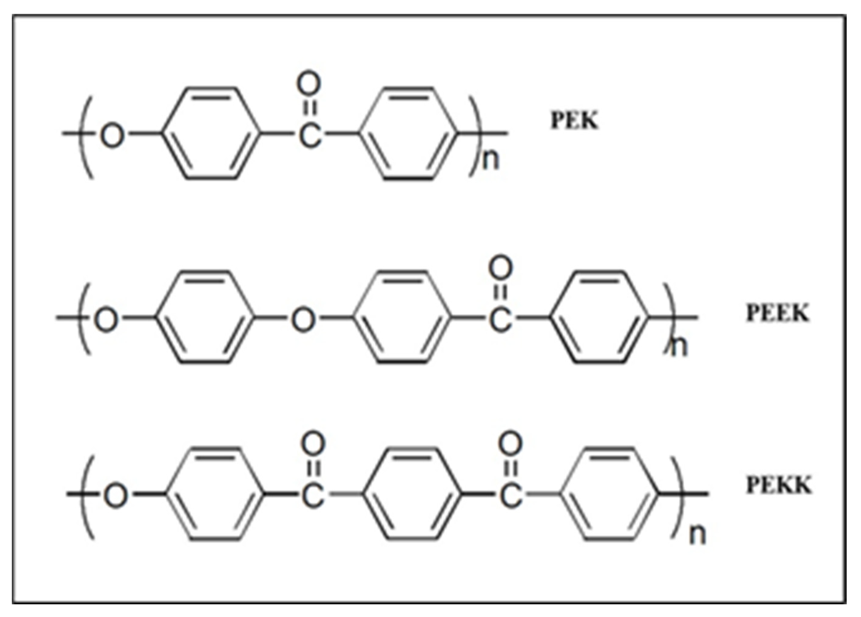
:1. Introduction
2. Materials and Methods
2.1. Materials
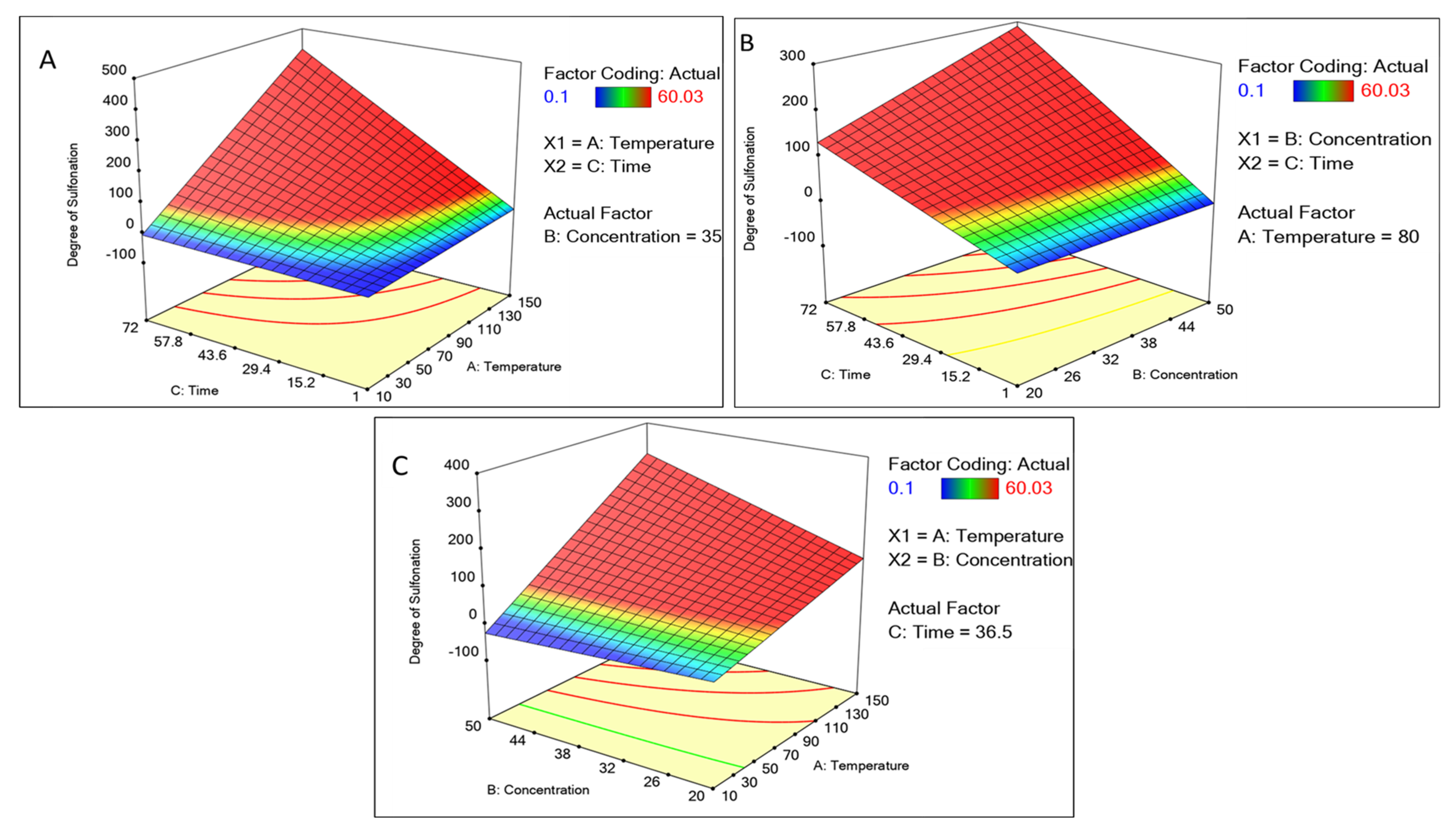
2.2. Statistical Experimental Design of Sulfonation
2.3. Synthesis of Sulfonation PEK (SPEK)
2.4. Characterizations of SPEK
2.4.1. Degree of Sulfonation by IEC
2.4.2. Chemical Interaction of SPEK
2.5. Membrane Casting
2.6. Water Uptake Studies
2.7. Pharmaco-Physicochemical Studies on SPEK Membrane
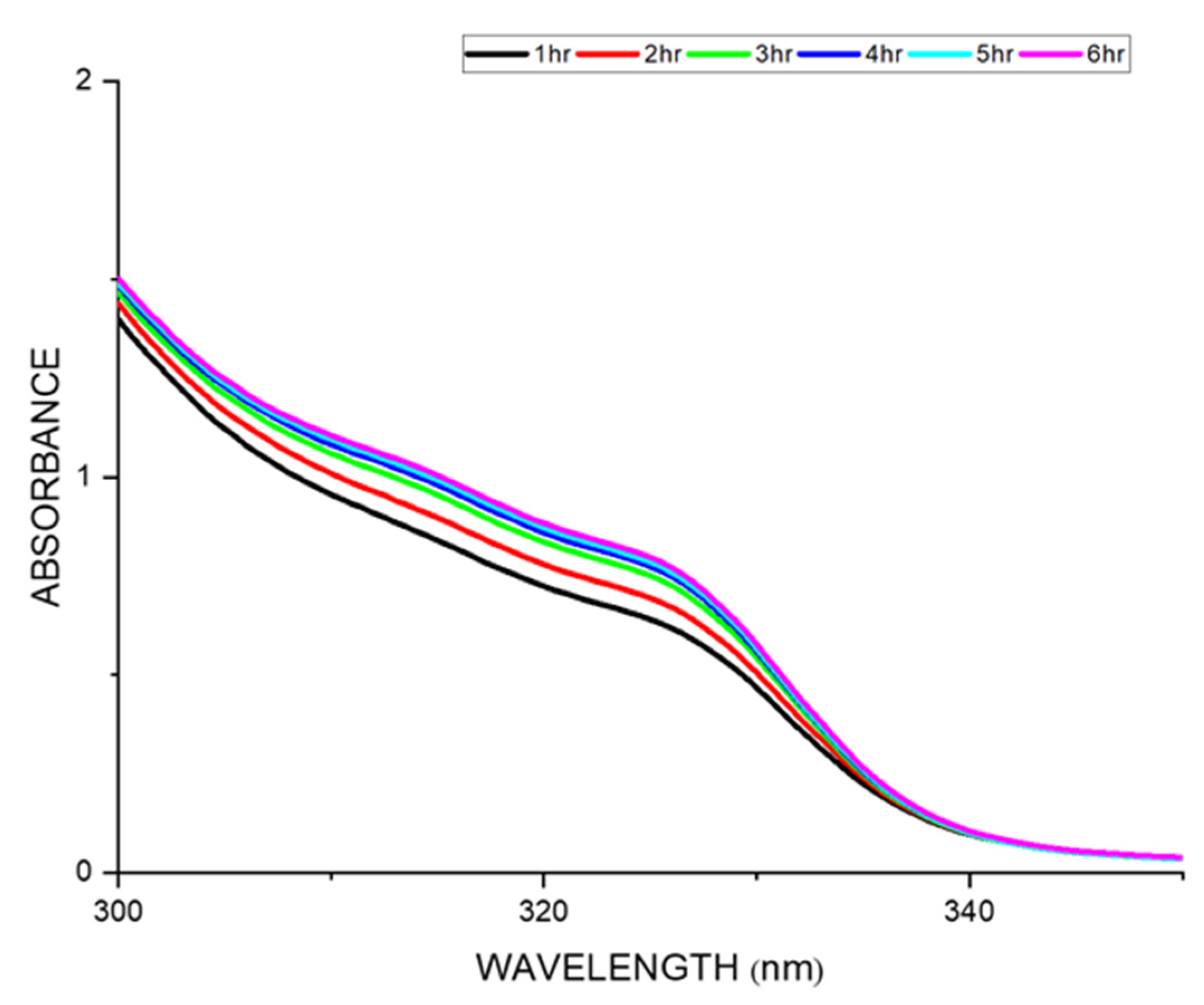
2.8. In Vitro Drug Release Studies from SPEK Membrane
2.9. Mathematical Models
2.10. Hemolysis
2.11. In Vitro Biocompatibility Studies
2.12. Statistical Analysis
3. Results and Discussion
3.1. Statistical Optimization of Sulfonation of PEK
3.2. Sulfonation
3.3. Solubility and Membrane Casting
3.4. Degree of Sulfonation
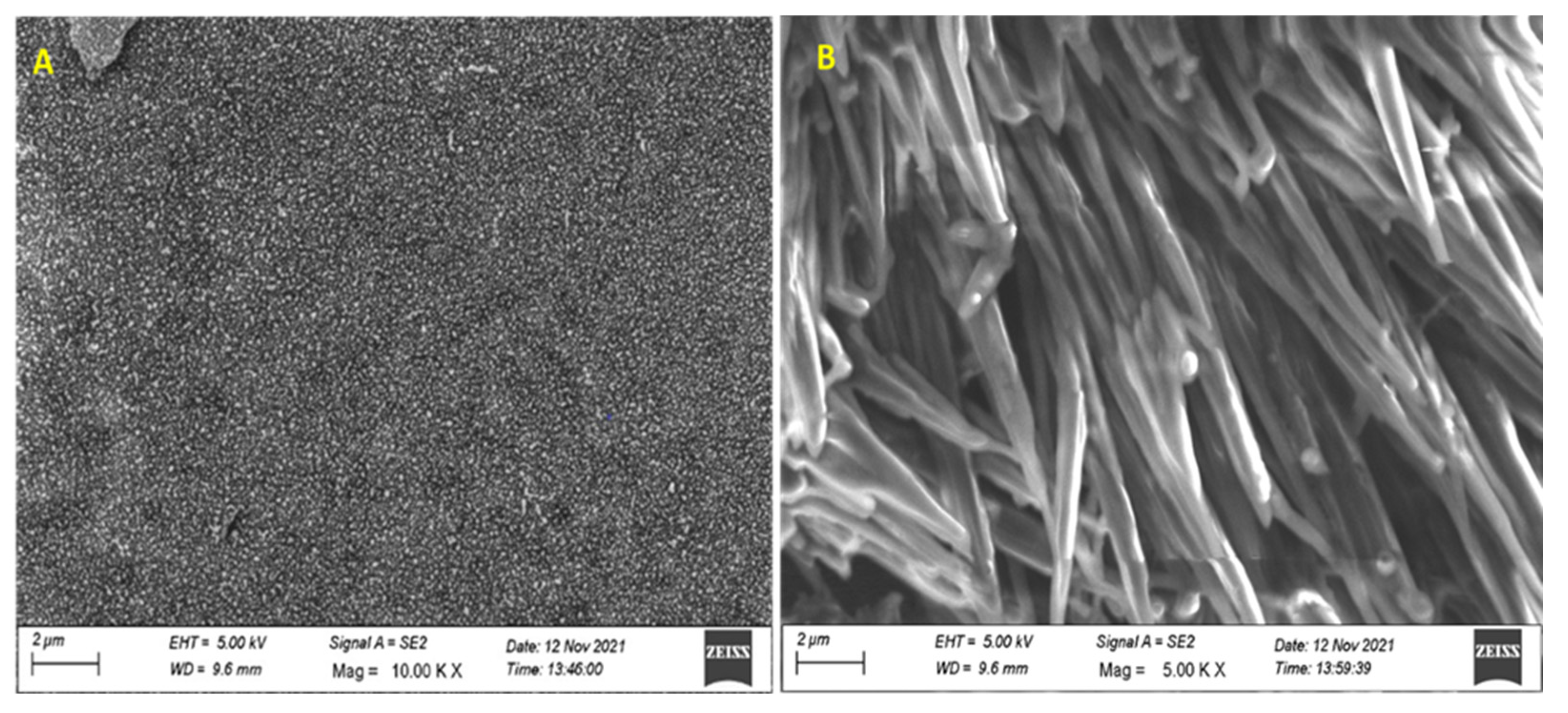
3.5. Characterization of SPEK
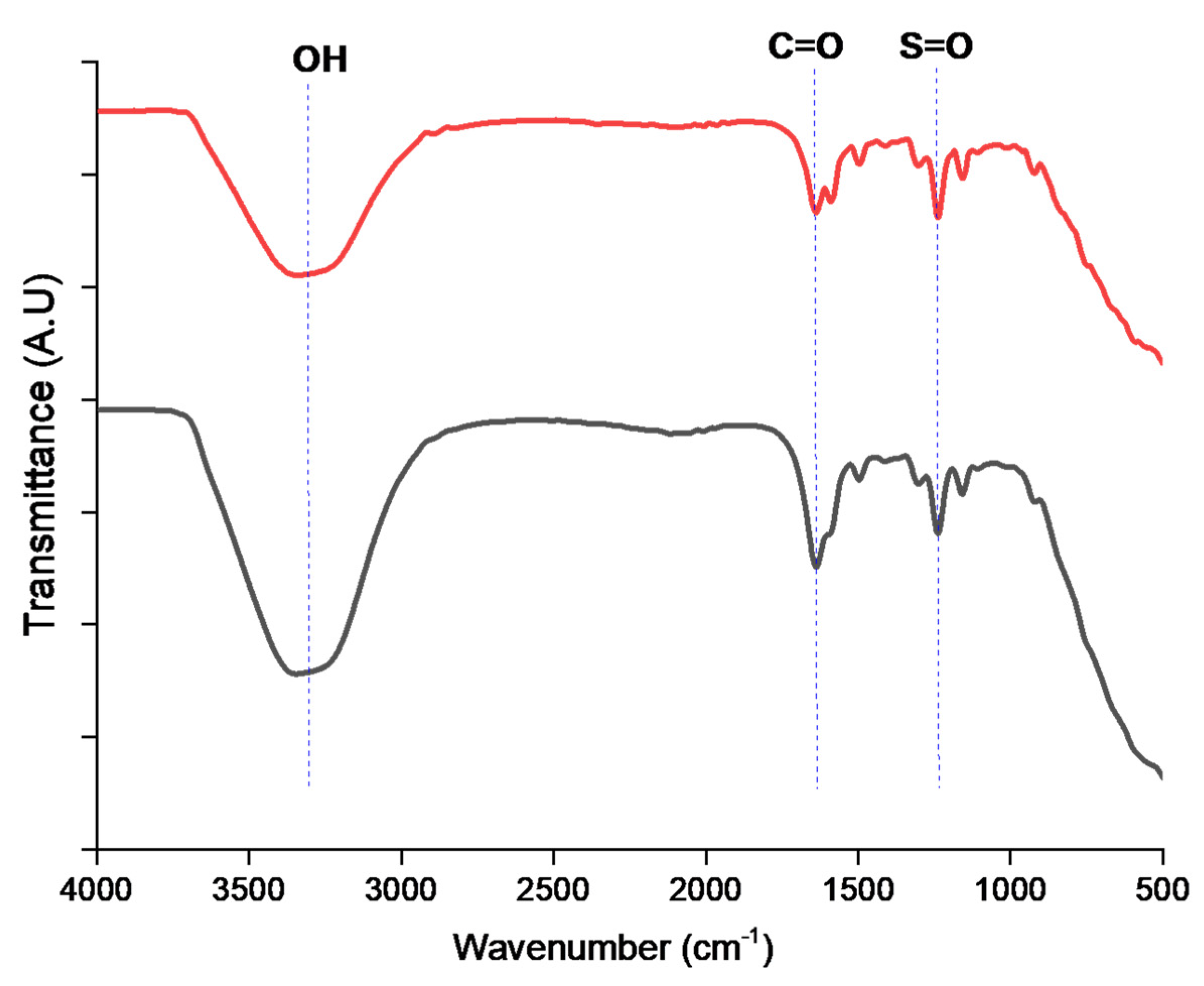
3.5.1. FTIR Spectrum Studies
3.5.2. TGA Analysis
3.6. Water Uptake Studies
3.7. Drug Release Kinetics
3.8. Distribution Coefficient, Diffusion Coefficient, and Permeability Coefficient
3.9. Mathematical Models of Drug Release
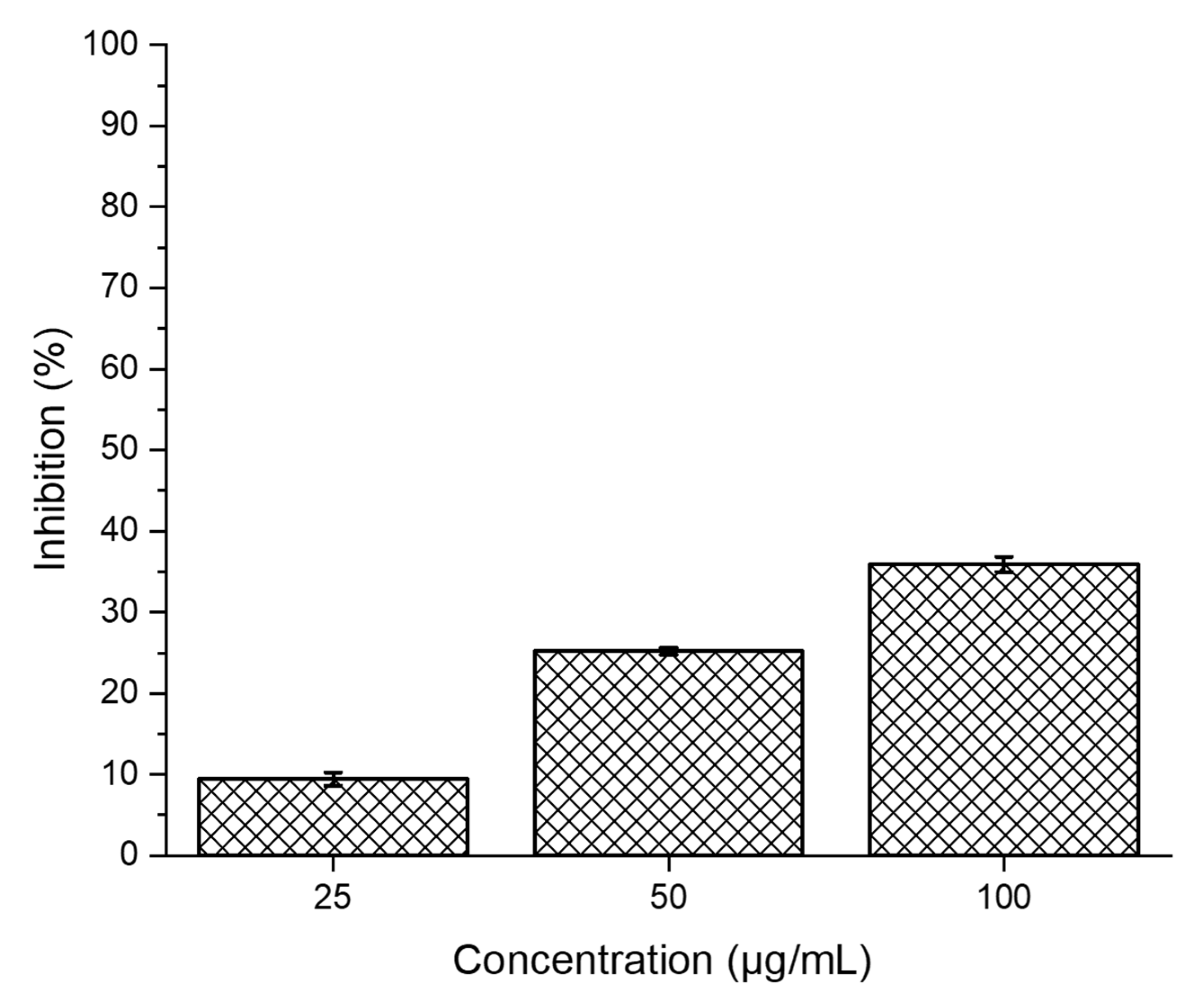
3.10. Hemolysis Studies of SPEK
3.11. In Vitro Cytotoxicity Studies of SPEK
4. Conclusions
Supplementary Materials
Author Contributions
Funding
Institutional Review Board Statement
Informed Consent Statement
Data Availability Statement
Acknowledgments
Conflicts of Interest
References
- Jones, D.S. Pharmaceutical Applications of Polymers for Drug Delivery; Rapra Technology Ltd.: Shropshire, UK, 2004; Volume 15, ISBN 978-1-85957-479-9. [Google Scholar]
- Adepu, S.; Ramakrishna, S. Controlled Drug Delivery Systems: Current Status and Future Directions. Molecules 2021, 26, 5905. [Google Scholar] [CrossRef]
- Ratnaparkhi, M.G.; Jyothi, P. Sustained Release Oral Drug Delivery System-an Overview. Int. J. Pharma Res. Rev. 2013, 2, 11–21. [Google Scholar]
- Patel, H.; Panchal, D.R.; Patel, U.; Brahmbhatt, T.; Suthar, M. Matrix Type Drug Delivery System: A Review. J. Pharm. Sci. Biosci. Res. 2011, 1, 143–151. [Google Scholar]
- Huang, X.; Brazel, C.S. On the Importance and Mechanisms of Burst Release in Matrix-Controlled Drug Delivery Systems. J. Control. Release 2001, 73, 121–136. [Google Scholar] [CrossRef]
- Nidhi, P.; Anamika, C.; Twinkle, S.; Mehul, S.; Hitesh, J. Controlled Drug Delivery System: A Review. Biol. Med. 2006, 3, 227–233. [Google Scholar]
- Baladhandapania, A.; Suresh, S.; Sindhu, S.; Ramani, P. Physico-Chemical Studies of Amoxycillin Loaded Sulfonated Polymer. Mater. Today Proc. 2018, 5, 16146–16151. [Google Scholar] [CrossRef]
- Baldoli, C.; Oldani, C.; Licandro, E.; Ramani, P.; Valerio, A.; Ferruti, P.; Falciola, L.; Mussini, P. Ferrocene Derivatives Supported on Poly(N-Vinylpyrrolidin-2-One) (PVP): Synthesis of New Water-Soluble Electrochemically Active Probes for Biomolecules. J. Organomet. Chem. 2007, 692, 1363–1371. [Google Scholar] [CrossRef]
- Irfan, M.; Idris, A. Overview of PES Biocompatible/Hemodialysis Membranes: PES–Blood Interactions and Modification Techniques. Mater. Sci. Eng. C 2015, 56, 574–592. [Google Scholar] [CrossRef] [PubMed]
- Wypych, G. PEK Polyetherketone. In Handbook of Polymers; Elsevier Science: Amsterdam, The Netherlands, 2012; pp. 364–366. [Google Scholar] [CrossRef]
- Ajeesh, G.; Bhowmik, S.; Sivakumar, V.; Varshney, L.; Kumar, V.; Abraham, M. Investigation on Polyetheretherketone Composite for Long Term Storage of Nuclear Waste. J. Nucl. Mater. 2015, 467, 855–862. [Google Scholar] [CrossRef]
- Krishna, M.S.; Balaji, S.; Raj, G.V.; A Pravin, P.; Kumar, M.S.; Kothurkar, N.K.; Ramani, P.; Narayanan, B.S.; Moorthy, A. Polymer-iron tungstate-reduced graphene oxide nanocomposites for microwave absorption. IOP Conf. Ser. Mater. Sci. Eng. 2019, 577, 012079. [Google Scholar] [CrossRef]
- Alqurashi, H.; Khurshid, Z.; Syed, A.U.Y.; Rashid Habib, S.; Rokaya, D.; Zafar, M.S. Polyetherketoneketone (PEKK): An Emerging Biomaterial for Oral Implants and Dental Prostheses. J. Adv. Res. 2021, 28, 87–95. [Google Scholar] [CrossRef] [PubMed]
- Katzer, A.; Marquardt, H.; Westendorf, J.; Wening, J.V.; Von Foerster, G. Polyetheretherketone—Cytotoxicity and Mutagenicity In Vitro. Biomaterials 2002, 23, 1749–1759. [Google Scholar] [CrossRef] [PubMed]
- Wang, W.; Luo, C.J.; Huang, J.; Edirisinghe, M. PEEK Surface Modification by Fast Ambient-Temperature Sulfonation for Bone Implant Applications. J. R. Soc. Interface 2019, 16, 20180955. [Google Scholar] [CrossRef] [PubMed]
- Nabipour, H.; Sadr, M.H.; Thomas, N. Synthesis, Controlled Release and Antibacterial Studies of Nalidixic Acid–Zinc Hydroxide Nitrate Nanocomposites. New J. Chem. 2016, 40, 238–244. [Google Scholar] [CrossRef]
- Bakhaidar, R.B.; Naveen, N.R.; Basim, P.; Murshid, S.S.; Kurakula, M.; Alamoudi, A.J.; Bukhary, D.M.; Jali, A.M.; Majrashi, M.A.; Alshehri, S.; et al. Response Surface Methodology (RSM) Powered Formulation Development, Optimization and Evaluation of Thiolated Based Mucoadhesive Nanocrystals for Local Delivery of Simvastatin. Polymers 2022, 14, 5184. [Google Scholar] [CrossRef]
- Rajewski, J.; Dobrzyńska-Inger, A. Application of Response Surface Methodology (RSM) for the Optimization of Chromium(III) Synergistic Extraction by Supported Liquid Membrane. Membranes 2021, 11, 854. [Google Scholar] [CrossRef]
- Nabipour, H.; Hossaini Sadr, M.; Rezanejade Bardajee, G. Release Behavior, Kinetic and Antimicrobial Study of Nalidixic Acid from [Zn2(Bdc)2(Dabco)] Metal-Organic Frameworks. J. Coord. Chem. 2017, 70, 2771–2784. [Google Scholar] [CrossRef]
- Banerjee, K.; Debroy, M.; Balla, V.K.; Bodhak, S. Recent Progress in 3D-Printed Polyaryletherketone (PAEK)-Based High-Performance Polymeric Implants for Musculoskeletal Reconstructions. J. Mater. Res. 2021, 36, 3877–3893. [Google Scholar] [CrossRef]
- Hans-Heinz Ulrich, G.R. Sulfonated Poly(Aryl Ether Ketone)s. Angew. Makromol. Chem. 1998, 263, 71–78. [Google Scholar] [CrossRef]
- Liu, D.; Dong, B.; Zhang, H.; Xie, Y.; Pang, J.; Jiang, Z. High Methanol Resistant Polyelectrolyte Membrane Based on Semi-Crystalline Poly(Ether Ketone) with Densely Sulfonated Side Chain for Direct Methanol Fuel Cell. J. Power Sources 2021, 482, 228982. [Google Scholar] [CrossRef]
- Yusuff, A.S.; Ishola, N.B.; Gbadamosi, A.O. Artificial Intelligence Techniques and Response Surface Methodology for the Optimization of Methyl Ester Sulfonate Synthesis from Used Cooking Oil by Sulfonation. ACS Omega 2023, 8, 19287–19301. [Google Scholar] [CrossRef] [PubMed]
- Chen, J.H.; Liu, Q.L.; Zhu, A.M.; Fang, J.; Zhang, Q.G. Dehydration of Acetic Acid Using Sulfonation Cardo Polyetherketone (SPEK-C) Membranes. J. Membr. Sci. 2008, 308, 171–179. [Google Scholar] [CrossRef]
- Oroujzadeh, M.; Mehdipour-Ataei, S.; Esfandeh, M. Proton Exchange Membranes with Microphase Separated Structure from from dual electrospun poly (ether ketone) mats: Producing ionic paths in a hydrophobic matrix. Chem. Eng. J. 2014, 269, 212–220. [Google Scholar] [CrossRef]
- Talley, S.J.; Yuan, X.; Moore, R.B. Thermoreversible Gelation of Poly(Ether Ether Ketone). ACS Macro Lett. 2017, 6, 262–266. [Google Scholar] [CrossRef]
- Lindsey, J.; Anderson, R.B.M. Sulfonation of Blocky Brominated PEEK to Prepare Hydrophilic-Hydrophobic Blocky Copolymers for Efficient Proton Conduction. Solid State Ion. 2019, 336, 47–56. [Google Scholar]
- Huang, R.Y.M.; Shao, P.; Burns, C.M.; Feng, X. Sulfonation of Poly(Ether Ether Ketone) (PEEK): Kinetic Study and Characterization. J. Appl. Polym. Sci. 2001, 82, 2651–2660. [Google Scholar] [CrossRef]
- Shibuya, N.; Porter, R.S. Kinetics of PEEK Sulfonation in Concentrated Sulfuric Acid. Macromolecules 1992, 25, 6495–6499. [Google Scholar] [CrossRef]
- Feng, S.; Pang, J.; Yu, X.; Wang, G.; Manthiram, A. High-Performance Semicrystalline Poly(Ether Ketone)-Based Proton Exchange Membrane. ACS Appl. Mater. Interfaces 2017, 9, 24527–24537. [Google Scholar] [CrossRef]
- Bruschi, M.L. Strategies to Modify the Drug Release from Pharmaceutical Systems. In Strategies to Modify the Drug Release from Pharmaceutical Systems; Woodhead Publishing: Sawston, UK, 2015; pp. 1–199. [Google Scholar] [CrossRef]
- Calori, I.R.; Braga, G.; de Jesus, P.D.C.C.; Bi, H.; Tedesco, A.C. Polymer Scaffolds as Drug Delivery Systems. Eur. Polym. J. 2020, 129, 109621. [Google Scholar] [CrossRef]
- Siepmann, J.; Siepmann, F. Mathematical Modeling of Drug Delivery. Int. J. Pharm. 2008, 364, 328–343. [Google Scholar] [CrossRef]
- Dash, S.M.P.; lilakanta, N.C. Kinetic Modeling on Drug Release from Controlled Drug Delivery Systems. Acta Pol. Pharm. 2010, 67, 217–223. [Google Scholar] [PubMed]
- Böyum, A. Isolation of Leucocytes from Human Blood. A Two-Phase System for Removal of Red Cells with Methylcellulose as Erythrocyte-Aggregating Agent. Scand. J. Clin. Lab. Investig. Suppl. 1968, 97, 9–29. [Google Scholar]
- Li, S.Q.; Zhu, R.R.; Zhu, H.; Xue, M.; Sun, X.Y.; Yao, S.D.; Wang, S.L. Nanotoxicity of TiO(2) Nanoparticles to Erythrocyte In Vitro. Food Chem. Toxicol. 2008, 46, 3626–3631. [Google Scholar] [CrossRef]
- Sadrjahani, M.; Gharehaghaji, A.A.; Javanbakht, M. Modeling and Optimization of Sulfonated Poly (Ether Ether Ketone) Nanofibrous Proton Exchange Membranes with Response Surface Methodology. Polym. Eng. Sci. 2018, 58, 619–631. [Google Scholar] [CrossRef]
- El Magri, A.; El Mabrouk, K.; Vaudreuil, S.; Chibane, H.; Touhami, M.E. Optimization of Printing Parameters for Improvement of Mechanical and Thermal Performances of 3D Printed Poly(Ether Ether Ketone) Parts. J. Appl. Polym. Sci. 2020, 137, 49087. [Google Scholar] [CrossRef]
- Zhang, Y.; Zhang, A.; Wang, S.; Li, S. Investigation of Sulfonation Degree and Temperature on Structure, Thermal and Membrane’s Properties of Sulfonated Poly (Ether Ether Ketone). Int. J. Hydrog. Energy 2023. [Google Scholar] [CrossRef]
- Rao, V.L. Polyether Ketones. J. Macromol. Sci. Part C 1995, 35, 661–712. [Google Scholar] [CrossRef]
- Litter, M.I.; Marvel, C.S. Polyaromatic Ether-Ketones and Polyaromatic Ether-Ketone Sulfonamides from 4-Phenoxybenzoyl Chloride and from 4,4′-Dichloroformyldiphenyl Ether. J. Polym. Sci. Polym. Chem. Ed. 1985, 23, 2205–2223. [Google Scholar] [CrossRef]
- Malik, R.S.; Tripathi, S.N.; Gupta, D.; Choudhary, V. Novel Anhydrous Composite Membranes Based on Sulfonated Poly (Ether Ketone) and Aprotic Ionic Liquids for High Temperature Polymer Electrolyte Membranes for Fuel Cell Applications. Int. J. Hydrog. Energy 2014, 39, 12826–12834. [Google Scholar] [CrossRef]
- Padinjarathil, H.; Mudradi, S.; Balasubramanian, R.; Drago, C.; Dattilo, S.; Kothurkar, N.K.; Ramani, P. Design of an Antibiotic-Releasing Polymer: Physicochemical Characterization and Drug Release Patterns. Membranes 2023, 13, 102. [Google Scholar] [CrossRef]
- Akhtar, F.H.; Abdulhamid, M.A.; Vovusha, H.; Ng, K.C.; Schwingenschlögl, U.; Szekely, G. Defining Sulfonation Limits of Poly(Ether-Ether-Ketone) for Energy-Efficient Dehumidification. J. Mater. Chem. A Mater. 2021, 9, 17740–17748. [Google Scholar] [CrossRef]
- Liu, X.; He, S.; Shi, Z.; Zhang, L.; Lin, J. Effect of Residual Casting Solvent Content on the Structure and Properties of Sulfonated Poly(Ether Ether Ketone) Membranes. J. Membr. Sci. 2015, 492, 48–57. [Google Scholar] [CrossRef]
- Sandro Dattilo, C.P.; Emanuele Francesco Mirabella, A.S.; Andrea Antonino Scamporrino, D.C.Z. Thermal Degradation Processes of Aromatic Poly(Ether Sulfone) Random Copolymers Bearing Pendant Carboxyl Groups. Polymers 2020, 12, 1810. [Google Scholar] [CrossRef] [PubMed]
- Patel, H.A.; Somani, R.S.; Bajaj, H.C.; Jasra, R.V. Nanoclays for Polymer Nanocomposites, Paints, Inks, Greases and Cosmetics Formulations, Drug Delivery Vehicle and Waste Water Treatment. Bull. Mater. Sci. 2006, 29, 133–145. [Google Scholar] [CrossRef]
- Thomas, D.; Nair, V.V.; Latha, M.S.; Thomas, K.K. Theoretical and experimental studies on theophylline release from hydrophilic alginate nanoparticles. Future J. Pharm. Sci. 2019, 5, 2. [Google Scholar] [CrossRef]
- Guo, C.; Lu, R.; Wang, X.; Chen, S. Antibacterial Activity, Bio-Compatibility and Osteogenic Differentiation of Graphene Oxide Coating on 3D-Network Poly-Ether-Ether-Ketone for Orthopaedic Implants. J. Mater. Sci. Mater. Med. 2021, 32, 135. [Google Scholar] [CrossRef]
- Kim, B.S.; Chiba, T.; Inoue, T. Morphology Development via Reaction-Induced Phase Separation in Epoxy/Poly(Ether Sulfone) Blends: Morphology Control Using Poly(Ether Sulfone) with Functional End-Groups. Polymer 1995, 36, 43–47. [Google Scholar] [CrossRef]
- Salimi, E. Development of Bioactive Sodium Alginate/Sulfonated Polyether Ether Ketone/Hydroxyapatite Nanocomposites: Synthesis and In-Vitro Studies. Carbohydr. Polym. 2021, 267, 118236. [Google Scholar] [CrossRef]


| Code | Factors | Low | High |
|---|---|---|---|
| A | Concentration of sulfuric acid (mL/g) | 20 | 50 |
| B | Temperature (°C) | 10 | 150 |
| C | Time (h) | 1 | 72 |
| Entry | Temperature (°C) | Concentration (mL/g) | Time (h) | Degree of Sulfonation (%) |
|---|---|---|---|---|
| 1 | 10 | 20 | 1 | 0.10 |
| 2 | 10 | 20 | 2 | 0.28 |
| 3 | 150 | 50 | 6 | 60.03 |
| 4 | 10 | 20 | 6 | 0.25 |
| 5 | 10 | 20 | 24 | 5.50 |
| 6 | 10 | 20 | 48 | 10.62 |
| 7 | 10 | 20 | 72 | 12.00 |
| 8 | 30 | 50 | 24 | 8.62 |
| 9 | 30 | 20 | 1 | 5.43 |
| 10 | 30 | 20 | 2 | 6.77 |
| 11 | 100 | 50 | 6 | 27.79 |
| 12 | 30 | 20 | 6 | 16.9 |
| 13 | 30 | 20 | 24 | 24 |
| 14 | 30 | 20 | 48 | 37 |
| 15 | 30 | 20 | 72 | 42 |
| 16 | 150 | 50 | 3 | 33.14 |
| 17 | 100 | 20 | 1 | 1.18 |
| 18 | 100 | 20 | 3 | 2.5 |
| 19 | 100 | 20 | 6 | 8.00 |
| 20 | 100 | 50 | 1 | 3.50 |
| S.no | Polymer: H2SO4 | Temperature (°C) | Time (h) | Degree of Sulfonation (%) |
|---|---|---|---|---|
| 1 | PEK (1:20) | 10 | 1–72 | 0.10–10.95 |
| RT | 1–72 | 3.43–40.02 | ||
| 100 | 1–72 | 4.75–57.00 | ||
| 150 | 1–6 | 4.63–15.89 | ||
| 2 | PEK (1:50) | RT | 1–24 | 3.45–8.00 |
| 100 | 1–6 | 7.00–37.95 | ||
| 150 | 3–6 | 20.14–69.13 | ||
| 3 | PEK (1:500) | RT | 1 | Failed |
Disclaimer/Publisher’s Note: The statements, opinions and data contained in all publications are solely those of the individual author(s) and contributor(s) and not of MDPI and/or the editor(s). MDPI and/or the editor(s) disclaim responsibility for any injury to people or property resulting from any ideas, methods, instructions or products referred to in the content. |
© 2023 by the authors. Licensee MDPI, Basel, Switzerland. This article is an open access article distributed under the terms and conditions of the Creative Commons Attribution (CC BY) license (https://creativecommons.org/licenses/by/4.0/).
Share and Cite
Padinjarathil, H.; Vilasini, V.; Balasubramanian, R.; Drago, C.; Dattilo, S.; Ramani, P. Sulfonated Polyether Ketone Membranes Embedded with Nalidixic Acid—An Emerging Controlled Drug Releaser. Polymers 2023, 15, 3631. https://doi.org/10.3390/polym15173631
Padinjarathil H, Vilasini V, Balasubramanian R, Drago C, Dattilo S, Ramani P. Sulfonated Polyether Ketone Membranes Embedded with Nalidixic Acid—An Emerging Controlled Drug Releaser. Polymers. 2023; 15(17):3631. https://doi.org/10.3390/polym15173631
Chicago/Turabian StylePadinjarathil, Himabindu, Vidya Vilasini, Rajalakshmi Balasubramanian, Carmelo Drago, Sandro Dattilo, and Prasanna Ramani. 2023. "Sulfonated Polyether Ketone Membranes Embedded with Nalidixic Acid—An Emerging Controlled Drug Releaser" Polymers 15, no. 17: 3631. https://doi.org/10.3390/polym15173631
APA StylePadinjarathil, H., Vilasini, V., Balasubramanian, R., Drago, C., Dattilo, S., & Ramani, P. (2023). Sulfonated Polyether Ketone Membranes Embedded with Nalidixic Acid—An Emerging Controlled Drug Releaser. Polymers, 15(17), 3631. https://doi.org/10.3390/polym15173631