
Evaluation of Poly(etheretherketone) Post’s Mechanical Strength in Comparison with Three Metal-Free Biomaterials: An In Vitro Study


Abstract
1. Introduction
2. Materials and Methods
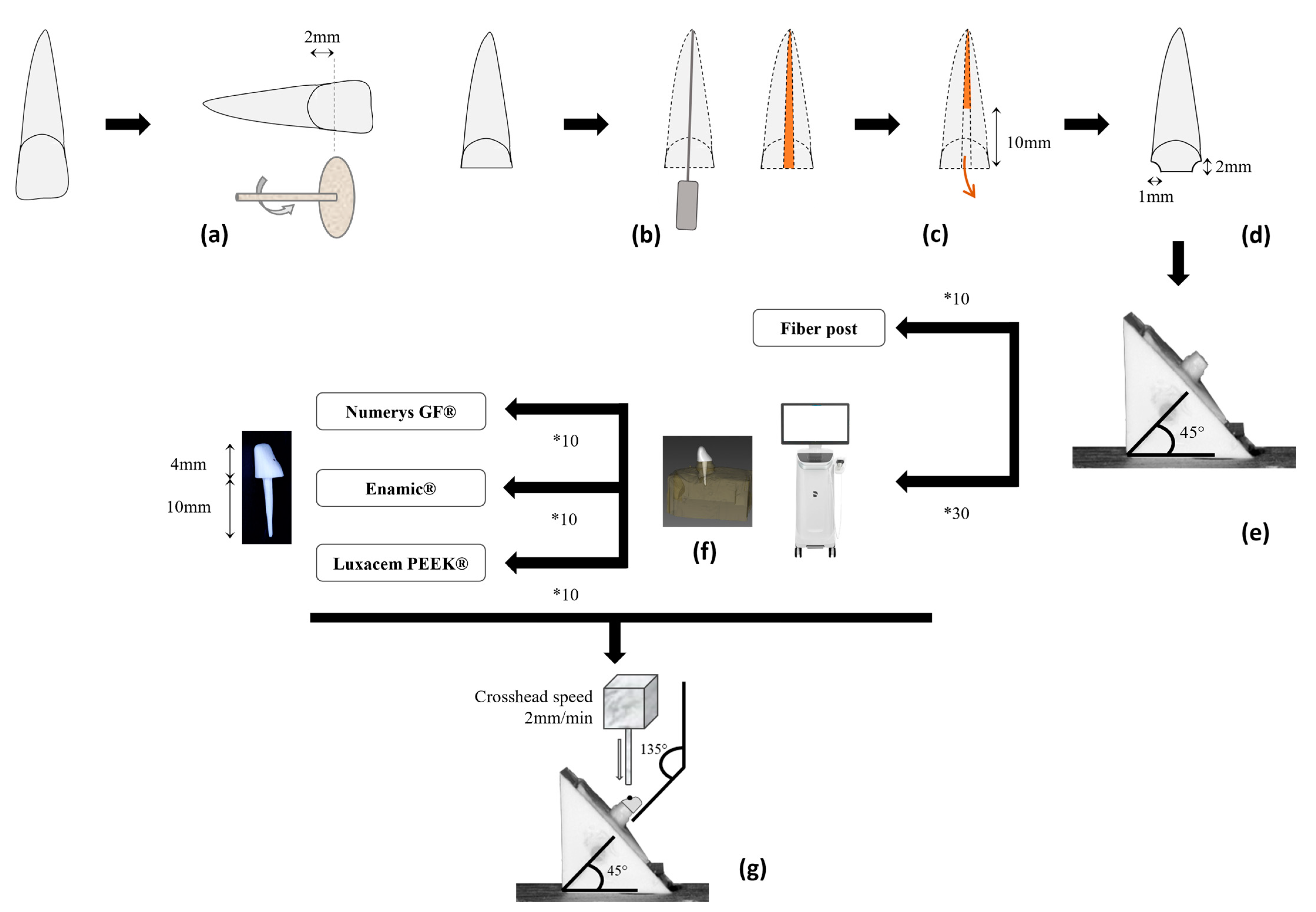
2.1. Teeth Collection
2.2. Teeth Preparation
2.3. Samples Design and Assembly
2.4. Fracture Tests
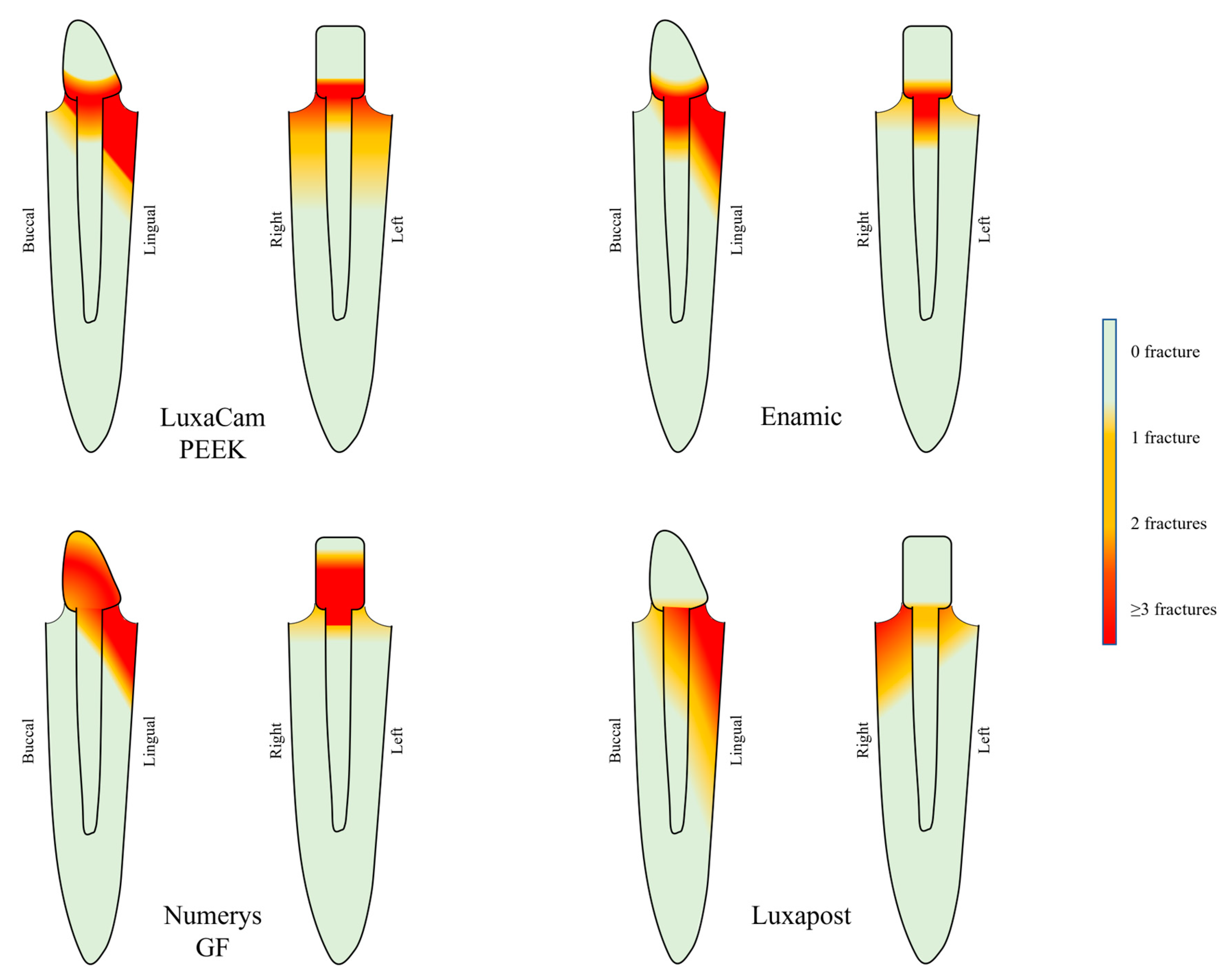
2.5. Fracture Mode
2.6. Statistical Analyses
3. Results
3.1. Fracture Tests
3.2. Fracture Mode
4. Discussion

| Year of Pub. | Authors | PEEK Manufacturer | Main Characteristics of the Protocol | Main Conclusions on PEEK |
|---|---|---|---|---|
| 2023 | Our study | DMG | Oblique compressive stress (2 mm/min) on PEEK posts and cores bonded to natural teeth (with tissue preparation and ferule effect). | Resistance of PEEK is lower than those of fiber posts. |
| 2023 | Ahmad et al. [59] | Juvora | Oblique compressive stress (1 mm/min) on PEEK posts and cores bonded to natural teeth (with tissue preparation and ferule effect) and pull-out tests. | Higher resistance for prefabricated PEEK posts with composite core than PEEK full posts and cores. Similar results for pull-out tests. |
| 2023 | Gontijo et al. [57] | Juvora | Oblique compressive stress (0.5 mm/min) on PEEK posts and cores bonded to bovine teeth (with tissue preparation and ferule effect). Moreover, simulation with finite element analysis. | Weakened roots restored with PEEK posts and cores are more resistant than those treated with prefabricated glass fiber posts. |
| 2023 | Kole et al. [60] | Juvora | Push-out stress on slices of PEEK posts and cores bonded to natural teeth (no ferule effect). | Zirconia post-cores appear to be a promising material. |
| 2023 | Lima et al. [55] | Juvora | Oblique compressive stress (1 mm/min) on PEEK posts and cores bonded to bovine teeth (with tissue preparation and ferule effect). Moreover, simulation with finite element analysis. | Fracture resistance of the tooth is not different with PEEK posts and cores. |
| 2023 | Saisho et al. [56] | Amann Girrbach | Oblique compressive stress (1 mm/min) on PEEK posts and cores bonded to natural teeth (no ferule effect) and pull-out tests. | No difference between the materials for compressive load. PEEK posts and cores showed lower bond strength to intracanal dentin. |
| 2023 | Zhao et al. [62] | Unspecified | Shear bond strength of PEEK-glass fibers composites cores on artificial molds. No posts; no use of natural teeth. | The mechanical properties of the composites were greatly improved. |
| 2022 | Attia et al. [72] | Bredent | Pull-out tests on PEEK posts and cores bonded to natural teeth (no ferule effect). | Precisions on bonding protocol, not on material choice for post and core (no control group). |
| 2022 | Gontijo et al. [53] | Juvora | Oblique compressive stress (0.5 mm/min) on PEEK posts and cores bonded to bovine teeth (with tissue preparation and ferule effect). | In presence of good bone condition, roots restored with PEEK posts and cores provide more reparable fractures and more resistant roots than those treated with prefabricated and anatomic glass fiber posts. |
| 2022 | Hallak et al. [73] | Informatic simulation | Simulation of masticatory forces with finite element analysis software on upper central incisors restored with PEEK posts. | Similar stress intensity and distribution between PEEK and glass fiber posts. |
| 2022 | Monteiro et al. [61] | Bredent | Pull-out tests on PEEK posts and cores bonded to bovine teeth (no ferule effect). | Good clinical options, but with a need for improvement. |
| 2022 | Pourkhalili et al. [74] | Bredent | Oblique compressive stress (0.5 mm/min) on PEEK posts and cores bonded to natural teeth (ferule effect not specified). | Mode of failure mostly repairable in the PEEK group. Resistance is lower than Ni-Cr alloys and greater than fiberglass posts. |
| 2022 | Yu et al. [52] | Informatic simulation | Simulation of mechanical load with finite element analysis software on upper central incisors restored with PEEK and Carbone-reinforced-PEEK posts. | Biomechanical behavior of the CFR-PEEK posts and cores was the closest to dentin. |
| 2021 | Haralur SB. [75] | Unspecified | Oblique compressive stress (0.5 mm/min) on PEEK posts and cores bonded to natural teeth (with tissue preparation). | Resistance of PEEK is higher than those of fiber posts. |
| 2021 | Ibrahim et al. [76] | Informatic simulation | Simulation of mechanical and thermal load with finite element analysis software on upper central incisors restored with PEEK posts. | Good resistance to masticatory forces for teeth restored with PEEK posts and favorable intra-radicular stress distribution. |
| 2021 | Özarslan et al. [77] | Juvora | Oblique compressive stress (1 mm/min) on PEEK posts and cores bonded to natural teeth (no ferule effect). | No superior features for PEEK in comparison with zirconia and glass-fiber. |
| 2020 | Benli et al. [78] | Juvora | Pull-out tests on PEEK posts bonded on natural teeth. No cores. | PEEK is a reliable option for dental posts. |
| 2020 | Çulhaoglu et al. [63] | Bredent | Shear bond strength of PEEK cores on resin. No posts; no use of natural teeth. | Acceptable resistance of resin bonding on PEEK surface. |
| 2020 | Li et al. [79] | Bredent | Pull-out tests of PEEK post and core restorations combined with polyvinylsiloxane attachments. No use of natural teeth. | The new post and core system showed favorable retention forces. |
| 2020 | Nahar et al. [80] | Informatic simulation | Simulation of mechanical load with finite element analysis software on upper central incisors restored with several PEEK posts (carbon fibers-reinforced (CFR), glass fibers-reinforced (GFR), PEKK). | CFR-PEEK is a good material for the fabrication of endodontic post. GFR-PEEK and PEKK materials can also be used. |
| 2020 | Sugano et al. [58] | Yasojima | Oblique compressive stress (1 mm/min) on PEEK posts and resin cores bonded to bovine teeth with flared canals (with ferule effect). | Higher stability is obtained with PEEK posts and glass fiber sleeves. |
| 2020 | Teixeira et al. [54] | DEGOS | Oblique compressive stress (0.5 mm/min) on PEEK posts and cores bonded to natural teeth (no ferule effect). | Resistance of PEEK is comparable to those of fiber posts. |
| 2020 | Tekin et al. [81] | Informatic simulation | Simulation of mechanical load and stress distribution with finite element analysis software on upper central incisors restored with PEEK posts and cores. | No difference in the stress value between PEEK and fiber posts. |
5. Conclusions
- Under oblique compressive tests, posts and cores designed from CAD/CAM blocks made of PEEK (80%) and TiO2 (20%) present a lower resistance in comparison to fiber posts.
- Prefabricated fiber posts and milled fiber posts present a similar behavior under oblique compressive tests.
- The Enamic material, used to design posts and cores, seems to be the most protective concerning root fracture.
Author Contributions
Funding
Institutional Review Board Statement
Data Availability Statement
Conflicts of Interest
References
- Naumann, M.; Schmitter, M.; Frankenberger, R.; Krastl, G. “Ferrule Comes First. Post Is Second!” Fake News and Alternative Facts? A Systematic Review. J. Endod. 2018, 44, 212–219. [Google Scholar] [CrossRef] [PubMed]
- Mamoun, J. Post and Core Build-Ups in Crown and Bridge Abutments: Bio-Mechanical Advantages and Disadvantages. J. Adv. Prosthodont. 2017, 9, 232–237. [Google Scholar] [CrossRef] [PubMed]
- Balevi, B. Metal or Fibre Posts and Root Fractures. Evid.-Based Dent. 2015, 16, 80–81. [Google Scholar] [CrossRef]
- Kumar, L.; Pal, B.; Pujari, P. An Assessment of Fracture Resistance of Three Composite Resin Core Build-up Materials on Three Prefabricated Non-Metallic Posts, Cemented in Endodontically Treated Teeth: An in Vitro Study. PeerJ 2015, 3, e795. [Google Scholar] [CrossRef] [PubMed]
- Gómez-Polo, M.; Llidó, B.; Rivero, A.; Del Río, J.; Celemín, A. A 10-Year Retrospective Study of the Survival Rate of Teeth Restored with Metal Prefabricated Posts versus Cast Metal Posts and Cores. J. Dent. 2010, 38, 916–920. [Google Scholar] [CrossRef] [PubMed]
- Başaran, G.; Göncü Başaran, E.; Ayna, E.; Değer, Y.; Ayna, B.; Tuncer, M.C. Microtensile Bond Strength of Root Canal Dentin Treated with Adhesive and Fiber-Reinforced Post Systems. Braz. Oral Res. 2019, 33, e027. [Google Scholar] [CrossRef]
- Martins, M.D.; Junqueira, R.B.; de Carvalho, R.F.; Lacerda, M.F.L.S.; Faé, D.S.; Lemos, C.A.A. Is a Fiber Post Better than a Metal Post for the Restoration of Endodontically Treated Teeth? A Systematic Review and Meta-Analysis. J. Dent. 2021, 112, 103750. [Google Scholar] [CrossRef]
- Dietschi, D.; Duc, O.; Krejci, I.; Sadan, A. Biomechanical Considerations for the Restoration of Endodontically Treated Teeth: A Systematic Review of the Literature—Part 1. Composition and Micro- and Macrostructure Alterations. Quintessence Int. 2007, 38, 11. [Google Scholar]
- Kurthukoti, A.J.; Paul, J.; Gandhi, K.; Rao, D.B.J. Fracture Resistance of Endodontically Treated Permanent Anterior Teeth Restored with Three Different Esthetic Post Systems: An in Vitro Study. J. Indian Soc. Pedod. Prev. Dent. 2015, 33, 296. [Google Scholar] [CrossRef]
- Ambica, K.; Mahendran, K.; Talwar, S.; Verma, M.; Padmini, G.; Periasamy, R. Comparative Evaluation of Fracture Resistance under Static and Fatigue Loading of Endodontically Treated Teeth Restored with Carbon Fiber Posts, Glass Fiber Posts, and an Experimental Dentin Post System: An In Vitro Study. J. Endod. 2013, 39, 96–100. [Google Scholar] [CrossRef]
- Verri, F.R.; Okumura, M.H.T.; Lemos, C.A.A.; de Faria Almeida, D.A.; de Souza Batista, V.E.; Cruz, R.S.; Oliveira, H.F.F.; Pellizzer, E.P. Three-Dimensional Finite Element Analysis of Glass Fiber and Cast Metal Posts with Different Alloys for Reconstruction of Teeth without Ferrule. J. Med. Eng. Technol. 2017, 41, 644–651. [Google Scholar] [CrossRef]
- King, P.A.; Setchell, D.J.; Rees, J.S. Clinical Evaluation of a Carbon Fibre Reinforced Carbon Endodontic Post. J. Oral Rehabil. 2003, 30, 785–789. [Google Scholar] [CrossRef] [PubMed]
- Zhou, L.; Wang, Q. Comparison of Fracture Resistance between Cast Posts and Fiber Posts: A Meta-Analysis of Literature. J. Endod. 2013, 39, 11–15. [Google Scholar] [CrossRef]
- Marchionatti, A.M.E.; Wandscher, V.F.; Rippe, M.P.; Kaizer, O.B.; Valandro, L.F.; Marchionatti, A.M.E.; Wandscher, V.F.; Rippe, M.P.; Kaizer, O.B.; Valandro, L.F. Clinical Performance and Failure Modes of Pulpless Teeth Restored with Posts: A Systematic Review. Braz. Oral Res. 2017, 31, e64. [Google Scholar] [CrossRef] [PubMed]
- Vaicelyte, A.; Janssen, C.; Le Borgne, M.; Grosgogeat, B. Cobalt–Chromium Dental Alloys: Metal Exposures, Toxicological Risks, CMR Classification, and EU Regulatory Framework. Crystals 2020, 10, 1151. [Google Scholar] [CrossRef]
- Alkhatri, R.; Saleh, A.R.M.; Kheder, W. Impact of Post and Core Materials on the Apical Extension of Root Fracture in Root Canal Treated Teeth. J. Mater. Res. Technol. 2021, 10, 730–737. [Google Scholar] [CrossRef]
- Falcão Spina, D.R.; Goulart da Costa, R.; Farias, I.C.; da Cunha, L.G.; Ritter, A.V.; Gonzaga, C.C.; Correr, G.M. CAD/CAM Post-and-Core Using Different Esthetic Materials: Fracture Resistance and Bond Strengths. Am. J. Dent. 2017, 30, 299–304. [Google Scholar]
- Skirbutis, G.; Dzingutė, A.; Masiliūnaitė, V.; Šulcaitė, G.; Žilinskas, J. PEEK Polymer’s Properties and Its Use in Prosthodontics. A Review. Stomatologija 2018, 20, 54–58. [Google Scholar]
- Papathanasiou, I.; Kamposiora, P.; Papavasiliou, G.; Ferrari, M. The Use of PEEK in Digital Prosthodontics: A Narrative Review. BMC Oral Health 2020, 20, 217. [Google Scholar] [CrossRef] [PubMed]
- Najeeb, S.; Zafar, M.S.; Khurshid, Z.; Siddiqui, F. Applications of Polyetheretherketone (PEEK) in Oral Implantology and Prosthodontics. J. Prosthodont. Res. 2016, 60, 12–19. [Google Scholar] [CrossRef]
- Delrieu, J.; Joniot, S.; Vergé, T.; Destruhaut, F.; Nasr, K.; Canceill, T. The Use of PEEK as an Occlusal Splint in a Patient with Histaminosis: A Case Report: Case Report of a PEEK Occlusal Splint. Spec. Care Dent. 2022, 42, 646–650. [Google Scholar] [CrossRef]
- Hahnel, S.; Wieser, A.; Lang, R.; Rosentritt, M. Biofilm Formation on the Surface of Modern Implant Abutment Materials. Clin. Oral Implant. Res. 2015, 26, 1297–1301. [Google Scholar] [CrossRef]
- Sobieraj, M.C.; Kurtz, S.M.; Rimnac, C.M. Notch Sensitivity of PEEK in Monotonic Tension. Biomaterials 2009, 30, 6485–6494. [Google Scholar] [CrossRef]
- Skirbutis, G. A Review of PEEK Polymer’s Properties and Its Use in Prosthodontics. Stomatologija 2017, 19, 19–23. [Google Scholar] [PubMed]
- Zoidis, P.; Papathanasiou, I.; Polyzois, G. The Use of a Modified Poly-Ether-Ether-Ketone (PEEK) as an Alternative Framework Material for Removable Dental Prostheses. A Clinical Report. J. Prosthodont. 2016, 25, 580–584. [Google Scholar] [CrossRef] [PubMed]
- Vaezi, M.; Yang, S. A Novel Bioactive PEEK/HA Composite with Controlled 3D Interconnected HA Network. Int. J. Bioprinting 2015, 1, 66–76. [Google Scholar] [CrossRef]
- Mobarak, E.H.; El-Badrawy, W.; Pashley, D.H.; Jamjoom, H. Effect of Pretest Storage Conditions of Extracted Teeth on Their Dentin Bond Strengths. J. Prosthet. Dent. 2010, 104, 92–97. [Google Scholar] [CrossRef]
- Al-Omiri, M.K.; Mahmoud, A.A.; Rayyan, M.R.; Abu-Hammad, O. Fracture Resistance of Teeth Restored with Post-Retained Restorations: An Overview. J. Endod. 2010, 36, 1439–1449. [Google Scholar] [CrossRef]
- Calabro, D.E.; Kojima, A.N.; Gallego Arias Pecorari, V.; Helena Coury Saraceni, C.; Blatz, M.B.; Özcan, M.; Mikail Melo Mesquita, A. A 10-Year Follow-Up of Different Intra-Radicular Retainers in Teeth Restored with Zirconia Crowns. Clin. Cosmet. Investig. Dent. 2019, 11, 409–417. [Google Scholar] [CrossRef]
- Makade, C.S.; Meshram, G.K.; Warhadpande, M.; Patil, P.G. A Comparative Evaluation of Fracture Resistance of Endodontically Treated Teeth Restored with Different Post Core Systems—An in-Vitro Study. J. Adv. Prosthodont. 2011, 3, 90–95. [Google Scholar] [CrossRef]
- Doshi, P.; Kanaparthy, A.; Kanaparthy, R.; Parikh, D.S. A Comparative Analysis of Fracture Resistance and Mode of Failure of Endodontically Treated Teeth Restored Using Different Fiber Posts: An In Vitro Study. J. Contemp. Dent. Pract. 2019, 20, 1195–1199. [Google Scholar] [CrossRef]
- Mehrvarzfar, P.; Rezvani, Y.; Jalalian, E. Comparison of Resilon and Gutta-Percha Filling Materials on Root Canal Fracture Resistance Following Restoring with Quartz Fiber Posts. J. Dent. 2012, 9, 156. [Google Scholar]
- Chauhan, N.; Saraswat, N.; Parashar, A.; Sandu, K.; Jhajharia, K.; Rabadiya, N. Comparison of the Effect for Fracture Resistance of Different Coronally Extended Post Length with Two Different Post Materials. J. Int. Soc. Prev. Community Dent. 2019, 9, 144. [Google Scholar] [CrossRef]
- Khaledi, A.A.R.; Sheykhian, S.; Khodaei, A. Evaluation of Retention of Two Different Cast Post-Core Systems and Fracture Resistance of the Restored Teeth. J. Dent. 2015, 16, 121–128. [Google Scholar]
- Mergulhão, V.; de Mendonça, L.; de Albuquerque, M.; Braz, R. Fracture Resistance of Endodontically Treated Maxillary Premolars Restored With Different Methods. Oper. Dent. 2019, 44, E1–E11. [Google Scholar] [CrossRef]
- Steiner, M.; Mitsias, M.E.; Ludwig, K.; Kern, M. In Vitro Evaluation of a Mechanical Testing Chewing Simulator. Dent. Mater. 2009, 25, 494–499. [Google Scholar] [CrossRef]
- Kumagae, N.; Komada, W.; Fukui, Y.; Okada, D.; Takahashi, H.; Yoshida, K.; Miura, H. Influence of the Flexural Modulus of Prefabricated and Experimental Posts on the Fracture Strength and Failure Mode of Composite Resin Cores. Dent. Mater. J. 2012, 31, 113–119. [Google Scholar] [CrossRef] [PubMed]
- Vafaee, F.; Khoshhal, M.; Rezaei, A.; Sooltani, F.; Jalalzadeh, M.; Yalpaniyan, A.; Firooz, F.; Izady, A.R.; Yarmohamadi, E. Multiple In Vitro Analyses of Fracture Resistance in Maxillary Central Incisors Restored with Fiber Posts. Iran. Endod. J. 2010, 5, 125–130. [Google Scholar]
- Kathuria, A.; Kavitha, M.; Khetarpal, S. Ex Vivo Fracture Resistance of Endodontically Treated Maxillary Central Incisors Restored with Fiber-Reinforced Composite Posts and Experimental Dentin Posts. J. Conserv. Dent. 2011, 14, 401–405. [Google Scholar] [CrossRef] [PubMed]
- Sammany, F.; Sultan, M.; Ide, N.A.-D. Stress and Strain Distribution in a Single Root Canal Post Made of Polyether Ether Ketone Using Three-Dimensional Finite Elements Method. Acad. J. Oral Dent. Med. 2019, 8, 1–7. [Google Scholar]
- Sarkis-Onofre, R.; de Castilho Jacinto, R.; Boscato, N.; Cenci, M.S.; Pereira-Cenci, T. Cast Metal vs. Glass Fibre Posts: A Randomized Controlled Trial with up to 3 Years of Follow Up. J. Dent. 2014, 42, 582–587. [Google Scholar] [CrossRef] [PubMed]
- Juloski, J.; Fadda, G.M.; Monticelli, F.; Fajó-Pascual, M.; Goracci, C.; Ferrari, M. Four-Year Survival of Endodontically Treated Premolars Restored with Fiber Posts. J. Dent. Res. 2014, 93, 52S–58S. [Google Scholar] [CrossRef]
- Fokkinga, W.A.; Kreulen, C.M.; Bronkhorst, E.M.; Creugers, N.H.J. Up to 17-Year Controlled Clinical Study on Post-and-Cores and Covering Crowns. J. Dent. 2007, 35, 778–786. [Google Scholar] [CrossRef]
- Wang, X.; Shu, X.; Zhang, Y.; Yang, B.; Jian, Y.; Zhao, K. Evaluation of Fiber Posts vs Metal Posts for Restoring Severely Damaged Endodontically Treated Teeth: A Systematic Review and Meta-Analysis. Quintessence Int. 2018, 50, 8–20. [Google Scholar] [CrossRef]
- Garcia, P.P.; Wambier, L.M.; de Geus, J.L.; da Cunha, L.F.; Correr, G.M.; Gonzaga, C.C. Do Anterior and Posterior Teeth Treated with Post-and-Core Restorations Have Similar Failure Rates? A Systematic Review and Meta-Analysis. J. Prosthet. Dent. 2019, 121, 887–894.e4. [Google Scholar] [CrossRef]
- De Souza Batista, V.E.; Bitencourt, S.B.; Bastos, N.A.; Pellizzer, E.P.; Goiato, M.C.; dos Santos, D.M. Influence of the Ferrule Effect on the Failure of Fiber-Reinforced Composite Post-and-Core Restorations: A Systematic Review and Meta-Analysis. J. Prosthet. Dent. 2020, 123, 239–245. [Google Scholar] [CrossRef]
- Naumann, M.; Koelpin, M.; Beuer, F.; Meyer-Lueckel, H. 10-Year Survival Evaluation for Glass-Fiber–Supported Postendodontic Restoration: A Prospective Observational Clinical Study. J. Endod. 2012, 38, 432–435. [Google Scholar] [CrossRef] [PubMed]
- Martino, N.; Truong, C.; Clark, A.E.; O’Neill, E.; Hsu, S.-M.; Neal, D.; Esquivel-Upshaw, J.F. Retrospective Analysis of Survival Rates of Post-and-Cores in a Dental School Setting. J. Prosthet. Dent. 2020, 123, 434–441. [Google Scholar] [CrossRef]
- Eid, R.; Juloski, J.; Ounsi, H.; Silwaidi, M.; Ferrari, M.; Salameh, Z. Fracture Resistance and Failure Pattern of Endodontically Treated Teeth Restored with Computer-Aided Design/ Computer-Aided Manufacturing Post and Cores: A Pilot Study. J. Contemp. Dent. Pract. 2019, 20, 56–63. [Google Scholar] [CrossRef]
- Borzangy, S.S.; Saker, S.; Al-Zordk, W.A.E.-G. Effect of Restauration Technique on Resistance to Fracture of Endodontically Treated Anterior Teeth with Flared Root Canals. J. Biomed. Res. 2019, 33, 131. [Google Scholar] [CrossRef] [PubMed]
- Aggarwal, R.; Gupta, S.; Tandan, A.; Gupta, N.K.; Dwivedi, R.; Aggarwal, R. Comparative Evaluation of Fracture Resistance of Various Post Systems Using Different Luting Agents under Tangential Loading. J. Oral Biol. Craniofacial Res. 2013, 3, 63–67. [Google Scholar] [CrossRef] [PubMed][Green Version]
- Yu, H.; Feng, Z.; Wang, L.; Mihcin, S.; Kang, J.; Bai, S.; Zhao, Y. Finite Element Study of PEEK Materials Applied in Post-Retained Restorations. Polymers 2022, 14, 3422. [Google Scholar] [CrossRef] [PubMed]
- Gontijo, I.G.; Ferretti, M.A.; Caldas, R.A.; Pecorari, V.G.A.; França, F.M.G.; Oliveira, L.K.N.; Aguiar, F.H.B. Biomechanical Behavior of Weakened Roots Restored with Custom-Made Post-and-Cores of Glass Fiber and Polyetheretherketone. J. Prosthodont. 2022, 1–8. [Google Scholar] [CrossRef]
- Teixeira, K.; Duque, T.; Maia, H.; Gonçalves, T. Fracture Resistance and Failure Mode of Custom-Made Post-and-Cores of Polyetheretherketone and Nano-Ceramic Composite. Oper. Dent. 2020, 45, 506–515. [Google Scholar] [CrossRef] [PubMed]
- Lima, M.O.; Ferretti, M.A.; Caldas, R.A.; Barão, V.A.R.; França, F.M.G.; Lima, D.A.N.L.; Martins, L.R.; Aguiar, F.H.B. Application of Polyetheretherketone (PEEK) Posts: Evaluation of Fracture Resistance and Stress Distribution in the Root: In Vitro and Finite Element Analyses. Braz. Oral Res. 2023, 37, e047. [Google Scholar] [CrossRef]
- Saisho, H.; Marcolina, G.; Perucelli, F.; Goulart da Costa, R.; Machado de Souza, E.; Rached, R.N. Fracture Strength, Pull-out Bond Strength, and Volume of Luting Agent of Tooth-Colored CAD-CAM Post-and-Cores. J. Prosthet. Dent. 2023, 129, 599–606. [Google Scholar] [CrossRef]
- Gontijo, I.G.; Ferretti, M.A.; Monteiro, L.C.; Caldas, R.A.; Pecorari, V.G.A.; França, F.M.G.; Aguiar, F.H.B. Influence of Alveolar Bone Height on the Biomechanical Behavior of Roots Restored with Custom-Made Posts-and-Cores. J. Esthet. Restor. Dent. 2023, 35, 677–686. [Google Scholar] [CrossRef] [PubMed]
- Sugano, K.; Komada, W.; Okada, D.; Miura, H. Evaluation of Composite Resin Core with Prefabricated Polyetheretherketone Post on Fracture Resistance in the Case of Flared Root Canals. Dent. Mater. J. 2020, 39, 924–932. [Google Scholar] [CrossRef]
- Ahmad, S.M.; Dawood, S.N.; Dalloo, G.A.M.; Al-Barazanchi, T.R.H. Evaluation of Mechanical Properties of Different Polyetheretherketone Endodontic Post Systems: An in Vitro Study. BMC Oral Health 2023, 23, 537. [Google Scholar] [CrossRef]
- Kole, S.; Ergun, G. Bond Strength of Various Post-Core Restorations with Different Lengths and Diameters Following Cycle Loading. J. Mech. Behav. Biomed. Mater. 2023, 142, 105804. [Google Scholar] [CrossRef]
- Monteiro, L.C.; Pecorari, V.G.A.; Gontijo, I.G.; Marchi, G.M.; Lima, D.A.N.L.; Aguiar, F.H.B. PEEK and Fiberglass Intra-Radicular Posts: Influence of Resin Cement and Mechanical Cycling on Push-out Bond Strength. Clin. Oral Investig. 2022, 26, 6907–6916. [Google Scholar] [CrossRef]
- Zhao, T.; Jiang, Z.; Ge, Y.; Yin, H.; Yang, Q.; Li, R.; Chen, Z.; Zhang, H.; Liu, X. Mechanical Properties, Biosafety, and Shearing Bonding Strength of Glass Fiber-Reinforced PEEK Composites Used as Post-Core Materials. J. Mech. Behav. Biomed. Mater. 2023, 145, 106047. [Google Scholar] [CrossRef] [PubMed]
- Çulhaoğlu, A.K.; Özkır, S.E.; Şahin, V.; Yılmaz, B.; Kılıçarslan, M.A. Effect of Various Treatment Modalities on Surface Characteristics and Shear Bond Strengths of Polyetheretherketone-Based Core Materials. J. Prosthodont. 2020, 29, 136–141. [Google Scholar] [CrossRef]
- Chen, Z.; Li, Y.; Deng, X.; Wang, X. A Novel Computer-Aided Method to Fabricate a Custom One-Piece Glass Fiber Dowel-and-Core Based on Digitized Impression and Crown Preparation Data: CAD/CAM of Custom One-Piece Glass Fiber Dowel-and-Core. J. Prosthodont. 2014, 23, 276–283. [Google Scholar] [CrossRef]
- Eid, R.Y.; Koken, S.; Baba, N.Z.; Ounsi, H.; Ferrari, M.; Salameh, Z. Effect of Fabrication Technique and Thermal Cycling on the Bond Strength of CAD/CAM Milled Custom Fit Anatomical Post and Cores: An In Vitro Study. J. Prosthodont. 2019, 28, 898–905. [Google Scholar] [CrossRef] [PubMed]
- Salameh, Z.; Ferrari, M.; Skienhe, H.; Ounsi, H.; Eid, R.; Azzam, K. Influence of Adaptation and Adhesion on the Retention of Computer-Aided Design/Computer-Aided Manufacturing Glass Fiber Posts to Root Canal. J. Contemp. Dent. Pract. 2019, 20, 1003–1008. [Google Scholar] [CrossRef]
- Santos Pantaleón, D.; Valenzuela, F.M.; Morrow, B.R.; Pameijer, C.H.; García-Godoy, F. Effect of Ferrule Location with Varying Heights on Fracture Resistance and Failure Mode of Restored Endodontically Treated Maxillary Incisors. J. Prosthodont. 2019, 28, 677–683. [Google Scholar] [CrossRef]
- Fontana, P.; Bohrer, T.; Wandscher, V.; Valandro, L.; Limberger, I.; Kaizer, O. Effect of Ferrule Thickness on Fracture Resistance of Teeth Restored With a Glass Fiber Post or Cast Post. Oper. Dent. 2019, 44, E299–E308. [Google Scholar] [CrossRef]
- Magne, P.; Lazari, P.C.; Carvalho, M.A.; Johnson, T.; Del Bel Cury, A.A. Ferrule-Effect Dominates Over Use of a Fiber Post When Restoring Endodontically Treated Incisors: An In Vitro Study. Oper. Dent. 2017, 42, 396–406. [Google Scholar] [CrossRef] [PubMed]
- Stavridakis, M.; Brokos, Y.; Krejci, I. Is the Glass Half Empty or Half Full? A Novel “Philosophical” Approach to the “Mystery” of the so-Called Ferrule Effect. Med. Hypotheses 2018, 115, 35–41. [Google Scholar] [CrossRef]
- Mishra, L.; Khan, A.S.; Velo, M.M.d.A.C.; Panda, S.; Zavattini, A.; Rizzante, F.A.P.; Arbildo Vega, H.I.; Sauro, S.; Lukomska-Szymanska, M. Effects of Surface Treatments of Glass Fiber-Reinforced Post on Bond Strength to Root Dentine: A Systematic Review. Materials 2020, 13, 1967. [Google Scholar] [CrossRef]
- Attia, M.A.; Shokry, T.E.; Abdel-Aziz, M. Effect of Different Surface Treatments on the Bond Strength of Milled Polyetheretherketone Posts. J. Prosthet. Dent. 2022, 127, 866–874. [Google Scholar] [CrossRef] [PubMed]
- Hallak, A.G.; Caldas, R.A.; Silva, I.D.; Miranda, M.E.; Brandt, W.C.; Vitti, R.P. Stress Distribution in Restorations with Glass Fiber and Polyetheretherketone Intraradicular Posts: An in Silico Analysis. Dent. Mater. J. 2022, 41, 376–381. [Google Scholar] [CrossRef]
- Pourkhalili, H.; Maleki, D. Fracture Resistance of Polyetheretherketone, Ni-Cr, and Fiberglass Postcore Systems: An in Vitro Study. Dent. Res. J. 2022, 19, 20. [Google Scholar] [CrossRef]
- Haralur, S.B. Fracture Resistance of Endodontically Treated Teeth Restored with Various Esthetic Posts. Technol. Health Care 2021, 29, 243–252. [Google Scholar] [CrossRef] [PubMed]
- Ibrahim, R.O.; Al-Zahawi, A.R.; Sabri, L.A. Mechanical and Thermal Stress Evaluation of PEEK Prefabricated Post with Different Head Design in Endodontically Treated Tooth: 3D-Finite Element Analysis. Dent. Mater. J. 2021, 40, 508–518. [Google Scholar] [CrossRef] [PubMed]
- Özarslan, M.; Büyükkaplan, U.Ş.; Özarslan, M.M. Comparison of the Fracture Strength of Endodontically Treated Teeth Restored with Polyether Ether Ketone, Zirconia and Glass-Fibre Post-Core Systems. Int. J. Clin. Pract. 2021, 75, e14440. [Google Scholar] [CrossRef] [PubMed]
- Benli, M.; Eker Gümüş, B.; Kahraman, Y.; Huck, O.; Özcan, M. Surface Characterization and Bonding Properties of Milled Polyetheretherketone Dental Posts. Odontology 2020, 108, 596–606. [Google Scholar] [CrossRef] [PubMed]
- Li, P.; Hasselbeck, D.; Unkovskiy, A.; Sharghi, F.; Spintzyk, S. Retentive Characteristics of a Polyetheretherketone Post-Core Restoration with Polyvinylsiloxane Attachments. Polymers 2020, 12, 2005. [Google Scholar] [CrossRef]
- Nahar, R.; Mishra, S.K.; Chowdhary, R. Evaluation of Stress Distribution in an Endodontically Treated Tooth Restored with Four Different Post Systems and Two Different Crowns—A Finite Element Analysis. J. Oral Biol. Craniofacial Res. 2020, 10, 719–726. [Google Scholar] [CrossRef]
- Tekin, S.; Adiguzel, O.; Cangul, S.; Atas, O.; Erpacal, B. Evaluation of the Use of PEEK Material in Post-Core and Crown Restorations Using Finite Element Analysis. Am. J. Dent. 2020, 33, 251–257. [Google Scholar] [PubMed]


| Biomaterials | Manufacturer | Composition | Elasticity Modulus (GPa) |
|---|---|---|---|
| LuxaCam PEEK | DMG | PEEK 80%; TiO2 20% | 3.8 |
| Enamic | Vita | Ceramic 84%; Polymers 14% | 30 |
| Numerys GF | Itena | Glass fibers 75–80%; Epoxy resin 20–25% | 25 |
| LuxaPost | DMG | Glass fibers; bis-GMA resin | 25 |
| LuxaCam PEEK | LuxaPost | Numerys GF | Enamic | |
|---|---|---|---|---|
| Maximal strength (MPa) | 9.48 ± 6.65 a | 15.88 ± 4.37 b | 15.35 ± 6.65 b | 6.05 ± 4.14 a |
| Tenacity (Pa/m²) | 6.43 ± 4.22 a,c | 12.58 ± 5.46 b | 9.69 ± 5.92 b,c | 3.18 ± 2.92 a |
| Fracture Mode | LuxaCam PEEK a,b | Luxapost b,c | Numerys GF a,c | Enamic d |
|---|---|---|---|---|
| Dental | 80% | 60% | 70% | 0 |
| Material | 10% | 20% | 0 | 60% |
| Mixed | 10% | 20% | 30% | 40% |
Disclaimer/Publisher’s Note: The statements, opinions and data contained in all publications are solely those of the individual author(s) and contributor(s) and not of MDPI and/or the editor(s). MDPI and/or the editor(s) disclaim responsibility for any injury to people or property resulting from any ideas, methods, instructions or products referred to in the content. |
© 2023 by the authors. Licensee MDPI, Basel, Switzerland. This article is an open access article distributed under the terms and conditions of the Creative Commons Attribution (CC BY) license (https://creativecommons.org/licenses/by/4.0/).
Share and Cite
Rakotoaridina, K.; Delrieu, J.; Pages, P.; Vergé, T.; Nasr, K.; Canceill, T. Evaluation of Poly(etheretherketone) Post’s Mechanical Strength in Comparison with Three Metal-Free Biomaterials: An In Vitro Study. Polymers 2023, 15, 3583. https://doi.org/10.3390/polym15173583
Rakotoaridina K, Delrieu J, Pages P, Vergé T, Nasr K, Canceill T. Evaluation of Poly(etheretherketone) Post’s Mechanical Strength in Comparison with Three Metal-Free Biomaterials: An In Vitro Study. Polymers. 2023; 15(17):3583. https://doi.org/10.3390/polym15173583
Chicago/Turabian StyleRakotoaridina, Kévin, Julien Delrieu, Paul Pages, Thierry Vergé, Karim Nasr, and Thibault Canceill. 2023. "Evaluation of Poly(etheretherketone) Post’s Mechanical Strength in Comparison with Three Metal-Free Biomaterials: An In Vitro Study" Polymers 15, no. 17: 3583. https://doi.org/10.3390/polym15173583
APA StyleRakotoaridina, K., Delrieu, J., Pages, P., Vergé, T., Nasr, K., & Canceill, T. (2023). Evaluation of Poly(etheretherketone) Post’s Mechanical Strength in Comparison with Three Metal-Free Biomaterials: An In Vitro Study. Polymers, 15(17), 3583. https://doi.org/10.3390/polym15173583