
Composite Fish Collagen-Hyaluronate Based Lyophilized Scaffolds Modified with Sodium Alginate for Potential Treatment of Chronic Wounds


Abstract
:1. Introduction
2. Materials and Methods
2.1. Materials
2.2. Methods
2.3. Preparation of Lyophilized Scaffolds
2.4. Physico-Chemical Characterization
2.4.1. Scanning Electron Microscopy (SEM)
2.4.2. X-ray Diffraction (XRD)
2.4.3. Attenuated Total Reflectance Fourier Transform Infrared Spectroscopy (ATR-FTIR)
2.4.4. Texture Analysis (TA)
Mechanical Strength (Hardness)
Adhesion
2.5. Exudate Handling Properties
2.5.1. Fluid Intake Evaluation
Swelling Studies
Porosity
2.5.2. Water Absorption (WA) and Equilibrium Water Content (EWC)
2.5.3. Fluid Loss Assessment
Water Vapor Transmission Rate (WVTR)
Evaporative Water Loss (EWL)
2.6. Protein Leaching
2.6.1. Standard Solution Preparation for Linearity and BSA Content Using HPLC
2.6.2. In Vitro BSA Dissolution Studies
2.6.3. Evaluation of BSA Release Mechanisms
2.7. MTT (Cell Viability) Assay
2.8. In Vitro Blood Clotting Assay
2.9. Statistical Analysis
3. Results and Discussion
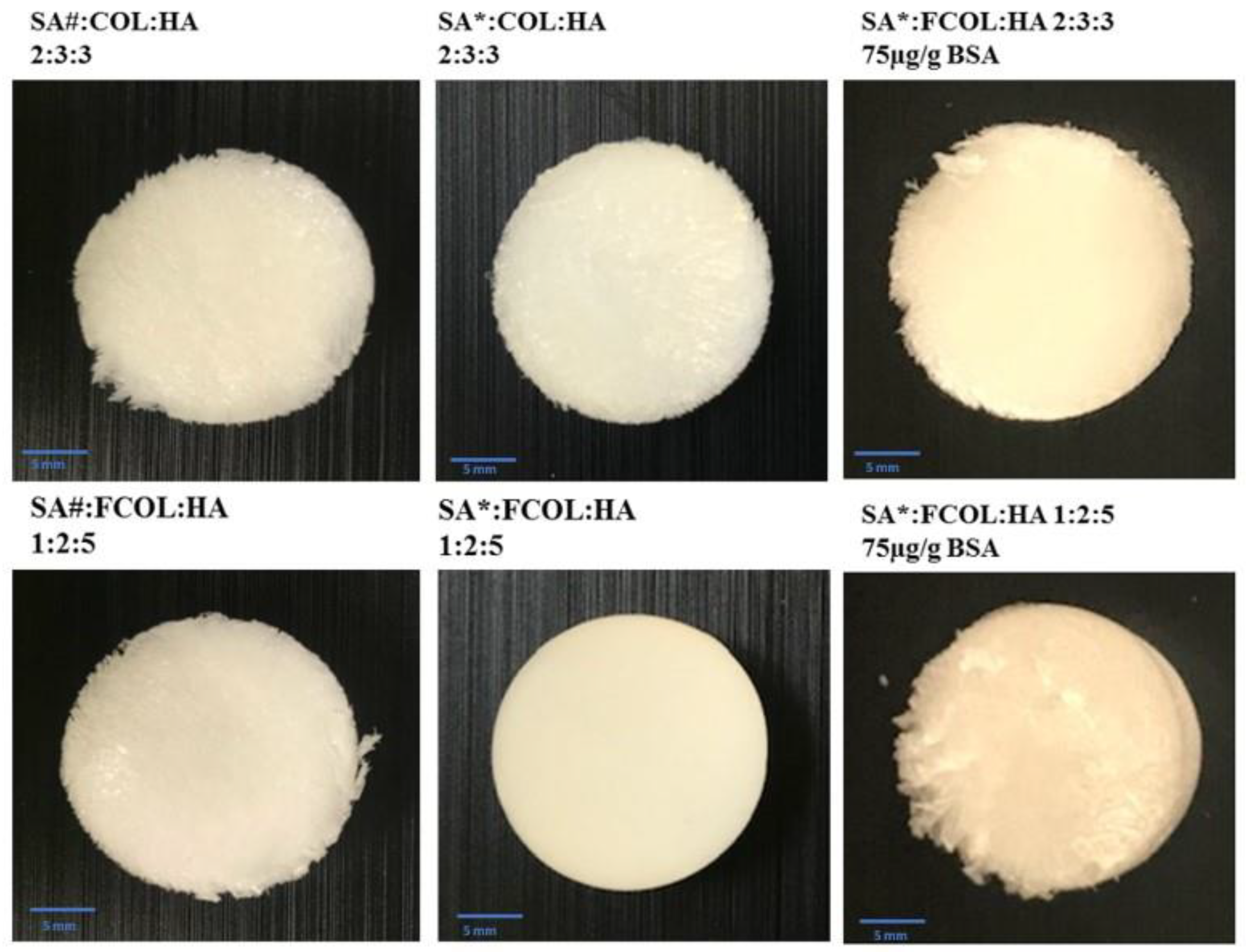
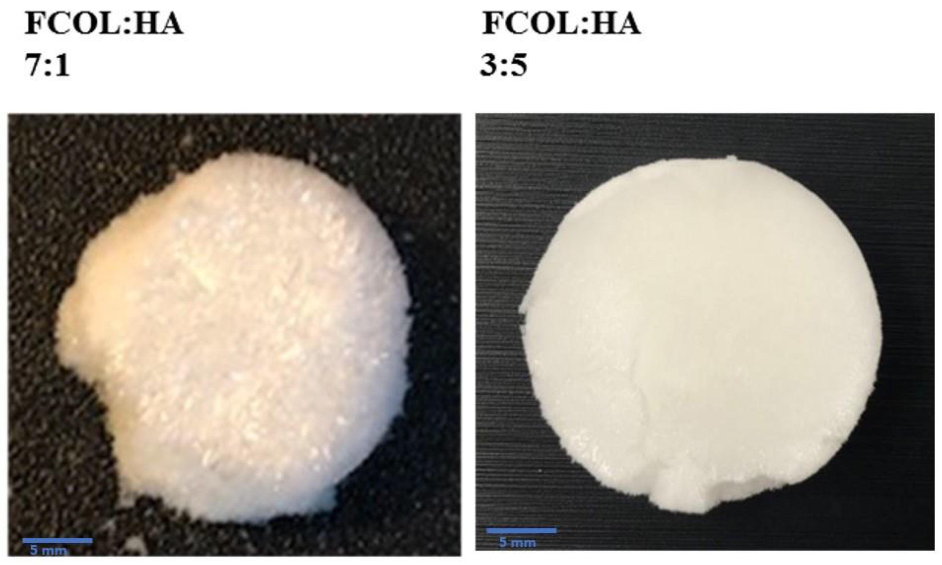
3.1. Visual Evaluations of Gels and Freeze-Dried Scaffolds
3.2. Physico-Chemical Characterization
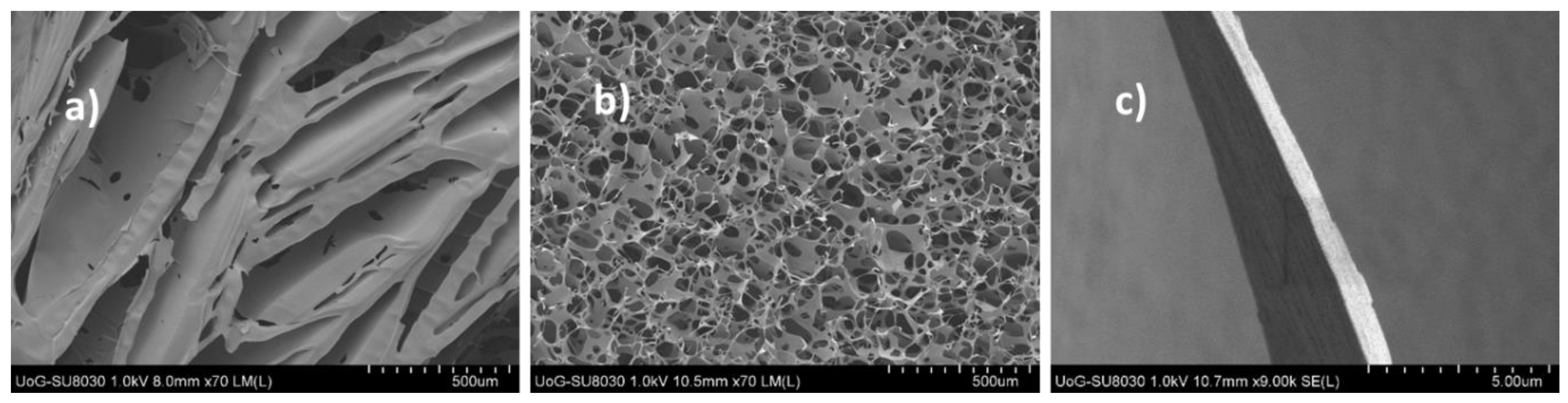
3.2.1. Scanning Electron Microscopy (SEM)
3.2.2. X-ray Diffraction (XRD)
3.2.3. Attenuated Total Reflectance Fourier Transform Infrared Spectroscopy (ATR-FTIR)
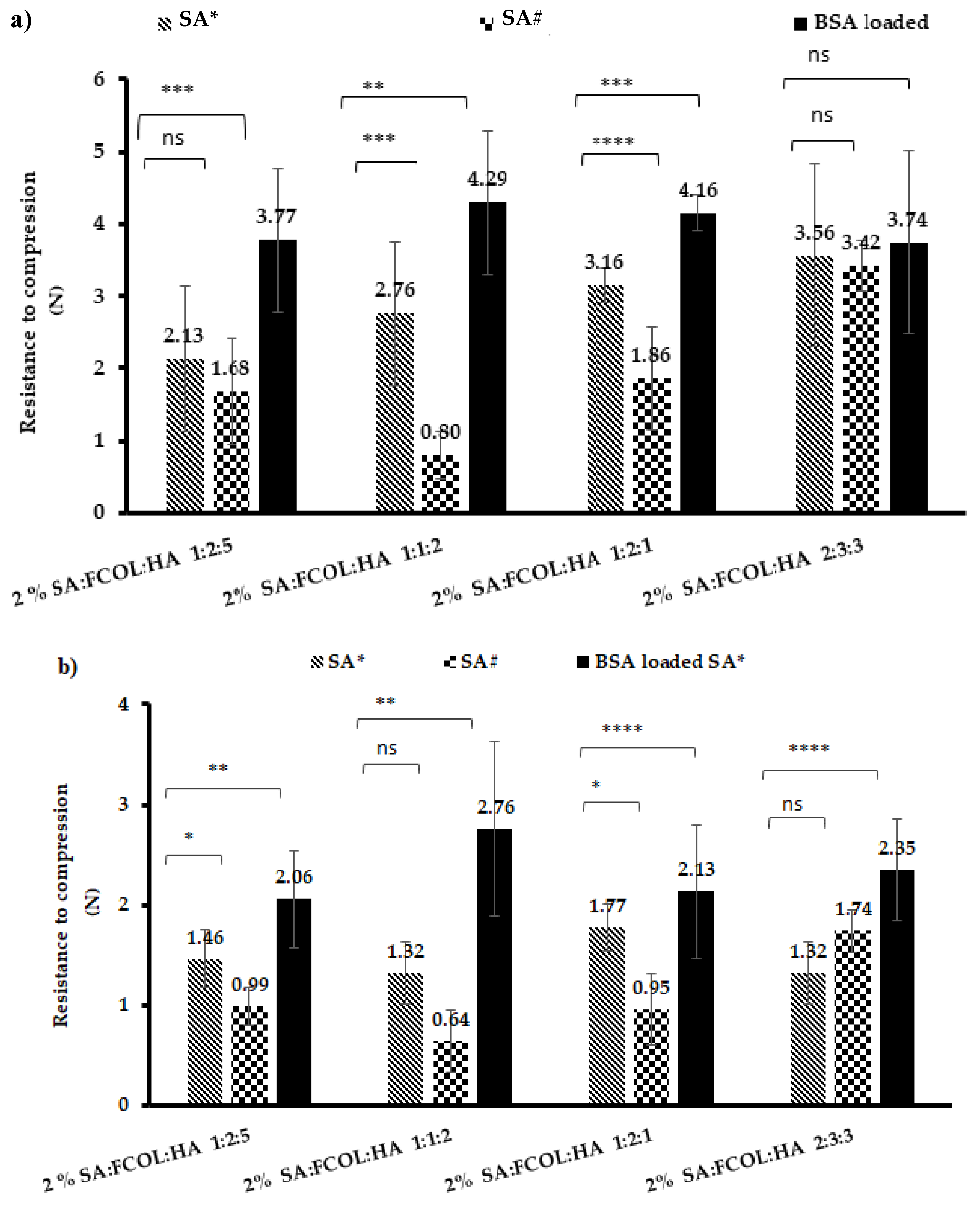
3.2.4. Texture Analysis
Hardness
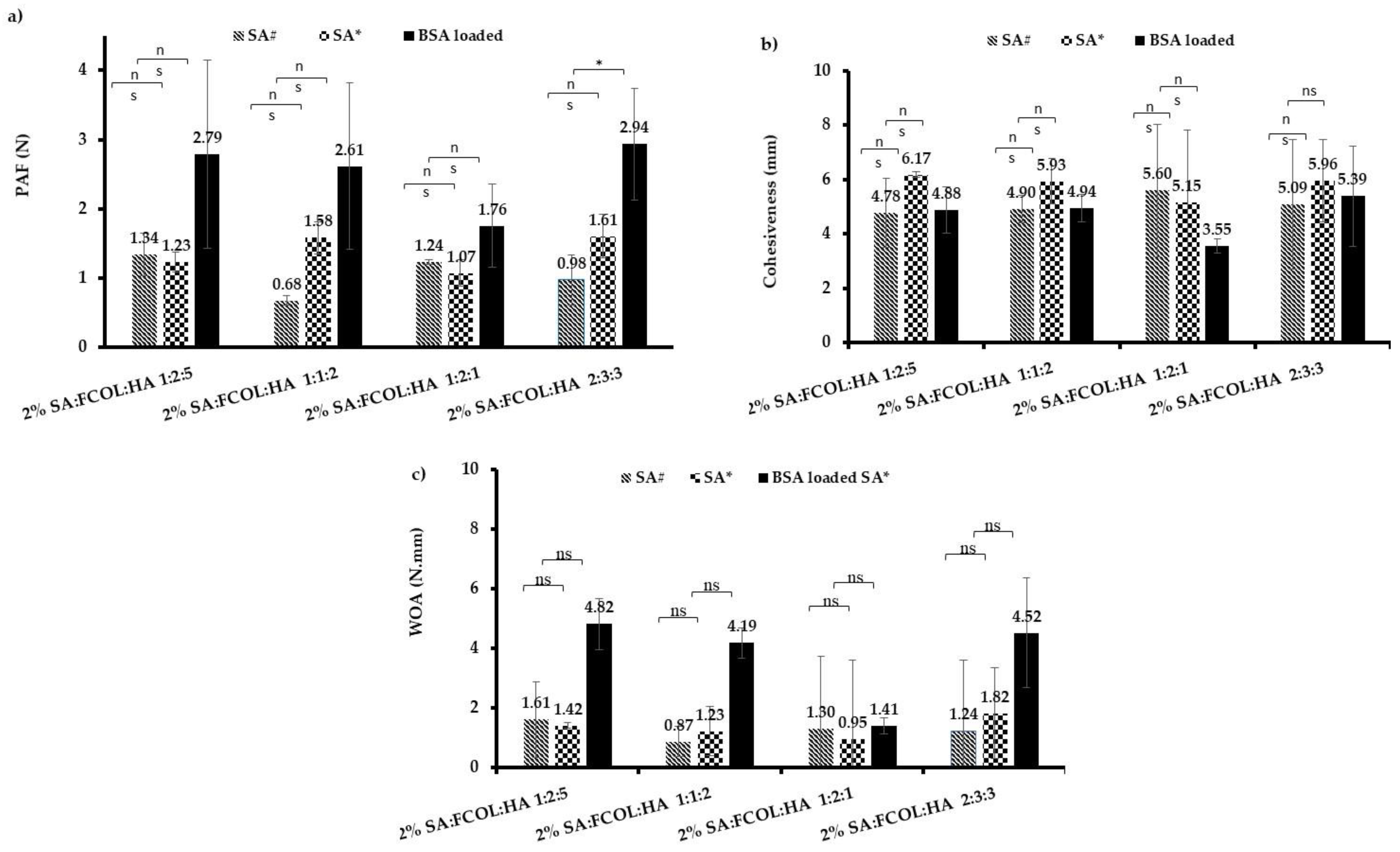
Adhesion
3.2.5. Exudate Handling Properties
Fluid Intake Evaluation
- Porosity
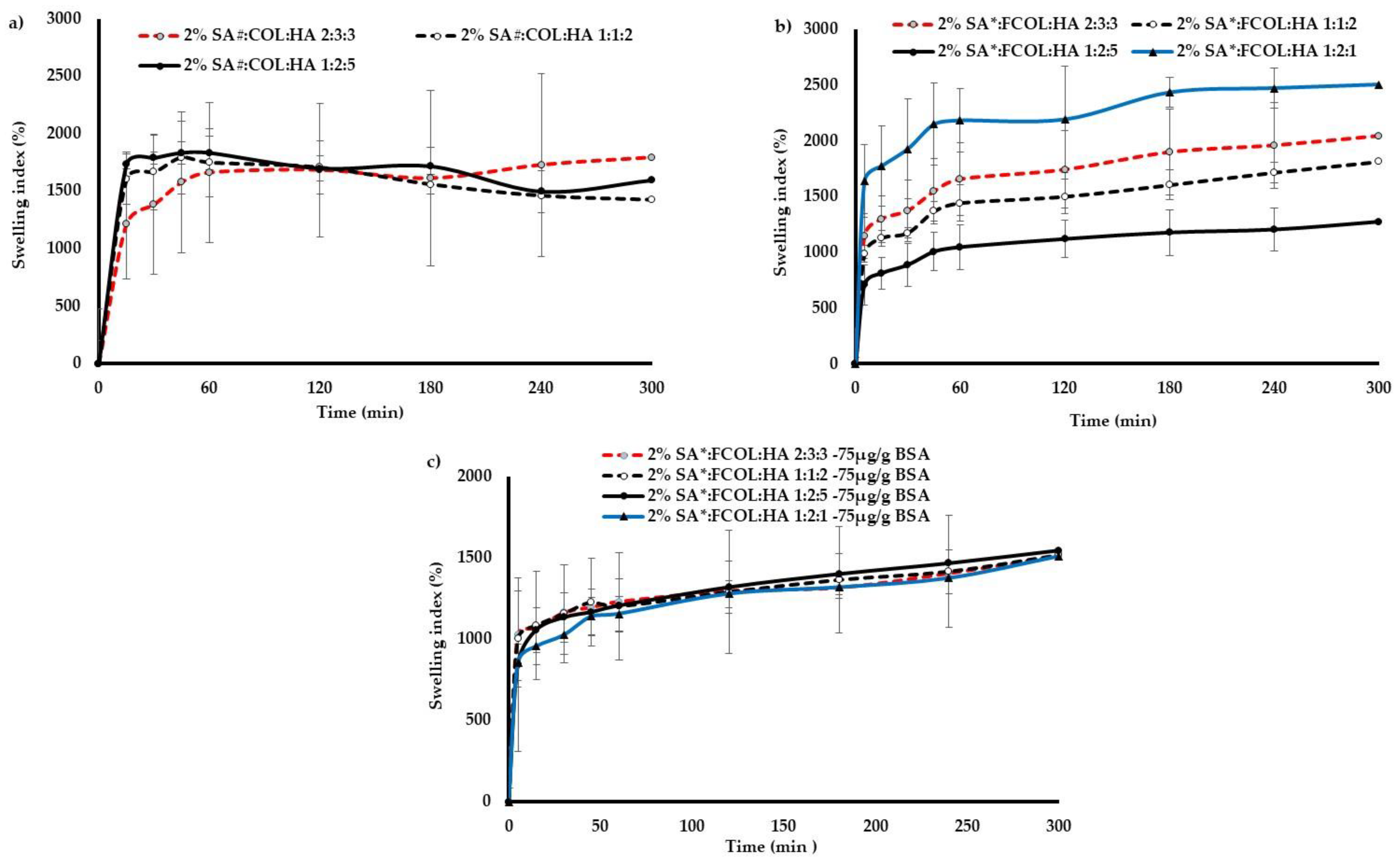
- Swelling
- Water Absorption (WA) and Equilibrium Water Content (EWC)
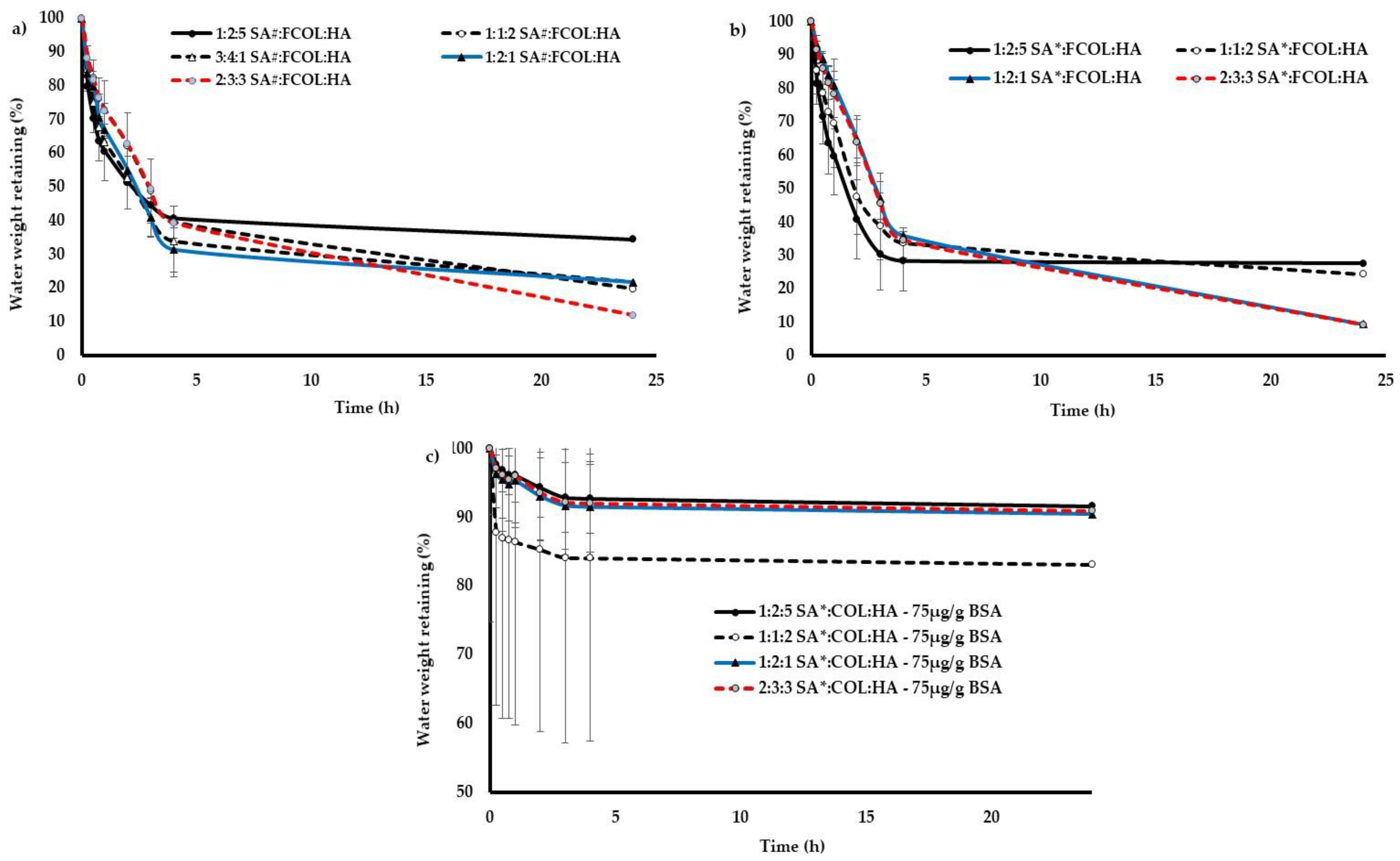
Fluid Loss Assessment
- Water Vapor Transmission Rate (WVTR)
- Evaporative Water Loss (EWL)
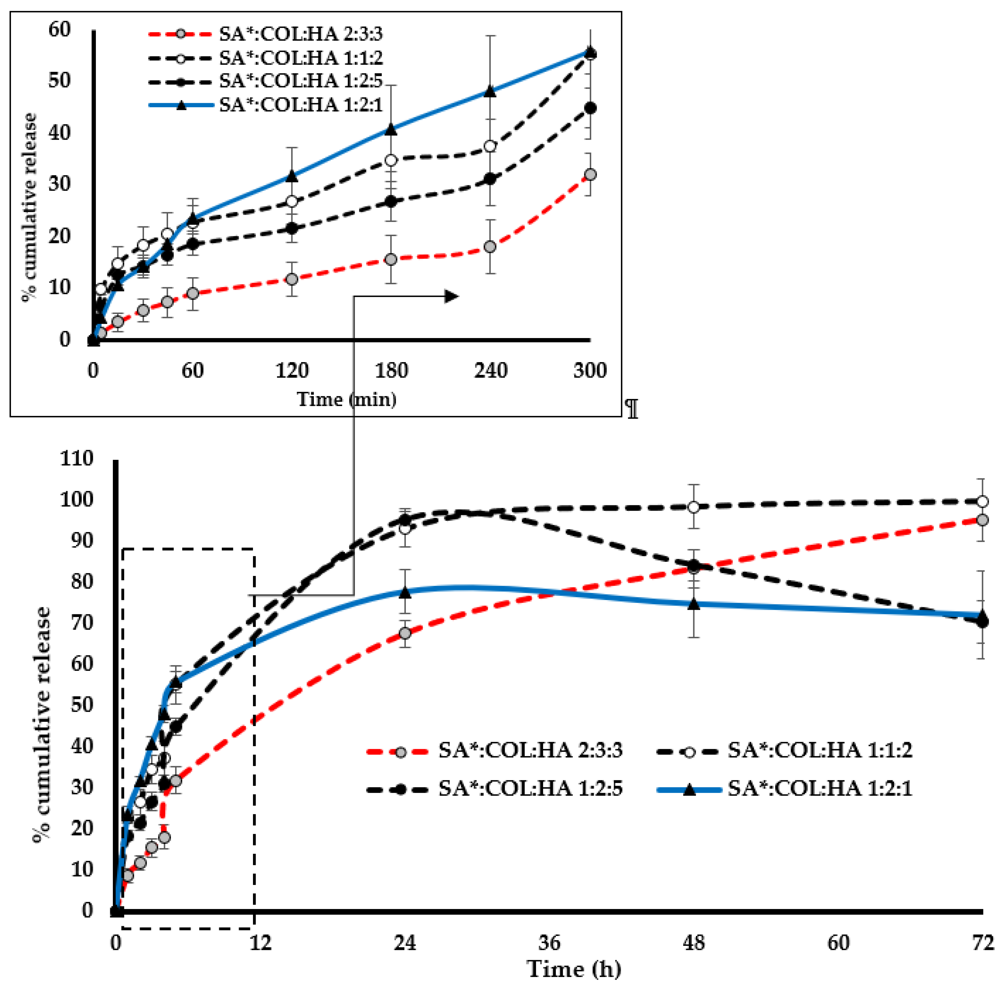
3.2.6. Protein Leaching
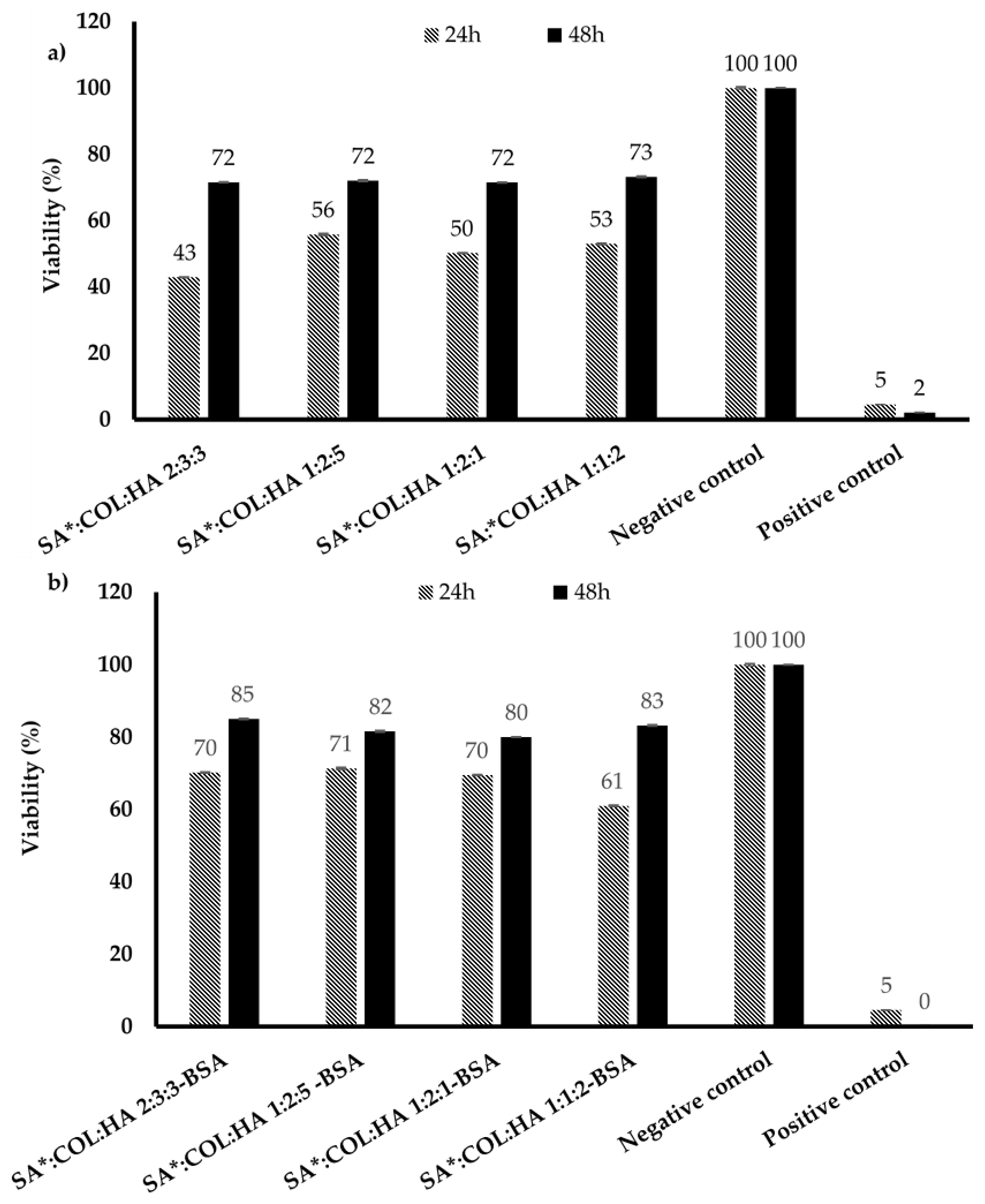
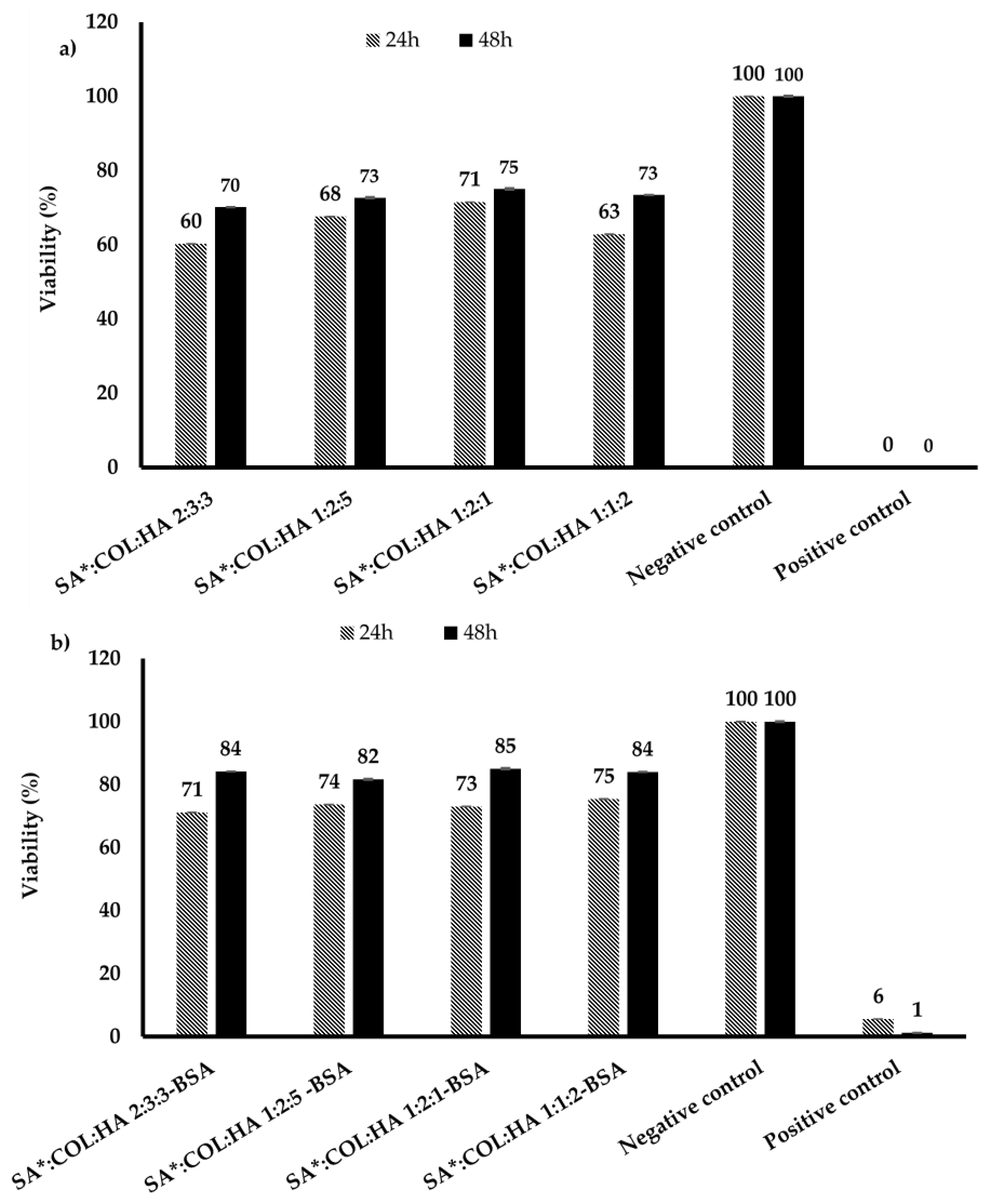
3.3. MTT Assay
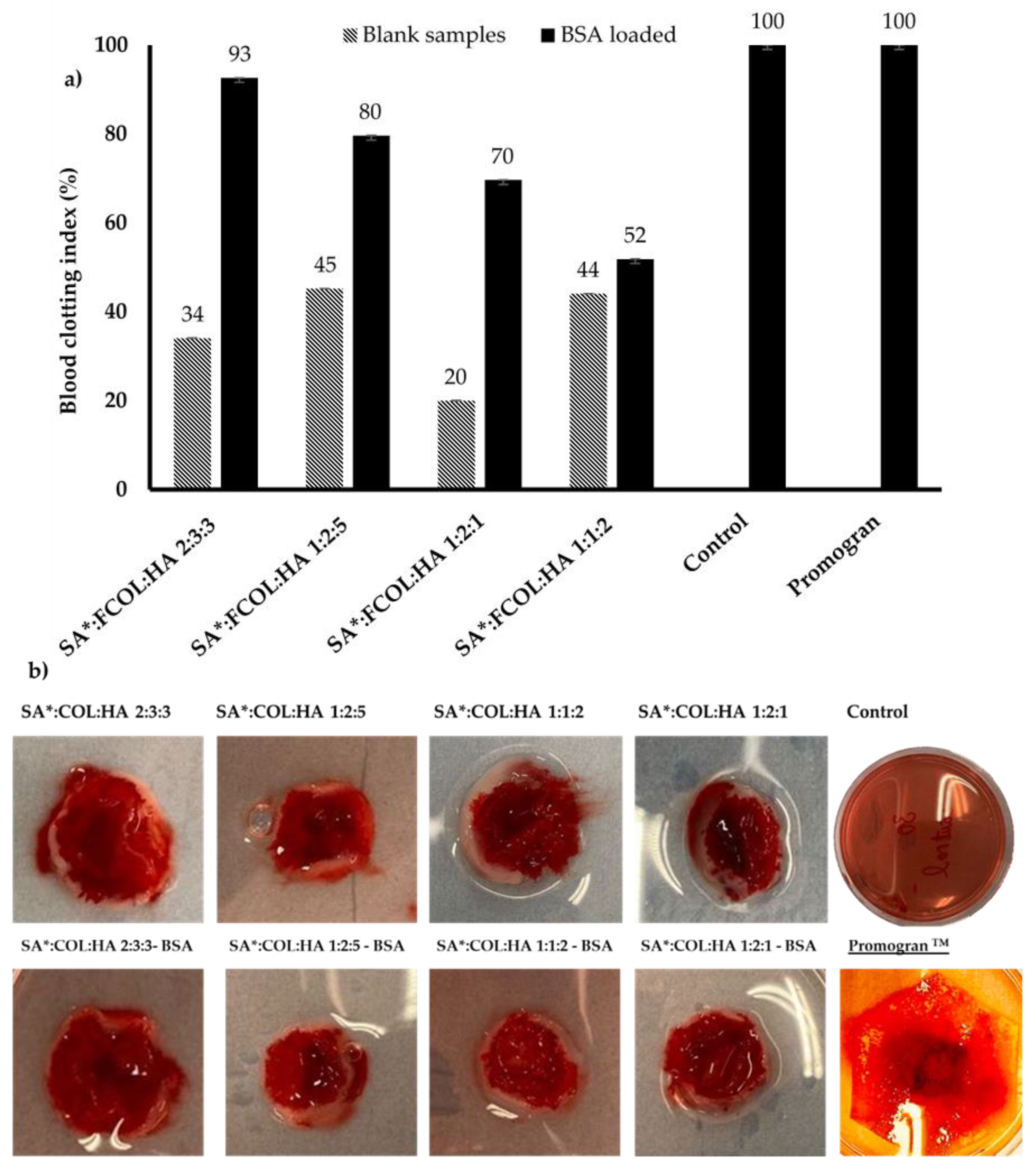
3.4. Whole Blood Clotting
4. Conclusions
Supplementary Materials
Author Contributions
Funding
Institutional Review Board Statement
Informed Consent Statement
Acknowledgments
Conflicts of Interest
References
- Opdenakker, G.; van Damme, J.; Vranckx, J.J. Immunomodulation as Rescue for Chronic Atonic Skin Wounds. Trends Immunol. 2018, 39, 341–354. [Google Scholar] [CrossRef] [PubMed]
- Govindaraju, P.; Todd, L.; Shetye, S.; Monslow, J.; Puré, E. CD44-dependent inflammation, fibrogenesis, and collagenolysis regulates extracellular matrix remodeling and tensile strength during cutaneous wound healing. Matrix Biol. 2019, 75–76, 314–330. [Google Scholar] [CrossRef]
- Fitridge, R.; Thompson, M. Mechanisms of Vascular Disease: A Reference Book for Vascular Specialists; University of Adelaide Press: Adelaide, Australia, 2011. [Google Scholar]
- Posnett, J.; Franks, P.J. The burden of chronic wounds in the UK. Nurs. Times 2008, 104, 44–45. [Google Scholar] [PubMed]
- Herrick, S.E.; Ireland, G.W.; Simon, D.; McCollum, C.N.; Ferguson, M.W.J. Venous ulcer fibroblasts compared with normal fibroblasts show differences in collagen but not fibronectin production under both normal and hypoxic conditions. J. Investig. Dermatol. 1996, 106, 187–193. [Google Scholar] [CrossRef] [Green Version]
- Maru, Y. Molecular biology of chronic myeloid leukemia. Cancer Sci. 2012, 103, 1601–1610. [Google Scholar] [CrossRef]
- Zhu, Y.K.; Liu, X.D.; Sköld, C.M.; Umino, T.; Wang, H.J.; Spurzem, J.R.; Kohyama, T.; Ertl, R.F.; Rennard, S.I. Synergistic neutrophil elastase-cytokine interaction degrades collagen in three-dimensional culture. Am. J. Physiol.-Lung Cell. Mol. Physiol. 2001, 281, 868–878. [Google Scholar] [CrossRef] [PubMed]
- Pastar, I.; Stojadinovic, O.; Yin, N.C.; Ramirez, H.; Nusbaum, A.G.; Sawaya, A.; Patel, S.B.; Khalid, L.; Isseroff, R.R.; Tomic-Canic, M. Epithelialization in Wound Healing: A Comprehensive Review. Adv. Wound Care 2014, 3, 445–464. [Google Scholar] [CrossRef] [PubMed] [Green Version]
- Gallo, N.; Natali, M.L.; Sannino, A.; Salvatore, L. An overview of the use of equine collagen as emerging material for biomedical applications. J. Funct. Biomater. 2020, 11, 79. [Google Scholar] [CrossRef] [PubMed]
- Stupin, V.A.; Gabitov, R.B.; Sinelnikova, T.G.; Silina, E.V. Biological mechanisms of chronic wound and diabetic foot healing: The role of collagen. Ser. J. Exp. Clin. Res. 2018, 19, 373–382. [Google Scholar] [CrossRef] [Green Version]
- Litwiniuk, M.; Krejner, A.; Speyrer, M.S.; Gauto, A.R.; Grzela, T. Hyaluronic Acid in Inflammation and Tissue Regeneration. Wounds 2016, 28, 78–88. [Google Scholar]
- Belattmania, Z.; Kaidi, S.; El Atouani, S.; Katif, C.; Bentiss, F.; Jama, C.; Reani, A.; Sabour, B.; Vasconcelos, V. Isolation and FTIR-ATR and 1H NMR characterization of alginates from the main alginophyte species of the atlantic coast of Morocco. Molecules 2020, 25, 4335. [Google Scholar] [CrossRef] [PubMed]
- Geahchan, S.; Baharlouei, P.; Rahman, M.A. Marine Collagen: A Promising Biomaterial for Wound Healing, Skin Anti-Aging, and Bone Regeneration. Mar. Drugs 2022, 20, 61. [Google Scholar] [CrossRef] [PubMed]
- Gauza-Włodarczyk, M.; Kubisz, L.; Włodarczyk, D. Amino acid composition in determination of collagen origin and assessment of physical factors effects. Int. J. Biol. Macromol. 2017, 104, 987–991. [Google Scholar] [CrossRef] [PubMed]
- Fleck, C.A.; Simman, R. Modern collagen wound dressings: Function and purpose. J. Am. Coll. Certif. Wound Spec. 2010, 2, 50–54. [Google Scholar] [CrossRef] [PubMed] [Green Version]
- Farias, S.; Boateng, J.S. In Vitro, ex vivo and in vivo evaluation of taste masked low dose acetylsalicylic acid loaded composite wafers as platforms for buccal administration in geriatric patients with dysphagia. Int. J. Pharm. 2020, 589, 119807. [Google Scholar] [CrossRef] [PubMed]
- Ahmed, A.; Getti, G.; Boateng, J. Medicated multi-targeted alginate-based dressings for potential treatment of mixed bacterial-fungal infections in diabetic foot ulcers. Int. J. Pharm. 2021, 606, 120903. [Google Scholar] [CrossRef]
- Akiyode, O.; Boateng, J. Composite biopolymer-basedwafer dressings loaded with microbial biosurfactants for potential application in chronicwounds. Polymers 2018, 10, 918. [Google Scholar] [CrossRef] [Green Version]
- Bhasarkar, J.; Bal, D. Kinetic investigation of a controlled drug delivery system based on alginate scaffold with embedded voids. J. Appl. Biomater. Funct. Mater. 2019, 17, 2280800018817462. [Google Scholar] [CrossRef]
- Okeke, O.C.; Boateng, J.S. Composite HPMC and sodium alginate based buccal formulations for nicotine replacement therapy. Int. J. Biol. Macromol. 2016, 91, 31–44. [Google Scholar] [CrossRef]
- Boateng, J.; Areago, D. Composite sodium alginate and chitosan based wafers for buccal delivery of macromolecules. Austin J. Anal. Pharm. Chem. 2014, 1, 1–7. [Google Scholar]
- Franks, F.; Auffret, T. Freeze-Drying of Pharmaceuticals and Biopharmaceuticals: Principles and Practice; Royal Society of Chemistry: London, UK, 2007. [Google Scholar]
- Abasalizadeh, F.; Moghaddam, S.V.; Alizadeh, E.; Kashani, E.; Fazljou, S.M.B.; Torbati, M.; Akbarzadeh, A. Alginate-based hydrogels as drug delivery vehicles in cancer treatment and their applications in wound dressing and 3D bioprinting. J. Biol. Eng. 2020, 14, 8. [Google Scholar] [CrossRef] [PubMed]
- Dianawati, D.; Mishra, V.; Shah, N.P. Survival of Bifidobacterium longum 1941 microencapsulated with proteins and sugars after freezing and freeze drying. Food Res. Int. 2013, 51, 503–509. [Google Scholar] [CrossRef]
- Subhan, F.; Ikram, M.; Shehzad, A.; Ghafoor, A. Marine Collagen: An Emerging Player in Biomedical applications. J. Food Sci. Technol. 2015, 52, 4703–4707. [Google Scholar] [CrossRef] [PubMed] [Green Version]
- Bozec, L.; Odlyha, M. Thermal denaturation studies of collagen by microthermal analysis and atomic force microscopy. Biophys. J. 2011, 10, 228–236. [Google Scholar] [CrossRef] [PubMed] [Green Version]
- Djalila, A.; Aicha, S. Development and characterization of biodegradables packaging obtained from biopolymers mixture. J. Macromol. Sci. Part A Pure Appl. Chem. 2018, 55, 11–16. [Google Scholar] [CrossRef]
- Sabaa, M.W.; Hanna, D.H.; Elella, M.H.A.; Mohamed, R.R. Encapsulation of bovine serum albumin within novel xanthan gum based hydrogel for protein delivery. Mater. Sci. Eng. C 2019, 94, 1044–1055. [Google Scholar] [CrossRef]
- Pawar, H.V.; Boateng, J.S.; Ayensu, I.; Tetteh, J. Multifunctional medicated lyophilised wafer dressing for effective chronic wound healing. J. Pharm. Sci. 2014, 10, 1720–1733. [Google Scholar] [CrossRef]
- Magne, D.; Weiss, P.; Bouler, J.M.; Laboux, O.; Daculsi, G. Study of the maturation of the organic (type I collagen) and mineral (nonstoichiometric apatite) constituents of a calcified tissue (dentin) as a function of location: A fourier transform infrared microspectroscopic investigation. J. Bone Miner. Res. 2001, 16, 750–757. [Google Scholar] [CrossRef]
- de Lederkremer, R.M.; Marino, C. Acids and other products of oxidation of sugars. Adv. Carbohydr. Chem. Biochem. 2003, 58, 199–306. [Google Scholar]
- Giubertoni, G.; Sofronov, O.O.; Bakker, H.J. Effect of intramolecular hydrogen-bond formation on the molecular conformation of amino acids. Commun. Chem. 2020, 3, 84. [Google Scholar] [CrossRef]
- Dianawati, D.; Mishra, V.; Shaha, N.P. Role of calcium alginate and mannitol in protecting Bifidobacterium. Appl. Environ. Microbiol. 2012, 78, 6914–6921. [Google Scholar] [CrossRef] [Green Version]
- Kong, J.; Yu, S. Fourier Transform Infrared Spectroscopic Analysis of Protein Secondary Structures Protein FTIR Data Analysis and Band Assignment. Acta Biochim. Biophys. Sin. 2007, 39, 549–559. [Google Scholar] [CrossRef] [PubMed] [Green Version]
- Narayan, R. Biomedical Materials; Springer: New York, NY, USA, 2009. [Google Scholar]
- Valey, S.J.; Barnett, S.E. A study of wound dressing adhesion. Clin. Mater. 1986, 1, 37–57. [Google Scholar] [CrossRef]
- Boateng, J.S.; Matthews, K.H.; Stevens, H.N.E.; Eccleston, G.M. Wound healing dressings and drug delivery systems: A review. J. Pharm. Sci. 2008, 97, 2892–2923. [Google Scholar] [CrossRef] [PubMed]
- Sionkowska, A.; Kaczmarek, B. Modification of 3D materials based on chitosan and collagen blends by sodium alginate. Mol. Cryst. Liq. Cryst. 2016, 640, 39–45. [Google Scholar] [CrossRef]
- Fu, Y.; Kao, W.J. Drug release kinetics and transport mechanisms of non-degradable and degradable polymeric delivery systems. Expert Opin. Drug Del. 2010, 7, 429–444. [Google Scholar] [CrossRef]
- Sartori, C. The Characterisation of Alginate Systems for Biomedical Applications. Ph.D. Thesis, Brunel University London, Uxbridge, UK, 1997; pp. 1–223. [Google Scholar]
- Aderibigbe, B.A.; Buyana, B. Alginate in wound dressings. Pharmaceutics 2018, 10, 42. [Google Scholar] [CrossRef] [Green Version]
- Zhang, Q.; Wu, T.M.; Chen, C.; Mukamel, S.; Zhuang, W. Molecular mechanism of water reorientational slowing down in concentrated ionic solutions. Proc. Natl. Acad. Sci. USA 2017, 11, 10023–10028. [Google Scholar] [CrossRef] [Green Version]
- Xu, R.; Xia, H.; He, W.; Li, Z.; Zhao, J.; Liu, B.; Wang, Y.; Lei, Q.; Kong, Y.; Bai, Y.; et al. Controlled water vapor transmission rate promotes wound-healing via wound re-epithelialization and contraction enhancement. Sci. Rep. 2016, 6, 24596. [Google Scholar] [CrossRef] [Green Version]
- Álvarez-Suárez, A.S.; Dastager, S.G.; Bogdanchikova, N.; Grande, D.; Pestryakov, A.; García-Ramos, J.C.; Pérez-González, G.L.; Juárez-Moreno, K.; Toledano-Magaña, Y.; Smolentseva, E.; et al. Electrospun fibers and sorbents as a possible basis for effective composite wound dressings. Micromachines 2020, 11, 441. [Google Scholar] [CrossRef] [Green Version]
- Shilpa, A.; Agrawal, S.S.; Ray, A.R. Controlled delivery of drugs from alginate matrix. J. Macromol. Sci.-Polym. Rev. 2003, 43, 187–221. [Google Scholar] [CrossRef]
- Shi, Y.; Zhang, H.; Zhang, X.; Chen, Z.; Zhao, D.; Ma, J. A comparative study of two porous sponge scaffolds prepared by collagen derived from porcine skin and fish scales as burn wound dressings in a rabbit model. Regen. Biomater. 2019, 7, 63–70. [Google Scholar] [CrossRef] [PubMed]
- Gwosdow, A.R.; Cunningham, J.J.; Lydon, M.; Rascati, R.; Berglund, L.G. Evaporative water losses through a temporary wound dressing under simulated wound conditions. J. Burn Care Rehabil. 1993, 14, 450–454. [Google Scholar] [CrossRef] [PubMed]
- Rehman, Q.; Akash, M.S.H.; Imran, I.; Rehman, K. Stability of Pharmaceutical Products. In Drug Stability and Chemical Kinetics; Springer: Singapore, 2020. [Google Scholar]
- Martínez, J.P.Q.; Ruiz, J.C.R.; Campos, M.R.S. Release kinetic studies of stevia rebaudiana extract capsules from sodium alginate and inulin by ionotropic gelation. Adv. Mater. Sci. Eng. 2018, 2018, 6354924. [Google Scholar]
- Zhang, F.; Yang, H.; Yang, Y.; Wang, H.; Li, X.; Wu, X. Stretchable and biocompatible bovine serum albumin fibrous films supported silver for accelerated bacteria-infected wound healing. Chem. Eng. J. 2021, 417, 129145. [Google Scholar] [CrossRef]
- Ghimire, S.; Sarkar, P.; Rigby, K.; Maan, A.; Mukherjee, S.; Crawford, K.E.; Mukhopadhyay, K. Polymeric materials for hemostatic wound healing. Pharmaceutics 2021, 13, 2127. [Google Scholar] [CrossRef]
- Paar, M.; Rossmann, C.; Nusshold, C.; Wagner, T.; Schlagenhauf, A.; Leschnik, B.; Oettl, K.; Koestenberger, M.; Cvirn, G.; Hallström, S. Anticoagulant action of low, physiologic, and high albumin levels in whole blood. PLoS ONE 2017, 12, e0182997. [Google Scholar] [CrossRef]


| Formulations | Constituent Amount (mg) | ||
|---|---|---|---|
| SA#/SA* | FCOL | HA | |
| 2-polymer composite gels | |||
| FCOL:HA 7:1 | 0 | 1750 | 250 |
| FCOL:HA 3:1 | 0 | 1500 | 500 |
| FCOL:HA 1:1 | 0 | 1000 | 1000 |
| FCOL:HA 5:3 | 0 | 1250 | 750 |
| FCOL:HA 3:5 | 0 | 750 | 1250 |
| 3-polymer composite gels | |||
| SA:FCOL:HA 3:4:1 | 750 | 1000 | 250 |
| SA: FCOL: HA 1:2:1 | 500 | 1000 | 500 |
| SA: FCOL: HA 1:1:2 | 500 | 500 | 1000 |
| SA: FCOL: HA 2:3:3 | 500 | 750 | 750 |
| SA: FCOL: HA 1:2:5 | 250 | 500 | 1250 |
| Sample | Low G Content SA (SA#) | High G Content SA (SA*) | ||||||
|---|---|---|---|---|---|---|---|---|
| Porosity (%) ± (SD) | EWC (%) ± (SD) | WA (%) ± (SD) | WVTR (g/m2 day −1) ± (SD) | Porosity (%) ± (SD) | EWC (%) ± (SD) | WA (%) ± (SD) | WVTR (g/m2 day −1) ± (SD) | |
| 2% SA:FCOL:HA 1:2:5 | 79 ± 2 | 86 ± 5 | 696 ± 267 | 2328 ± 22 | 83 ± 2 | 88 ± 2 | 843 ± 210 | 2429 ± 53 |
| 2% SA:FCOL:HA 1:2:1 | 76 ± 6 | 89 ± 4 | 948 ± 382 | 2340 ± 35 | 94 ± 16 | 94 ± 1 | 1382 ± 20 | 2183 ± 45 |
| 2% SA:FCOL:HA 2:3:3 | 70 ± 4 | 90 ± 2 | 956 ± 266 | 2354 ± 67 | 94 ± 14 | 93 ± 1 | 1252 ± 56 | 2255 ± 41 |
| 2% SA:FCOL:HA 1:1:2 | 77 ± 3 | 91 ± 2 | 1015 ± 287 | 2333 ± 63 | 96 ± 9 | 88 ± 2 | 793 ± 132 | 2393 ± 38 |
| 2% SA:FCOL:HA 3:4:1 | 76 ± 3 | 92 ± 1 | 1243 ± 299 | 2368 ± 38 | - | - | - | - |
| 2% SA:FCOL:HA 2:3:3 75 µg/g BSA | - | - | - | - | 88 ± 4 | 80 ± 1 | 405 ± 46 | 2527 ± 38 |
| 2% SA:FCOL:HA 1:2:5 75 µg/g BSA | - | - | - | - | 88 ± 5 | 79 ± 1 | 380 ± 14 | 2488 ± 14 |
| 2% SA:FCOL:HA 1:2:1 75 µg/g BSA | - | - | - | - | 89 ± 3 | 80 ± 1 | 404 ± 34 | 2475 ± 78 |
| 2% SA:FCOL:HA 1:1:2 75 µg/g BSA | - | - | - | - | 91 ± 7 | 82 ± 5 | 493 ± 213 | 2405 ± 69 |
| Higuchi | Korsmeyer–Peppas | Zero Order | First Order | ||||||
|---|---|---|---|---|---|---|---|---|---|
| KH | R2 | KK-P | n | R2 | K0 | R2 | K1 | R2 | |
| SA*:FCOL:HA 2:3:3 | 1.559 | 0.890 | 0.121 | 0.596 | 0.971 | 0.023 | 0.897 | 0.001 | 0.067 |
| SA*:FCOL:HA 1:1:2 | 2.615 | 0.941 | 0.447 | 0.478 | 0.897 | 0.094 | 0.795 | 0.001 | 0.036 |
| SA*:FCOL:HA 1:2:5 | 1.318 | 0.798 | 0.366 | 0.476 | 0.913 | 0.018 | 0.602 | 0.001 | 0.052 |
| SA*:FCOL:HA 1:2:1 | 1.162 | 0.772 | 0.359 | 0.495 | 0.896 | 0.016 | 0.571 | 0.001 | 0.023 |
Publisher’s Note: MDPI stays neutral with regard to jurisdictional claims in published maps and institutional affiliations. |
© 2022 by the authors. Licensee MDPI, Basel, Switzerland. This article is an open access article distributed under the terms and conditions of the Creative Commons Attribution (CC BY) license (https://creativecommons.org/licenses/by/4.0/).
Share and Cite
Afzali, M.; Boateng, J.S. Composite Fish Collagen-Hyaluronate Based Lyophilized Scaffolds Modified with Sodium Alginate for Potential Treatment of Chronic Wounds. Polymers 2022, 14, 1550. https://doi.org/10.3390/polym14081550
Afzali M, Boateng JS. Composite Fish Collagen-Hyaluronate Based Lyophilized Scaffolds Modified with Sodium Alginate for Potential Treatment of Chronic Wounds. Polymers. 2022; 14(8):1550. https://doi.org/10.3390/polym14081550
Chicago/Turabian StyleAfzali, Meena, and Joshua Siaw Boateng. 2022. "Composite Fish Collagen-Hyaluronate Based Lyophilized Scaffolds Modified with Sodium Alginate for Potential Treatment of Chronic Wounds" Polymers 14, no. 8: 1550. https://doi.org/10.3390/polym14081550
APA StyleAfzali, M., & Boateng, J. S. (2022). Composite Fish Collagen-Hyaluronate Based Lyophilized Scaffolds Modified with Sodium Alginate for Potential Treatment of Chronic Wounds. Polymers, 14(8), 1550. https://doi.org/10.3390/polym14081550