
Fluorescent Polymers Conspectus


Abstract
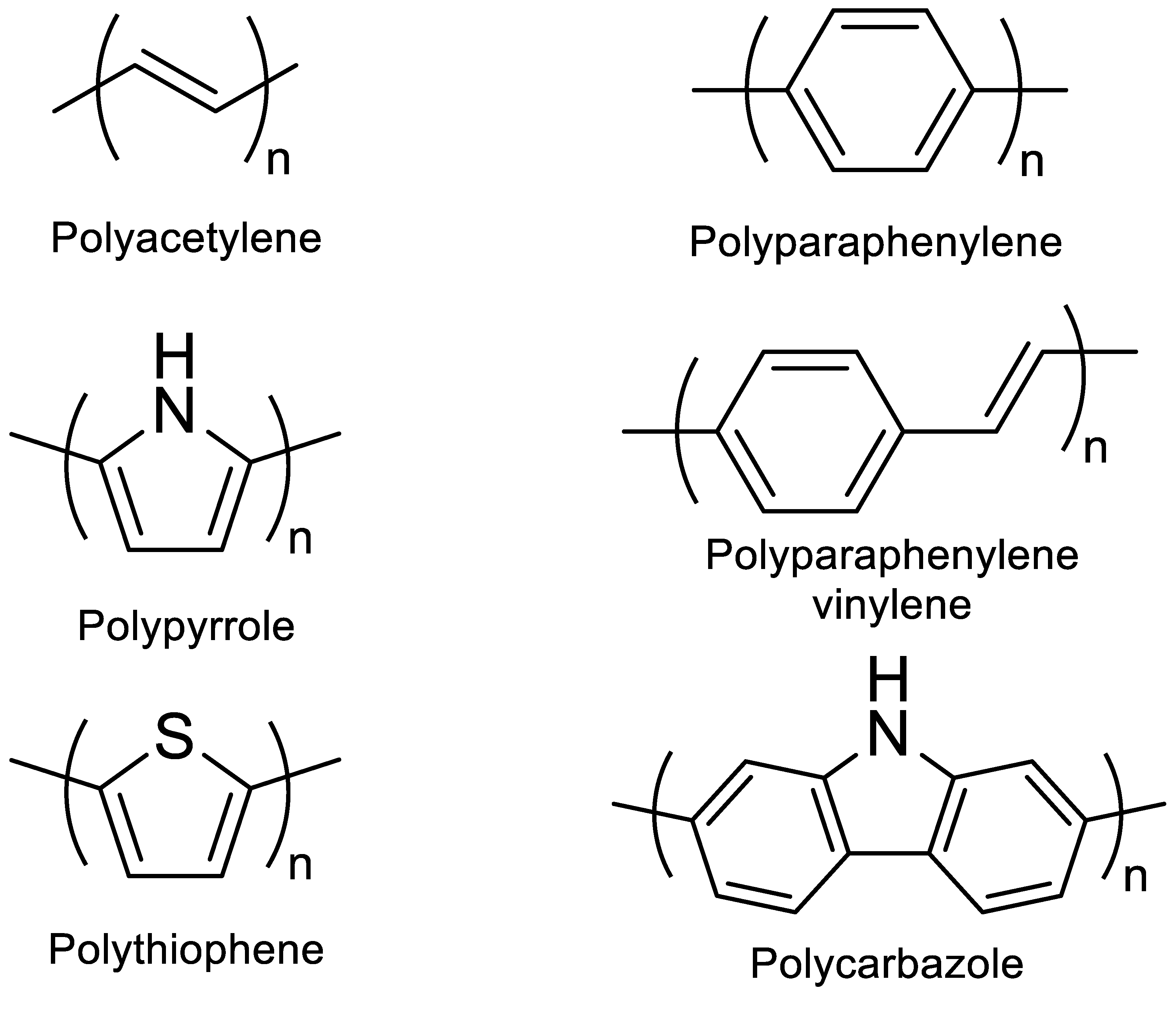
:
1. Introduction
2. Polymers Containing Fluorophores
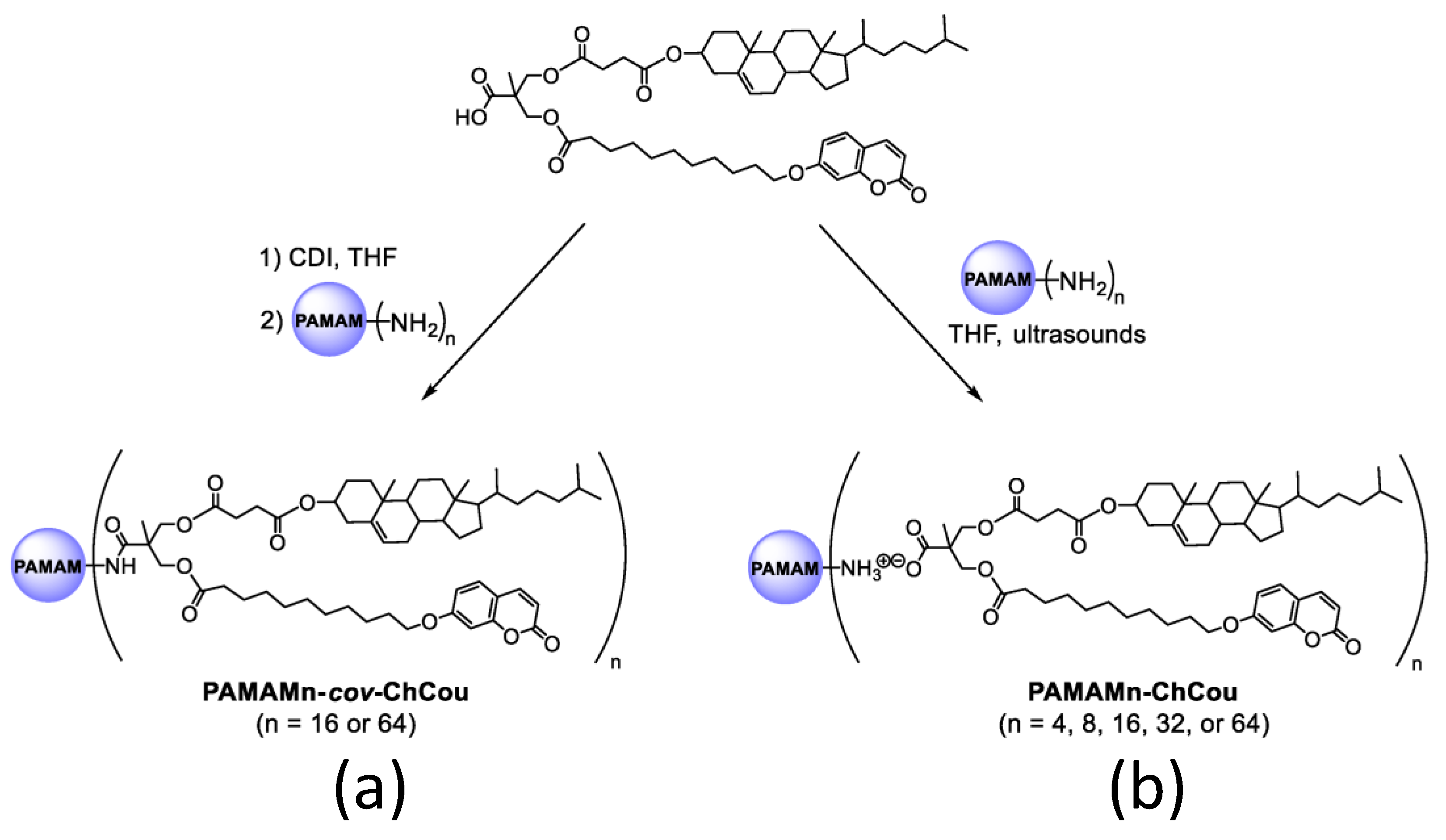
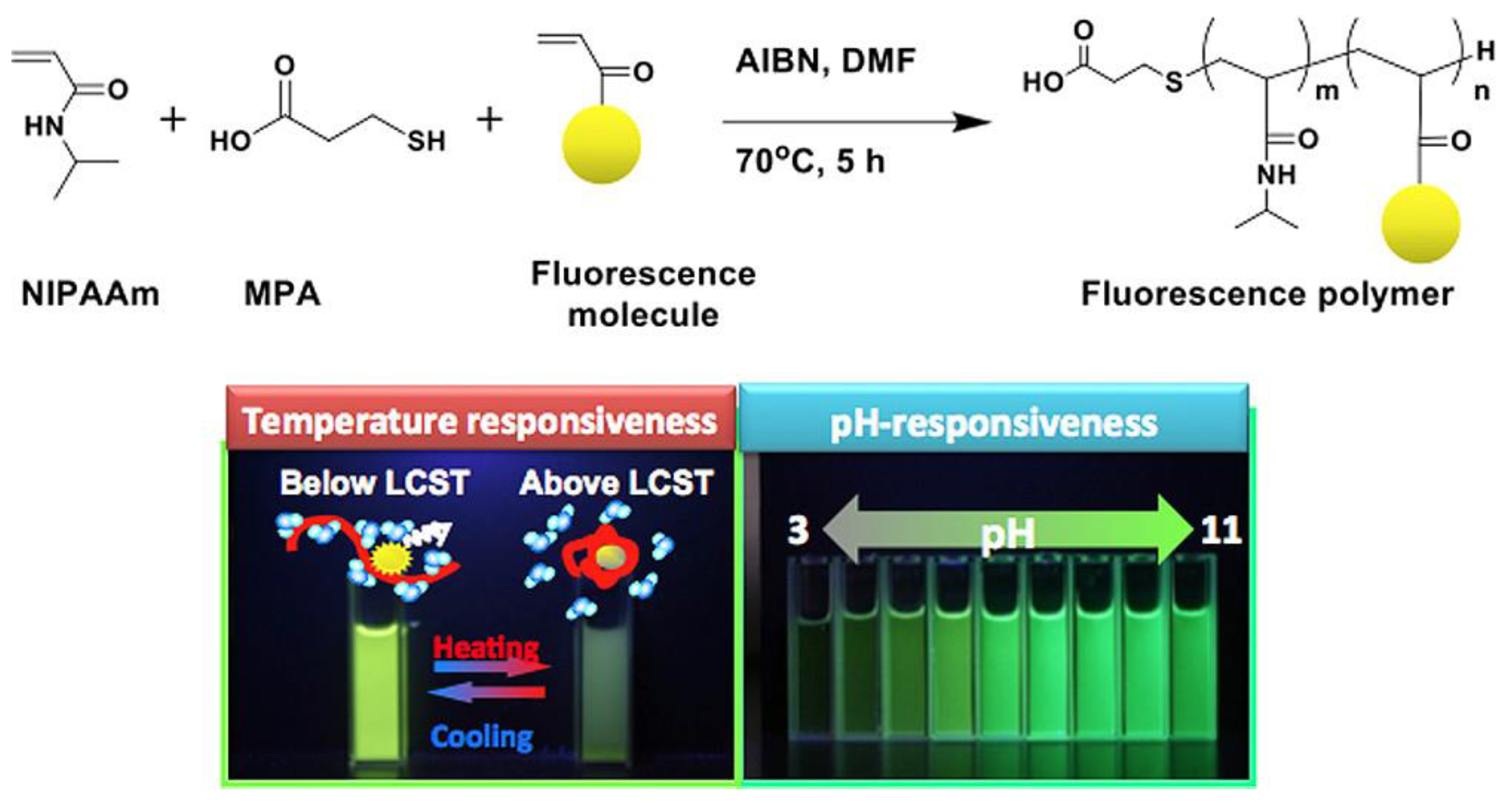
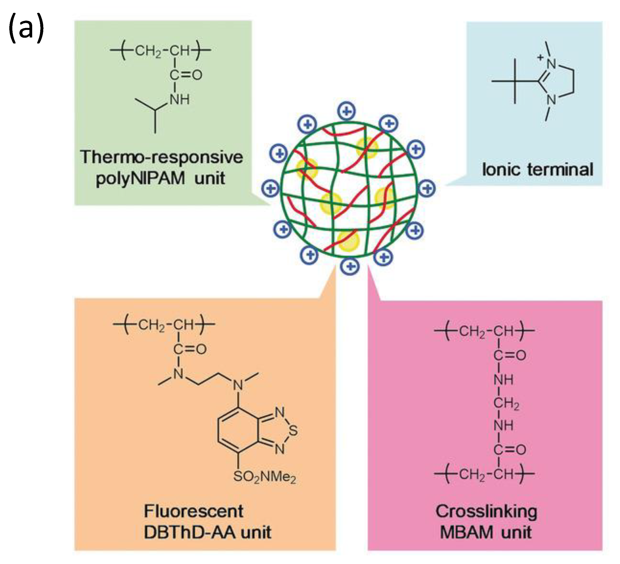
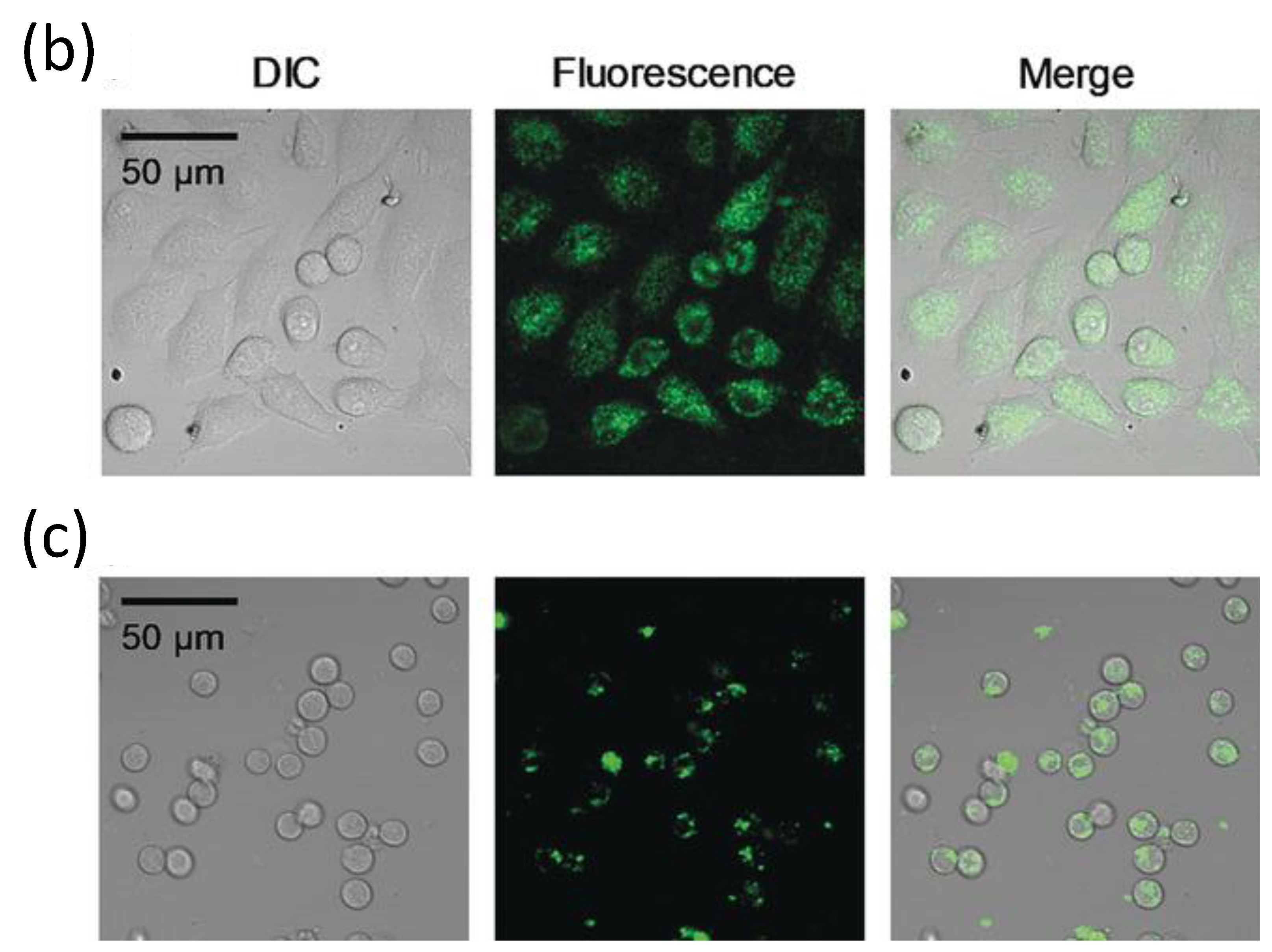
2.1. Non-Conjugated Polymers Containing Fluorophores
2.2. Conjugated Polymers Containing Fluorophores
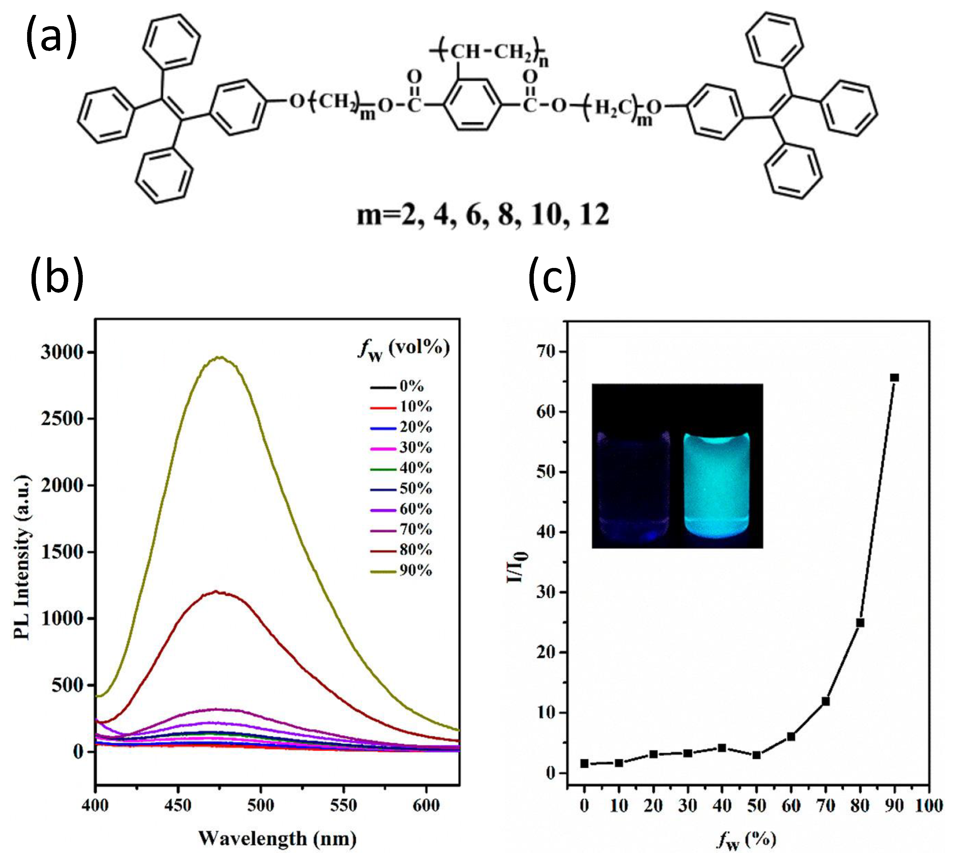
3. Aggregation-Induced Emission Macromolecules
4. Conclusions and Future Outlook
Author Contributions
Funding
Data Availability Statement
Acknowledgments
Conflicts of Interest
References
- Braslavsky, S.E. Glossary of terms used in photochemistry, 3rd edition (IUPAC Recommendations 2006). Pure Appl. Chem. 2007, 79, 293–465. [Google Scholar] [CrossRef]
- Lakowicz, J.R. Principles of Fluorescence Spectroscopy; Lakowicz, J.R., Ed.; Springer: Boston, MA, USA, 2006; ISBN 978-0-387-46312-4. [Google Scholar]
- Guilbault, G.G.; Norris, J.D. Practical Fluorescence, 2nd ed.; CRC Press: Boca Raton, FL, USA, 2020; ISBN 9781003066514. [Google Scholar]
- McNaught, A.D.; Wilkinson, A.; IUPAC. Compendium of Chemical Terminology (The ’Gold Book’), 2nd ed.; Blackwell Scientific Publications: Oxford, UK, 1997. [Google Scholar]
- Jin, Y.; Gao, X. Plasmonic fluorescent quantum dots. Nat. Nanotechnol. 2009, 4, 571–576. [Google Scholar] [CrossRef] [PubMed]
- Huang, C.-C.; Yang, Z.; Lee, K.-H.; Chang, H.-T. Synthesis of Highly Fluorescent Gold Nanoparticles for Sensing Mercury(II). Angew. Chem. Int. Ed. 2007, 119, 6948–6952. [Google Scholar] [CrossRef]
- Valeur, B.; Berberan-Santos, M.N. A Brief History of Fluorescence and Phosphorescence before the Emergence of Quantum Theory. J. Chem. Educ. 2011, 88, 731–738. [Google Scholar] [CrossRef]
- Baker, S.N.; Baker, G.A. Luminescent Carbon Nanodots: Emergent Nanolights. Angew. Chem. Int. Ed. 2010, 49, 6726–6744. [Google Scholar] [CrossRef]
- El-Safty, S.A.; Shenashen, M.; Ismael, M.; Khairy, M.; Awual, R. Mesoporous aluminosilica sensors for the visual removal and detection of Pd(II) and Cu(II) ions. Microporous Mesoporous Mater. 2013, 166, 195–205. [Google Scholar] [CrossRef]
- Crego-Calama, M.; Reinhoudt, D.N. New Materials for Metal Ion Sensing by Self-Assembled Monolayers on Glass. Adv. Mater. 2001, 13, 1171–1174. [Google Scholar] [CrossRef]
- Huang, C.-C.; Yang, Z.; Lee, K.-H.; Chang, H.-T. Synthesis of Highly Fluorescent Gold Nanoparticles for Sensing Mercury(II). Angew. Chem. Int. Ed. 2007, 46, 6824–6828. [Google Scholar] [CrossRef]
- Larson, D.R.; Zipfel, W.R.; Williams, R.M.; Clark, S.W.; Bruchez, M.P.; Wise, F.W.; Webb, W.W. Water-Soluble Quantum Dots for Multiphoton Fluorescence Imaging In Vivo. Science 2003, 300, 1434–1436. [Google Scholar] [CrossRef]
- Sharker, S.; Do, M. Nanoscale Carbon-Polymer Dots for Theranostics and Biomedical Exploration. J. Nanotheranostics 2021, 2, 118–130. [Google Scholar] [CrossRef]
- Chatterjee, D.P.; Pakhira, M.; Nandi, A.K. Fluorescence in Nonfluorescent” Polymers. ACS Omega 2020, 5, 30747–30766. [Google Scholar] [CrossRef] [PubMed]
- Ren, J.; Stagi, L.; Innocenzi, P. Fluorescent carbon dots in solid-state: From nanostructures to functional devices. Prog. Solid State Chem. 2021, 62, 100295. [Google Scholar] [CrossRef]
- AlSalhi, M.S.; Alam, J.; Dass, L.A.; Raja, M. Recent Advances in Conjugated Polymers for Light Emitting Devices. Int. J. Mol. Sci. 2011, 12, 2036–2054. [Google Scholar] [CrossRef] [PubMed]
- Park, J.M.; Nam, S.H.; Hong, K.-I.; Jeun, Y.E.; Ahn, H.S.; Jang, W.-D. Stimuli-responsive fluorescent dyes for electrochemically tunable multi-color-emitting devices. Sens. Actuators B Chem. 2021, 332, 129534. [Google Scholar] [CrossRef]
- Yao, Z.; Zong, W.; Wang, K.; Yuan, P.; Liu, Y.; Xu, S.; Cao, S. Triphenylamine-carbazole alternating copolymers bearing thermally activated delayed fluorescent emitting and host pendant groups for solution-processable OLEDs. React. Funct. Polym. 2021, 163, 104898. [Google Scholar] [CrossRef]
- Minotto, A.; Bulut, I.; Rapidis, A.G.; Carnicella, G.; Patrini, M.; Lunedei, E.; Anderson, H.L.; Cacialli, F. Towards efficient near-infrared fluorescent organic light-emitting diodes. Light. Sci. Appl. 2021, 10, 18. [Google Scholar] [CrossRef]
- Li, T.; Yu, L.; Jin, D.; Chen, B.; Li, L.; Chen, L.; Li, Y. A colorimetric and fluorescent probe for fluoride anions based on a phenanthroimidazole–cyanine platform. Anal. Methods 2013, 5, 1612–1616. [Google Scholar] [CrossRef]
- Weldeab, A.O.; Li, L.; Cekli, S.; Abboud, K.A.; Schanze, K.S.; Castellano, R.K. Pyridine-terminated low gap π-conjugated oligomers: Design, synthesis, and photophysical response to protonation and metalation. Org. Chem. Front. 2018, 5, 3170–3177. [Google Scholar] [CrossRef]
- Saremi, B.; Bandi, V.; Kazemi, S.; Hong, Y.; D’Souza, F.; Yuan, B. Exploring NIR Aza-BODIPY-Based Polarity Sensitive Probes with ON-and-OFF Fluorescence Switching in Pluronic Nanoparticles. Polymers 2020, 12, 540. [Google Scholar] [CrossRef] [Green Version]
- Hayashi, T.; Kawamoto, K.; Inada, N.; Uchiyama, S. Cationic Fluorescent Nanogel Thermometers based on Thermoresponsive Poly(N-isopropylacrylamide) and Environment-Sensitive Benzofurazan. Polymers 2019, 11, 1305. [Google Scholar] [CrossRef] [Green Version]
- Zhao, W.-X.; Zhou, C.; Peng, H.-S. Ratiometric Luminescent Nanoprobes Based on Ruthenium and Terbium-Containing Metallopolymers for Intracellular Oxygen Sensing. Polymers 2019, 11, 1290. [Google Scholar] [CrossRef] [PubMed] [Green Version]
- Zhang, X.; Yang, S.; Chen, W.; Li, Y.; Wei, Y.; Luo, A. Magnetic Fluorescence Molecularly Imprinted Polymer Based on FeOx/ZnS Nanocomposites for Highly Selective Sensing of Bisphenol A. Polymers 2019, 11, 1210. [Google Scholar] [CrossRef] [PubMed] [Green Version]
- Yamagishi, H.; Matsui, T.; Kitayama, Y.; Aikyo, Y.; Tong, L.; Kuwabara, J.; Kanbara, T.; Morimoto, M.; Irie, M.; Yamamoto, Y. Fluorescence Switchable Conjugated Polymer Microdisk Arrays by Cosolvent Vapor Annealing. Polymers 2021, 13, 269. [Google Scholar] [CrossRef] [PubMed]
- Shchapova, E.; Nazarova, A.; Gurkov, A.; Borvinskaya, E.; Rzhechitskiy, Y.; Dmitriev, I.; Meglinski, I.; Timofeyev, M. Application of PEG-Covered Non-Biodegradable Polyelectrolyte Microcapsules in the Crustacean Circulatory System on the Example of the Amphipod Eulimnogammarus verrucosus. Polymers 2019, 11, 1246. [Google Scholar] [CrossRef] [Green Version]
- Li, Q.; Zhang, W.-C.; Wang, C.-F.; Chen, S. In situ access to fluorescent dual-component polymers towards optoelectronic devices via inhomogeneous biphase frontal polymerization. RSC Adv. 2015, 5, 102294–102299. [Google Scholar] [CrossRef]
- Liu, Y.; Wang, Y.-M.; Zhu, W.-Y.; Zhang, C.-H.; Tang, H.; Jiang, J.-H. Conjugated polymer nanoparticles-based fluorescent biosensor for ultrasensitive detection of hydroquinone. Anal. Chim. Acta. 2018, 1012, 60–65. [Google Scholar] [CrossRef]
- Lichon, L.; Kotras, C.; Myrzakhmetov, B.; Arnoux, P.; Daurat, M.; Nguyen, C.; Durand, D.; Bouchmella, K.; Ali, L.M.; Durand, J.-O.; et al. Polythiophenes with Cationic Phosphonium Groups as Vectors for Imaging, siRNA Delivery, and Photodynamic Therapy. Nanomaterials 2020, 10, 1432. [Google Scholar] [CrossRef]
- Hu, L.; Chen, Z.; Liu, Y.; Tian, B.; Guo, T.; Liu, R.; Wang, C.; Ying, L. In Vivo Bioimaging and Photodynamic Therapy Based on Two-Photon Fluorescent Conjugated Polymers Containing Dibenzothiophene-S,S-dioxide Derivatives. ACS Appl. Mater. Interfaces 2020, 12, 57281–57289. [Google Scholar] [CrossRef]
- Li, C.; Xu, Y.; Liu, Y.; Ren, Z.; Ma, Y.; Yan, S. Highly efficient white-emitting thermally activated delayed fluorescence polymers: Synthesis, non-doped white OLEDs and electroluminescent mechanism. Nano Energy 2019, 65, 104057. [Google Scholar] [CrossRef]
- Jiang, J.; Zhang, P.; Liu, L.; Li, Y.; Zhang, Y.; Wu, T.; Xie, H.; Zhang, C.; Cui, J.; Chen, J. Dual photochromics-contained photoswitchable multistate fluorescent polymers for advanced optical data storage, encryption, and photowritable pattern. Chem. Eng. J. 2021, 425, 131557. [Google Scholar] [CrossRef]
- Wang, M.; Jiang, K.; Gao, Y.; Liu, Y.; Zhang, Z.; Zhao, W.; Ji, H.; Zheng, T.; Feng, H. A facile fabrication of conjugated fluorescent nanoparticles and micro-scale patterned encryption via high resolution inkjet printing. Nanoscale 2021, 13, 14337–14345. [Google Scholar] [CrossRef] [PubMed]
- Abdollahi, A.; Alidaei-Sharif, H.; Roghani-Mamaqani, H.; Herizchi, A. Photoswitchable fluorescent polymer nanoparticles as high-security anticounterfeiting materials for authentication and optical patterning. J. Mater. Chem. C 2020, 8, 5476–5493. [Google Scholar] [CrossRef]
- Ru, Y.; Zhang, X.; Song, W.; Liu, Z.; Feng, H.; Wang, B.; Guo, M.; Wang, X.; Luo, C.; Yang, W.; et al. A new family of thermoplastic photoluminescence polymers. Polym. Chem. 2016, 7, 6250–6256. [Google Scholar] [CrossRef]
- Zhai, Y.-L.; Wang, Q.-B.; Yu, H.; Ji, X.-Y.; Zhang, X. Enhanced Two-Photon Fluorescence and Fluorescence Imaging of Novel Probe for Calcium Ion by Self-Assembly with Conjugated Polymer. Polymers 2019, 11, 1643. [Google Scholar] [CrossRef] [PubMed] [Green Version]
- Ostos, F.J.; Iasilli, G.; Carlotti, M.; Pucci, A. High-Performance Luminescent Solar Concentrators Based on Poly(Cyclohexylmethacrylate) (PCHMA) Films. Polymers 2020, 12, 2898. [Google Scholar] [CrossRef] [PubMed]
- Wang, B.; Yoshida, K.; Sato, K.; Anzai, J.-I. Phenylboronic Acid-Functionalized Layer-by-Layer Assemblies for Biomedical Applications. Polymers 2017, 9, 202. [Google Scholar] [CrossRef] [PubMed] [Green Version]
- Chen, J.; Li, J.; Xu, L.; Hong, W.; Yang, Y.; Chen, X. The Glass-Transition Temperature of Supported PMMA Thin Films with Hydrogen Bond/Plasmonic Interface. Polymers 2019, 11, 601. [Google Scholar] [CrossRef] [PubMed] [Green Version]
- Yin, M.; Zhang, C.; Li, J.; Li, H.; Deng, Q.; Wang, S. Highly Sensitive Detection of Benzoyl Peroxide Based on Organoboron Fluorescent Conjugated Polymers. Polymers 2019, 11, 1655. [Google Scholar] [CrossRef] [Green Version]
- Gou, Z.; Zhang, X.; Zuo, Y.; Lin, W. Synthesis of Silane-Based Poly(thioether) via Successive Click Reaction and Their Applications in Ion Detection and Cell Imaging. Polymers 2019, 11, 1235. [Google Scholar] [CrossRef] [Green Version]
- Luthjens, L.H.; Yao, T.; Warman, J.M. A Polymer-Gel Eye-Phantom for 3D Fluorescent Imaging of Millimetre Radiation Beams. Polymers 2018, 10, 1195. [Google Scholar] [CrossRef] [Green Version]
- Bidan, G.; Billon, M.; Livache, T.; Mathis, G.; Roget, A.; Torres-Rodriguez, L. Conducting polymers as a link between biomolecules and microelectronics. Synth. Met. 1999, 102, 1363–1365. [Google Scholar] [CrossRef]
- Lange, U.; Roznyatovskaya, N.V.; Mirsky, V.M. Conducting polymers in chemical sensors and arrays. Anal. Chim. Acta. 2008, 614, 1–26. [Google Scholar] [CrossRef] [PubMed]
- Livache, T.; Bazin, H.; Mathis, G. Conducting polymers on microelectronic devices as tools for biological analyses. Clin. Chim. Acta. 1998, 278, 171–176. [Google Scholar] [CrossRef]
- Disney, M.D.; Zheng, J.; Swager, A.T.M.; Seeberger, P.H. Detection of Bacteria with Carbohydrate-Functionalized Fluorescent Polymers. J. Am. Chem. Soc. 2004, 126, 13343–13346. [Google Scholar] [CrossRef] [PubMed]
- Yan, J.-J.; Wang, Z.K.; Lin, X.-S.; Hong, C.-Y.; Liang, H.-J.; Pan, C.-Y.; You, Y.-Z. Polymerizing Nonfluorescent Monomers without Incorporating any Fluorescent Agent Produces Strong Fluorescent Polymers. Adv. Mater. 2012, 24, 5617–5624. [Google Scholar] [CrossRef] [PubMed]
- Hong, Y.; Lam, J.W.Y.; Tang, B.Z. Aggregation-induced emission. Chem. Soc. Rev. 2011, 40, 5361–5388. [Google Scholar] [CrossRef] [PubMed] [Green Version]
- Würthner, F. Aggregation-Induced Emission (AIE): A Historical Perspective. Angew. Chem. Int. Ed. 2020, 59, 14192–14196. [Google Scholar] [CrossRef] [PubMed]
- Hu, R.; Qin, A.; Tang, B.Z. AIE polymers: Synthesis and applications. Prog. Polym. Sci. 2020, 100, 101176. [Google Scholar] [CrossRef]
- Hu, R.; Kang, Y.; Tang, B.Z. Recent advances in AIE polymers. Polym. J. 2016, 48, 359–370. [Google Scholar] [CrossRef]
- Hu, R.; Yang, X.; Qin, A.; Tang, B.Z. AIE polymers in sensing, imaging and theranostic applications. Mater. Chem. Front. 2021, 5, 4073–4088. [Google Scholar] [CrossRef]
- Zhan, R.; Pan, Y.; Manghnani, P.; Liu, B. AIE Polymers: Synthesis, Properties, and Biological Applications. Macromol. Biosci. 2017, 17, 1600433. [Google Scholar] [CrossRef] [PubMed]
- Thomas, S.W.; Joly, G.D.; Swager, T.M. Chemical Sensors Based on Amplifying Fluorescent Conjugated Polymers. Chem. Rev. 2007, 107, 1339–1386. [Google Scholar] [CrossRef] [PubMed]
- Alvarez, A.; Costa-Fernández, J.M.; Pereiro, R.; Sanz-Medel, A.; Salinas-Castillo, A. Fluorescent conjugated polymers for chemical and biochemical sensing. TrAC Trends Anal. Chem. 2011, 30, 1513–1525. [Google Scholar] [CrossRef]
- Braunecker, W.A.; Matyjaszewski, K. Controlled/living radical polymerization: Features, developments, and perspectives. Prog. Polym. Sci. 2007, 32, 93–146. [Google Scholar] [CrossRef]
- Grubbs, R.B.; Grubbs, R.H. 50th Anniversary Perspective: Living Polymerization—Emphasizing the Molecule in Macromolecules. Macromolecules 2017, 50, 6979–6997. [Google Scholar] [CrossRef]
- Sánchez-Ruiz, A.; Sousa-Hervés, A.; Tolosa, B.J.; Navarro, A.; García-Martínez, J.C. Aggregation-Induced Emission Properties in Fully π-Conjugated Polymers, Dendrimers, and Oligomers. Polymers 2021, 13, 213. [Google Scholar] [CrossRef]
- Zhao, J.; Pan, X.; Zhu, J.; Zhu, X. Novel AIEgen-Functionalized Diselenide-Crosslinked Polymer Gels as Fluorescent Probes and Drug Release Carriers. Polymers 2020, 12, 551. [Google Scholar] [CrossRef] [PubMed] [Green Version]
- Wu, F.; Misra, M.; Mohanty, A.K. Challenges and new opportunities on barrier performance of biodegradable polymers for sustainable packaging. Prog. Polym. Sci. 2021, 117, 101395. [Google Scholar] [CrossRef]
- Rai, P.; Mehrotra, S.; Priya, S.; Gnansounou, E.; Sharma, S.K. Recent advances in the sustainable design and applications of biodegradable polymers. Bioresour. Technol. 2021, 325, 124739. [Google Scholar] [CrossRef]
- Namsheer, K.; Rout, C.S. Conducting polymers: A comprehensive review on recent advances in synthesis, properties and applications. RSC Adv. 2021, 11, 5659–5697. [Google Scholar] [CrossRef]
- de Leon, A.C.C.; da Silva Ítalo, G.; Pangilinan, K.D.; Chen, Q.; Caldona, E.B.; Advincula, R.C. High performance polymers for oil and gas applications. React. Funct. Polym. 2021, 162, 104878. [Google Scholar] [CrossRef]
- Nandi, M.; Maiti, B.; Srikanth, K.; De, P. Supramolecular Interaction-Assisted Fluorescence and Tunable Stimuli-Responsiveness of l-Phenylalanine-Based Polymers. Langmuir 2017, 33, 10588–10597. [Google Scholar] [CrossRef] [PubMed]
- Kwak, G.; Lee, W.-E.; Jeong, H.; Sakaguchi, T.; Fujiki, M. Swelling-Induced Emission Enhancement in Substituted Acetylene Polymer Film with Large Fractional Free Volume: Fluorescence Response to Organic Solvent Stimuli. Macromolecules 2008, 42, 20–24. [Google Scholar] [CrossRef]
- Saha, B.; Bauri, K.; Bag, A.; Ghorai, P.K.; De, P. Conventional fluorophore-free dual pH- and thermo-responsive luminescent alternating copolymer. Polym. Chem. 2016, 7, 6895–6900. [Google Scholar] [CrossRef]
- Dou, C.; Han, L.; Zhao, S.; Zhang, H.; Wang, Y. Multi-Stimuli-Responsive Fluorescence Switching of a Donor−Acceptor π-Conjugated Compound. J. Phys. Chem. Lett. 2011, 2, 666–670. [Google Scholar] [CrossRef]
- Lavrenova, A.; Balkenende, D.W.R.; Sagara, Y.; Schrettl, S.; Simon, Y.C.; Weder, C. Mechano- and Thermoresponsive Photoluminescent Supramolecular Polymer. J. Am. Chem. Soc. 2017, 139, 4302–4305. [Google Scholar] [CrossRef] [PubMed] [Green Version]
- Sagara, Y.; Yamane, S.; Mitani, M.; Weder, C.; Kato, T. Mechanoresponsive Luminescent Molecular Assemblies: An Emerging Class of Materials. Adv. Mater. 2016, 28, 1073–1095. [Google Scholar] [CrossRef]
- Pucci, A.; Bizzarri, R.; Ruggeri, G. Polymer composites with smart optical properties. Soft Matter 2011, 7, 3689–3700. [Google Scholar] [CrossRef]
- Sagara, Y.; Kato, T. Mechanically induced luminescence changes in molecular assemblies. Nat. Chem. 2009, 1, 605–610. [Google Scholar] [CrossRef]
- Estupiñán, D.; Gegenhuber, T.; Blinco, J.; Barner-Kowollik, C.; Barner, L. Self-Reporting Fluorescent Step-Growth RAFT Polymers Based on Nitrile Imine-Mediated Tetrazole-ene Cycloaddition Chemistry. ACS Macro Lett. 2017, 6, 229–234. [Google Scholar] [CrossRef]
- Deng, H.; Su, Y.; Hu, M.; Jin, X.; He, L.; Pang, Y.; Dong, R.; Zhu, X. Multicolor Fluorescent Polymers Inspired from Green Fluorescent Protein. Macromolecules 2015, 48, 5969–5979. [Google Scholar] [CrossRef]
- Matyjaszewski, K. Atom Transfer Radical Polymerization (ATRP): Current Status and Future Perspectives. Macromolecules 2012, 45, 4015–4039. [Google Scholar] [CrossRef]
- Worrell, B.T.; Malik, J.A.; Fokin, V.V. Direct Evidence of a Dinuclear Copper Intermediate in Cu(I)-Catalyzed Azide-Alkyne Cycloadditions. Science 2013, 340, 457–460. [Google Scholar] [CrossRef] [PubMed] [Green Version]
- Chalfie, M.; Tu, Y.; Euskirchen, G.; Ward, W.W.; Prasher, D.C. Green fluorescent protein as a marker for gene expression. Science 1994, 263, 802–805. [Google Scholar] [CrossRef] [PubMed] [Green Version]
- Elsabahy, M.; Wooley, K.L. Design of polymeric nanoparticles for biomedical delivery applications. Chem. Soc. Rev. 2012, 41, 2545–2561. [Google Scholar] [CrossRef] [PubMed] [Green Version]
- Concellón, A.; Anselmo, M.S.; Hernández-Ainsa, S.; Romero, P.; Marcos, M.; Serrano, J.L. Micellar Nanocarriers from Dendritic Macromolecules Containing Fluorescent Coumarin Moieties. Polymers 2020, 12, 2872. [Google Scholar] [CrossRef] [PubMed]
- Palmerston Mendes, L.; Pan, J.; Torchilin, V.P. Dendrimers as Nanocarriers for Nucleic Acid and Drug Delivery in Cancer Therapy. Molecules 2017, 22, 1401. [Google Scholar] [CrossRef] [Green Version]
- Lucey, B.P.; Nelson-Rees, W.A.; Hutchins, G.M. Henrietta Lacks, HeLa Cells, and Cell Culture Contamination. Arch. Pathol. Lab. Med. 2009, 133, 1463–1467. [Google Scholar] [CrossRef]
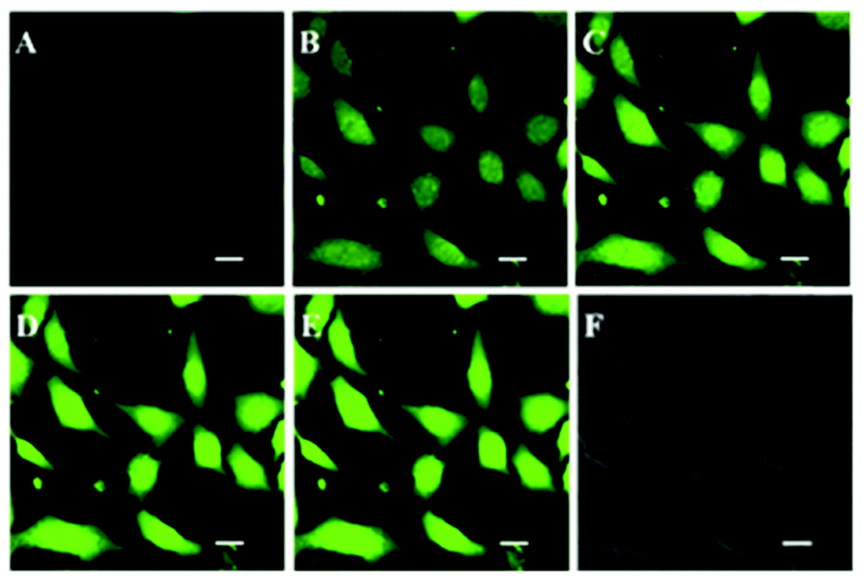
- Yamada, A.; Hiruta, Y.; Wang, J.; Ayano, E.; Kanazawa, H. Design of Environmentally Responsive Fluorescent Polymer Probes for Cellular Imaging. Biomacromolecules 2015, 16, 2356–2362. [Google Scholar] [CrossRef]
- Hiruta, Y.; Shimamura, M.; Matsuura, M.; Maekawa, Y.; Funatsu, T.; Suzuki, Y.; Ayano, E.; Okano, T.; Kanazawa, H. Temperature-Responsive Fluorescence Polymer Probes with Accurate Thermally Controlled Cellular Uptakes. ACS Macro Lett. 2014, 3, 281–285. [Google Scholar] [CrossRef]
- Hiruta, Y.; Funatsu, T.; Matsuura, M.; Wang, J.; Ayano, E.; Kanazawa, H. pH/temperature-responsive fluorescence polymer probe with pH-controlled cellular uptake. Sens. Actuators B Chem. 2015, 207, 724–731. [Google Scholar] [CrossRef]
- Okabe, K.; Inada, N.; Gota, C.; Harada, Y.; Funatsu, T.; Uchiyama, S. Intracellular temperature mapping with a fluorescent polymeric thermometer and fluorescence lifetime imaging microscopy. Nat. Commun. 2012, 3, 705. [Google Scholar] [CrossRef] [PubMed] [Green Version]
- Gota, C.; Okabe, K.; Funatsu, T.; Harada, Y.; Uchiyama, S. Hydrophilic Fluorescent Nanogel Thermometer for Intracellular Thermometry. J. Am. Chem. Soc. 2009, 131, 2766–2767. [Google Scholar] [CrossRef] [PubMed]
- Uchiyama, S.; Tsuji, T.; Kawamoto, K.; Okano, K.; Fukatsu, E.; Noro, T.; Ikado, K.; Yamada, S.; Shibata, Y.; Hayashi, T.; et al. A Cell-Targeted Non-Cytotoxic Fluorescent Nanogel Thermometer Created with an Imidazolium-Containing Cationic Radical Initiator. Angew. Chem. Int. Ed. 2018, 57, 5413–5417. [Google Scholar] [CrossRef] [PubMed]
- Uchiyama, S.; Kimura, K.; Gota, C.; Okabe, K.; Kawamoto, K.; Inada, N.; Yoshihara, T.; Tobita, S. Environment-Sensitive Fluorophores with Benzothiadiazole and Benzoselenadiazole Structures as Candidate Components of a Fluorescent Polymeric Thermometer. Chem.-A Eur. J. 2012, 18, 9552–9563. [Google Scholar] [CrossRef]
- Patil, A.O.; Heeger, A.J.; Wudl, F. Optical properties of conducting polymers. Chem. Rev. 1988, 88, 183–200. [Google Scholar] [CrossRef]
- Andrew, T.L.; Swager, T.M. Structure-Property relationships for exciton transfer in conjugated polymers. J. Polym. Sci. Part B Polym. Phys. 2011, 49, 476–498. [Google Scholar] [CrossRef]
- Das, T.K.; Prusty, S. Review on Conducting Polymers and Their Applications. Polym. Plast. Technol. Eng. 2012, 51, 1487–1500. [Google Scholar] [CrossRef]
- Chiang, C.K.; Fincher, C.R., Jr.; Park, Y.W.; Heeger, A.J.; Shirakawa, H.; Louis, E.J.; Gau, S.C.; MacDiarmid, A.G. Electrical Conductivity in Doped Polyacetylene. Phys. Rev. Lett. 1977, 39, 1098–1101. [Google Scholar] [CrossRef]
- Hudson, B.S. Polyacetylene: Myth and Reality. Materials 2018, 11, 242. [Google Scholar] [CrossRef] [Green Version]
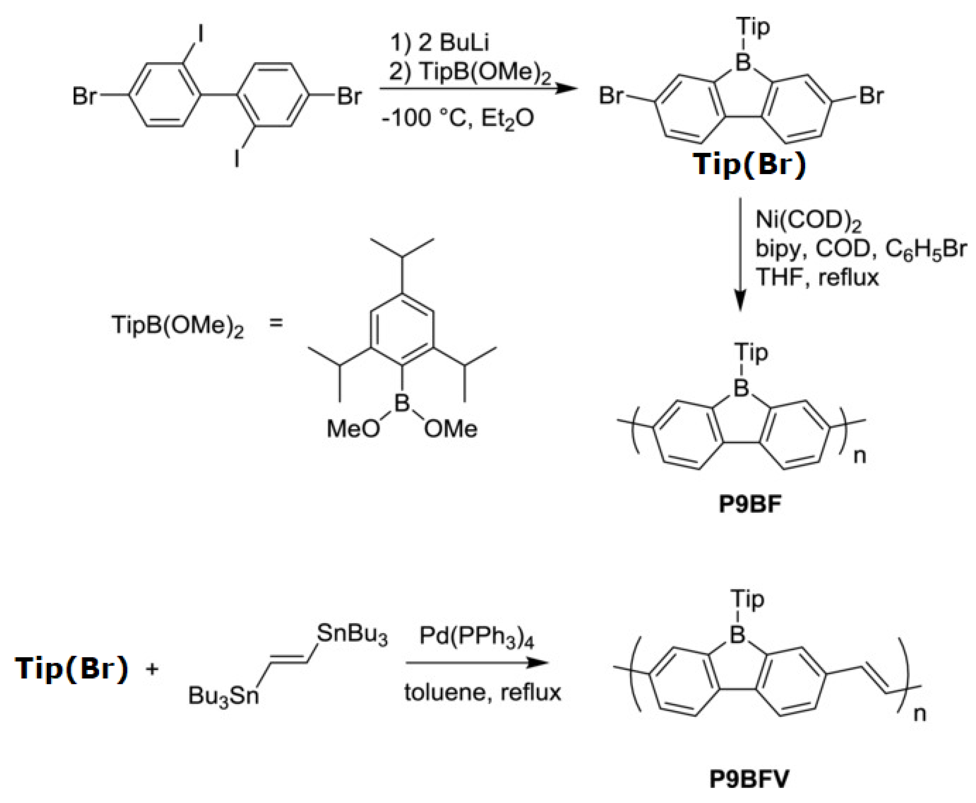
- He, X.; Baumgartner, T. Conjugated main-group polymers for optoelectronics. RSC Adv. 2013, 3, 11334–11350. [Google Scholar] [CrossRef]
- Adams, I.A.; Rupar, P.A. A Poly(9-Borafluorene) Homopolymer: An Electron-Deficient Polyfluorene with “Turn-On” Fluorescence Sensing of NH3 Vapor. Macromol. Rapid Commun. 2015, 36, 1336–1340. [Google Scholar] [CrossRef] [PubMed]
- Kukla, A.; Shirshov, Y.; Piletsky, S. Ammonia sensors based on sensitive polyaniline films. Sens. Actuators B Chem. 1996, 37, 135–140. [Google Scholar] [CrossRef]
- Hibbard, T.; Crowley, K.; Kelly, F.; Ward, F.; Holian, J.; Watson, A.; Killard, A.J. Point of Care Monitoring of Hemodialysis Patients with a Breath Ammonia Measurement Device Based on Printed Polyaniline Nanoparticle Sensors. Anal. Chem. 2013, 85, 12158–12165. [Google Scholar] [CrossRef] [PubMed]
- Timmer, B.; Olthuis, W.; Van Den Berg, A. Ammonia sensors and their applications—A review. Sens. Actuators B Chem. 2005, 107, 666–677. [Google Scholar] [CrossRef]
- Zhou, Z.-H.; Yamamoto, T. Research on carbon—carbon coupling reactions of haloaromatic compounds mediated by zerovalent nickel complexes. Preparation of cyclic oligomers of thiophene and benzene and stable anthrylnickel(II) complexes. J. Organomet. Chem. 1991, 414, 119–127. [Google Scholar] [CrossRef]
- Stille, J.K. The Palladium-Catalyzed Cross-Coupling Reactions of Organotin Reagents with Organic Electrophiles[New Synthetic Methods(58)]. Angew. Chem. Int. Ed. 1986, 25, 508–524. [Google Scholar] [CrossRef]
- Tang, T.; Ahumada, G.; Bielawski, C.W. Direct laser writing of poly(phenylene vinylene) on poly(barrelene). Polym. Chem. 2020, 11, 5437–5443. [Google Scholar] [CrossRef]
- Selimis, A.; Mironov, V.; Farsari, M. Direct laser writing: Principles and materials for scaffold 3D printing. Microelectron. Eng. 2015, 132, 83–89. [Google Scholar] [CrossRef]
- Chen, J.; Fan, Z.; Zhang, C.; Duan, H.; Fan, L.-J. Amphiphilic Conjugated Polyelectrolyte-Based Sensing System for Visually Observable Detection of Neomycin with High Sensitivity. ACS Appl. Polym. Mater. 2021, 3, 2088–2097. [Google Scholar] [CrossRef]
- Wu, P.; Tan, C. Biological Sensing and Imaging Using Conjugated Polymers and Peptide Substrates. Protein Pept. Lett. 2021, 28, 2–10. [Google Scholar] [CrossRef] [PubMed]
- Pak, Y.L.; Wang, Y.; Xu, Q. Conjugated polymer based fluorescent probes for metal ions. Coord. Chem. Rev. 2021, 433, 213745. [Google Scholar] [CrossRef]
- Zhao, X.; Dai, X.; Zhao, S.; Cui, X.; Gong, T.; Song, Z.; Meng, H.; Zhang, X.; Yu, B. Aptamer-based fluorescent sensors for the detection of cancer biomarkers. Spectrochim. Acta Part A Mol. Biomol. Spectrosc. 2021, 247, 119038. [Google Scholar] [CrossRef] [PubMed]
- Wang, T.; Zhang, N.; Bai, W.; Bao, Y. Fluorescent chemosensors based on conjugated polymers with N-heterocyclic moieties: Two decades of progress. Polym. Chem. 2020, 11, 3095–3114. [Google Scholar] [CrossRef]
- Zhang, L.; Huang, X.; Cao, Y.; Xinyan, H.; Ding, L. Fluorescent Binary Ensemble Based on Pyrene Derivative and Sodium Dodecyl Sulfate Assemblies as a Chemical Tongue for Discriminating Metal Ions and Brand Water. ACS Sens. 2017, 2, 1821–1830. [Google Scholar] [CrossRef] [PubMed]
- Ghosh, R.; Chatterjee, D.P.; Das, S.; Mukhopadhyay, T.K.; Datta, A.; Nandi, A.K. Influence of Hofmeister I− on Tuning Optoelectronic Properties of Ampholytic Polythiophene by Varying pH and Conjugating with RNA. Langmuir 2017, 33, 12739–12749. [Google Scholar] [CrossRef]
- Chang, C.-C.; Wang, G.; Takarada, T.; Maeda, M. Iodine-Mediated Etching of Triangular Gold Nanoplates for Colorimetric Sensing of Copper Ion and Aptasensing of Chloramphenicol. ACS Appl. Mater. Interfaces 2017, 9, 34518–34525. [Google Scholar] [CrossRef]
- Han, J.; Wang, B.; Bender, M.; Kushida, S.; Seehafer, K.; Bunz, U.H.F. Poly(aryleneethynylene) Tongue That Identifies Nonsteroidal Anti-Inflammatory Drugs in Water: A Test Case for Combating Counterfeit Drugs. ACS Appl. Mater. Interfaces 2017, 9, 790–797. [Google Scholar] [CrossRef]
- Han, J.; Wang, B.; Bender, M.; Seehafer, K.; Bunz, U.H.F. Water-Soluble Poly(p-aryleneethynylene)s: A Sensor Array Discriminates Aromatic Carboxylic Acids. ACS Appl. Mater. Interfaces 2016, 8, 20415–20421. [Google Scholar] [CrossRef]
- Singh, A.; Bains, D.; Hassen, W.M.; Singh, N.; Dubowski, J.J. Formation of a Au/Au9Ga4 Alloy Nanoshell on a Bacterial Surface through Galvanic Displacement Reaction for High-Contrast Imaging. ACS Appl. Bio Mater. 2020, 3, 477–485. [Google Scholar] [CrossRef] [Green Version]
- Xiao, Q.; Mai, B.; Nie, Y.; Yuan, C.; Xiang, M.; Shi, Z.; Wu, J.; Leung, W.; Xu, C.; Yao, S.Q.; et al. In Vitro and In Vivo Demonstration of Ultraefficient and Broad-Spectrum Antibacterial Agents for Photodynamic Antibacterial Chemotherapy. ACS Appl. Mater. Interfaces 2021, 13, 11588–11596. [Google Scholar] [CrossRef] [PubMed]
- Liu, L.; Wang, X.; Zhu, S.; Li, L. Different Surface Interactions between Fluorescent Conjugated Polymers and Biological Targets. ACS Appl. Bio Mater. 2021, 4, 1211–1220. [Google Scholar] [CrossRef] [PubMed]
- Ourari, A.; Ouari, K.; Moumeni, W.; Sibous, L.; Bouet, G.M.; Khan, M.A. Unsymmetrical Tetradentate Schiff Base Complexes Derived from 2,3-diaminophenol and Salicylaldehyde or 5-bromosalicylaldehyde. Transit. Met. Chem. 2006, 31, 169–175. [Google Scholar] [CrossRef]
- Wu, Y.; Tan, Y.; Wu, J.; Chen, S.; Chen, Y.Z.; Zhou, X.; Jiang, Y.; Tan, C. Fluorescence Array-Based Sensing of Metal Ions Using Conjugated Polyelectrolytes. ACS Appl. Mater. Interfaces 2015, 7, 6882–6888. [Google Scholar] [CrossRef] [PubMed]
- Zhou, Y.; Yang, Q.; Zhang, D.; Gan, N.; Li, Q.; Cuan, J. Detection and removal of antibiotic tetracycline in water with a highly stable luminescent MOF. Sens. Actuators B Chem. 2018, 262, 137–143. [Google Scholar] [CrossRef]
- Liu, M.-K.; Liu, Y.-Y.; Bao, D.-D.; Zhu, G.; Yang, G.-H.; Geng, J.-F.; Li, H.-T. Effective Removal of Tetracycline Antibiotics from Water using Hybrid Carbon Membranes. Sci. Rep. 2017, 7, srep43717. [Google Scholar] [CrossRef] [PubMed] [Green Version]
- Daghrir, R.; Drogui, P. Tetracycline antibiotics in the environment: A review. Environ. Chem. Lett. 2013, 11, 209–227. [Google Scholar] [CrossRef]
- Malik, A.H.; Iyer, P.K. Conjugated Polyelectrolyte Based Sensitive Detection and Removal of Antibiotics Tetracycline from Water. ACS Appl. Mater. Interfaces 2017, 9, 4433–4439. [Google Scholar] [CrossRef] [PubMed]
- Hammerer, F.; Poyer, F.; Fourmois, L.; Chen, S.; Garcia, G.; Teulade-Fichou, M.-P.; Maillard, P.; Mahuteau-Betzer, F. Mitochondria-targeted cationic porphyrin-triphenylamine hybrids for enhanced two-photon photodynamic therapy. Bioorganic Med. Chem. 2018, 26, 107–118. [Google Scholar] [CrossRef]
- Deng, W.; Wu, Q.; Sun, P.; Yuan, P.; Lu, X.; Fan, Q.; Huang, W. Zwitterionic diketopyrrolopyrrole for fluorescence/photoacoustic imaging guided photodynamic/photothermal therapy. Polym. Chem. 2018, 9, 2805–2812. [Google Scholar] [CrossRef]
- Di Maria, F.; Lodola, F.; Zucchetti, E.; Benfenati, F.; Lanzani, G. The evolution of artificial light actuators in living systems: From planar to nanostructured interfaces. Chem. Soc. Rev. 2018, 47, 4757–4780. [Google Scholar] [CrossRef] [PubMed]
- Simelane, N.W.N.; Kruger, C.A.; Abrahamse, H. Targeted Nanoparticle Photodynamic Diagnosis and Therapy of Colorectal Cancer. Int. J. Mol. Sci. 2021, 22, 9779. [Google Scholar] [CrossRef] [PubMed]
- Simelane, N.W.N.; Kruger, C.A.; Abrahamse, H. Photodynamic diagnosis and photodynamic therapy of colorectal cancer in vitro and in vivo. RSC Adv. 2020, 10, 41560–41576. [Google Scholar] [CrossRef]
- Lan, M.; Zhao, S.; Xie, Y.; Zhao, J.; Guo, L.; Niu, G.; Li, Y.; Sun, H.; Zhang, H.; Liu, W.; et al. Water-Soluble Polythiophene for Two-Photon Excitation Fluorescence Imaging and Photodynamic Therapy of Cancer. ACS Appl. Mater. Interfaces 2017, 9, 14590–14595. [Google Scholar] [CrossRef] [PubMed]
- Liu, S.; Feng, G.; Tang, B.Z.; Liu, B. Recent advances of AIE light-up probes for photodynamic therapy. Chem. Sci. 2021, 12, 6488–6506. [Google Scholar] [CrossRef] [PubMed]
- Alam, P.; Leung, N.L.; Zhang, J.; Kwok, R.T.; Lam, J.W.; Tang, B.Z. AIE-based luminescence probes for metal ion detection. Co-ord. Chem. Rev. 2021, 429, 213693. [Google Scholar] [CrossRef]
- Roy, E.; Nagar, A.; Chaudhary, S.; Pal, S. Advanced Properties and Applications of AIEgens-Inspired Smart Materials. Ind. Eng. Chem. Res. 2020, 59, 10721–10736. [Google Scholar] [CrossRef]
- Zhang, J.; He, B.; Hu, Y.; Alam, P.; Zhang, H.; Lam, J.W.Y.; Tang, B.Z. Stimuli-Responsive AIEgens. Adv. Mater. 2021, 33, 2008071. [Google Scholar] [CrossRef]
- Bai, H.; He, W.; Chau, J.H.; Zheng, Z.; Kwok, R.T.; Lam, J.W.; Tang, B.Z. AIEgens for microbial detection and antimicrobial therapy. Biomaterials 2021, 268, 120598. [Google Scholar] [CrossRef]
- Luo, J.; Xie, Z.; Xie, Z.; Lam, J.W.Y.; Cheng, L.; Chen, H.; Qiu, C.; Kwok, H.S.; Zhan, X.; Liu, Y.; et al. Aggregation-Induced Emission of 1-Methyl-1,2,3,4,5-Pentaphenylsilole. Chem. Commun. 2001, 1740–1741. [Google Scholar] [CrossRef]
- Mei, J.; Leung, N.L.C.; Kwok, R.T.K.; Lam, J.W.Y.; Tang, B.Z. Aggregation-Induced Emission: Together We Shine, United We Soar! Chem. Rev. 2015, 115, 11718–11940. [Google Scholar] [CrossRef] [PubMed]
- Wang, J.; Jiang, Q.; Cao, S.; Sun, C.; Zhang, Y.; Qiu, Y.; Wang, H.; Yin, G.; Liao, Y.; Xie, X. Z/E Effect on Phase Behavior of Main-Chain Liquid Crystalline Polymers Bearing AIEgens. Macromolecules 2021, 54, 10740–10749. [Google Scholar] [CrossRef]
- Mu, B.; Zhang, Z.; Quan, X.; Hao, X.; Tian, W. Perylene Bisimide-Based Luminescent Liquid Crystals with Tunable Solid-State Light Emission. ACS Appl. Mater. Interfaces 2021, 13, 57786–57795. [Google Scholar] [CrossRef] [PubMed]
- Li, J.; Wang, J.-H.; Cao, X.-J.; Li, X.-D.; Ren, X.-K.; Yu, Z.-Q. Peripherally Modified Tetraphenylethene: Emerging as a Room-Temperature Luminescent Disc-Like Nematic Liquid Crystal. ACS Appl. Mater. Interfaces 2021, 13, 35207–35213. [Google Scholar] [CrossRef] [PubMed]
- Yang, Q.; Zhu, J.-C.; Li, Z.-X.; Chen, X.-S.; Jiang, Y.-X.; Luo, Z.-W.; Wang, P.; Xie, H.-L. Luminescent Liquid Crystals Based on Carbonized Polymer Dots and Their Polarized Luminescence Application. ACS Appl. Mater. Interfaces 2021, 13, 26522–26532. [Google Scholar] [CrossRef] [PubMed]
- Li, D.; Chen, J.; Hong, M.; Wang, Y.; Haddleton, D.M.; Li, G.-Z.; Zhang, Q. Cationic Glycopolymers with Aggregation-Induced Emission for the Killing, Imaging, and Detection of Bacteria. Biomacromolecules 2021, 22, 2224–2232. [Google Scholar] [CrossRef] [PubMed]
- Guo, Y.; Shi, D.; Luo, Z.-W.; Xu, J.-R.; Li, M.-L.; Yang, L.-H.; Yu, Z.-Q.; Chen, E.-Q.; Xie, H.-L. High Efficiency Luminescent Liquid Crystalline Polymers Based on Aggregation-Induced Emission and “Jacketing” Effect: Design, Synthesis, Photophysical Property, and Phase Structure. Macromolecules 2017, 50, 9607–9616. [Google Scholar] [CrossRef]
- White, T.J.; Broer, D.J. Programmable and adaptive mechanics with liquid crystal polymer networks and elastomers. Nat. Mater. 2015, 14, 1087–1098. [Google Scholar] [CrossRef]
- Peng, H.-S.; Chiu, D.T. Soft fluorescent nanomaterials for biological and biomedical imaging. Chem. Soc. Rev. 2015, 44, 4699–4722. [Google Scholar] [CrossRef] [Green Version]
- Wu, C.; Chiu, D.T. Highly Fluorescent Semiconducting Polymer Dots for Biology and Medicine. Angew. Chem. Int. Ed. 2013, 52, 3086–3109. [Google Scholar] [CrossRef] [Green Version]
- Reisch, A.; Klymchenko, A.S. Fluorescent Polymer Nanoparticles Based on Dyes: Seeking Brighter Tools for Bioimaging. Small 2016, 12, 1968–1992. [Google Scholar] [CrossRef] [Green Version]
- Du, W.; Liu, X.; Liu, L.; Lam, J.W.Y.; Tang, B.Z. Photoresponsive Polymers with Aggregation-Induced Emission. ACS Appl. Polym. Mater. 2021, 3, 2290–2309. [Google Scholar] [CrossRef]
- Liu, H.; Xiong, L.; Kwok, R.T.K.; He, X.; Lam, J.W.Y.; Tang, B.Z. AIE Bioconjugates for Biomedical Applications. Adv. Opt. Mater. 2020, 8, 2000162. [Google Scholar] [CrossRef]
- Cong, Z.; Xie, S.; Jiang, Z.; Zheng, S.; Wang, W.; Wang, W.; Song, H. In vivo photodynamic therapy based on Near-Infrared AIE cationic polymers. Chem. Eng. J. 2021, 431, 133748. [Google Scholar] [CrossRef]
- Su, X.; Gao, Q.; Wang, D.; Han, T.; Tang, B.Z. One-Step Multicomponent Polymerizations for the Synthesis of Multifunctional AIE Polymers. Macromol. Rapid Commun. 2021, 42, e2000471. [Google Scholar] [CrossRef]
- Yuan, Y.; Zhang, C.-J.; Liu, B. A Photoactivatable AIE Polymer for Light-Controlled Gene Delivery: Concurrent Endo/Lysosomal Escape and DNA Unpacking. Angew. Chem. Int. Ed. 2015, 54, 11419–11423. [Google Scholar] [CrossRef]
- Zheng, Y.; Lu, H.; Jiang, Z.; Guan, Y.; Zou, J.; Wang, X.; Cheng, R.; Gao, H. Low-power white light triggered AIE polymer nanoparticles with high ROS quantum yield for mitochondria-targeted and image-guided photodynamic therapy. J. Mater. Chem. B 2017, 5, 6277–6281. [Google Scholar] [CrossRef]
- Porteous, C.M.; Logan, A.; Evans, C.; Ledgerwood, E.C.; Menon, D.K.; Aigbirhio, F.; Smith, R.A.; Murphy, M.P. Rapid uptake of lipophilic triphenylphosphonium cations by mitochondria in vivo following intravenous injection: Implications for mitochondria-specific therapies and probes. Biochim. Biophys. Acta (BBA)-Gen. Subj. 2010, 1800, 1009–1017. [Google Scholar] [CrossRef]
- Zalmi, G.A.; Jadhav, R.W.; Mirgane, H.A.; Bhosale, S.V. Recent Advances in Aggregation-Induced Emission Active Materials for Sensing of Biologically Important Molecules and Drug Delivery System. Molecules 2021, 27, 150. [Google Scholar] [CrossRef]
- Pei, Y.; Wang, Z.; Wang, C. Recent Progress in Polymeric AIE-Active Drug Delivery Systems: Design and Application. Mol. Pharm. 2021, 18, 3951–3965. [Google Scholar] [CrossRef]
- Kumar, K.S.S.; Girish, Y.R.; Ashrafizadeh, M.; Mirzaei, S.; Rakesh, K.P.; Gholami, M.H.; Zabolian, A.; Hushmandi, K.; Orive, G.; Kadumudi, F.B.; et al. AIE-featured tetraphenylethylene nanoarchitectures in biomedical application: Bioimaging, drug delivery and disease treatment. Co-ord. Chem. Rev. 2021, 447, 214135. [Google Scholar] [CrossRef]
- Jain, K.K. Drug Delivery Systems-An Overview. Methods Pharmacol. Toxicol. 2008, 437, 1–50. [Google Scholar] [CrossRef]
- Liow, S.S.; Dou, Q.; Kai, D.; Li, Z.; Sugiarto, S.; Yu, C.Y.Y.; Kwok, R.T.K.; Chen, X.; Wu, Y.-L.; Ong, S.T.; et al. Long-Term Real-Time In Vivo Drug Release Monitoring with AIE Thermogelling Polymer. Small 2017, 13, 1603404. [Google Scholar] [CrossRef] [PubMed]
- Zhou, T.; Hu, R.; Wang, L.; Qiu, Y.; Zhang, G.; Deng, Q.; Zhang, H.; Yin, P.; Situ, B.; Zhan, C.; et al. An AIE-Active Conjugated Polymer with High ROS-Generation Ability and Biocompatibility for Efficient Photodynamic Therapy of Bacterial Infections. Angew. Chem. Int. Ed. 2020, 59, 9952–9956. [Google Scholar] [CrossRef]
- Griffith, R.S.; Black, H.R. Cephalothin—A New Antibiotic. JAMA J. Am. Med. Assoc. 1964, 189, 823–828. [Google Scholar] [CrossRef]


| Polymer | Mw a | Đ | λex (nm) | λem (nm) |
|---|---|---|---|---|
| P(NIPAAm-co-FL) | 52,994 | 1.979 | 490 | 515 |
| P(NIPAAm-co-CO) | 47,494 | 1.873 | 376 | 460 |
| P(NIPAAm-co-RH) | 45,905 | 2.028 | 540 | 588 |
| P(NIPAAm-co-DA) | 43,596 | 1.984 | 335 | 526 |
Publisher’s Note: MDPI stays neutral with regard to jurisdictional claims in published maps and institutional affiliations. |
© 2022 by the authors. Licensee MDPI, Basel, Switzerland. This article is an open access article distributed under the terms and conditions of the Creative Commons Attribution (CC BY) license (https://creativecommons.org/licenses/by/4.0/).
Share and Cite
Ahumada, G.; Borkowska, M. Fluorescent Polymers Conspectus. Polymers 2022, 14, 1118. https://doi.org/10.3390/polym14061118
Ahumada G, Borkowska M. Fluorescent Polymers Conspectus. Polymers. 2022; 14(6):1118. https://doi.org/10.3390/polym14061118
Chicago/Turabian StyleAhumada, Guillermo, and Magdalena Borkowska. 2022. "Fluorescent Polymers Conspectus" Polymers 14, no. 6: 1118. https://doi.org/10.3390/polym14061118
APA StyleAhumada, G., & Borkowska, M. (2022). Fluorescent Polymers Conspectus. Polymers, 14(6), 1118. https://doi.org/10.3390/polym14061118