


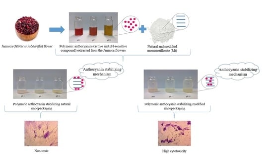
Active and pH-Sensitive Nanopackaging Based on Polymeric Anthocyanin/Natural or Organo-Modified Montmorillonite Blends: Characterization and Assessment of Cytotoxicity


Abstract
1. Introduction
2. Experimental
2.1. Materials
2.2. Manufacture of Potential Active and pH-Sensitive Nanopackaging
2.3. Characterizations of Potential Active and pH-Sensitive Nanopackaging
2.3.1. X-Ray Diffraction (XRD)
2.3.2. Thermogravimetric Analysis (TGA)
2.3.3. Field Emission Scanning Electron Microscopy (FESEM)
2.3.4. Moisture Content (MC)
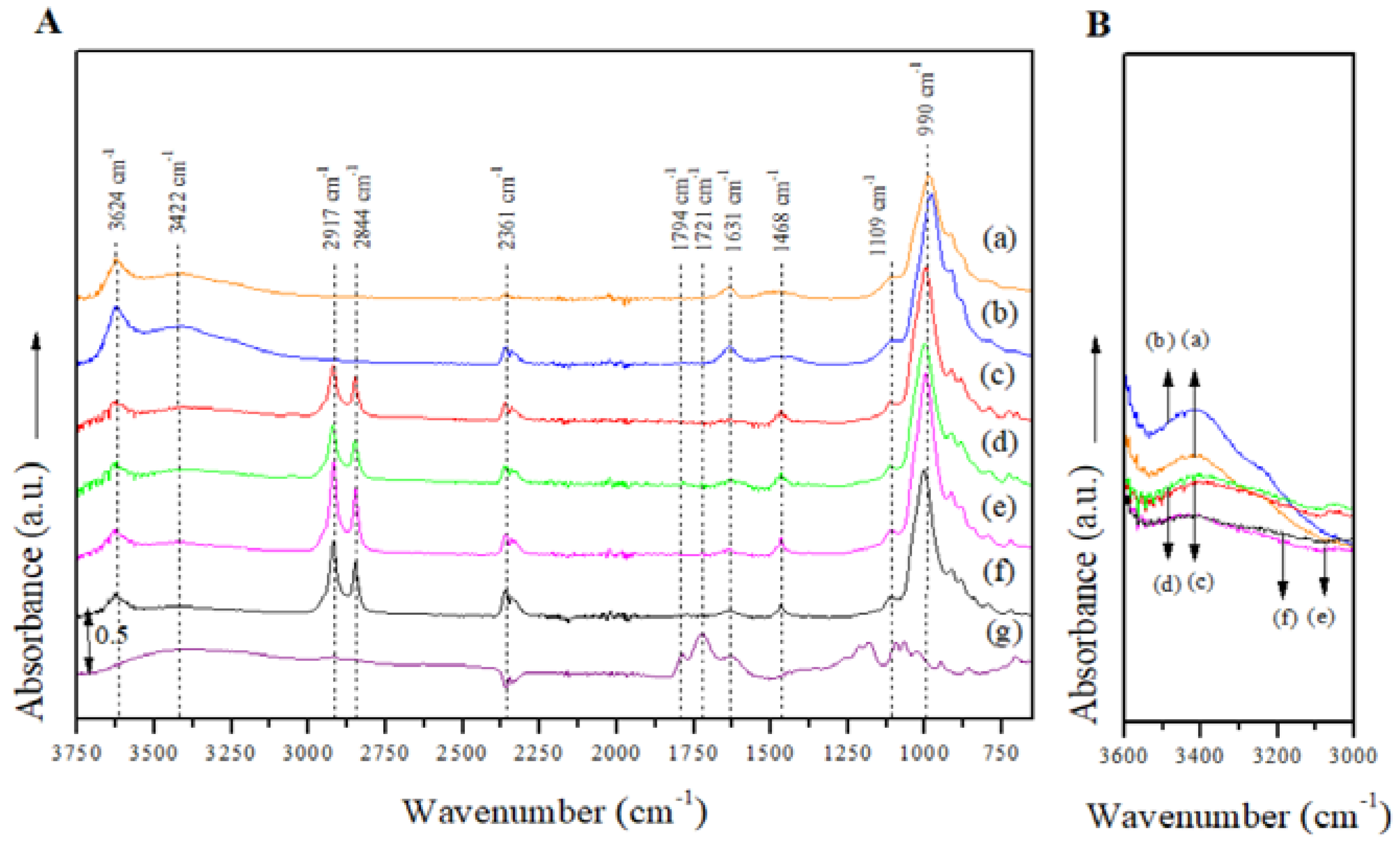
2.3.5. Attenuated Total Reflectance Fourier Transform Infrared (ATR/FTIR) Spectroscopy
2.3.6. Raman Spectroscopy
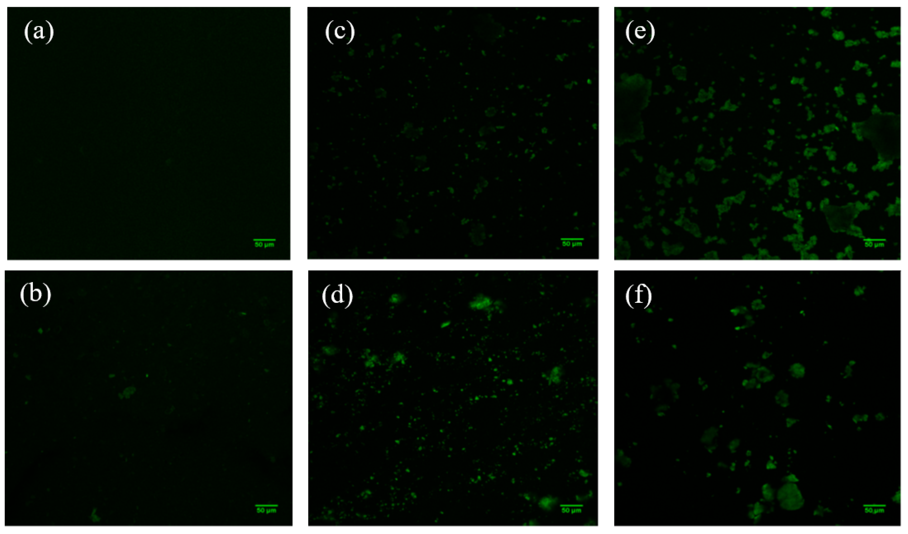
2.3.7. Confocal Laser Scanning Microscopy (CLSM)
2.3.8. Color
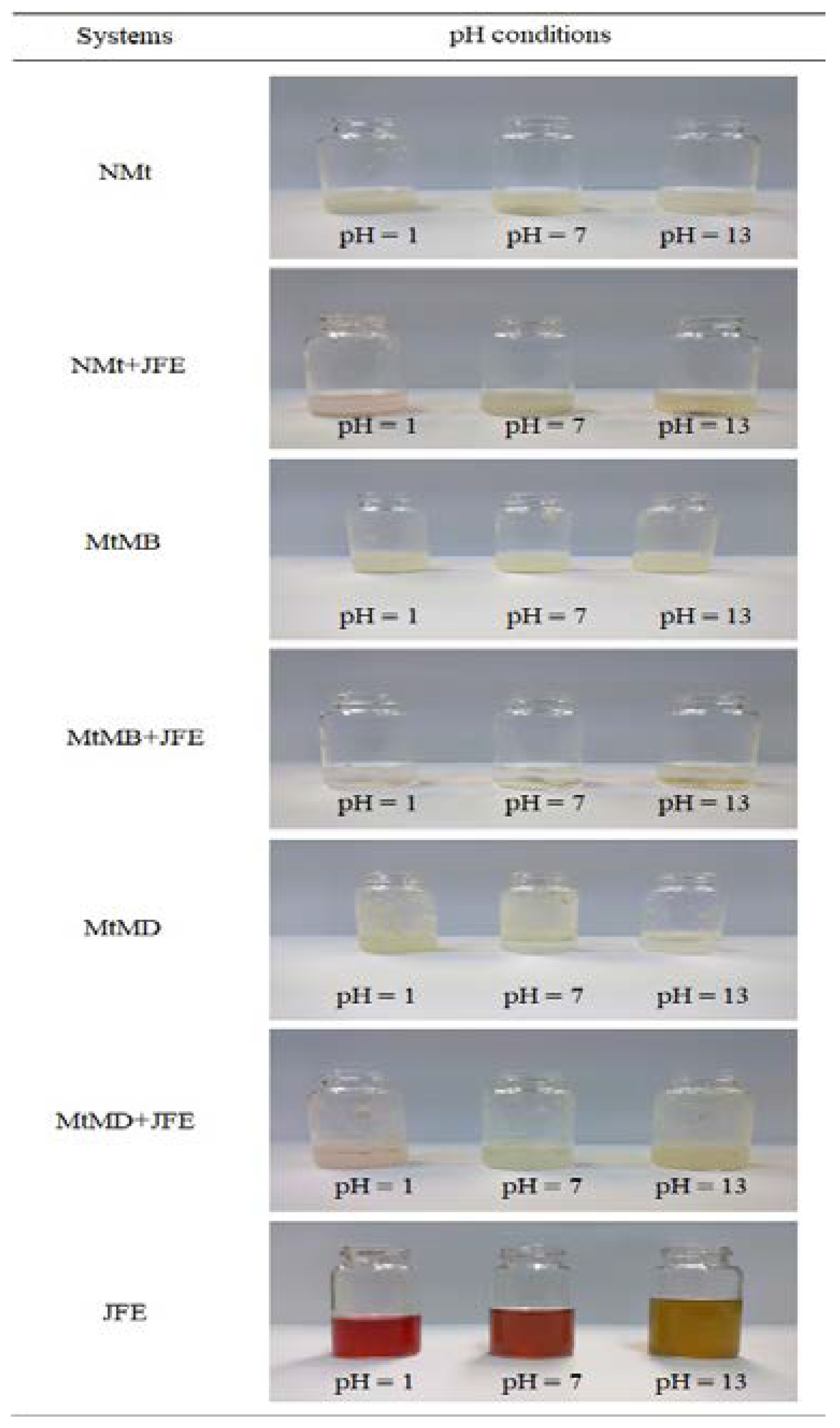
2.3.9. Response to pH Changes
2.3.10. DPPH• Antioxidant Activity
2.3.11. Antimicrobial Activity
2.3.12. Cytotoxicity Assay
2.3.13. Cell Morphology
2.4. Statistic Analysis
3. Results and Discussion
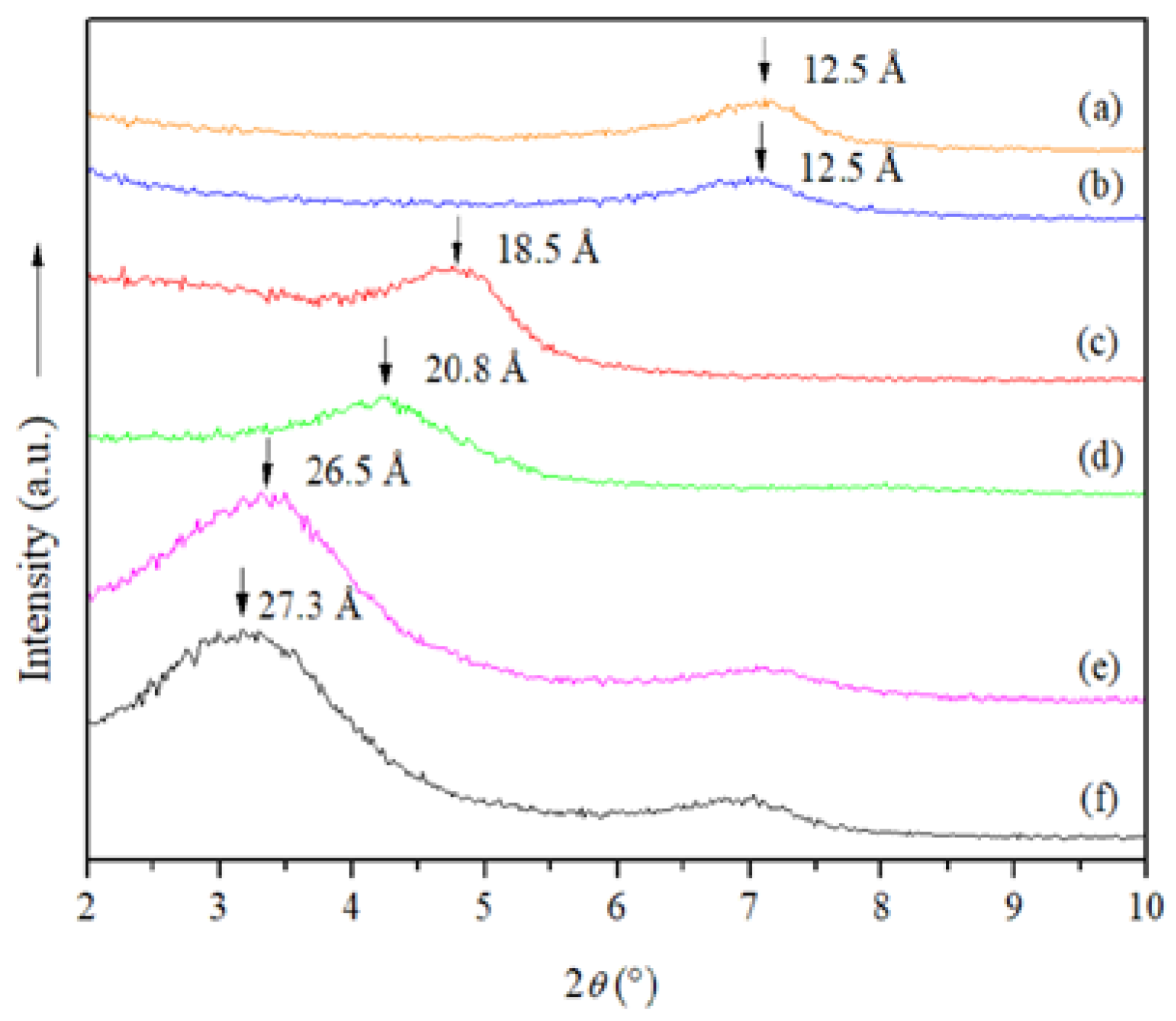
3.1. X-Ray Diffraction (XRD)
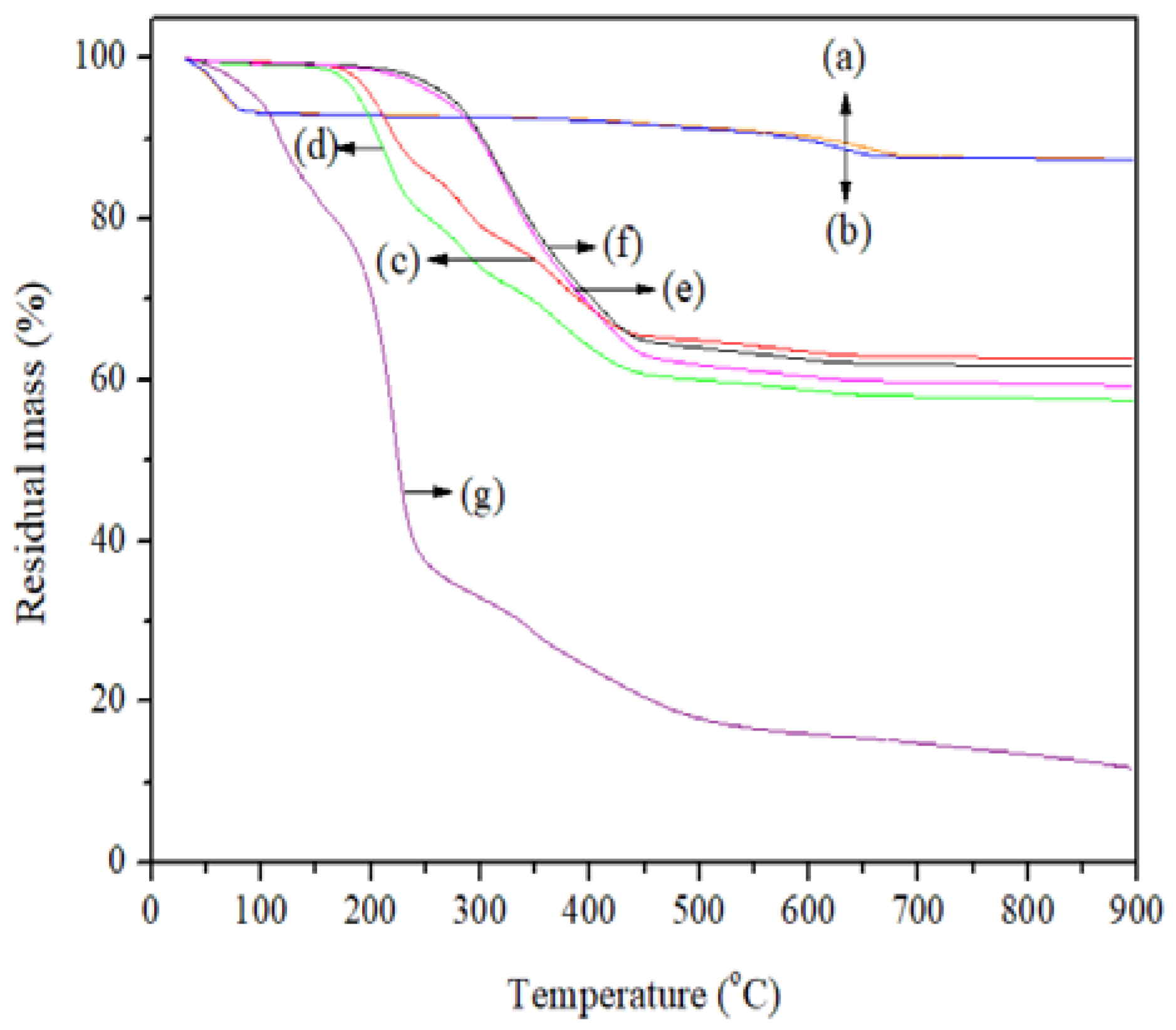
3.2. Thermogravimetric Analysis (TGA)
3.3. Field Emission Scanning Electron Microscopy (FESEM)
3.4. Moisture Content (MC)
3.5. Attenuated Total Reflectance Fourier Transform Infrared (ATR/FTIR) Spectroscopy
3.6. Raman Spectroscopy and Confocal Laser Scanning Microscopy (CLSM)
3.7. Color
3.8. Response to pH Changes
3.9. DPPH• Antioxidant Activity and Antimicrobial Activity
3.10. Cytotoxicity Assay and Cell Morphology
4. Conclusions
Author Contributions
Funding
Institutional Review Board Statement
Data Availability Statement
Acknowledgments
Conflicts of Interest
References
- Castañeda-Ovando, A.; de Lourdes Pacheco-Hernández, M.; Páez-Hernández, M.E.; Rodríguez, J.A.; Galán-Vidal, C.A. Chemical Studies of Anthocyanins: A Review. Food Chem. 2009, 113, 859–871. [Google Scholar] [CrossRef]
- Gonçalves, A.C.; Falcão, A.; Alves, G.; Lopes, J.A.; Silva, L.R. Employ of Anthocyanins in Nanocarriers for Nano Delivery: In Vitro and in Vivo Experimental Approaches for Chronic Diseases. Pharmaceutics 2022, 14, 2272. [Google Scholar] [CrossRef]
- Jabeur, I.; Pereira, E.; Barros, L.; Calhelha, R.C.; Soković, M.; Oliveira, M.B.P.P.; Ferreira, I.C.F.R. Hibiscus sabdariffa L. as a Source of Nutrients, Bioactive Compounds and Colouring Agents. Food Res. Int. 2017, 100, 717–723. [Google Scholar] [CrossRef]
- Ali, B.H.; Cahliková, L.; Opletal, L.; Karaca, T.; Manoj, P.; Ramkumar, A.; Al Suleimani, Y.M.; Al Za’abi, M.; Nemmar, A.; Chocholousova-Havlikova, L.; et al. Effect of Aqueous Extract and Anthocyanins of Calyces of Hibiscus sabdariffa (Malvaceae) in Rats with Adenine-Induced Chronic Kidney Disease. J. Pharm. Pharmacol. 2017, 69, 1219–1229. [Google Scholar] [CrossRef] [PubMed]
- Mozaffari-Khosravi, H.; Jalali-Khanabadi, B.-A.; Afkhami-Ardekani, M.; Fatehi, F.; Noori-Shadkam, M. The Effects of Sour Tea (Hibiscus sabdariffa) on Hypertension in Patients with Type II Diabetes. J. Hum. Hypertens. 2008, 23, 48. [Google Scholar] [CrossRef]
- Panaitescu, M.; Lengyel, E. Monitoring the Antibacterial Activity of Hibiscus sabdariffa Extracts. Manag. Sustain. Dev. 2017, 9, 31–34. [Google Scholar] [CrossRef]
- Abedi-Firoozjah, R.; Yousefi, S.; Heydari, M.; Seyedfatehi, F.; Jafarzadeh, S.; Mohammadi, R.; Rouhi, M.; Garavand, F. Application of Red Cabbage Anthocyanins as pH-Sensitive Pigments in Smart Food Packaging and Sensors. Polymers 2022, 14, 1629. [Google Scholar] [CrossRef]
- Gutiérrez, T.J. Active and Intelligent Films Made from Starchy Sources/Blackberry Pulp. J. Polym. Environ. 2018, 26, 2374–2391. [Google Scholar] [CrossRef]
- Gutiérrez, T.J. Advanced Materials Made from Reactive and Functional Polymers: Editor’s Insights. In Reactive and Functional Polymers Volume Three; Gutiérrez, T.J., Ed.; Springer International Publishing: Cham, Switzerland, 2020; pp. 1–4. [Google Scholar]
- Bracone, M.; Merino, D.; González, J.; Alvarez, V.A.; Gutiérrez, T.J. Nanopackaging from Natural Fillers and Biopolymers for the Development of Active and Intelligent Films. In Natural Polymers: Derivatives, Blends and Composites; Ikram, S., Ahmed, S., Eds.; Nova Science Publishers: New York, NY, USA, 2016; pp. 119–155. ISBN 978-1-63485-831-1. [Google Scholar]
- Gutiérrez, T.J.; Suniaga, J.; Monsalve, A.; García, N.L. Influence of Beet Flour on the Relationship Surface-Properties of Edible and Intelligent Films Made from Native and Modified Plantain Flour. Food Hydrocoll. 2016, 54, 234–244. [Google Scholar] [CrossRef]
- Merz, B.; Capello, C.; Leandro, G.C.; Moritz, D.E.; Monteiro, A.R.; Valencia, G.A. A Novel Colorimetric Indicator Film Based on Chitosan, Polyvinyl Alcohol and Anthocyanins from Jambolan (Syzygium cumini) Fruit for Monitoring Shrimp Freshness. Int. J. Biol. Macromol. 2020, 153, 625–632. [Google Scholar] [CrossRef]
- Gutiérrez, T.J.; Alvarez, V.A. Bionanocomposite Films Developed from Corn Starch and Natural and Modified Nano-Clays with or without Added Blueberry Extract. Food Hydrocoll. 2018, 77, 407–420. [Google Scholar] [CrossRef]
- Gutiérrez, T.J.; Toro-Márquez, L.A.; Merino, D.; Mendieta, J.R. Hydrogen-Bonding Interactions and Compostability of Bionanocomposite Films Prepared from Corn Starch and Nano-Fillers with and without Added Jamaica Flower Extract. Food Hydrocoll. 2019, 89, 283–293. [Google Scholar] [CrossRef]
- Gutiérrez, T.J.; Herniou-Julien, C.; Álvarez, K.; Alvarez, V.A. Structural Properties and in Vitro Digestibility of Edible and pH-Sensitive Films Made from Guinea Arrowroot Starch and Wastes from Wine Manufacture. Carbohydr. Polym. 2018, 184, 135–143. [Google Scholar] [CrossRef] [PubMed]
- Gutiérrez, T.J. Surface and Nutraceutical Properties of Edible Films Made from Starchy Sources with and without Added Blackberry Pulp. Carbohydr. Polym. 2017, 165, 169–179. [Google Scholar] [CrossRef] [PubMed]
- Toro-Márquez, L.A.; Merino, D.; Gutiérrez, T.J. Bionanocomposite Films Prepared from Corn Starch with and without Nanopackaged Jamaica (Hibiscus sabdariffa) Flower Extract. Food Bioprocess Technol. 2018, 11, 1955–1973. [Google Scholar] [CrossRef]
- Gutiérrez, T.J. Are Modified Pumpkin Flour/Plum Flour Nanocomposite Films Biodegradable and Compostable? Food Hydrocoll. 2018, 83, 397–410. [Google Scholar] [CrossRef]
- Sinela, A.; Rawat, N.; Mertz, C.; Achir, N.; Fulcrand, H.; Dornier, M. Anthocyanins Degradation during Storage of Hibiscus sabdariffa Extract and Evolution of Its Degradation Products. Food Chem. 2017, 214, 234–241. [Google Scholar] [CrossRef]
- Ribeiro, H.L.; de Oliveira, A.V.; de Brito, E.S.; Ribeiro, P.R.V.; Souza Filho, M.d.s.M.; Azeredo, H.M.C. Stabilizing Effect of Montmorillonite on Acerola Juice Anthocyanins. Food Chem. 2018, 245, 966–973. [Google Scholar] [CrossRef]
- de Moura, S.C.S.R.; Berling, C.L.; Germer, S.P.M.; Alvim, I.D.; Hubinger, M.D. Encapsulating Anthocyanins from Hibiscus sabdariffa L. Calyces by Ionic Gelation: Pigment Stability during Storage of Microparticles. Food Chem. 2018, 241, 317–327. [Google Scholar] [CrossRef]
- Gutiérrez, T.J.; Álvarez, K. Biopolymers as Microencapsulation Materials in the Food Industry. In Advances in Physicochemical Properties of Biopolymers; Masuelli, M., Renard, D., Eds.; Bentham Science Publishers: Sharjah, United Arab Emirates, 2017; pp. 296–322. [Google Scholar]
- Gutiérrez, T.J.; Ponce, A.G.; Alvarez, V.A. Nano-Clays from Natural and Modified Montmorillonite with and without Added Blueberry Extract for Active and Intelligent Food Nanopackaging Materials. Mater. Chem. Phys. 2017, 194, 283–292. [Google Scholar] [CrossRef]
- Maisanaba, S.; Ortuño, N.; Jordá-Beneyto, M.; Aucejo, S.; Jos, Á. Development, Characterization and Cytotoxicity of Novel Silane-Modified Clay Minerals and Nanocomposites Intended for Food Packaging. Appl. Clay Sci. 2017, 138, 40–47. [Google Scholar] [CrossRef]
- Wang, M.K.; Wang, S.L.; Wang, W.M. Rapid Estimation of Cation-Exchange Capacities of Soils and Clays with Methylene Blue Exchange. Soil Sci. Soc. Am. J. 1996, 60, 138–141. [Google Scholar] [CrossRef]
- Dai, J.; Gupte, A.; Gates, L.; Mumper, R.J. A Comprehensive Study of Anthocyanin-Containing Extracts from Selected Blackberry Cultivars: Extraction Methods, Stability, Anticancer Properties and Mechanisms. Food Chem. Toxicol. 2009, 47, 837–847. [Google Scholar] [CrossRef] [PubMed]
- ASTM D1925-70; Test Method for Yellowness Index of Plastics. ASTM International: West Conshohocken, PA, USA, 2021. Available online: https://www.astm.org/d1925-70r88e01.html (accessed on 26 October 2022).
- Hsu, C.-L.; Chen, W.; Weng, Y.-M.; Tseng, C.-Y. Chemical Composition, Physical Properties, and Antioxidant Activities of Yam Flours as Affected by Different Drying Methods. Food Chem. 2003, 83, 85–92. [Google Scholar] [CrossRef]
- García-Tejeda, Y.V.; López-González, C.; Pérez-Orozco, J.P.; Rendón-Villalobos, R.; Jiménez-Pérez, A.; Flores-Huicochea, E.; Solorza-Feria, J.; Bastida, C.A. Physicochemical and Mechanical Properties of Extruded Laminates from Native and Oxidized Banana Starch during Storage. LWT Food Sci. Technol. 2013, 54, 447–455. [Google Scholar] [CrossRef]
- Molyneux, P. The Use of the Stable Free Radical Diphenylpicrylhydrazyl (DPPH) for Estimating Antioxidant Activity. Songklanakarin J. Sci. Technol. 2004, 26, 211–219. [Google Scholar]
- Mosmann, T. Rapid Colorimetric Assay for Cellular Growth and Survival: Application to Proliferation and Cytotoxicity Assays. J. Immunol. Methods 1983, 65, 55–63. [Google Scholar] [CrossRef]
- Leon, I.E.; Porro, V.; Di Virgilio, A.L.; Naso, L.G.; Williams, P.A.M.; Bollati-Fogolín, M.; Etcheverry, S.B. Antiproliferative and Apoptosis-Inducing Activity of an Oxidovanadium(IV) Complex with the Flavonoid Silibinin against Osteosarcoma Cells. JBIC J. Biol. Inorg. Chem. 2014, 19, 59–74. [Google Scholar] [CrossRef]
- Tunç, S.; Duman, O. Preparation and Characterization of Biodegradable Methyl Cellulose/Montmorillonite Nanocomposite Films. Appl. Clay Sci. 2010, 48, 414–424. [Google Scholar] [CrossRef]
- Tunç, S.; Duman, O. Preparation of Active Antimicrobial Methyl Cellulose/Carvacrol/Montmorillonite Nanocomposite Films and Investigation of Carvacrol Release. LWT Food Sci. Technol. 2011, 44, 465–472. [Google Scholar] [CrossRef]
- de Azeredo, H.M.C. Antimicrobial Nanostructures in Food Packaging. Trends Food Sci. Technol. 2013, 30, 56–69. [Google Scholar] [CrossRef]
- Ouellet-Plamondon, C.M.; Stasiak, J.; Al-Tabbaa, A. The Effect of Cationic, Non-Ionic and Amphiphilic Surfactants on the Intercalation of Bentonite. Colloids Surf. A Physicochem. Eng. Asp. 2014, 444, 330–337. [Google Scholar] [CrossRef]
- Zhu, J.; Qing, Y.; Wang, T.; Zhu, R.; Wei, J.; Tao, Q.; Yuan, P.; He, H. Preparation and Characterization of Zwitterionic Surfactant-Modified Montmorillonites. J. Colloid Interface Sci. 2011, 360, 386–392. [Google Scholar] [CrossRef] [PubMed]
- Merino, D.; Ollier, R.; Lanfranconi, M.; Alvarez, V. Preparation and Characterization of Soy Lecithin-Modified Bentonites. Appl. Clay Sci. 2016, 127, 17–22. [Google Scholar] [CrossRef]
- Zhang, J.; Gupta, R.K.; Wilkie, C.A. Controlled Silylation of Montmorillonite and Its Polyethylene Nanocomposites. Polymer 2006, 47, 4537–4543. [Google Scholar] [CrossRef]
- Öztop, B.; Shahwan, T. Modification of a Montmorillonite–Illite Clay Using Alkaline Hydrothermal Treatment and Its Application for the Removal of Aqueous Cs+ Ions. J. Colloid Interface Sci. 2006, 295, 303–309. [Google Scholar] [CrossRef]
- D’Amico, D.A.; Ollier, R.P.; Alvarez, V.A.; Schroeder, W.F.; Cyras, V.P. Modification of Bentonite by Combination of Reactions of Acid-Activation, Silylation and Ionic Exchange. Appl. Clay Sci. 2014, 99, 254–260. [Google Scholar] [CrossRef]
- Madejová, J.; Komadel, P. Baseline Studies of the Clay Minerals Society Source Clays: Infrared Methods. Clays Clay Miner. 2001, 49, 410–432. [Google Scholar] [CrossRef]
- Farmer, V.C.; Russell, J.D. The Infra-Red Spectra of Layer Silicates. Spectrochim. Acta 1964, 20, 1149–1173. [Google Scholar] [CrossRef]
- Nayak, P.S.; Singh, B.K. Instrumental Characterization of Clay by XRF, XRD and FTIR. Bull. Mater. Sci. 2007, 30, 235–238. [Google Scholar] [CrossRef]
- Pereira, V.A.; de Arruda, I.N.Q.; Stefani, R. Active Chitosan/PVA Films with Anthocyanins from Brassica oleraceae (Red Cabbage) as Time–Temperature Indicators for Application in Intelligent Food Packaging. Food Hydrocoll. 2015, 43, 180–188. [Google Scholar] [CrossRef]
- Zhang, Y.; Hu, Y.; Lin, J.; Fan, Y.; Li, Y.; Lv, Y.; Liu, X. Excitation Wavelength Independence: Toward Low-Threshold Amplified Spontaneous Emission from Carbon Nanodots. ACS Appl. Mater. Interfaces 2016, 8, 25454–25460. [Google Scholar] [CrossRef]
- Obón, J.M.; Castellar, M.R.; Alacid, M.; Fernández-López, J.A. Production of a Red–Purple Food Colorant from Opuntia Stricta Fruits by Spray Drying and Its Application in Food Model Systems. J. Food Eng. 2009, 90, 471–479. [Google Scholar] [CrossRef]
- Steyn, W.J. Prevalence and Functions of Anthocyanins in Fruits. In Anthocyanins: Biosynthesis, Functions, and Applications; Winefield, C., Davies, K., Gould, K., Eds.; Springer: New York, NY, USA, 2009; pp. 86–105. ISBN 978-0-387-77335-3. [Google Scholar]
- Garzón, G.A. Anthocyanins as Natural Colorants and Bioactive Compounds: A Review. Acta Biol. Colomb. 2008, 13, 27–36. [Google Scholar]
- Wong, D.W.S. Química de Los Alimentos: Mecanismos y Teoría; Acribia: Zaragoza, Spain, 1994; ISBN 8420007757. [Google Scholar]
- Badui, S. Química de Los Alimentos, 5th ed.; Longman de México Editores: Mexico City, Mexico, 1999. [Google Scholar]
- Hutchings, J.B. Food Color and Appearance, 2nd ed.; Aspen Publishers, Inc.: Boston, MA, USA, 1999. [Google Scholar]
- Abreu, A.S.; Oliveira, M.; de Sá, A.; Rodrigues, R.M.; Cerqueira, M.A.; Vicente, A.A.; Machado, A.V. Antimicrobial Nanostructured Starch Based Films for Packaging. Carbohydr. Polym. 2015, 129, 127–134. [Google Scholar] [CrossRef] [PubMed]


| Parameter | NMt | NMt + JFE | MtMB | MtMB + JFE | MtMD | MtMD + JFE |
|---|---|---|---|---|---|---|
| Δid (Å) | - | 0.0 ± 0.1 a | 6.0 ± 0.1 b | 8.3 ± 0.1 c | 14.0 ± 0.1 d | 14.8 ± 0.1 e |
| XJFE | - | 0.00 ± 0.01 a | - | 0.10 ± 0.01 c | - | 0.05 ± 0.01 b |
| MC (%) | 5.6 ± 0.7 d | 9.5 ± 0.4 e | 1.8 ± 0.2 b | 2.6 ± 0.2 b,c | 1.4 ± 0.1 a | 2.3 ± 0.3 b |
| L* | 83 ± 5 d | 49 ± 2 a | 65 ± 5 b | 69 ± 2 b,c | 60.4 ± 0.6 b | 68 ± 5 b,c |
| a* | 1.9 ± 0.1 c | 1.48 ± 0.07 b | 1.20 ± 0.08 a | 1.19 ± 0.01 a | 1.43 ± 0.08 b | 1.99 ± 0.01 c |
| b* | 10.6 ± 0.7 d | 6.3 ± 0.1 a | 11.7 ± 0.7 d | 8.2 ± 0.1 c | 12.0 ± 0.3 d,e | 7.70 ± 0.09 b |
| Color difference (ΔE) | 0.00 ± 0.00 a | 34 ± 2 d | 18 ± 5 b | 14 ± 2 b | 22.4 ± 0.6 b,c | 15 ± 5 b |
| WhitenessIndex (WI) | 80 ± 4 d | 49 ± 2 a | 63 ± 5 b | 68 ± 2 b,c | 58.6 ± 0.7 b | 67 ± 4 b,c |
| C* | 10.8 ± 0.7 d | 6.5 ± 0.1 a | 11.7 ± 0.7 d | 8.3 ± 0.1 c | 12.1 ± 0.4 d,e | 7.95 ± 0.09 b |
| h (o) | 190.4 ± 0.2 d | 193.1 ± 0.4 e | 185.84 ± 0.01 a | 188.22 ± 0.01 c | 186.8 ± 0.2 b | 194.5 ± 0.2 f |
Publisher’s Note: MDPI stays neutral with regard to jurisdictional claims in published maps and institutional affiliations. |
© 2022 by the authors. Licensee MDPI, Basel, Switzerland. This article is an open access article distributed under the terms and conditions of the Creative Commons Attribution (CC BY) license (https://creativecommons.org/licenses/by/4.0/).
Share and Cite
Gutiérrez, T.J.; León, I.E.; Ponce, A.G.; Alvarez, V.A. Active and pH-Sensitive Nanopackaging Based on Polymeric Anthocyanin/Natural or Organo-Modified Montmorillonite Blends: Characterization and Assessment of Cytotoxicity. Polymers 2022, 14, 4881. https://doi.org/10.3390/polym14224881
Gutiérrez TJ, León IE, Ponce AG, Alvarez VA. Active and pH-Sensitive Nanopackaging Based on Polymeric Anthocyanin/Natural or Organo-Modified Montmorillonite Blends: Characterization and Assessment of Cytotoxicity. Polymers. 2022; 14(22):4881. https://doi.org/10.3390/polym14224881
Chicago/Turabian StyleGutiérrez, Tomy J., Ignacio E. León, Alejandra G. Ponce, and Vera A. Alvarez. 2022. "Active and pH-Sensitive Nanopackaging Based on Polymeric Anthocyanin/Natural or Organo-Modified Montmorillonite Blends: Characterization and Assessment of Cytotoxicity" Polymers 14, no. 22: 4881. https://doi.org/10.3390/polym14224881
APA StyleGutiérrez, T. J., León, I. E., Ponce, A. G., & Alvarez, V. A. (2022). Active and pH-Sensitive Nanopackaging Based on Polymeric Anthocyanin/Natural or Organo-Modified Montmorillonite Blends: Characterization and Assessment of Cytotoxicity. Polymers, 14(22), 4881. https://doi.org/10.3390/polym14224881