


Progressive Trends on the Biomedical Applications of Metal Organic Frameworks


 ,
,  and
and 
Abstract
1. Introduction
2. Classification of MOFs
2.1. Water-Soluble MOFs
2.2. Water-Stable MOFs

| S. No. | MOF | Metal | Ligand | Use | Targeting Agent | References |
|---|---|---|---|---|---|---|
| 1 | FMOF-1 | Ag | 3,5-bis(trifluoromethyl)-1,2,4-triazolate | Adsorption | CO2, Water | [20] |
| 2 | Al2(OH)2TCPP-Co | Al | 4,4′,4″,4‴-(porphyrin-5,10,15,20-tetrayl) tetrabenzoate | Catalysis | Reduction of carbon dioxide | [20] |
| 3 | CAU-1 | Al | 1,4-benzenedicarboxylate (terephthalate) | Organic Adsorption | Nitrobenzene | [21] |
| 4 | CAU-10 | Al | 1,3-benzene dicarboxylic acid | Sensing applications | Water | [22] |
| 5 | MIL-121 | Al | 1,2,4,5-benzene tetracarboxylic acid, pyromellitic acid | Sensing applications Adsorption conductivity | Hippuric acid, Cu(II) Li and Na | [22] |
| 6 | MIL-96 | Al | 1,3,5,-bezenetricarboxylate | Adsorption | Fluoride, CO2, NO2, p-HBA | [23,24] |
| 7 | Co-ZiF-9 | Co | Benzimidazole | Catalysis | Oxygen evolution reaction | [22] |
| 8 | ZIF-67 | Co | 2-methylimidazole | Adsorption | Benzotriazole, CO2, Methylene blue, Neutral rhodamine, Methyl orange | [25] |
| 9 | MIL-101 | Cr/Al | (O2C)-C6H4-(CO2) | Organic Adsorption, Catalyst, Separation | Ukraine, cyanosilylation reaction, Knoevenagel condensation, CO2/CH4 Separation | [26,27] |
| 10 | Cu(i)-MOF | Cu | 1-benzimidazolyl-3,5-bis(4-pyridyl) benzene | Sensing applications | Water and formaldehyde | [22] |
| 11 | HKUST-1 | Cu | 1,3,5-benzenetricarboxylate | Sensing applications, Adsorption | Water NH3, CO2, NO2 | [28,29] |
| 12 | PCP-33 | Cu | 3,5-bis(2H-tetrazol-5-yl)-benzoic acid | Gas Adsorption | C2H2 | [30] |
| 13 | MIL-68 | Fe | 1,4-benzenedicarboxylate(terephthalate) | Organic Adsorption, Catalyst | Phenol, reduction of Cr(VI), condensation between alcohols and o-aminothio-phenols | [31,32] |
| 14 | PCF-1 | In | 4′-phosphonobiphenyl-3,5-dicarboxylate | Sensing applications | Methylviologen and Cu2+ ions | [33] |
| 15 | AEMOF-1 | Mg | 2,5-dihydroxy-terephthalic acid; N,N-dimethylacetamide | Sensing applications | Water | [34] |
| 16 | MOF-74/PES | Mg | 2,5-dioxide-1,4-benzenedicarboxylate | Membrane Separation | Ultrafiltration (BSA rejection) | [35] |
| 17 | PCMOF-10 | Mg | 2,5-dicarboxy-1,4-benzene-diphosphonic aci | Proton conduction | [22] | |
| 18 | MAF-X25 ox | Mn | 1H,5H-benzo(1,2-d:4,5-d0)bistriazole | Gas Adsorption | CO2 | [36] |
| 19 | Na-HIPAA | Na | Hydroxyphosphonoacetate | Proton conduction | [22] | |
| 20 | BFMOF-1 | Pb | N,N-dimethylacetamide | Sensing applications | H2S | [37] |
| 21 | Tb-DSOA | Tb | 2,2′-disulfonate-4,4′-oxydibenzoic acid | Proton conduction | [22] | |
| 22 | NH2-MIL-125(Ti) | Ti | (O2C)-C6H4-(CO2) | Sensing applications, Adsorption | Water CO2, isoprene | [38,39] |
| 23 | MAF-6 | Zn | 2-ethylimidazolate | Organic Adsorption | Methanol, ethanol, benzene | [40] |
| 24 | ZIF-7 | Zn | Benzimidazole | Membrane Separation, Adsorption | H2/CO2 Separation, Ethane, CH4, CO2 | [41] |
| 25 | ZIF-8 | Zn | 2-methylimidazole | Organic Adsorption, Sensing, Proton conduction Membrane Separation | Phthalic acid, Water sensing, Furfural | [22,42] |
| 26 | ZIF-90 | Zn | 2-carboxaldehyde imidazolate | Membrane Separation Adsorption and Heat storage | H2/CH4 and H2/CO2 Separation Hg(II) | [43] |
| 27 | MENU-500 | Zn, Mo | Benzene tribenzoate; Tetrabutylammonium Ion | Catalysis | Hydrogen evolution reaction | [44] |
| 28 | NU-1000 | Zr | 1,3,6,8-tetrakis(p-benzoic acid)pyrene | Bioimaging | [45] | |
| 29 | NU-1100 | Zr | 4-[2-[3,6,8-tris [2-(4-carboxyphenyl)-ethynyl]-pyren-1-yl]ethynyl]-benzoic acid | Gas Adsorption | H2, CH4 | [46] |
| 30 | PCN-222 | Zr | (Tetrakis(4-carboxyphenyl)porphyrin | Catalysis | Cycloaddition Reactions | [46] |
| 31 | UiO-66 | Zr | Benzene-1,4-dicarboxylic acid | Drug Delivery, Gas Absorption, Membrane separation | Pulmonary drugs | [47,48] |
| 32 | UiO-67 | Zr | Biphenyldicarboxylate | Catalyst Organic Adsorption | Friedel–Crafts Alkylation Toluene | [49,50] |
| 33 | CdEDDA | Cd | EDDA | Sensing applications | Hg(II) | [22] |
| 34 | Co-MOF-74 | Co | 2,5-dioxide-1,4-benzenedicarboxylate | Adsorption and catalyst | CO2, Cycloaddition reaction | [51] |
| 35 | FIR-54 | Zn | tris(4-(1H-imidazole-1-yl)phenyl)amine and Dimethylformamide | Adsorption | Chromium | [52] |
| 36 | MIL-53 | Cr/Fe/Al | (O2C)-C6H4-(CO2) | Membrane Separation Adsorption | Dyes, Heavy metal | [53] |
| 37 | Ni-MOF-74 | Ni | 2,5-dioxide-1,4-benzenedicarboxylate | Adsorption, catalyst | CO2 | [54] |
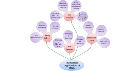
3. Biological Applications of MOF
3.1. MOFs for Biosensing
3.1.1. Enzyme and Protein Biosensing
3.1.2. DNA and RNA Sensing
3.2. Biomedical Imaging of MOFs
3.2.1. Intracellular RNA DNA Bioimaging
3.2.2. MR Imaging (MRI)
3.2.3. X-ray Computed Tomography Imaging
3.2.4. Positron Emission Tomography (PET) Imaging
3.3. MOF as a Drug Delivery System (DDS)
- Surface Adsorption
- Pore Encapsulation
- Covalent Binding
- Functional Molecules as the Building Block
3.4. Miscellaneous Biomedical Applications of MOFs
4. Critical Issues and Role of MOFs in Biological Applications
4.1. Role of Synthesis Techniques
4.2. Role of Organic Linkers
4.2.1. Amino Acids, Peptides, and Protein
4.2.2. Miscellaneous
5. Opportunities and Challenges
Author Contributions
Funding
Acknowledgments
Conflicts of Interest
References
- Li, B.; Wen, H.M.; Zhou, W.; Chen, B. Porous metal-organic frameworks for gas storage and separation: What, how, and why? J. Phys. Chem. Lett. 2014, 5, 3468–3479. [Google Scholar] [CrossRef] [PubMed]
- Shi, X.; Shan, Y.; Du, M.; Pang, H. Synthesis and application of metal-organic framework films. Coord. Chem. Rev. 2021, 444, 214060. [Google Scholar] [CrossRef]
- Imaz, I.; Rubio-Martínez, M.; An, J.; Solé-Font, I.; Rosi, N.L.; Maspoch, D. Metal-biomolecule frameworks (MBioFs). Chem. Commun. 2011, 47, 7287–7302. [Google Scholar] [CrossRef] [PubMed]
- Seki, K.; Takamizawa, S.; Mori, W. Characterization of microporous copper(II) dicarboxylates (fumarate, terephthalate, and trans-1,4-cyclohexanedicarboxylate) by gas adsorption. Chem. Lett. 2001, 30, 122–123. [Google Scholar] [CrossRef]
- Horcajada, P.; Serre, C.; Vallet-Regí, M.; Sebban, M.; Taulelle, F.; Férey, G. Metal–Organic Frameworks as Efficient Materials for Drug Delivery. Angew. Chem. 2006, 118, 6120–6124. [Google Scholar] [CrossRef]
- Wu, P.; Wang, J.; He, C.; Zhang, X.; Wang, Y.; Liu, T.; Duan, C. Luminescent metal-organic frameworks for selectively sensing nitric oxide in an aqueous solution and in living cells. Adv. Funct. Mater 2012, 22, 1698–1703. [Google Scholar] [CrossRef]
- Horcajada, P.; Chalati, T.; Serre, C.; Gillet, B.; Sebrie, C.; Baati, T.; Eubank, J.F.; Heurtaux, D.; Clayette, P.; Kreuz, C.; et al. Porous metal–organic-framework nanoscale carriers as a potential platform for drug delivery and imaging. Nat. Mater. 2010, 9, 172–178. [Google Scholar] [CrossRef]
- Imaz, I.; Rubio-Martínez, M.; García-Fernández, L.; García, F.; Ruiz-Molina, D.; Hernando, J.; Puntes, V.; Maspoch, D. Coordination polymer particles as potential drug delivery systems. Chem. Commun. 2010, 46, 4737. [Google Scholar] [CrossRef]
- Chen, Y.; Lykourinou, V.; Vetromile, C.; Hoang, T.; Ming, L.J.; Larsen, R.W.; Ma, S. How can proteins enter the interior of a MOF? investigation of cytochrome c translocation into a MOF consisting of mesoporous cages with microporous windows. J. Am. Chem. Soc. 2012, 134, 13188–13191. [Google Scholar] [CrossRef]
- Lykourinou, V.; Chen, Y.; Wang, X.-S.; Meng, L.; Hoang, T.; Ming, L.-J.; Musselman, R.L.; Ma, S. Immobilization of MP-11 into a Mesoporous MetalÀOrganic Framework, MP-11@mesoMOF: A New Platform for Enzymatic Catalysis. J. Am. Chem. Soc. 2011, 133, 10382–10385. [Google Scholar] [CrossRef]
- Shieh, F.K.; Wang, S.C.; Yen, C.I.; Wu, C.C.; Dutta, S.; Chou, L.Y.; Morabito, J.V.; Hu, P.; Hsu, M.H.; Wu, K.C.W.; et al. Imparting Functionality to Biocatalysts via Embedding Enzymes into Nanoporous Materials by a de Novo Approach: Size-Selective Sheltering of Catalase in Metal-Organic Framework Microcrystals. J. Am. Chem. Soc. 2015, 137, 4276–4279. [Google Scholar] [CrossRef] [PubMed]
- Lyu, F.; Zhang, Y.; Zare, R.N.; Ge, J.; Liu, Z. One-Pot Synthesis of Protein-Embedded Metal−Organic Frameworks with Enhanced Biological Activities. ACS Publ. 2014, 14, 5761–5765. [Google Scholar] [CrossRef] [PubMed]
- Zhang, X.; Wang, B.; Alsalme, A.; Xiang, S.; Zhang, Z.; Chen, B. Design and applications of water-stable metal-organic frameworks: Status and challenges. Coord. Chem. Rev. 2020, 423, 213507. [Google Scholar] [CrossRef]
- Zhang, S.; Liu, Y.; Li, D.; Wang, Q.; Ran, F. Water-soluble MOF nanoparticles modified polyethersulfone membrane for improving flux and molecular retention. Appl. Surf. Sci. 2020, 505, 144553. [Google Scholar] [CrossRef]
- Law, K.Y. Definitions for hydrophilicity, hydrophobicity, and superhydrophobicity: Getting the basics right. J. Phys. Chem. Lett. 2014, 5, 686–688. [Google Scholar] [CrossRef]
- Guo, W.; Errington, J.R. Effect of Surface Hydrophilicity on the Interfacial Properties of a Model Octane-Water-Silica System. J. Phys. Chem. C 2019, 123, 19649–19658. [Google Scholar] [CrossRef]
- Li, R.Y.; Wang, Z.S.; Yuan, Z.Y.; Van Horne, C.; Freger, V.; Lin, M.; Cai, R.K.; Chen, J.P. A comprehensive review on water stable metal-organic frameworks for large-scale preparation and applications in water quality management based on surveys made since 2015. Crit. Rev. Environ. Sci. Technol. 2021, 52, 4038–4071. [Google Scholar] [CrossRef]
- Rowe, M.D.; Tham, D.H.; Kraft, S.L.; Boyes, S.G. Polymer-modified gadolinium metal-organic framework nanoparticles used as multifunctional nanomedicines for the targeted imaging and treatment of cancer. Biomacromolecules 2009, 10, 983–993. [Google Scholar] [CrossRef]
- Yuan, S.; Feng, L.; Wang, K.; Pang, J.; Bosch, M.; Lollar, C.; Sun, Y.; Qin, J.; Yang, X.; Zhang, P.; et al. Stable Metal–Organic Frameworks: Design, Synthesis, and Applications. Adv. Mater. 2018, 30, 1704303. [Google Scholar] [CrossRef]
- Moghadam, P.Z.; Ivy, J.F.; Arvapally, R.K.; dos Santos, A.M.; Pearson, J.C.; Zhang, L.; Tylianakis, E.; Ghosh, P.; Oswald, I.W.H.; Kaipa, U.; et al. Adsorption and molecular siting of CO2, water, and other gases in the superhydrophobic, flexible pores of FMOF-1 from experiment and simulation. Chem. Sci. 2017, 8, 3989–4000. [Google Scholar] [CrossRef]
- Xie, L.; Liu, D.; Huang, H.; Yang, Q.; Zhong, C. Efficient capture of nitrobenzene from waste water using metal–organic frameworks. Chem. Eng. J. 2014, 246, 142–149. [Google Scholar] [CrossRef]
- Wang, C.; Liu, X.; Demir, N.K.; Chen, J.P.; Li, K. Applications of water stable metal–organic frameworks. Chem. Soc. Rev. 2016, 45, 5107–5134. [Google Scholar] [CrossRef] [PubMed]
- Azhar, M.R.; Abid, H.R.; Tade, M.O.; Periasamy, V.; Sun, H.; Wang, S. Cascade applications of robust MIL-96 metal organic frameworks in environmental remediation: Proof of concept. Chem. Eng. J. 2018, 341, 262–271. [Google Scholar] [CrossRef]
- Abid, H.R.; Rada, Z.H.; Shang, J.; Wang, S. Synthesis, characterization, and CO2 adsorption of three metal-organic frameworks (MOFs): MIL-53, MIL-96, and amino-MIL-53. Polyhedron 2016, 120, 103–111. [Google Scholar] [CrossRef]
- Zhong, G.; Liu, D.; Zhang, J. The application of ZIF-67 and its derivatives: Adsorption, separation, electrochemistry and catalysts. J. Mater. Chem. A 2018, 6, 1887–1899. [Google Scholar] [CrossRef]
- Henschel, A.; Gedrich, K.; Kraehnert, R.; Kaskel, S. Catalytic properties of MIL-101. Chem. Commun. 2008, 35, 4192–4194. [Google Scholar] [CrossRef] [PubMed]
- Serra-Crespo, P.; Ramos-Fernandez, E.V.; Gascon, J.; Kapteijn, F. Synthesis and characterization of an amino functionalized MIL-101(Al): Separation and catalytic properties. Chem. Mater. 2011, 23, 2565–2572. [Google Scholar] [CrossRef]
- Borfecchia, E.; Maurelli, S.; Gianolio, D.; Groppo, E.; Chiesa, M.; Bonino, F.; Lamberti, C. Insights into adsorption of NH 3 on HKUST-1 metal-organic framework: A multitechnique approach. J. Phys. Chem. C 2012, 116, 19839–19850. [Google Scholar] [CrossRef]
- Supronowicz, B.; Mavrandonakis, A.; Heine, T. Interaction of small gases with the unsaturated metal centers of the HKUST-1 metal organic framework. J. Phys. Chem. C 2013, 117, 14570–14578. [Google Scholar] [CrossRef]
- Duan, J.; Jin, W.; Krishna, R. Natural gas purification using a porous coordination polymer with water and chemical stability. Inorg. Chem. 2015, 54, 4279–4284. [Google Scholar] [CrossRef]
- Jing, F.; Liang, R.; Xiong, J.; Chen, R.; Zhang, S.; Li, Y.; Wu, L. MIL-68(Fe) as an efficient visible-light-driven photocatalyst for the treatment of a simulated waste-water contain Cr(VI) and Malachite Green. Appl. Catal. B Environ. 2017, 206, 9–15. [Google Scholar] [CrossRef]
- Wang, D.; Albero, J.; García, H.; Li, Z. Visible-light-induced tandem reaction of o-aminothiophenols and alcohols to benzothiazoles over Fe-based MOFs: Influence of the structure elucidated by transient absorption spectroscopy. J. Catal. 2017, 349, 156–162. [Google Scholar] [CrossRef]
- Dan, W.; Liu, X.; Deng, M.; Ling, Y.; Chen, Z.; Zhou, Y. A highly stable indium phosphonocarboxylate framework as a multifunctional sensor for Cu2+ and methylviologen ions. Dalt. Trans. 2015, 44, 3794–3800. [Google Scholar] [CrossRef] [PubMed]
- Douvali, A.; Tsipis, A.C.; Eliseeva, S.V.; Petoud, S.; Papaefstathiou, G.S.; Malliakas, C.D.; Papadas, I.; Armatas, G.S.; Margiolaki, I.; Kanatzidis, M.G.; et al. Turn-On Luminescence Sensing and Real-Time Detection of Traces of Water in Organic Solvents by a Flexible Metal-Organic Framework. Angew. Chem. 2015, 127, 1671–1676. [Google Scholar] [CrossRef]
- Sotto, A.; Orcajo, G.; Arsuaga, J.M.; Calleja, G.; Landaburu-Aguirre, J. Preparation and characterization of MOF-PES ultrafiltration membranes. J. Appl. Polym. Sci. 2015, 132, 41633. [Google Scholar] [CrossRef]
- Wang, B.; Xie, L.H.; Wang, X.; Liu, X.M.; Li, J.; Li, J.R. Applications of metal–organic frameworks for green energy and environment: New advances in adsorptive gas separation, storage and removal. Green Energy Environ. 2018, 3, 191–228. [Google Scholar] [CrossRef]
- Cui, J.; Wong, Y.-L.; Zeller, M.; Hunter, A.D.; Xu, Z. Pd Uptake and H 2 S Sensing by an Amphoteric Metal-Organic Framework with a Soft Core and Rigid Side Arms. Angew. Chem. 2014, 126, 14666–14670. [Google Scholar] [CrossRef]
- Zhang, Y.; Chen, Y.; Zhang, Y.; Cong, H.; Fu, B.; Wen, S.; Ruan, S. A novel humidity sensor based on NH2-MIL-125(Ti) metal organic framework with high responsiveness. J. Nanoparticle Res. 2013, 15, 2014. [Google Scholar] [CrossRef]
- Sun, Y.; Liu, Y.; Caro, J.; Guo, X.; Song, C.; Liu, Y. In-Plane Epitaxial Growth of Highly c-Oriented NH2-MIL-125(Ti) Membranes with Superior H2/CO2 Selectivity. Angew. Chemie—Int. Ed. 2018, 57, 16088–16093. [Google Scholar] [CrossRef]
- He, C.T.; Jiang, L.; Ye, Z.M.; Krishna, R.; Zhong, Z.S.; Liao, P.Q.; Xu, J.; Ouyang, G.; Zhang, J.P.; Chen, X.M. Exceptional Hydrophobicity of a Large-Pore Metal-Organic Zeolite. J. Am. Chem. Soc. 2015, 137, 7217–7223. [Google Scholar] [CrossRef]
- Chang, H.; Wang, Y.; Xiang, L.; Liu, D.; Wang, C.; Pan, Y. Improved H2/CO2 separation performance on mixed-linker ZIF-7 polycrystalline membranes. Chem. Eng. Sci. 2018, 192, 85–93. [Google Scholar] [CrossRef]
- Khan, N.A.; Jung, B.K.; Hasan, Z.; Jhung, S.H. Adsorption and removal of phthalic acid and diethyl phthalate from water with zeolitic imidazolate and metal–organic frameworks. J. Hazard. Mater. 2015, 282, 194–200. [Google Scholar] [CrossRef] [PubMed]
- Bhattacharjee, S.; Lee, Y.R.; Ahn, W.S. Post-synthesis functionalization of a zeolitic imidazolate structure ZIF-90: A study on removal of Hg(II) from water and epoxidation of alkenes. CrystEngComm 2015, 17, 2575–2582. [Google Scholar] [CrossRef]
- Qin, J.S.; Du, D.Y.; Guan, W.; Bo, X.J.; Li, Y.F.; Guo, L.P.; Su, Z.M.; Wang, Y.Y.; Lan, Y.Q.; Zhou, H.C. Ultrastable Polymolybdate-Based Metal-Organic Frameworks as Highly Active Electrocatalysts for Hydrogen Generation from Water. J. Am. Chem. Soc. 2015, 137, 7169–7177. [Google Scholar] [CrossRef] [PubMed]
- Webber, T.E.; Desai, S.P.; Combs, R.L.; Bingham, S.; Lu, C.C.; Penn, R.L. Size Control of the MOF NU-1000 through Manipulation of the Modulator/Linker Competition. Cryst. Growth Des. 2020, 20, 2965–2972. [Google Scholar] [CrossRef]
- Carrasco, S.; Sanz-Marco, A.; Martín-Matute, B. Fast and Robust Synthesis of Metalated PCN-222 and Their Catalytic Performance in Cycloaddition Reactions with CO2. Organometallics 2019, 38, 3429–3435. [Google Scholar] [CrossRef]
- Jarai, B.M.; Stillman, Z.; Attia, L.; Decker, G.E.; Bloch, E.D.; Fromen, C.A. Evaluating UiO-66 Metal-Organic Framework Nanoparticles as Acid-Sensitive Carriers for Pulmonary Drug Delivery Applications. ACS Appl. Mater. Interfaces 2020, 12, 38989–39004. [Google Scholar] [CrossRef] [PubMed]
- Liu, X. Metal-organic framework UiO-66 membranes. Front. Chem. Sci. Eng. 2019, 14, 216–232. [Google Scholar] [CrossRef]
- Das, A.; Anbu, N.; Sk, M.; Dhakshinamoorthy, A.; Biswas, S. Highly Active Urea-Functionalized Zr(IV)-UiO-67 Metal-Organic Framework as Hydrogen Bonding Heterogeneous Catalyst for Friedel-Crafts Alkylation. Inorg. Chem. 2019, 58, 5163–5172. [Google Scholar] [CrossRef]
- Vo, T.K.; Le, V.N.; Quang, D.T.; Song, M.; Kim, D.; Kim, J. Rapid defect engineering of UiO-67 (Zr) via microwave-assisted continuous-flow synthesis: Effects of modulator species and concentration on the toluene adsorption. Microporous Mesoporous Mater. 2020, 306, 110405. [Google Scholar] [CrossRef]
- Cho, H.Y.; Yang, D.A.; Kim, J.; Jeong, S.Y.; Ahn, W.S. CO2 adsorption and catalytic application of Co-MOF-74 synthesized by microwave heating. Catal. Today 2012, 185, 35–40. [Google Scholar] [CrossRef]
- Chen, Y.; Bai, X.; Ye, Z. Recent Progress in Heavy Metal Ion Decontamination Based on Metal–Organic Frameworks. Nanomaterials 2020, 10, 1481. [Google Scholar] [CrossRef] [PubMed]
- Tomar, S.; Singh, V.K. Review on synthesis and application of MIL-53. Mater. Today Proc. 2021, 43, 3291–3296. [Google Scholar] [CrossRef]
- Xiao, T.; Liu, D. The most advanced synthesis and a wide range of applications of MOF-74 and its derivatives. Microporous Mesoporous Mater. 2019, 283, 88–103. [Google Scholar] [CrossRef]
- Banerjee, D.; Hu, Z.; Li, J. Luminescent metal–organic frameworks as explosive sensors. Dalt. Trans. 2014, 43, 10668–10685. [Google Scholar] [CrossRef] [PubMed]
- Lu, W.; Wei, Z.; Gu, Z.Y.; Liu, T.F.; Park, J.; Park, J.; Tian, J.; Zhang, M.; Zhang, Q.; Gentle, T.; et al. Tuning the structure and function of metal–organic frameworks via linker design. Chem. Soc. Rev. 2014, 43, 5561–5593. [Google Scholar] [CrossRef] [PubMed]
- Desai, A.V.; Sharma, S.; Let, S.; Ghosh, S.K. N-donor linker based metal-organic frameworks (MOFs): Advancement and prospects as functional materials. Coord. Chem. Rev. 2019, 395, 146–192. [Google Scholar] [CrossRef]
- Pan, Y.; Liu, Y.; Zeng, G.; Zhao, L.; Lai, Z. Rapid synthesis of zeolitic imidazolate framework-8 (ZIF-8) nanocrystals in an aqueous system. Chem. Commun. 2011, 47, 2071–2073. [Google Scholar] [CrossRef]
- Gross, A.F.; Sherman, E.; Vajo, J.J. Aqueous room temperature synthesis of cobalt and zinc sodalite zeolitic imidizolate frameworks. Dalt. Trans. 2012, 41, 5458–5460. [Google Scholar] [CrossRef]
- Häckl, K.; Kunz, W. Some aspects of green solvents. Comptes Rendus Chim. 2018, 21, 572–580. [Google Scholar] [CrossRef]
- Duan, C.; Yu, Y.; Xiao, J.; Zhang, X.; Li, L.; Yang, P.; Wu, J.; Xi, H. Water-based routes for synthesis of metal-organic frameworks: A review. Sci. China Mater. 2020, 63, 667–685. [Google Scholar] [CrossRef]
- Huo, J.; Brightwell, M.; Scientific, T.F.; El Hankari, S.; Garai, A. A versatile, industrially relevant, aqueous room temperature synthesis of HKUST-1 with high space-time yield Polyaniline gel nanoconposites View project Fluorescent MOFs View project. Artic. J. Mater. Chem. A 2013, 1, 15220–15223. [Google Scholar] [CrossRef]
- Cheng, X.; Zhang, A.; Hou, K.; Liu, M.; Wang, Y.; Song, C.; Zhang, G.; Guo, X. Size- and morphology-controlled NH2-MIL-53(Al) prepared in DMF-water mixed solvents. Dalt. Trans. 2013, 42, 13698–13705. [Google Scholar] [CrossRef]
- Morozova, S.M.; Sharsheeva, A.; Morozov, M.I.; Vinogradov, A.V.; Hey-Hawkins, E. Bioresponsive metal–organic frameworks: Rational design and function. Coord. Chem. Rev. 2021, 431, 213682. [Google Scholar] [CrossRef]
- Qiu, G.H.; Lu, W.Z.; Hu, P.P.; Jiang, Z.H.; Bai, L.P.; Wang, T.R.; Li, M.M.; Chen, J.X. A metal-organic framework based PCR-free biosensor for the detection of gastric cancer associated microRNAs. J. Inorg. Biochem. 2017, 177, 138–142. [Google Scholar] [CrossRef] [PubMed]
- Liu, C.S.; Sun, C.X.; Tian, J.Y.; Wang, Z.W.; Ji, H.F.; Song, Y.; Zhang, S.; Zhang, Z.H.; He, L.H.; Du, M. Highly stable aluminum-based metal-organic frameworks as biosensing platforms for assessment of food safety. Biosens. Bioelectron. 2017, 91, 804–810. [Google Scholar] [CrossRef]
- Zhao, H.Q.; Qiu, G.H.; Liang, Z.; Li, M.M.; Sun, B.; Qin, L.; Yang, S.P.; Chen, W.H.; Chen, J.X. A zinc(II)-based two-dimensional MOF for sensitive and selective sensing of HIV-1 ds-DNA sequences. Anal. Chim. Acta 2016, 922, 55–63. [Google Scholar] [CrossRef]
- Qin, L.; Lin, L.X.; Fang, Z.P.; Yang, S.P.; Qiu, G.H.; Chen, J.X.; Chen, W.H. A water-stable metal–organic framework of a zwitterionic carboxylate with dysprosium: A sensing platform for Ebolavirus RNA sequences. Chem. Commun. 2015, 52, 132–135. [Google Scholar] [CrossRef]
- Dong, S.; Zhang, D.; Cui, H.; Huang, T. ZnO/porous carbon composite from a mixed-ligand MOF for ultrasensitive electrochemical immunosensing of C-reactive protein. Sens. Actuators B Chem. 2019, 284, 354–361. [Google Scholar] [CrossRef]
- Xia, C.; Xu, Y.; Cao, M.M.; Liu, Y.P.; Xia, J.F.; Jiang, D.Y.; Zhou, G.H.; Xie, R.J.; Zhang, D.F.; Li, H.L. A selective and sensitive fluorescent probe for bilirubin in human serum based on europium(III) post-functionalized Zr(IV)-Based MOFs. Talanta 2020, 212, 120795. [Google Scholar] [CrossRef]
- Bhardwaj, N.; Bhardwaj, S.K.; Mehta, J.; Kim, K.H.; Deep, A. MOF-bacteriophage biosensor for highly sensitive and specific detection of staphylococcus aureus. ACS Appl. Mater. Interfaces 2017, 9, 33589–33598. [Google Scholar] [CrossRef] [PubMed]
- Liu, S.; Bai, J.; Huo, Y.; Ning, B.; Peng, Y.; Li, S.; Han, D.; Kang, W.; Gao, Z. A zirconium-porphyrin MOF-based ratiometric fluorescent biosensor for rapid and ultrasensitive detection of chloramphenicol. Biosens. Bioelectron. 2020, 149, 111801. [Google Scholar] [CrossRef] [PubMed]
- He, J.; Yang, H.; Zhang, Y.; Yu, J.; Miao, L.; Song, Y.; Wang, L. Smart Nanocomposites of Cu-Hemin Metal-Organic Frameworks for Electrochemical Glucose Biosensing. Sci. Rep. 2016, 6, 36637. [Google Scholar] [CrossRef]
- Cesewski, E.; Johnson, B.N. Electrochemical biosensors for pathogen detection. Biosens. Bioelectron. 2020, 159, 112214. [Google Scholar] [CrossRef]
- Ebralidze, I.I.; Laschuk, N.O.; Poisson, J.; Zenkina, O.V. Colorimetric Sensors and Sensor Arrays. In Nanomaterials Design for Sensing Applications; Zenkina, O., Ed.; Elsevier: Amsterdam, The Netherlands, 2019; pp. 1–39. [Google Scholar] [CrossRef]
- Roda, A.; Mirasoli, M.; Michelini, E.; Di Fusco, M.; Zangheri, M.; Cevenini, L.; Roda, B.; Simoni, P. Progress in chemical luminescence-based biosensors: A critical review. Biosens. Bioelectron. 2016, 76, 164–179. [Google Scholar] [CrossRef] [PubMed]
- Metkar, S.K.; Girigoswami, K. Diagnostic biosensors in medicine—A review. Biocatal. Agric. Biotechnol. 2019, 17, 271–283. [Google Scholar] [CrossRef]
- Zhang, G.Y.; Zhuang, Y.H.; Shan, D.; Su, G.F.; Cosnier, S.; Zhang, X.J. Zirconium-based porphyrinic metal-organic framework (PCN-222): Enhanced photoelectrochemical response and its application for label-free phosphoprotein detection. Anal. Chem. 2016, 88, 11207–11212. [Google Scholar] [CrossRef]
- Jiang, Z.W.; Zhao, T.T.; Li, C.M.; Li, Y.F.; Huang, C.Z. 2D MOF-Based Photoelectrochemical Aptasensor for SARS-CoV-2 Spike Glycoprotein Detection. ACS Appl. Mater. Interfaces 2021, 13, 49754–49761. [Google Scholar] [CrossRef]
- Wang, L.; Hu, Z.; Wu, S.; Pan, J.; Xu, X.; Niu, X. A peroxidase-mimicking Zr-based MOF colorimetric sensing array to quantify and discriminate phosphorylated proteins. Anal. Chim. Acta 2020, 1121, 26–34. [Google Scholar] [CrossRef]
- Wang, Y.; Hu, Y.; He, Q.; Yan, J.; Xiong, H.; Wen, N.; Cai, S.; Peng, D.; Liu, Y.; Liu, Z. Metal-organic frameworks for virus detection. Biosens. Bioelectron. 2020, 169, 112604. [Google Scholar] [CrossRef]
- Lin, X.; Lian, X.; Luo, B.; Huang, X.C. A highly sensitive and stable electrochemical HBV DNA biosensor based on ErGO-supported Cu-MOF. Inorg. Chem. Commun. 2020, 119, 108095. [Google Scholar] [CrossRef]
- Han, Q.; Zhang, D.; Zhang, R.; Tang, J.; Xu, K.; Shao, M.; Li, Y.; Du, P.; Zhang, R.; Yang, D.; et al. DNA-functionalized metal-organic framework ratiometric nanoprobe for MicroRNA detection and imaging in live cells. Sens. Actuators B Chem. 2022, 361, 131676. [Google Scholar] [CrossRef]
- Rahmati, Z.; Roushani, M. SARS-CoV-2 virus label-free electrochemical nanohybrid MIP-aptasensor based on Ni3(BTC)2 MOF as a high-performance surface substrate. Microchim. Acta 2022, 189, 287. [Google Scholar] [CrossRef] [PubMed]
- Wang, H.S. Metal–organic frameworks for biosensing and bioimaging applications. Coord. Chem. Rev. 2017, 349, 139–155. [Google Scholar] [CrossRef]
- Wang, H.S.; Li, J.; Li, J.Y.; Wang, K.; Ding, Y.; Xia, X.H. Lanthanide-based metal-organic framework nanosheets with unique fluorescence quenching properties for two-color intracellular adenosine imaging in living cells. NPG Asia Mater. 2017, 9, e354. [Google Scholar] [CrossRef]
- Zhou, J.; Tian, G.; Zeng, L.; Song, X.; Bian, X.W. Nanoscaled Metal-Organic Frameworks for Biosensing, Imaging, and Cancer Therapy. Adv. Healthc. Mater. 2018, 7, 1800022. [Google Scholar] [CrossRef]
- Cadoni, E.; Manicardi, A.; Madder, A. PNA-Based MicroRNA Detection Methodologies. Molecules 2020, 25, 1296. [Google Scholar] [CrossRef]
- Peller, M.; Lanza, A.; Wuttke, S. MRI-Active Metal-Organic Frameworks: Concepts for the Translation from Lab to Clinic. Adv. Ther. 2021, 4, 2100067. [Google Scholar] [CrossRef]
- Taylor, K.M.L.; Jin, A.; Lin, W. Surfactant-assisted synthesis of nanoscale gadolinium metal–organic frameworks for potential multimodal imaging. Angew. Chem. Int. Ed. 2008, 47, 7722–7725. [Google Scholar] [CrossRef]
- Rieter, W.J.; Taylor, K.M.L.; An, H.; Lin, W.; Lin, W. Nanoscale metal-organic frameworks as potential multimodal contrast enhancing agents. J. Am. Chem. Soc. 2006, 128, 9024–9025. [Google Scholar] [CrossRef]
- Wu, M.X.; Yang, Y.W. Metal–Organic Framework (MOF)-Based Drug/Cargo Delivery and Cancer Therapy. Adv. Mater. 2017, 29, 1606134. [Google Scholar] [CrossRef] [PubMed]
- Jia, M.; Yang, X.; Chen, Y.; He, M.; Zhou, W.; Lin, J.; An, L.; Yang, S. Grafting of Gd-DTPA onto MOF-808 to enhance MRI performance for guiding photothermal therapy. J. Mater. Chem. B 2021, 9, 8631–8638. [Google Scholar] [CrossRef] [PubMed]
- Yang, Y.; Liu, J.; Liang, C.; Feng, L.; Fu, T.; Dong, Z.; Chao, Y.; Li, Y.; Lu, G.; Chen, M.; et al. Nanoscale Metal-Organic Particles with Rapid Clearance for Magnetic Resonance Imaging-Guided Photothermal Therapy. ACS Nano 2016, 10, 2774–2781. [Google Scholar] [CrossRef] [PubMed]
- Chowdhuri, A.R.; Bhattacharya, D.; Sahu, S.K. Magnetic nanoscale metal organic frameworks for potential targeted anticancer drug delivery, imaging and as an MRI contrast agent. Dalt. Trans. 2016, 45, 2963–2973. [Google Scholar] [CrossRef]
- Gao, X.; Zhai, M.; Guan, W.; Liu, J.; Liu, Z.; Damirin, A. Controllable synthesis of a smart multifunctional nanoscale metal-organic framework for magnetic resonance/optical imaging and targeted drug delivery. ACS Appl. Mater. Interfaces 2017, 9, 3455–3462. [Google Scholar] [CrossRef]
- Qin, L.; Sun, Z.Y.; Cheng, K.; Liu, S.W.; Pang, J.X.; Xia, L.M.; Chen, W.H.; Cheng, Z.; Chen, J.X. Zwitterionic Manganese and Gadolinium Metal-Organic Frameworks as Efficient Contrast Agents for in Vivo Magnetic Resonance Imaging. ACS Appl. Mater. Interfaces 2017, 9, 41378–41386. [Google Scholar] [CrossRef]
- Banerjee, S.; Lollar, C.T.; Xiao, Z.; Fang, Y.; Zhou, H.C. Biomedical Integration of Metal–Organic Frameworks. Trends Chem. 2020, 2, 467–479. [Google Scholar] [CrossRef]
- Zhang, T.; Wang, L.; Ma, C.; Wang, W.; Ding, J.; Liu, S.; Zhang, X.; Xie, Z. BODIPY-containing nanoscale metal–organic frameworks as contrast agents for computed tomography. J. Mater. Chem. B 2017, 5, 2330–2336. [Google Scholar] [CrossRef]
- Shang, W.; Zeng, C.; Du, Y.; Hui, H.; Liang, X.; Chi, C.; Wang, K.; Wang, Z.; Tian, J. Core–Shell Gold Nanorod@Metal–Organic Framework Nanoprobes for Multimodality Diagnosis of Glioma. Adv. Mater. 2017, 29, 1604381. [Google Scholar] [CrossRef]
- Yang, J.; Yang, Y.W. Metal–Organic Frameworks for Biomedical Applications. Small 2020, 16, 1906846. [Google Scholar] [CrossRef]
- Chen, D.; Yang, D.; Dougherty, C.A.; Lu, W.; Wu, H.; He, X.; Cai, T.; Van Dort, M.E.; Ross, B.D.; Hong, H. In Vivo Targeting and Positron Emission Tomography Imaging of Tumor with Intrinsically Radioactive Metal-Organic Frameworks Nanomaterials. ACS Nano 2017, 11, 4315–4327. [Google Scholar] [CrossRef] [PubMed]
- Zhang, S.; Pei, X.; Gao, H.; Chen, S.; Wang, J. Metal-organic framework-based nanomaterials for biomedical applications. Chin. Chem. Lett. 2020, 31, 1060–1070. [Google Scholar] [CrossRef]
- Duan, D.; Liu, H.; Xu, M.; Chen, M.; Han, Y.; Shi, Y.; Liu, Z. Size-Controlled Synthesis of Drug-Loaded Zeolitic Imidazolate Framework in Aqueous Solution and Size Effect on Their Cancer Theranostics in Vivo. ACS Appl. Mater. Interfaces 2018, 10, 42165–42174. [Google Scholar] [CrossRef]
- He, Z.; Huang, X.; Wang, C.; Li, X.; Liu, Y.; Zhou, Z.; Wang, S.; Zhang, F.; Wang, Z.; Jacobson, O.; et al. A Catalase-Like Metal-Organic Framework Nanohybrid for O 2 -Evolving Synergistic Chemoradiotherapy. Angew. Chem. 2019, 131, 8844–8848. [Google Scholar] [CrossRef]
- Park, J.; Xu, M.; Li, F.; Zhou, H.C. 3D Long-Range Triplet Migration in a Water-Stable Metal-Organic Framework for Upconversion-Based Ultralow-Power in Vivo Imaging. J. Am. Chem. Soc. 2018, 140, 5493–5499. [Google Scholar] [CrossRef] [PubMed]
- Zhang, H.; Shang, Y.; Li, Y.H.; Sun, S.K.; Yin, X.B. Smart Metal-Organic Framework-Based Nanoplatforms for Imaging-Guided Precise Chemotherapy. ACS Appl. Mater. Interfaces 2019, 11, 1886–1895. [Google Scholar] [CrossRef]
- Brock, D.J.; Kustigian, L.; Jiang, M.; Graham, K.; Wang, T.Y.; Erazo-Oliveras, A.; Najjar, K.; Zhang, J.; Rye, H.; Pellois, J.P. Efficient cell delivery mediated by lipid-specific endosomal escape of supercharged branched peptides. Traffic 2018, 19, 421–435. [Google Scholar] [CrossRef]
- He, L.; Wang, T.; An, J.; Li, X.; Zhang, L.; Li, L.; Li, G.; Wu, X.; Su, Z.; Wang, C. Carbon nanodots@zeolitic imidazolate framework-8 nanoparticles for simultaneous pH-responsive drug delivery and fluorescence imaging. CrystEngComm 2014, 16, 3259–3263. [Google Scholar] [CrossRef]
- Dekrafft, K.E.; Xie, Z.; Cao, G.; Tran, S.; Liqing, M.; Zhou, O.Z.; Lin, W. Iodinated Nanoscale Coordination Polymers as Potential Contrast Agents for Computed Tomography. Angew. Chem. Int. Ed. 2009, 48, 9901–9904. [Google Scholar] [CrossRef]
- Taylor, K.M.L.; Rieter, W.J.; Lin, W. Manganese-based nanoscale metal-organic frameworks for magnetic resonance imaging. J. Am. Chem. Soc. 2008, 130, 14358–14359. [Google Scholar] [CrossRef]
- Sun, Y.; Zheng, L.; Yang, Y.; Qian, X.; Fu, T.; Li, X.; Yang, Z.; Yan, H.; Cui, C.; Tan, W. Metal–Organic Framework Nanocarriers for Drug Delivery in Biomedical Applications. Nano-Micro Lett. 2020, 12, 103. [Google Scholar] [CrossRef] [PubMed]
- Huxford, R.C.; Della Rocca, J.; Lin, W. Metal–organic frameworks as potential drug carriers. Curr. Opin. Chem. Biol. 2010, 14, 262–268. [Google Scholar] [CrossRef] [PubMed]
- Singh, R. Geetanjali Metal organic frameworks for drug delivery. In Applications of Nanocomposite Materials in Drug Delivery; Inamuddin, Asiri, A., Mohammad, A., Eds.; Elsevier: Amsterdam, The Netherlands, 2018; pp. 605–617. [Google Scholar] [CrossRef]
- Beg, S.; Rahman, M.; Jain, A.; Saini, S.; Midoux, P.; Pichon, C.; Jalees Ahmad, F.; Akhter, S. Nanoporous metal organic frameworks as hybrid polymer–metal composites for drug delivery and biomedical applications. Drug Discov. Today 2017, 22, 625–637. [Google Scholar] [CrossRef] [PubMed]
- Miller, S.R.; Heurtaux, D.; Baati, T.; Horcajada, P.; Grenèche, J.M.; Serre, C. Biodegradable therapeutic MOFs for the delivery of bioactive molecules. Chem. Commun. 2010, 46, 4526–4528. [Google Scholar] [CrossRef] [PubMed]
- Zhang, D.H.; Yuwen, L.X.; Peng, L.J. Parameters affecting the performance of immobilized enzyme. J. Chem. 2013, 2013, 946248. [Google Scholar] [CrossRef]
- Pisklak, T.J.; Macías, M.; Coutinho, D.H.; Huang, R.S.; Balkus, K.J. Hybrid materials for immobilization of MP-11 catalyst. Top. Catal. 2006, 38, 269–278. [Google Scholar] [CrossRef]
- Zhuang, J.; Kuo, C.H.; Chou, L.Y.; Liu, D.Y.; Weerapana, E.; Tsung, C.K. Optimized metal-organic-framework nanospheres for drug delivery: Evaluation of small-molecule encapsulation. ACS Nano 2014, 8, 2812–2819. [Google Scholar] [CrossRef]
- Jung, S.; Kim, Y.; Kim, S.J.; Kwon, T.H.; Huh, S.; Park, S. Bio-functionalization of metal–organic frameworks by covalent protein conjugation. Chem. Commun. 2011, 47, 2904–2906. [Google Scholar] [CrossRef]
- Saunders, C.D.L.; Burford, N.; Werner-Zwanziger, U.; McDonald, R. Preparation and comprehensive characterization of [Hg6(alanine) 4(NO3)4]·H2O. Inorg. Chem. 2008, 47, 3693–3699. [Google Scholar] [CrossRef]
- Katsoulidis, A.P.; Park, K.S.; Antypov, D.; Martí-Gastaldo, C.; Miller, G.J.; Warren, J.E.; Robertson, C.M.; Blanc, F.; Darling, G.R.; Berry, N.G.; et al. Guest-adaptable and water-stable peptide-based porous materials by imidazolate side chain control. Angew. Chemie—Int. Ed. 2014, 53, 193–198. [Google Scholar] [CrossRef]
- Anderson, S.L.; Stylianou, K.C. Biologically derived metal organic frameworks. Coord. Chem. Rev. 2017, 349, 102–128. [Google Scholar] [CrossRef]
- Rojas, S.; Devic, T.; Horcajada, P. Metal organic frameworks based on bioactive components. J. Mater. Chem. B 2017, 5, 2560–2573. [Google Scholar] [CrossRef]
- An, J.; Geib, S.J.; Rosi, N.L. Cation-triggered drug release from a porous zinc-adeninate metal-organic framework. J. Am. Chem. Soc. 2009, 131, 8376–8377. [Google Scholar] [CrossRef]
- Vassal, G.; Gouyette, A.; Hartmann, O.; Pico, J.L.; Lemerle, J. Pharmacokinetics of high-dose busulfan in children. Cancer Chemother. Pharmacol. 1989, 24, 386–390. [Google Scholar] [CrossRef] [PubMed]
- Sehouli, J.; Oskay-Özcelik, G. Current role and future aspects of topotecan in relapsed ovarian cancer. Curr. Med. Res. Opin. 2009, 25, 639–651. [Google Scholar] [CrossRef] [PubMed]
- Nicum, S.J.; O’Brien, M.E.R. Topotecan for the treatment of small-cell lung cancer. Expert Rev. Anticancer. Ther. 2007, 7, 795–801. [Google Scholar] [CrossRef]
- Park, K.S.; Ni, Z.; Cô, A.P.; Choi, J.Y.; Huang, R.; Uribe-Romo, F.J.; Chae, H.K.; O’keeffe, M.; Yaghi, O.M. Exceptional chemical and thermal stability of zeolitic imidazolate frameworks. Proc. Natl. Acad. Sci. USA 2006, 3, 10186–10191. [Google Scholar] [CrossRef]
- Bezerra Vasconcelos, I.; Gonçalves-Silva, T.; Rodrigues, N.M. Cytotoxicity and slow release of the anti-cancer drug doxorubicin from ZIF-8 Potential of the lectin/inhibitor isolated from Crataeva tapia bark (CrataBL) for controlling Callosobruchus maculatus larvae development View project. RSC Adv. 2014, 2, 9437. [Google Scholar] [CrossRef]
- Sun, C.Y.; Qin, C.; Wang, X.L.; Yang, G.S.; Shao, K.Z.; Lan, Y.Q.; Su, Z.M.; Huang, P.; Wang, C.G.; Wang, E.B. Zeolitic imidazolate framework-8 as efficient pH-sensitive drug delivery vehicle. Dalt. Trans. 2012, 41, 6906–6909. [Google Scholar] [CrossRef]
- Giliopoulos, D.; Zamboulis, A.; Giannakoudakis, D.; Bikiaris, D.; Triantafyllidis, K. Polymer/Metal Organic Framework (MOF) Nanocomposites for Biomedical Applications. Molecules 2020, 25, 185. [Google Scholar] [CrossRef]
- Zheng, H.; Zhang, Y.; Liu, L.; Wan, W.; Guo, P.; Nyström, A.M.; Zou, X. One-pot Synthesis of Metal-Organic Frameworks with Encapsulated Target Molecules and Their Applications for Controlled Drug Delivery. J. Am. Chem. Soc. 2016, 138, 962–968. [Google Scholar] [CrossRef] [PubMed]
- Sava Gallis, D.F.; Butler, K.S.; Agola, J.O.; Pearce, C.J.; Mcbride, A.A. Antibacterial Countermeasures via Metal-Organic Framework-Supported Sustained Therapeutic Release. ACS Appl. Mater. Interfaces 2019, 11, 7782–7791. [Google Scholar] [CrossRef] [PubMed]
- Cai, W.; Gao, H.; Chu, C.; Wang, X.; Wang, J.; Zhang, P.; Lin, G.; Li, W.; Liu, G.; Chen, X. Engineering phototheranostic nanoscale metal-organic frameworks for multimodal imaging-guided cancer therapy. ACS Appl. Mater. Interfaces 2017, 9, 2040–2051. [Google Scholar] [CrossRef] [PubMed]
- Chen, X.; Zhang, M.; Li, S.; Li, L.; Zhang, L.; Wang, T.; Yu, M.; Mou, Z.; Wang, C. Facile synthesis of polypyrrole@metal–organic framework core–shell nanocomposites for dual-mode imaging and synergistic chemo-photothermal therapy of cancer cells. J. Mater. Chem. B 2017, 5, 1772–1778. [Google Scholar] [CrossRef]
- Taylor-Pashow, K.M.L.; Della Rocca, J.; Xie, Z.; Tran, S.; Lin, W. Postsynthetic modifications of iron-carboxylate nanoscale metal-organic frameworks for imaging and drug delivery. J. Am. Chem. Soc. 2009, 131, 14261–14263. [Google Scholar] [CrossRef]
- Wang, X.G.; Dong, Z.Y.; Cheng, H.; Wan, S.S.; Chen, W.H.; Zou, M.Z.; Huo, J.W.; Deng, H.X.; Zhang, X.Z. A multifunctional metal–organic framework based tumor targeting drug delivery system for cancer therapy. Nanoscale 2015, 7, 16061–16070. [Google Scholar] [CrossRef]
- Hu, Q.; Yu, J.; Liu, M.; Liu, A.; Dou, Z.; Yang, Y. A low cytotoxic cationic metal-organic framework carrier for controllable drug release. J. Med. Chem. 2014, 57, 5679–5685. [Google Scholar] [CrossRef]
- Chen, Q.; Chen, Q.W.; Zhuang, C.; Tang, P.P.; Lin, N.; Wei, L.Q. Controlled release of drug molecules in metal–organic framework material HKUST-1. Inorg. Chem. Commun. 2017, 79, 78–81. [Google Scholar] [CrossRef]
- Anand, R.; Borghi, F.; Manoli, F.; Manet, I.; Agostoni, V.; Reschiglian, P.; Gref, R.; Monti, S. Host-guest interactions in Fe(III)-trimesate MOF nanoparticles loaded with doxorubicin. J. Phys. Chem. B 2014, 118, 8532–8539. [Google Scholar] [CrossRef]
- Wang, S.; Chen, Y.; Wang, S.; Li, P.; Mirkin, C.A.; Farha, O.K. DNA-Functionalized Metal-Organic Framework Nanoparticles for Intracellular Delivery of Proteins. J. Am. Chem. Soc. 2019, 141, 2215–2219. [Google Scholar] [CrossRef]
- Chen, Y.; Li, P.; Modica, J.A.; Drout, R.J.; Farha, O.K. Acid-Resistant Mesoporous Metal-Organic Framework toward Oral Insulin Delivery: Protein Encapsulation, Protection, and Release. J. Am. Chem. Soc. 2018, 140, 5678–5681. [Google Scholar] [CrossRef] [PubMed]
- Wang, Z.C.; Zhang, Y.; Li, Z.Y. A Low Cytotoxic Metal–Organic Framework Carrier: pH-Responsive 5-Fluorouracil Delivery and Anti-Cervical Cancer Activity Evaluation. J. Clust. Sci. 2018, 29, 1285–1290. [Google Scholar] [CrossRef]
- Zhao, H.X.; Zou, Q.; Sun, S.K.; Yu, C.; Zhang, X.; Li, R.J.; Fu, Y.Y. Theranostic metal–organic framework core–shell composites for magnetic resonance imaging and drug delivery. Chem. Sci. 2016, 7, 5294–5301. [Google Scholar] [CrossRef] [PubMed]
- Filippousi, M.; Turner, S.; Leus, K.; Siafaka, P.I.; Tseligka, E.D.; Vandichel, M.; Nanaki, S.G.; Vizirianakis, I.S.; Bikiaris, D.N.; Van Der Voort, P.; et al. Biocompatible Zr-based nanoscale MOFs coated with modified poly(ε-caprolactone) as anticancer drug carriers. Int. J. Pharm. 2016, 509, 208–218. [Google Scholar] [CrossRef]
- He, C.; Lu, K.; Liu, D.; Lin, W. Nanoscale metal–organic frameworks for the Co-delivery of cisplatin and pooled siRNAs to enhance therapeutic efficacy in drug-resistant ovarian cancer cells. J. Am. Chem. Soc. 2014, 136, 5181–5184. [Google Scholar] [CrossRef]
- Gao, P.; Chen, Y.; Pan, W.; Li, N.; Liu, Z.; Tang, B. Antitumor Agents Based on Metal–Organic Frameworks. Angew. Chemie 2021, 133, 16901–16914. [Google Scholar] [CrossRef]
- Ni, K.; Lan, G.; Chan, C.; Quigley, B.; Lu, K.; Aung, T.; Guo, N.; La Riviere, P.; Weichselbaum, R.R.; Lin, W. Nanoscale metal-organic frameworks enhance radiotherapy to potentiate checkpoint blockade immunotherapy. Nat. Commun. 2018, 9, 2351. [Google Scholar] [CrossRef]
- Prajapati, N.; Karan, A.; Khezerlou, E.; DeCoster, M.A. The Immunomodulatory Potential of Copper and Silver Based Self-Assembled Metal Organic Biohybrids Nanomaterials in Cancer Theranostics. Front. Chem. 2021, 8, 1296. [Google Scholar] [CrossRef]
- Darder, M.; Karan, A.; del Real, G.; DeCoster, M.A. Cellulose-based biomaterials integrated with copper-cystine hybrid structures as catalysts for nitric oxide generation. Mater. Sci. Eng. C 2020, 108, 110369. [Google Scholar] [CrossRef]
- Xiao, J.; Chen, S.; Yi, J.; Zhang, H.F.; Ameer, G.A. A Cooperative Copper Metal–Organic Framework-Hydrogel System Improves Wound Healing in Diabetes. Adv. Funct. Mater. 2017, 27, 1604872. [Google Scholar] [CrossRef]
- Yao, X.; Zhu, G.; Zhu, P.; Ma, J.; Chen, W.; Liu, Z.; Kong, T. Omniphobic ZIF-8@Hydrogel Membrane by Microfluidic-Emulsion-Templating Method for Wound Healing. Adv. Funct. Mater. 2020, 30, 1909389. [Google Scholar] [CrossRef]
- Abdelhamid, H.N.; Mathew, A.P. Cellulose–metal organic frameworks (CelloMOFs) hybrid materials and their multifaceted Applications: A review. Coord. Chem. Rev. 2022, 451, 214263. [Google Scholar] [CrossRef]
- Stock, N.; Biswas, S. Synthesis of metal-organic frameworks (MOFs): Routes to various MOF topologies, morphologies, and composites. Chem. Rev. 2012, 112, 933–969. [Google Scholar] [CrossRef] [PubMed]
- Dong, J.; Dao, X.Y.; Zhang, X.Y.; Zhang, X.D.; Sun, W.Y. Sensing Properties of NH2-MIL-101 Series for Specific Amino Acids via Turn-On Fluorescence. Molecules 2021, 26, 5336. [Google Scholar] [CrossRef]
- Sikka, R.; Kumar, P.; Lee, J.; Sonne, C. Aqueous-phase biofunctionalized NH2-MIL-53(Al) MOF for biosensing applications. J. Porous Mater. 2022, 29, 515–522. [Google Scholar] [CrossRef]
- Lv, M.; Cao, X.; Tian, M.; Jiang, R.; Gao, C.; Xia, J.; Wang, Z. A novel electrochemical biosensor based on MIL-101-NH2 (Cr) combining target-responsive releasing and self-catalysis strategy for p53 detection. Biosens. Bioelectron. 2022, 214, 114518. [Google Scholar] [CrossRef]
- Wang, H.S.; Wang, Y.H.; Ding, Y. Development of biological metal–organic frameworks designed for biomedical applications: From bio-sensing/bio-imaging to disease treatment. Nanoscale Adv. 2020, 2, 3788–3797. [Google Scholar] [CrossRef]
- Tibbetts, I.; Kostakis, G.E. Recent bio-advances in metal-organic frameworks. Molecules 2020, 25, 1291. [Google Scholar] [CrossRef]
- Martí-Gastaldo, C.; Warren, J.E.; Briggs, M.E.; Armstrong, J.A.; Thomas, K.M.; Rosseinsky, M.J. Sponge-Like Behaviour in Isoreticular Cu(Gly-His-X) Peptide-Based Porous Materials. Chem.—A Eur. J. 2015, 21, 16027–16034. [Google Scholar] [CrossRef]
- Bilal, M.; Adeel, M.; Rasheed, T.; Iqbal, H.M.N. Multifunctional metal–organic frameworks-based biocatalytic platforms: Recent developments and future prospects. J. Mater. Res. Technol. 2019, 8, 2359–2371. [Google Scholar] [CrossRef]
- Salgado, E.N.; Radford, R.J.; Tezcan, F.A. Metal-directed protein self-assembly. Acc. Chem. Res. 2010, 43, 661–672. [Google Scholar] [CrossRef] [PubMed]
- Bailey, J.B.; Zhang, L.; Chiong, J.A.; Ahn, S.; Tezcan, F.A. Synthetic Modularity of Protein-Metal-Organic Frameworks. J. Am. Chem. Soc. 2017, 139, 8160–8166. [Google Scholar] [CrossRef] [PubMed]
- Maddigan, N.K.; Tarzia, A.; Huang, D.M.; Sumby, C.J.; Bell, S.G.; Falcaro, P.; Doonan, C.J. Protein surface functionalisation as a general strategy for facilitating biomimetic mineralisation of ZIF-8. Chem. Sci. 2018, 9, 4217–4223. [Google Scholar] [CrossRef] [PubMed]
- Liang, K.; Ricco, R.; Doherty, C.M.; Styles, M.J.; Bell, S.; Kirby, N.; Mudie, S.; Haylock, D.; Hill, A.J.; Doonan, C.J.; et al. Biomimetic mineralization of metal-organic frameworks as protective coatings for biomacromolecules. Nat. Commun. 2015, 6, 7240. [Google Scholar] [CrossRef]
- Chen, W.; Lv, Y.; Xie, R.-J. Color-Tunable and High-Efficiency Dye-Encapsulated Metal–Organic Framework Composites Used for Smart White-Light-Emitting Diodes. ACS Publ. 2018, 10, 18910–18917. [Google Scholar] [CrossRef]
- Shen, X.; Yan, B. Photofunctional hybrids of lanthanide functionalized bio-MOF-1 for fluorescence tuning and sensing. J. Colloid Interface Sci. 2015, 451, 63–68. [Google Scholar] [CrossRef]
- Schmidt, B.V.K.J.; Schmidt, B.V.K.J. Metal-Organic Frameworks in Polymer Science: Polymerization Catalysis, Polymerization Environment, and Hybrid Materials. Macromol. Rapid Commun. 2020, 41, 1900333. [Google Scholar] [CrossRef]
- McKinlay, A.C.; Morris, R.E.; Horcajada, P.; Férey, G.; Gref, R.; Couvreur, P.; Serre, C. BioMOFs: Metal–Organic Frameworks for Biological and Medical Applications. Angew. Chem. Int. Ed. 2010, 49, 6260–6266. [Google Scholar] [CrossRef]
- Simon-Yarza, T.; Mielcarek, A.; Couvreur, P.; Serre, C. Nanoparticles of Metal-Organic Frameworks: On the Road to In Vivo Efficacy in Biomedicine. Adv. Mater. 2018, 30, 1707365. [Google Scholar] [CrossRef]
- Liu, Y.; Zhao, Y.; Chen, X. Bioengineering of metal-organic frameworks for nanomedicine. Theranostics 2019, 9, 3122–3133. [Google Scholar] [CrossRef]


| S. No. | MOF Formula | Quality of MOF | Metal Ion | Ligand | Targeting Agent/Detection | Source of Targeting Agent | Work Mechanism | Method | Detection Limit | Reference |
|---|---|---|---|---|---|---|---|---|---|---|
| 1. | [Cu(dcbb)2 (H2O)2]∙ 10H2O}n | Water stable | Cu | 1-(3,5-dicarboxybenzyl)-4,4′-bipyridinium bromide) | miR-185, miR-20a, miR-92b, miR-25 miR-210 | Plasma of gastric carcinoma patients | Electrostatic/π-stack- ing interactions | Fluorescence quenching | 172 ± 5 pM 321 ± 8 pM 91 ± 7 pM 91 ± 7 pM 132 ± 12 pM | [65] |
| 2. | {[Al4(OH)4 (H2O)(NTB)2(HCOO)3] (HCOO) (NMF)14.5 (H2O)4}n | Water stable | Al | 4,4′,4″-nitrilotribenzoic acid | Vomitoxin | Wine | NA | Electrochemical techniques | 0.70 pg mL−1 | [66] |
| 3. | {[Al4(OH)4 (H2O)(NTB)2(HCOO)3] (HCOO) (NMF)14.5 (H2O)4}n | Water stable | Al | 4,4′,4″-nitrilotribenzoic acid | Salbutamol | Pork | NA | Electrochemical techniques | 0.40 pg mL−1 | [66] |
| 4. | {[Zn(Cbdcp)(H2O)3]·H2O}n | Water stable | Zn | {Na3[Na9(Cbdcp)6(H2O)18]} | HIV ds-DNA | NA | Electrostatic, π-stacking and/or hydrogen bonding interactions | NA | 10 pM | [67] |
| 5. | {[Dy(Cmdcp)(H2O)3](NO3)·2H2O}n | Water stable | Dy | Zwitterionic carboxylate ligand | Ebola virus RNA sequences | NA | Electrostatic, π -stacking and/or hydrogen bonding interactions | Fluorescent detection | 160 pM | [68] |
| 6. | Zn-BDC-TED | NA | Zn | Terephthalic acid and triethylenediamine | C-reactive protein (CRP) | Human serum | conductivity | Electrochemical immune sensor | 5.0 pg/mL | [69] |
| 7. | Zr-MOF:Eu3+ | NA | Zr and Eu | Benzene tetracarboxylic acid | Bilirubin | Human serum | Fluorescent resonant energy transfer | Fluorescent probe | 1.5 μM | [70] |
| 8. | NH2-MIL-53(Fe) | Water dispersible | Fe | 2-aminobenzene-1,4-dicarboxylic acid | Staphylococcus aureus | Pastry cream | NA | Photoluminescence | 31 CFU/mL | [71] |
| 9. | zirconium-porphyrin MOF (PCN-222) | Highly stable | Zr | meso-tetra(4-carboxyphenyl)porphyrin | chloramphenicol | Milk, shrimp | π-π stacking interactions | Ratiometric fluorescent sensing | 0.08 pg/mL | [72] |
| 10. | Cu-hemin MOFs | NA | Cu | Hemin | Glucose | Human serum | NA | Electrochemical sensor | 2.73 μM | [73] |
| S. No. | MOFs | Metal | Ligand | Integration Method | Function | References |
|---|---|---|---|---|---|---|
| 1 | NH2-MIL-53 | Fe3+ | 2-Amino-terephthalic acid [H2NC6H3-1,4-(CO2H)2] | Surface amendment and encapsulation | Magnetic resonance imaging (MRI) of target cell | [96] |
| 2 | PCN-TTA-UC | Al3+ | 4,4′-(9,10-Anthracenediyl) dipyridine [C24H16N2] | Triplet-triplet annihilation alteration | Bio-imaging (In vivo) | [106] |
| 3 | UiO-66 | Zr4+ | Terephthalic acid [C6H4(CO2H)2] | BODIPY attachment | Computed tomography (CT) scan | [99] |
| 4 | UiO-66 | 89Zr | Terephthalic acid [C6H4(CO2H)2] | Radioactivity as secondary binding unit | Photodynamic therapy (PET) scan (In vivo) | [102] |
| 5 | Gd-MOFs | Gd3+ | 5-bromobenzene 1,3-dicarboxylic acid [C16H10Br2O8] | Encapsulation | Computed tomography (Chemotherapy) | [107] |
| 6 | TTA-UC MOF | Zr4+ | 4, 4′-Bis (alpha, alpha′-dimethylbenzyl) diphenylamine [C30H31N] | Optical bio-imaging | [106] | |
| 7 | ZIF-8 | Zn2+ | Imidazole [C3H4N2] | Encapsulation | NIR response to cell | [108] |
| 8 | PPy@MIL-100(Fe) | Fe3+ | Trimesic acid [C6H3(CO2H] | Micro emulsion encapsulation | Near infrared imaging (NIR) (T2 cancer cell) | [96] |
| 9 | C-dot@ ZIF-8 (nUiO-67)-[Ru(bpy)3]2+ | Zn2+ | 2,2′-Bipyridine [C10H8N2] | Optical imaging | [109] | |
| 10 | NMOF-1 | Tb3+ | 1,3,5-Benzene tricarboxylate (BTC) [C9H6O6] | Luminescent surface modification | Magnetic resonance imaging | [7] |
| 11 | Gd-NMOF | Gd3+ | 1,2,3,4,5,6-Cyclohexanehexacarboxylic acid [C12H12O12] | Magnetic resonance imaging | [90] | |
| 12 | Cu-NMOF | Cu2+ | 2,3,5,6 tetra-iodo 1,4-benzenedicarboxylic acid[C8H2I4O4] | Encapsulation | Computed tomography | [110] |
| 13 | Fe-MOF | Fe3+ | 1,3,5-Benzene tricarboxylate (BTC) [C9H6O6] | Magnetic resonance imaging | [7] | |
| 14 | Fe-NMOF | Fe3+ | 2-Amino-terephthalic acid [H2NC6H3-1,4-(CO2H)2] | Surface modification | Optical imaging | [7] |
| 15 | Mn-MOF | Mn2+ | 1,3,5-Benzene tricarboxylate (BTC) [C9H6O6] | Magnetic resonance imaging | [111] |
| S. No. | Name of MOFs | Name of Drug | Biological Test System | Mechanism | Drug Loading % | References |
|---|---|---|---|---|---|---|
| 1 | ZIF-8 | Doxorubicin | Breast cancer cell lines | Encapsulation | 20% wt | [133] |
| 2 | ZIF-8 | Ceftazidime | Escherichia coli | NA | ~10.8% wt | [134] |
| 3 | MIL-100(Fe) | Indocyanine green | MCF-7 cells/xenograft tumors | π–π interaction | 40% wt | [135] |
| 4 | MIL-100 (Fe) | Doxorubicin | HepG-2 cells | NA | 29% wt | [136] |
| 5 | MIL-100 (Fe) | Metformin hydrochloride | PBS Buffer | pH-cleavable bonds | 35% wt | [128] |
| 6 | MIL-101 (Fe) | BODIPY | HT-29 human colon adenocarcinoma cells | NA | 11.6 wt % | [137] |
| 7 | MIL-101 (Fe) | Doxorubicin | H-22 tumor-bearing mice | NA | 82.2% wt | [138] |
| 8 | MOF-74 (Fe) | Ibuprofen | PC12 cells | Ion exchange | 15.9% wt | [139] |
| 9 | HKUST-1 | Ibuprofen, anethole and guaiacol | PBS buffer | NA | 0.34 g/g, 0.38 g/g and 0.40 g/g | [140] |
| 10 | MIL-100(Fe) | Doxorubicin | Tris Buffer | Host–Guest Interactions | 9% wt | [141] |
| 11 | NU-1000 | Insulin | Nucleic acids | NA | 34% wt | [142] |
| 12 | NU-1000 | Insulin | PBS Buffer | NA | 40% wt | [143] |
| 13 | Zn-MOF | 5-Fluorouracil | PBS Buffer | pH-controlled | 44.6% wt | [144] |
| 14 | UiO-66@ Fe3O4 | Doxorubicin | 3T3, HeLa | π–π interaction | 66.3% wt | [145] |
| 15 | UiO-66 | Cisplatin | HSC-3 and U-87 MG cancer cell | Encapsulation | 48 mg/g | [146] |
| 16 | UiO-68 | Cisplatin | SKOV-3 cells | Encapsulation | 2.3 ± 1.2 wt% | [147] |
Publisher’s Note: MDPI stays neutral with regard to jurisdictional claims in published maps and institutional affiliations. |
© 2022 by the authors. Licensee MDPI, Basel, Switzerland. This article is an open access article distributed under the terms and conditions of the Creative Commons Attribution (CC BY) license (https://creativecommons.org/licenses/by/4.0/).
Share and Cite
Awasthi, G.; Shivgotra, S.; Nikhar, S.; Sundarrajan, S.; Ramakrishna, S.; Kumar, P. Progressive Trends on the Biomedical Applications of Metal Organic Frameworks. Polymers 2022, 14, 4710. https://doi.org/10.3390/polym14214710
Awasthi G, Shivgotra S, Nikhar S, Sundarrajan S, Ramakrishna S, Kumar P. Progressive Trends on the Biomedical Applications of Metal Organic Frameworks. Polymers. 2022; 14(21):4710. https://doi.org/10.3390/polym14214710
Chicago/Turabian StyleAwasthi, Gaurav, Sahil Shivgotra, Shibyendu Nikhar, Subramanian Sundarrajan, Seeram Ramakrishna, and Pawan Kumar. 2022. "Progressive Trends on the Biomedical Applications of Metal Organic Frameworks" Polymers 14, no. 21: 4710. https://doi.org/10.3390/polym14214710
APA StyleAwasthi, G., Shivgotra, S., Nikhar, S., Sundarrajan, S., Ramakrishna, S., & Kumar, P. (2022). Progressive Trends on the Biomedical Applications of Metal Organic Frameworks. Polymers, 14(21), 4710. https://doi.org/10.3390/polym14214710