Article

Influence of TiO2 and ZrO2 Nanoparticles on Adhesive Bond Strength and Viscosity of Dentin Polymer: A Physical and Chemical Evaluation

1
Department of Prosthetic Dental Sciences, College of Dentistry, King Saud University, P.O. Box 21069, Riyadh 11475, Saudi Arabia
2
College of Dentistry, King Saud University, P.O. Box 21069, Riyadh 11475, Saudi Arabia
3
Department of Restorative Dentistry, Division of Operative Dentistry, College of Dentistry, King Saud University, Riyadh 60169, Saudi Arabia
4
Faculty of Dentistry, University of Toronto, Toronto, ON M5G 1G6, Canada
*
Author to whom correspondence should be addressed.
Polymers 2021, 13(21), 3794; https://doi.org/10.3390/polym13213794
Submission received: 2 October 2021 / Revised: 28 October 2021 / Accepted: 29 October 2021 / Published: 2 November 2021
(This article belongs to the Special Issue Bioactive Polymer Composites and Their Clinical Applications)

Abstract

:
The present study aimed to formulate an experimental adhesive (EA) and reinforce it with 5 wt.% titanium dioxide (TiO2) or zirconium oxide (ZrO2) to yield 5% TiO2 and 5% ZrO2 adhesives, respectively, and then analyze the impact of this reinforcement on various mechanical properties of the adhesives. The EA contained a blend of monomers such as bisphenol A glycol dimethacrylate (BisGMA), triethylene glycol dimethacrylate (TEGDMA), 2-hydroxyethyl methacrylate (HEMA), and ethyl 4-dimethylamino benzoate and camphorquinone. The EA included ethyl 4-dimethylamino benzoate and camphorquinone photo-initiators, and diphenyliodonium hexafluorophosphate (DPIHP) was also included to act as an electron initiator. The TiO2 and ZrO2 nanoparticles were incorporated into the EA post-synthesis. To characterize the filler nanoparticles, scanning electron microscopy (SEM) and line-energy dispersive X-ray (EDX) spectroscopy were performed. The adhesives were characterized by analyzing their rheological properties, shear-bond strength (SBS), and interfacial failure types. Further, the resin–dentin interface was also analyzed via SEM. The TiO2 nanoparticles were spherically shaped on the SEM micrographs, while the ZrO2 nanoparticles were seen as non-uniformly shaped agglomerates. The EDX mapping demonstrated the presence of Ti and oxygen for TiO2 and Zr and oxygen for the ZrO2 nanoparticles. Both 5% TiO2 and 5% ZrO2 adhesives revealed decreased viscosity as compared with the EA. The 5% TiO2 adhesive demonstrated higher SBS values for both non-thermocycled (NTC) and thermocycled samples (NTC: 25.35 ± 1.53, TC: 23.89 ± 1.95 MPa), followed by the 5% ZrO2 adhesive group (NTC: 23.10 ± 2.22, TC: 20.72 ± 1.32 MPa). The bulk of the failures (>70%) were of adhesive type in all groups. The SEM analysis of the resin–dentin interface revealed the development of a hybrid layer and resin tags (of variable depth) for the EA and 5% TiO2 groups. However, for the 5% ZrO2 group, the hybrid layer and resin tag establishment appeared compromised. Reinforcement of the EA with TiO2 or ZrO2 caused an increase in the adhesive’s SBS (with the 5% TiO2 group demonstrating the highest values) in comparison with the EA (without nanoparticles). However, both nanoparticle-containing adhesives revealed decreased viscosity compared with the EA (without nanoparticles). Further studies investigating the impact of diverse filler concentrations on the properties of adhesives are suggested.

1. Introduction

Adhesion is described in the literature as the sticking or attachment of two surfaces to each other [1]. The concept of adhesion has revolutionized dentistry [2] as it has led to the development of restorative materials that involve minimal intervention [3]. Dentin adhesives are one such material that were introduced in the late 1940s, but since then, they have been modified in order to optimize their function [4]. Regrettably, the bond formed between the adhesive and dentin tissue is unstable and can lose strength with prolonged use, resulting in the restoration’s failure [5]. There are several reasons for this gradual decline in the bond strength of dentin adhesive. One reason pertains to the composition of dental tissue: adhesion to dentin is more complex, when compared with enamel, as dentin is collagenous, hydrophilic, and ever-regenerating [1]. Another reason is the conventional composition of dentin adhesives. This composition plays a role in failure due to breaking of the covalent polymeric bonds of the material via hydrolytic degradation, causing a loss of bond strength and the eventual failure of the adhesive bond [6]. To address this, researchers have made attempts to improve dentin adhesives by adding bioactive inorganic fillers that could improve the bond strength of the adhesives [7]. Previous studies revealed that the addition of inorganic fillers such as hydroxyapatite (HA), silica (SiO2), and graphene oxide (GO) can augment the mechanical properties of dentin adhesives [8,9,10]. However, the quest for a filler material that can boost the mechanical properties of dentin adhesive—more than alternative materials—continues.
Titanium dioxide (TiO2) is a trace element that is primarily added to different foods because of its high refractive index (RI) [11]. TiO2 is also a thermally stable material and is able to resist chemical attacks [12]. Furthermore, TiO2 nanoparticles have a large surface area (due to their nanosize) and possess high biocompatibility [13]. In dentistry, TiO2 nanoparticles have been used to improve the fracture resistance of endodontically treated teeth [14], osseointegration of dental implants [15], antibacterial potential of a material [13], and bonding to human teeth by incorporating them in the adhesives [16]. In a recent study, the addition of TiO2 nanoparticles in experimental dental adhesives demonstrated comparable water sorption, solubility, and biocompatibility with unaltered adhesives, advocating the use of these particles in adhesive dentistry [17]. In another study, it was reported that self-adhesive resin cements containing TiO2 displayed a superior degree of conversion (DC) to their unaltered counterparts [18]. The above-mentioned studies verify the potential of TiO2 nanoparticles as fillers in dentin adhesives. A major concern with the use of TiO2 is their potential cytotoxicity, which is associated with the human body being exposed to it in large quantities [19]. The quantity of TiO2 included in dentin adhesives generally ranges from 5 to 10 wt.%, which makes the adhesives less likely to cause any cytotoxicity. Therefore, the potential advantages of the inclusion of TiO2 still outweigh its problems.
Zirconium oxide (ZrO2) was introduced as a hip replacement prosthesis material and as an alternative to alumina or titanium [20]. ZrO2 possesses high biocompatibility and admirable aesthetic and mechanical properties [20]. In dentistry, Zr-based materials are commonly used in the manufacturing of dental prostheses [21], dental implants [22], and yttria-stabilized tetragonal zirconia polycrystal (Y-TZP) endodontic posts [23]. In a previous study, zirconia-based fillers resulted in the improvement of tensile bond strength and toughness of composites [24]. In another study, it was confirmed that the incorporation of Zr-based nanoparticles at 5, 10, 15, and 20 wt.% in the dental adhesive resulted in an increased bond strength with dentin (with higher nanofiller concentrations causing a more efficient increase in the bond strength) [25]. The potential of Zr-based nanoparticles to be used as filler in dentin adhesives is evident from the aforementioned studies, and it would thus be of interest to investigate the influence of their addition on various properties of dentin adhesives. A concerning property of Zr-based materials is the fact that ZrO2 degrades at lower temperatures (aging) [26]. However, one factor that influences its degradation is the grain size (larger-sized particles cause degradation and smaller sized particles improve its stability) [27]. As only nanosized ZrO2 particles are used in dentin adhesives, the potential to achieve improved bond strength in ZrO2-containing adhesives and their beneficial properties advocate their use in dentin adhesives.
Therefore, the aim of the current study was to prepare an experimental adhesive and analyze the impact of reinforcing it with two different nanoparticle groups (TiO2 and ZrO2). The influence of the integration of TiO2 and ZrO2 nanoparticles in the EA was studied by analyzing the rheological properties and shear-bond strength (SBS) of the adhesives. We hypothesized that the insertion of these filler nanoparticles would improve rheological properties, SBS, and dentin interaction of the adhesives.

2. Materials and Methods

The present study received approval from the institutional review board and ethics committee at King Saud University with approval no. E-20-5545. Furthermore, it was designed according to the principles stated in the Helsinki Declaration of 1964 and its later amendments, and we carefully applied all of the associated ethical protocols. The maxillary premolar teeth used in the experiments of this study were collected from the Oral Surgery Clinics of the Dental Hospital of King Saud University, Riyadh, Saudi Arabia, after obtaining written informed consent from the patients. Only the teeth which were restoration or defect-free were included in this study. These teeth were stored in 10% formalin and used within 30 days post-collection for the experiments.

2.1. Preparation of the EA and Its Reinforcement with Filler Nanoparticles

We prepared the EA by following the previous recommendations of Almutairi et al. [10]. Both of the filler nanoparticles were commercially acquired (TiO2: particle mixture-634662-Merck SA, Darmstadt, Germany; and ZrO2: 643028-Merck SA, Darmstadt, Germany). Post-EA synthesis, 5 wt.% of TiO2 and ZrO2 nanoparticles were added to the EA, and to ensure their homogenized dispersal, they were sonicated in a centrifuge. These newly prepared adhesives (EA, 5% TiO2, and 5% ZrO2) were stored at 4 °C and used for further experiments within three weeks of their production.

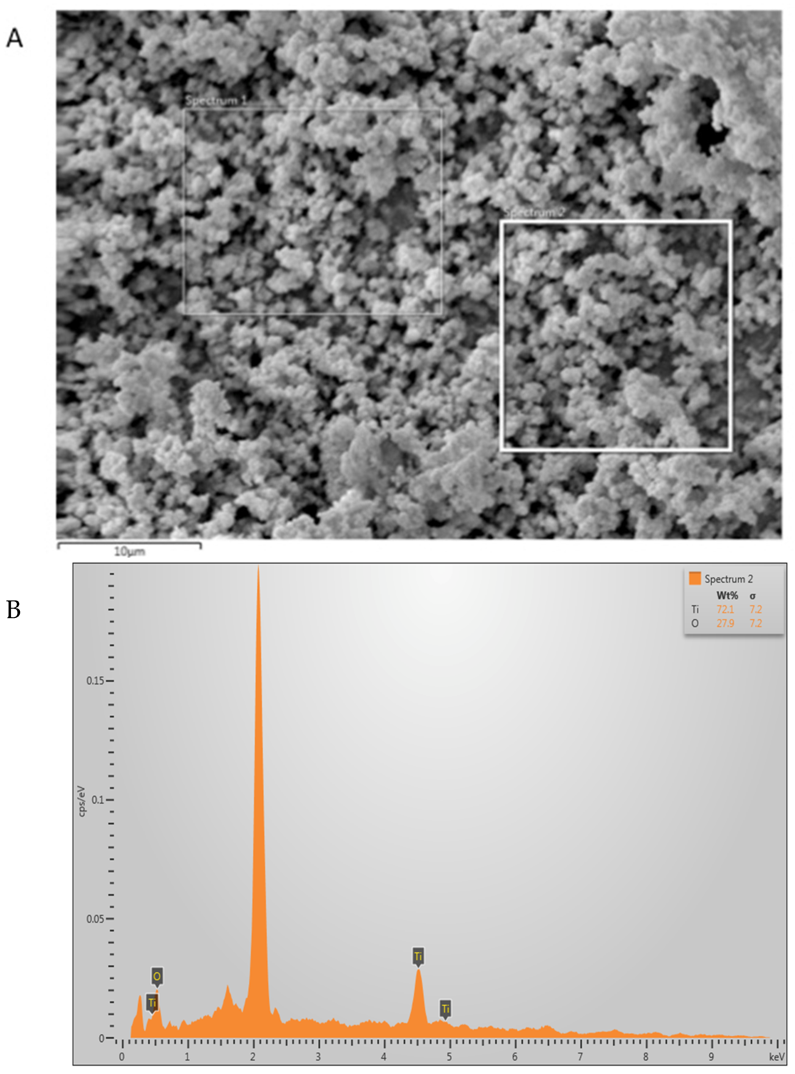
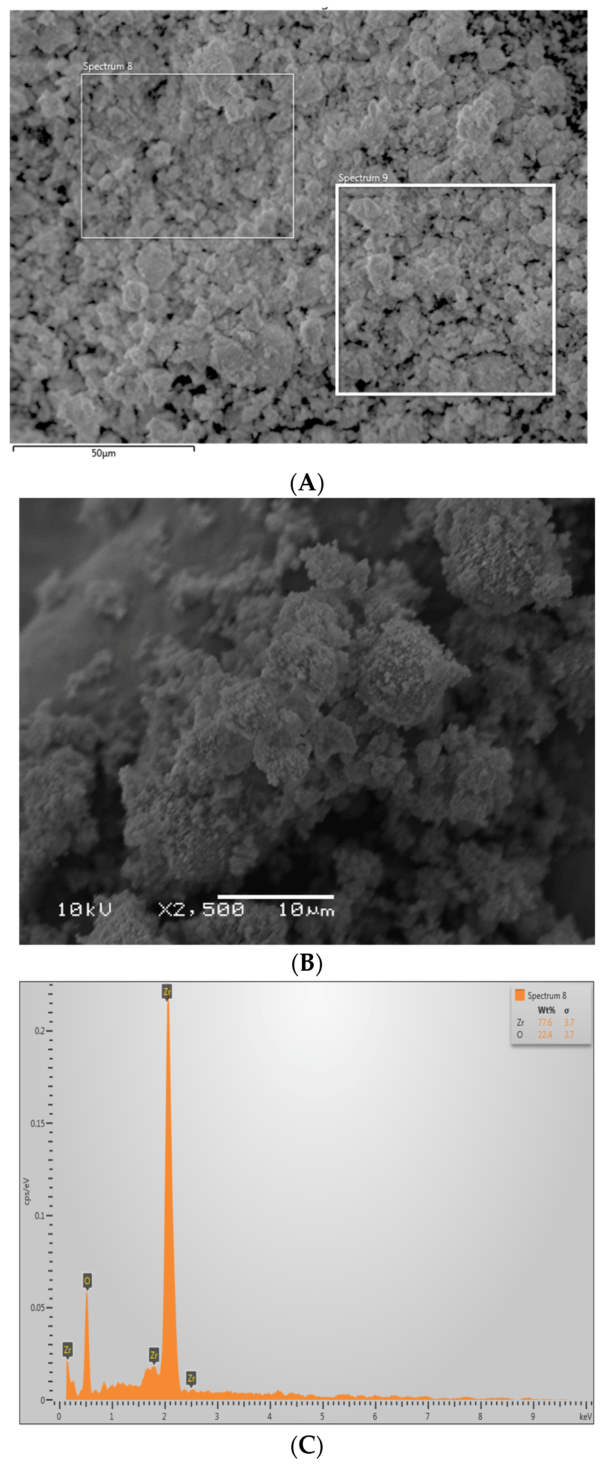
2.2. Scanning Electron Microscopy (SEM) and Energy Dispersive X-ray (EDX) Analysis

Characterization of the TiO2 and ZrO2 filler nanoparticles was carried out utilizing SEM and EDX analysis. A small quantity of these nanoparticles was mounted on the aluminum stubs, coated with gold, and then observed via an SEM (FEI Quanta 250, Scanning Electron Microscope, OR, USA), which operated at an accelerating voltage of 30 kV. These samples were observed at various magnifications (based on convenience). EDX spectroscopy was also implemented to characterize these filler nanoparticles further and assess their elemental distribution.

2.3. Assessment of the Rheological Properties of the Adhesives

The adhesives were first characterized by considering changes in their rheological properties using an MCR-72 rheometer (Anton Paar, Graz, Austria). These rheological properties were evaluated using rotation mode in a frequency sweep pre-set state of 8 mm (parallel plate) and 0.25 mm (opening). The samples were assessed over a wide range of angular frequencies, extending between 0.1 and 100 rad/s at 25 °C.

2.4. SBS Testing and Interfacial Fracture Types of the Adhesives

Seventy-five teeth (N = 75) were collected and then equally and randomly divided into three groups such that each adhesive group (EA, 5% TiO2, and 5% ZrO2) received 25 tooth samples (n = 25). Each of these teeth was prepared for bonding, and then SBS testing was carried out following the prior recommendations of Almutairi et al. [10]. Briefly, the dentin surfaces of the tooth samples were first flattened using a slow-speed diamond disc under running water. The exposed dentin surfaces were then polished using a 600-grit silicone paper under running water and the samples were positioned in the machine such that the shearing stamp loaded the compomer cylinder at a 90° angle. The crosshead speed was 1 mm/min and the cell load capability was 1 kN unit failure. A fitted crescent-shaped copper piece guaranteed the safety of the composite cylinder, which was necessary as each specimen provided two experimental interfaces. The SBS testing values were estimated (in megapascals, MPa) by dividing the peak force at the point of failure (in Newtons) by the bonding area in N/mm2, as suggested by Kensche et al. [28]. The SBS was evaluated using a universal testing machine (TIRAtest 2720, TIRA GmbH, Schalkau, Germany). Pre-SBS testing, of the twenty-five samples in each group, ten samples were thermocycled (TC), ten remained non-TC (NTC), and the remaining five were tested for the analysis of the resin–dentin interface. For TC specimens, distilled water baths were carried out for 10,000 cycles at 5 °C and 55 °C with a dwell time of 5 s (THE-1100, SD Mechatronik GmbH, Germany). For the NTC specimens, the bonded samples were stored for 1 day in distilled water prior to sectioning.
In the current study, the interfacial bond failure types were also assessed. This examination was conducted with a digital microscope (Hirox KH 7700, Tokyo, Japan). We divided the failure types into three categories: adhesive, cohesive, and/or mixed.

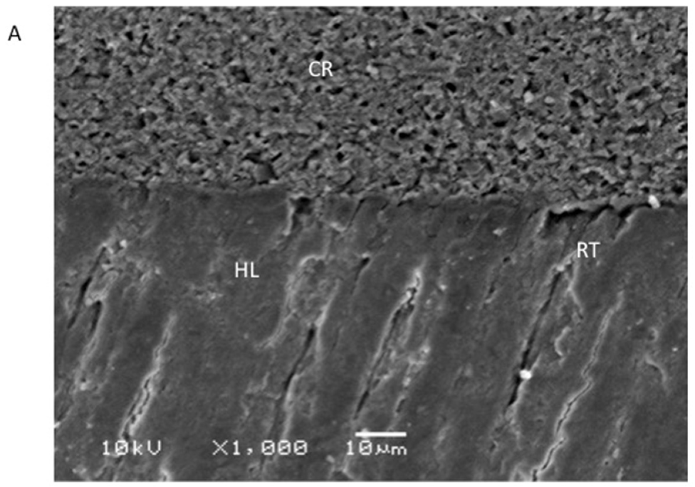
2.5. Evaluation of the Resin–Dentin Interface

Five bonded specimens from each adhesive group were first sectioned by means of a slow-speed isomet saw (Buehler Isomet 2000 Precision saw, Lake Bluff, IL, USA). This helped to form 1 x 1 mm beams. SEM and EDX spectroscopy were again employed in this study to analyze the bonded resin–dentin interface. With the help of a polisher (Beuhler Polisher, Lake Bluff, IL, USA), wet polishing of the beams was executed. This step was followed by their washing and placement in an ultrasonic bath (Bandelin Digital-Sigma-Aldrich Darmstadt, Germany) containing distilled water for 5 min. The conditioning of the samples was then performed using 36% phosphoric acid (DeTrey conditioner, Dentsply, PA, USA) followed by their washing with distilled water and sodium hypochlorite (5.25%) and solution immersion (for 15 min). Cleaning of the specimens was then carried out using distilled water, and they were then dehydrated using ethanol solutions of varying concentrations (80–100%). Gold coating of the specimens was achieved, and the samples were then analyzed using an SEM (FEI Quanta 250, Scanning Electron Microscope, OR, USA) to appraise the resin–dentin interface. The SEM was again operated at an accelerating voltage of 30 kV, and a range of magnifications was utilized.

2.6. Statistical Analysis

The outcomes of the SBS testing were gathered, computed on excel sheets, and assessed using SPSS-20.0 (IBM, Chicago, IL, USA). These values were used to calculate mean and standard deviations. The normality of the data was first checked via the Kolmogorov–Smirnov test. On observing non-normal distribution, ANOVA and post-hoc multiple comparison non-parametric tests were then applied to further analyze the results. The statistical significance level was set at 1%.

3. Results

3.1. Outcomes of the Characterization of the Filler Nanoparticles

The TiO2 nanoparticles demonstrated irregular shape on the SEM micrograph (Figure 1A). These filler nanoparticles were spherically shaped in an agglomerated form with an average particle size ranging between 500 and 1000 nm. The EDX mapping verified the occurrence of titanium and oxygen in the TiO2 nanoparticles (Figure 1B). The ZrO2 nanoparticles were also seen as irregularly shaped on SEM micrographs (Figure 2A,B). These nanoparticles also exhibited agglomerated form, with the particles seen sticking to each other with an average particle size of 500 nm (Figure 2B). The EDX mapping for ZrO2 nanoparticles revealed the presence of Zr and oxygen (Figure 2C), which are the two essential elements for ZrO2.

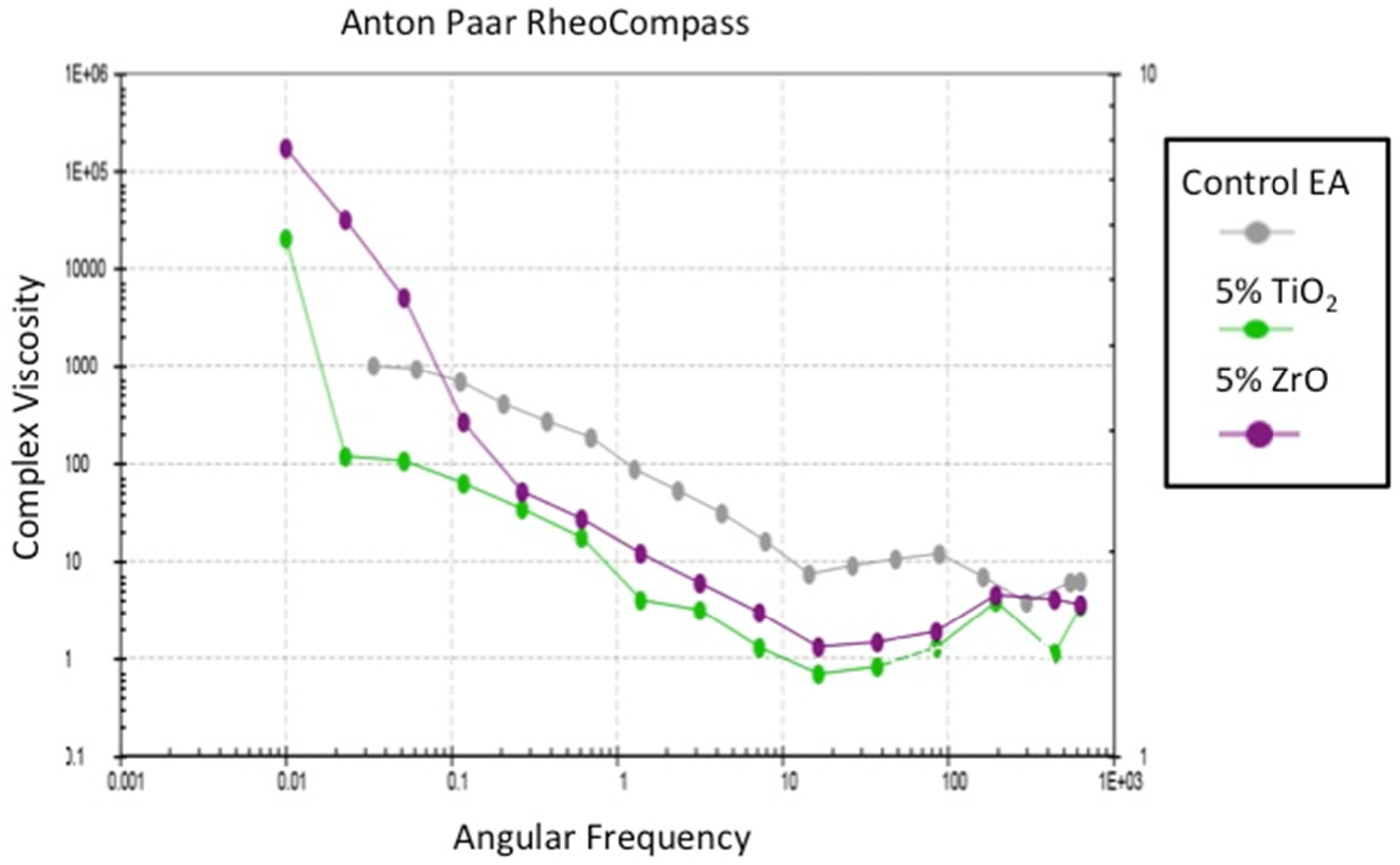
3.2. Outcomes of the Assessment of the Rheological Properties

The rheological assessment of the adhesives revealed that a decrease in the viscosity was seen when the angular frequency was increased (Figure 3). This decrease was most evident for the 5% ZrO2, followed by the 5% TiO2. The EA adhesive demonstrated a smaller decrease in the viscosity compared with the other two adhesive groups, although this change in the EA was also noticeable. All three adhesives studied here demonstrated non-Newtonian behavior (shear-thinning or pseudo-plasticity). The observed reduction in viscosity for filler-reinforced adhesive groups could mean that the incorporation of TiO2 and ZrO2 caused an improved fluidity of the resin/filler mixture; nevertheless, a Newtonian plateau was not witnessed, even at low angular frequencies. In the present study, although the EA group revealed greater viscosity than the other two groups, this trend was not consistently observed, and an overlap was seen between the viscosities of all of the adhesive groups at higher frequencies (Figure 3).

3.3. Outcomes of the SBS Testing and Failure Types Investigation

The results of the SBS assessment appeared to be in favor of the 5% TiO2 group (both NTC samples: 25.35 ± 1.53 MPa and TC samples: 23.89 ± 1.95 MPa) followed by the 5% ZrO2 adhesive group (NTC samples: 23.10 ± 2.22 MPa and TC samples: 20.72 ± 1.32 MPa (Table 1). The EA group revealed the lowest SBS values (NTC samples: 21.03 ± 2.44 MPa and TC samples: 17.62 ± 1.70 MPa) compared with the other two adhesive groups. Statistically significant results (p < 0.01) were witnessed upon intergroup comparison when the SBS test values for the NTC samples of the EA group were compared with the other two groups. A similar trend was observed concerning the intergroup comparison of the TC samples, and again the EA group’s SBS values were significantly different (p < 0.01) from the other two groups. The intergroup comparison between NTC and TC samples was statistically significant (p < 0.01) for all of the adhesive groups (Table 1).
Failures due to adhesion were most commonly found in the present study, accounting for 70 to 100% (in some instances) of the total failures (Table 1). The mixed type was the next most common failure, while cohesive failure was only seen for the NTC samples of the 5% ZrO2 group (Table 1).

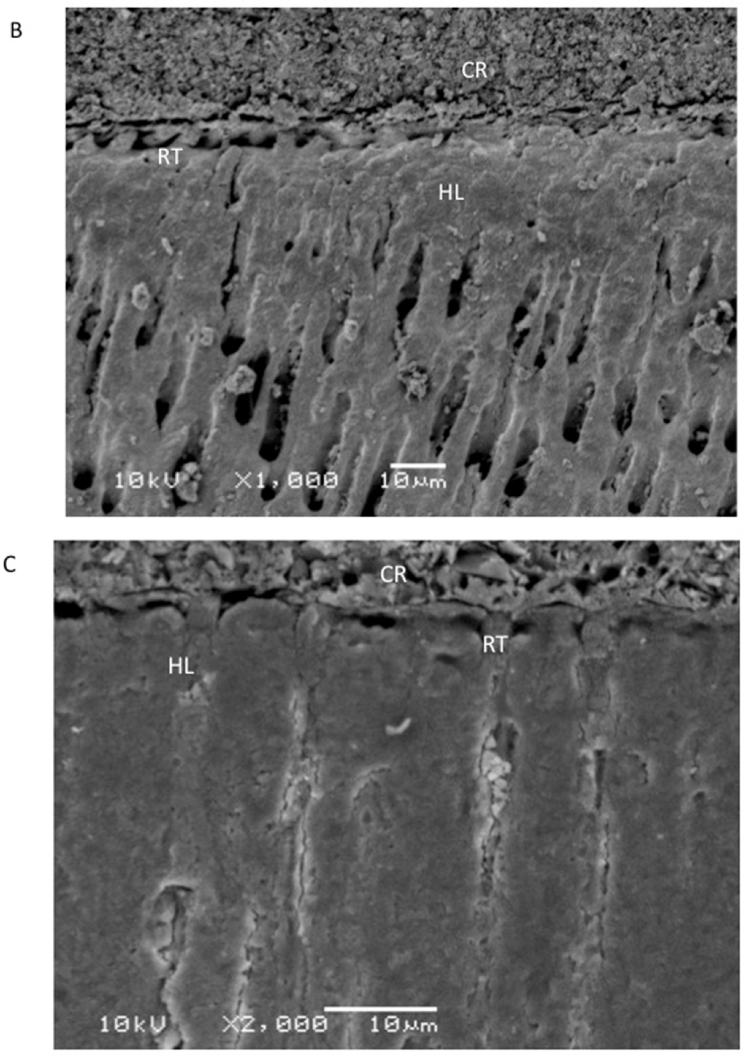
3.4. Outcomes of the Evaluation of the Resin–Dentin Interface

The SEM analysis revealed the establishment of a hybrid layer and resin tags (of variable depth) for the EA group (Figure 4A). For the 5% TiO2 group, again, comparable hybrid layer and resin tag development was seen on the SEM micrograph (Figure 4B). However, for the 5% ZrO2 group, it was noted that, although the hybrid layer and resin tag formation was perceived, it was not comparable to the other two groups and seemed to be compromised (Figure 4C).

4. Discussion

Based on the results of the current study, we partly accept the hypothesis that the incorporation of filler nanoparticles improves the SBS of adhesives. The hypothesis was also partly rejected, as the 5% TiO2 and 5% ZrO2 adhesives demonstrated less viscosity as compared with the EA (without filler nanoparticles). Several previous studies in the literature positively affirmed that the integration of inorganic fillers can amplify the mechanical properties of the adhesives studied [8,10,29]. TiO2 is mostly used in dentistry to coat the outer surface of dental implants as it offers excellent biocompatibility and remarkable mechanical properties [30]. TiO2 is also a strong antibacterial material that reduces the incidence of peri-implantitis (a common problem in dental implantology) [15]. In addition, the inclusion of TiO2 as a filler has resulted in an improvement of its physicochemical properties [31], and its inclusion in the composition of adhesive resin cement improved the DC of the material [18]. Similarly, ZrO2 is a bioinert and biocompatible material [32]. In dentistry, both Zr and Ti are preferred materials for the fabrication and coating of implants as they do not inhibit the formation and differentiation of osteoblasts, thus promoting osseointegration [33,34]. Zr-based fillers have been shown to result in an improvement to the mechanical properties of dental adhesives [25]. Keeping in mind the beneficial properties of these two nanoparticle groups, it was decided to incorporate 5 wt.% of TiO2 and ZrO2 in the EA and to probe the impact of their incorporation on different adhesive properties.
The SEM micrograph of TiO2 nanoparticles revealed that these particles were spherical in shape and present in an agglomerated form (Figure 1A). These findings are similar to a prior study that also demonstrated that TiO2 nanoparticles appear to be spherical in shape when observed under an SEM [35]. EDX mapping in the current study revealed the occurrence of Ti and oxygen (with Ti being more prevalent than oxygen, Figure 1B), which is again similar to the previous findings of Bhattacharya et al. [35]. The SEM micrograph of ZrO2 also led to comparable findings to previous studies [36,37], where Zr-based nanoparticles also demonstrated non-uniform morphology. EDX mapping for ZrO2 nanoparticles revealed the presence of Zr and oxygen, similar to previous studies [37,38].
In the present study, the rheological properties of all of the adhesives were also analyzed. The results demonstrated that, for all of the adhesives, a decrease in viscosity was observed when the angular frequency increased (Figure 3). This decrease in viscosity was more pronounced for the nanoparticle-containing adhesives as opposed to the EA. The results of this study are in line with an earlier study where an adhesive modified with nanofillers revealed lower viscosity, as compared with the control group [39]. Another former study also reported similar findings and associated decreased viscosity with the adhesive’s increasing filler content [40]. It should be kept in mind that the assessment of rheological properties is inconsistent and can be affected by the material’s handling [41], although the investigation of this characteristic was not part of this study.
In this study, half of the samples/groups selected for SBS testing were TC, while the remaining half remained NTC. The TC provided a vigorous challenge to the material in an in vitro setting which was identical to in vivo conditions. The ISO standard number 11,405 states that providing dental materials with a TC challenge with temperature ranges of 5–55 °C is appropriate to assess the aging of samples over a limited period of time [42]. We detected a reduced bond strength for all of the adhesives after they were TC, a result comparable to the formerly published literature [9,43]. The SBS testing experiments in this study revealed higher values for the nanoparticle-containing adhesives (with 5% TiO2 demonstrating the highest SBS values), as compared with the EA. These findings are similar to a former study that demonstrated an increase in the SBS of zirconia and TiO2 nanoparticle-containing adhesives [44]. TiO2 has been suggested as a reinforcing filler for dental materials due to its advantageous properties [45,46]. In addition, Zr-based nanoparticles disperse well in a material and improve its biocompatibility with organic polymers [47], which could have resulted in the improvement to the SBS observed in the present study. Another factor that could have played an important part in this finding could be the nano size of the two inorganic fillers used in this study. It is known that inorganic nanoparticles have high surface area [48] that can improve adhesion at the interface [49]. As the fillers were also nanosized, this could resulted in an improvement to the interfacial adhesion, resulting in greater SBS.
The bulk of the interfacial failures in this study were of the adhesive type (>70%). This finding is in line with the findings of an earlier study where adhesive-type failures were most commonly detected after the incorporation of filler nanoparticles in the composition of the adhesive [40]. It is pertinent to mention that dentin is collagenous in nature and this could have played a major part in the observation of adhesive-type failures in the current study, as the bonding of adhesives to dentin is challenging and is still considered a puzzling task in dentistry [50].
The resin–dentin interface was also analyzed (via SEM) in this study. It was witnessed that hybrid layers and resin tags (of variable depth) were formed for the EA (Figure 4A) and the 5% TiO2 group (Figure 4B), whereas the 5% ZrO2 group demonstrated compromised hybrid layer and resin tag formation (Figure 4C). Earlier studies demonstrated that the thickness of the adhesive layer and the length of the resin tag are not essential determinants of the bond strength, which is actually dependent on the adhesive’s composition [51,52]. Therefore, we still advocate the addition of ZrO2 nanoparticles to adhesives due to the improved SBS and comparable rheological properties to the other adhesive groups observed in the current study.
Although the results of the present study are encouraging, readers should exercise caution while interpreting them. The outcomes are possibly limited to the particular adhesive fabricated in the study and thus may not be applicable to commercially available adhesives. In addition, the TiO2 and ZrO2 contents were arbitrarily determined; however, different content percentages of these nanoparticles may show different properties. Moreover, the SBS test is a static test for bond strength while, clinically, material interfaces are commonly exposed to fatigue loads, which could influence the bond strength and durability of modified adhesives. We observed an improved SBS for the two nanoparticle-containing adhesives; however, both demonstrated lower viscosity compared with the EA. Future studies probing the impact of the integration of various other concentrations of these nanoparticle groups are recommended. We suggest further exploration of the effect of the incorporation of 2.5 wt.% TiO2 and ZrO2 on the mechanical properties of adhesives to further improve understanding of the relationship between filler concentrations and SBS and rheological properties.

5. Conclusions

Reinforcing the EA with filler nanoparticles (TiO2 and ZrO2) caused an increase in the adhesive’s SBS and the 5% TiO2 group demonstrated the highest values. The SBS values for the 5% TiO2 group were significantly different from the EA but were non-significantly different from the 5% ZrO2 group (for both NTC and TC samples). In addition, both nanoparticle-containing adhesive groups demonstrated comparable viscosity to the EA; nevertheless, their values were less than that of the control group. Additional studies examining the impact of diverse filler concentrations on the various properties of adhesives are suggested.

Author Contributions

Conceptualization, F.V., A.A., H.I.T. and A.H.A.; methodology, A.H.A., A.A., S.A.-S., A.S.A.-Q. and T.A.; validation, F.V., I.F., H.I.T., S.A.-S. and T.A.; formal analysis, A.A., A.H.A., M.B., S.S. and I.F.; investigation, A.H.A., A.S.A.-Q., M.B., A.A. and T.A.; data curation, F.V., A.A., S.S. and A.H.A.; writing—original draft preparation, I.F., F.V., A.S.A.-Q., M.B., H.I.T. and A.A.; writing—review and editing, I.F. and F.V.; supervision, F.V. and T.A.; funding acquisition, T.A. All authors have read and agreed to the published version of the manuscript.

Funding

The authors are grateful to the Researchers Supporting Project at King Saud University for funding through Researchers Supporting Project No. (RSP-2021-044).

Institutional Review Board Statement

The research ethics review committee of King Saud University, College of Dentistry, approved the research protocol, E-20-5545. All of the recommendations of the Helsinki Declaration and its later amendments were strictly followed. The teeth extracted for orthodontic treatments, which were free from any apparent defects, were gathered after attaining the patients’ written informed consent and were utilized for the experiments in our study.

Informed Consent Statement

All of the recommendations of the Helsinki Declaration and its later amendments were strictly followed. The teeth extracted for orthodontic treatments, which were free from any apparent defects, were gathered after attaining the patients’ written informed consent and were utilized for the experiments in our study.

Data Availability Statement

The data analyzed in this study are available on request form the corresponding author.

Acknowledgments

The authors are grateful to the Researchers Supporting Project at King Saud University for funding through Researchers Supporting Project No. (RSP-2021-044).

Conflicts of Interest

The authors declare no conflict of interest.

References

  1. Zhao, Z.W.Q.; Zhao, J.; Zhao, B.; Ma, Z.; Zhang, C. Adhesion in teeth. Front. Mater. 2021, 7, 615225. [Google Scholar] [CrossRef]
  2. Perdigao, J. Current perspectives on dental adhesion: (1) Dentin adhesion—Not there yet. Jpn. Dent. Sci. Rev. 2020, 56, 190–207. [Google Scholar] [CrossRef] [PubMed]
  3. FDI, World Dental Federation. FDI policy statement on Minimal Intervention Dentistry (MID) for managing dental caries: Adopted by the General Assembly: September 2016, Poznan, Poland. Int. Dent. J. 2017, 67, 6–7. [Google Scholar] [CrossRef]
  4. Sofan, E.; Sofan, A.; Palaia, G.; Tenore, G.; Romeo, U.; Migliau, G. Classification review of dental adhesive systems: From the IV generation to the universal type. Ann. Stomatol. 2017, 8, 1–17. [Google Scholar]
  5. Ferracane, J.L. Models of Caries Formation around Dental Composite Restorations. J. Dent. Res. 2017, 96, 364–371. [Google Scholar] [CrossRef] [PubMed] [Green Version]
  6. Iliev, G.; Hardan, L.; Kassis, C.; Bourgi, R.; Cuevas-Suarez, C.E.; Lukomska-Szymanska, M.; Mancino, D.; Haikel, Y.; Kharouf, N. Shelf Life and Storage Conditions of Universal Adhesives: A Literature Review. Polymers 2021, 13, 2708. [Google Scholar] [CrossRef] [PubMed]
  7. Farooq, I.; Ali, S.; Al-Saleh, S.; AlHamdan, E.M.; AlRefeai, M.H.; Abduljabbar, T.; Vohra, F. Synergistic Effect of Bioactive Inorganic Fillers in Enhancing Properties of Dentin Adhesives-A Review. Polymers 2021, 13, 2169. [Google Scholar] [CrossRef]
  8. AlFawaz, Y.F.; Almutairi, B.; Kattan, H.F.; Zafar, M.S.; Farooq, I.; Naseem, M.; Vohra, F.; Abduljabbar, T. Dentin Bond Integrity of Hydroxyapatite Containing Resin Adhesive Enhanced with Graphene Oxide Nano-Particles-An SEM, EDX, Micro-Raman, and Microtensile Bond Strength Study. Polymers 2020, 12, 2978. [Google Scholar] [CrossRef] [PubMed]
  9. Alhenaki, A.M.; Attar, E.A.; Alshahrani, A.; Farooq, I.; Vohra, F.; Abduljabbar, T. Dentin Bond Integrity of Filled and Unfilled Resin Adhesive Enhanced with Silica Nanoparticles-An SEM, EDX, Micro-Raman, FTIR and Micro-Tensile Bond Strength Study. Polymers 2021, 13, 1093. [Google Scholar] [CrossRef]
  10. Almutairi, B.; Kattan, H.F.; BinMahfooz, A.M.; Qutub, O.A.; Basunbul, G.; ArRejaie, A.S.; Farooq, I.; Vohra, F.; Abduljabbar, T. Synergistic effect of graphene oxide/calcium phosphate nanofiller in a dentin adhesive on its dentin bond integrity and degree of conversion. A scanning electron microscopy, energy dispersive X-ray spectroscopy, Fourier transform infrared, micro-Raman, and bond strength study. Microsc. Res. Tech. 2021, 84, 2082–2094. [Google Scholar]
  11. Weir, A.; Westerhoff, P.; Fabricius, L.; Hristovski, K.; von Goetz, N. Titanium dioxide nanoparticles in food and personal care products. Environ. Sci. Technol. 2012, 46, 2242–2250. [Google Scholar] [CrossRef] [Green Version]
  12. Musial, J.; Krakowiak, R.; Mlynarczyk, D.T.; Goslinski, T.; Stanisz, B.J. Titanium Dioxide Nanoparticles in Food and Personal Care Products-What Do We Know about Their Safety? Nanomaterials 2020, 10, 1110. [Google Scholar] [CrossRef]
  13. Besinis, A.; De Peralta, T.; Handy, R.D. The antibacterial effects of silver, titanium dioxide and silica dioxide nanoparticles compared to the dental disinfectant chlorhexidine on Streptococcus mutans using a suite of bioassays. Nanotoxicology 2014, 8, 1–16. [Google Scholar] [CrossRef] [Green Version]
  14. Jowkar, Z.; Hamidi, S.A.; Shafiei, F.; Ghahramani, Y. The Effect of Silver, Zinc Oxide, and Titanium Dioxide Nanoparticles Used as Final Irrigation Solutions on the Fracture Resistance of Root-Filled Teeth. Clin. Cosmet. Investig. Dent. 2020, 12, 141–148. [Google Scholar] [CrossRef] [PubMed] [Green Version]
  15. Jafari, S.; Mahyad, B.; Hashemzadeh, H.; Janfaza, S.; Gholikhani, T.; Tayebi, L. Biomedical Applications of TiO2 Nanostructures: Recent Advances. Int. J. Nanomed. 2020, 15, 3447–3470. [Google Scholar] [CrossRef]
  16. Sun, J.; Petersen, E.J.; Watson, S.S.; Sims, C.M.; Kassman, A.; Frukhtbeyn, S.; Skrtic, D.; Ok, M.T.; Jacobs, D.S.; Reipa, V.; et al. Biophysical characterization of functionalized titania nanoparticles and their application in dental adhesives. Acta Biomater. 2017, 53, 585–597. [Google Scholar] [CrossRef] [PubMed] [Green Version]
  17. Esteban Florez, F.L.; Kraemer, H.; Hiers, R.D.; Sacramento, C.M.; Rondinone, A.J.; Silverio, K.G.; Khajotia, S.S. Sorption, solubility and cytotoxicity of novel antibacterial nanofilled dental adhesive resins. Sci. Rep. 2020, 10, 13503. [Google Scholar] [CrossRef] [PubMed]
  18. Ramos-Tonello, C.M.; Lisboa-Filho, P.N.; Arruda, L.B.; Tokuhara, C.K.; Oliveira, R.C.; Furuse, A.Y.; Rubo, J.H.; Borges, A.F.S. Titanium dioxide nanotubes addition to self-adhesive resin cement: Effect on physical and biological properties. Dent. Mater. 2017, 33, 866–875. [Google Scholar] [CrossRef] [Green Version]
  19. Skocaj, M.; Filipic, M.; Petkovic, J.; Novak, S. Titanium dioxide in our everyday life; is it safe? Radiol. Oncol. 2011, 45, 227–247. [Google Scholar] [CrossRef] [Green Version]
  20. Madfa, A.A.A.-S.F.A.; Al-Quadami, N.H.; Al-Sanabani, J.S.; Amran, A.G. Use of Zirconia in Dentistry: An Overview. Open Biomat. J. 2014, 5, 1–9. [Google Scholar] [CrossRef]
  21. Grech, J.A.E. Zirconia in dental prosthetics: A literature review. J. Mater. Res. Technol. 2019, 8, 4956–4964. [Google Scholar] [CrossRef]
  22. Apratim, A.; Eachempati, P.; Krishnappa Salian, K.K.; Singh, V.; Chhabra, S.; Shah, S. Zirconia in dental implantology: A review. J. Int. Soc. Prev. Community Dent. 2015, 5, 147–156. [Google Scholar] [CrossRef] [Green Version]
  23. Arena, A.; Prete, F.; Rambaldi, E.; Bignozzi, M.C.; Monaco, C.; Di Fiore, A.; Chevalier, J. Nanostructured Zirconia-Based Ceramics and Composites in Dentistry: A State-of-the-Art Review. Nanomaterials 2019, 9, 1393. [Google Scholar] [CrossRef] [PubMed] [Green Version]
  24. Halder, S.; Ahemad, S.; Das, S.; Wang, J. Epoxy/Glass Fiber Laminated Composites Integrated with Amino Functionalized ZrO22 for Advanced Structural Applications. ACS Appl. Mater. Interfaces 2016, 8, 1695–1706. [Google Scholar] [CrossRef] [PubMed]
  25. Lohbauer, U.; Wagner, A.; Belli, R.; Stoetzel, C.; Hilpert, A.; Kurland, H.D.; Grabow, J.; Muller, F.A. Zirconia nanoparticles prepared by laser vaporization as fillers for dental adhesives. Acta Biomater. 2010, 6, 4539–4546. [Google Scholar] [CrossRef]
  26. Cionca, N.; Hashim, D.; Mombelli, A. Zirconia dental implants: Where are we now, and where are we heading? Periodontol. 2000. 2017, 73, 241–258. [Google Scholar] [CrossRef] [PubMed]
  27. Lughi, V.; Sergo, V. Low temperature degradation aging of zirconia: A critical review of the relevant aspects in dentistry. Dent. Mater. 2010, 26, 807–820. [Google Scholar] [CrossRef]
  28. Kensche, A.; Dahne, F.; Wagenschwanz, C.; Richter, G.; Viergutz, G.; Hannig, C. Shear bond strength of different types of adhesive systems to dentin and enamel of deciduous teeth in vitro. Clin. Oral. Investig. 2016, 20, 831–840. [Google Scholar] [CrossRef]
  29. Al-Hamdan, R.S.; Almutairi, B.; Kattan, H.F.; Alsuwailem, N.A.; Farooq, I.; Vohra, F.; Abduljabbar, T. Influence of Hydroxyapatite Nanospheres in Dentin Adhesive on the Dentin Bond Integrity and Degree of Conversion: A Scanning Electron Microscopy (SEM), Raman, Fourier Transform-Infrared (FTIR), and Microtensile Study. Polymers 2020, 12, 2948. [Google Scholar] [CrossRef]
  30. Lubojanski, A.; Dobrzynski, M.; Nowak, N.; Rewak-Soroczynska, J.; Sztyler, K.; Zakrzewski, W.; Dobrzynski, W.; Szymonowicz, M.; Rybak, Z.; Wiglusz, K.; et al. Application of Selected Nanomaterials and Ozone in Modern Clinical Dentistry. Nanomaterials 2021, 11, 259. [Google Scholar] [CrossRef]
  31. Sturmer, M.; Garcia, I.M.; Souza, V.S.; Visioli, F.; Scholten, J.D.; Samuel, S.M.W.; Leitune, V.C.B.; Collares, F.M. Titanium dioxide nanotubes with triazine-methacrylate monomer to improve physicochemical and biological properties of adhesives. Dent. Mater. 2021, 37, 223–235. [Google Scholar] [CrossRef]
  32. Yousefi, K.; Manesh, H.D.; Khalifeh, A.R.; Gholami, A. Fabrication and Characterization of a Nanofast Cement for Dental Restorations. Biomed. Res. Int. 2021, 2021, 7343147. [Google Scholar] [CrossRef]
  33. Kobayashi, E.; Matsumoto, S.; Doi, H.; Yoneyama, T.; Hamanaka, H. Mechanical properties of the binary titanium-zirconium alloys and their potential for biomedical materials. J. Biomed. Mater. Res. 1995, 29, 943–950. [Google Scholar] [CrossRef] [PubMed]
  34. Bona, A.D.; Pecho, O.E.; Alessandretti, R. Zirconia as a Dental Biomaterial. Materials 2015, 8, 4978–4991. [Google Scholar] [CrossRef] [Green Version]
  35. Bhattacharya, K.; Davoren, M.; Boertz, J.; Schins, R.P.; Hoffmann, E.; Dopp, E. Titanium dioxide nanoparticles induce oxidative stress and DNA-adduct formation but not DNA-breakage in human lung cells. Part. Fibre Toxicol. 2009, 6, 17. [Google Scholar] [CrossRef] [PubMed] [Green Version]
  36. Behbahani, A.R.S.; Esmaeilifar, A. Hydrothermal synthesis of zirconia nanoparticles from commercial zirconia. Proc. Eng. 2012, 42, 908–917. [Google Scholar] [CrossRef] [Green Version]
  37. Majedi, A.A.A.; Davar, F. Green synthesis of zirconia nanoparticles using the modified Pechini method and characterization of its optical and electrical properties. J. Sol.-Gel. Sci. Tech. 2016, 77, 542–552. [Google Scholar] [CrossRef]
  38. Zinatloo-Ajabshir, S.S.-N.M. Zirconia Nanostructures: Novel Facile Surfactant-Free Preparation and Characterization. App. Ceram. Technol. 2016, 13, 108–115. [Google Scholar] [CrossRef] [Green Version]
  39. AlHamdan, E.M.; Al-Saleh, S.; AlRefeai, M.H.; Farooq, I.; Abrar, E.; Vohra, F.; Abduljabbar, T. Adhesive bond integrity of Y-TZP post with calcium fluoride infiltrated resin dentin adhesive: An SEM, EDX, FTIR and micro-Raman study. Surf. Interface Anal. 2021, 53, 956–962. [Google Scholar] [CrossRef]
  40. Alqarawi, F.K.; Alkahtany, M.F.; Almadi, K.H.; Ben Gassem, A.A.; Alshahrani, F.A.; AlRefeai, M.H.; Farooq, I.; Vohra, F.; Abduljabbar, T. Influence of Different Conditioning Treatments on the Bond Integrity of Root Dentin to rGO Infiltrated Dentin Adhesive. SEM, EDX, FTIR and MicroRaman Study. Polymers 2021, 13, 1555. [Google Scholar] [CrossRef]
  41. Lee, J.H.; Um, C.M.; Lee, I.B. Rheological properties of resin composites according to variations in monomer and filler composition. Dent. Mater. 2006, 22, 515–526. [Google Scholar] [CrossRef] [PubMed]
  42. International Organization for Standardization. ISO/TS 11405 Dentistry—Testing of Adhesion to Tooth Structure, 3rd ed.; International Organization for Standardization: Geneva, Switzerland, 2015. [Google Scholar]
  43. Helvatjoglu-Antoniades, M.; Koliniotou-Kubia, E.; Dionyssopoulos, P. The effect of thermal cycling on the bovine dentine shear bond strength of current adhesive systems. J. Oral Rehabil. 2004, 31, 911–917. [Google Scholar] [CrossRef]
  44. Felemban, N.H.; Ebrahim, M.I. The influence of adding modified zirconium oxide-titanium dioxide nano-particles on mechanical properties of orthodontic adhesive: An in vitro study. BMC Oral Health 2017, 17, 43. [Google Scholar] [CrossRef] [PubMed] [Green Version]
  45. Xia, Y.; Zhang, F.; Xie, H.; Gu, N. Nanoparticle-reinforced resin-based dental composites. J. Dent. 2008, 36, 450–455. [Google Scholar] [CrossRef]
  46. Garcia-Contreras, R.; Scougall-Vilchis, R.J.; Contreras-Bulnes, R.; Sakagami, H.; Morales-Luckie, R.A.; Nakajima, H. Mechanical, antibacterial and bond strength properties of nano-titanium-enriched glass ionomer cement. J. Appl. Oral Sci. 2015, 23, 321–328. [Google Scholar] [CrossRef] [PubMed] [Green Version]
  47. Sun, L.G.R.; Gordaninejad, F.; Suhr, J. Energy absorption capability of nanocomposites: A review. CompSci Tech. 2009, 69, 2392–2409. [Google Scholar] [CrossRef]
  48. Ashraf, M.A.; Peng, W.; Zare, Y.; Rhee, K.Y. Effects of Size and Aggregation/Agglomeration of Nanoparticles on the Interfacial/Interphase Properties and Tensile Strength of Polymer Nanocomposites. Nanoscale Res. Lett. 2018, 13, 214. [Google Scholar] [CrossRef] [PubMed]
  49. Zare, Y.; Rhee, K.Y. Dependence of Z Parameter for Tensile Strength of Multi-Layered Interphase in Polymer Nanocomposites to Material and Interphase Properties. Nanoscale Res. Lett. 2017, 12, 42. [Google Scholar] [CrossRef] [Green Version]
  50. Peumans, M.; Kanumilli, P.; De Munck, J.; Van Landuyt, K.; Lambrechts, P.; Van Meerbeek, B. Clinical effectiveness of contemporary adhesives: A systematic review of current clinical trials. Dent. Mater. 2005, 21, 864–881. [Google Scholar] [CrossRef]
  51. Kharouf, N.A.T.; Eid, A.; Maguina, L.; Zghal, J.; Sekayan, N.; Bourgi, R.; Hardan, L.; Sauro, S.; Haikel, Y.; Mancino, D. Does Adhesive Layer Thickness and Tag Length Influence Short/Long-Term Bond Strength of Universal Adhesive Systems? An In Vitro Study. Appl. Sci. 2021, 11, 2635. [Google Scholar] [CrossRef]
  52. Anchieta, R.B.; Oliveira, F.G.; Sundfeld, R.H.; Rahal, V.; Machado, L.S.; Alexandre, R.S.; Sundefeld, M.L.; Rocha, E.P. Analysis of hybrid layer thickness, resin tag length and their correlation with microtensile bond strength using a total etch adhesive to intact dentin. Acta Odontol. Latinoam. 2011, 24, 272–278. [Google Scholar] [PubMed]
Figure 1. High-magnification (A) SEM image of the nano-titanium oxide (TiO2) particles. The TiO2 powder showed agglomerated particles of various irregularly sized polygonal crystals, ranging from 500 nm to 1000 nm. (B) Representative EDX graph showing high percentage of Ti and oxygen.
Polymers 13 03794 g001
Figure 2. High magnification (A,B) SEM image of the nano-zirconium oxide (ZrO2) particles. The ZrO2 powder showed agglomerated particles of irregularly sized crystals around 500 nm. (C) Representative EDX graph showing high percentage of Zr and oxygen (O).
Polymers 13 03794 g002
Figure 3. Rheological properties of the experimental control (Control EA), 5%-TiO2, and 5% ZrO2 adhesives. Complex viscosity is shown for angular frequencies of 0.001 to 1000 rads/s.
Polymers 13 03794 g003
Figure 4. High-magnification images of resin–dentin interface showing: (A) composite resin (CR), hybrid layer (HL), and normal resin tag (RT) formation in control samples (EA). (B) Normal resin tag formation and standard dentin penetration with TiO incorporated dentin adhesive, (C) RT formation and dentin penetration was compromised in ZrO2-containing EA (few resin tags).
Polymers 13 03794 g004aPolymers 13 03794 g004b
Table 1. Shear-bond strength (SBS) and failure mode analysis among the study groups.
SBS (MPa) (Mean ± SD)Failure Mode Analysis (%)
Group
(n = 10)
NTCTCp-Value *AdhesiveCohesiveMixed
Control EA
(0% particles)
21.03 ± 2.44 a,A-<0.0110000
-17.62 ± 1.70 a,B10000
5% TiO225.35 ± 1.53 b,A 80020
-23.89 ± 1.95 b,B10000
5% ZrO223.10 ± 2.22 b,A 701020
-20.72 ± 1.32 b,B80020
TC: thermocycling, NTC: no thermocycling, TiO2: titanium oxide, ZrO2: zirconium oxide, Control EA, experimental adhesive with no nanofiller composite. * ANOVA. Dissimilar lower case letters in the same column indicate statistical significance. Dissimilar capital letters in a row (same group) indicate statistical significance.
Publisher’s Note: MDPI stays neutral with regard to jurisdictional claims in published maps and institutional affiliations.

