Review

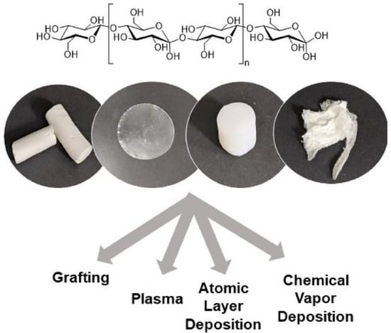
Production and Surface Modification of Cellulose Bioproducts

Fiber and Biopolymer Research Institute, Texas Tech University, Lubbock, TX 79409-5019, USA
*
Author to whom correspondence should be addressed.
Polymers 2021, 13(19), 3433; https://doi.org/10.3390/polym13193433
Submission received: 3 September 2021 / Revised: 29 September 2021 / Accepted: 2 October 2021 / Published: 7 October 2021

Abstract

:
Petroleum-based synthetic plastics play an important role in our life. As the detrimental health and environmental effects of synthetic plastics continue to increase, the renewable, degradable and recyclable properties of cellulose make subsequent products the “preferred environmentally friendly” alternatives, with a small carbon footprint. Despite the fact that the bioplastic industry is growing rapidly with many innovative discoveries, cellulose-based bioproducts in their natural state face challenges in replacing synthetic plastics. These challenges include scalability issues, high cost of production, and most importantly, limited functionality of cellulosic materials. However, in order for cellulosic materials to be able to compete with synthetic plastics, they must possess properties adequate for the end use and meet performance expectations. In this regard, surface modification of pre-made cellulosic materials preserves the chemical profile of cellulose, its mechanical properties, and biodegradability, while diversifying its possible applications. The review covers numerous techniques for surface functionalization of materials prepared from cellulose such as plasma treatment, surface grafting (including RDRP methods), and chemical vapor and atomic layer deposition techniques. The review also highlights purposeful development of new cellulosic architectures and their utilization, with a specific focus on cellulosic hydrogels, aerogels, beads, membranes, and nanomaterials. The judicious choice of material architecture combined with a specific surface functionalization method will allow us to take full advantage of the polymer’s biocompatibility and biodegradability and improve existing and target novel applications of cellulose, such as proteins and antibodies immobilization, enantiomers separation, and composites preparation.

Graphical Abstract

1. Introduction

As the ubiquity of synthetic plastic waste and its harmful effects on the environment, aquatic and terrestrial life, and human health increase, the concept of making materials from renewable natural sources has become popular. The magnitude of plastic pollution is vast, and it has become one of the most pressing societal concerns. Only around 9% of the 6300 metric tons of plastics made in 2015 have been recycled, 12% incinerated, and the rest has accumulated in the environment [1]. Much of this problem emerges from excessive consumption of single-use plastics as they are convenient, affordable, and easily accessible. Plastic waste accumulation also increases due to the lack of economically attractive and energy-efficient recycling options and almost indefinite shelf-life of most plastics. If the current production and recycling trends persist, scientists predict that approximately 12,000 metric tons of plastics will accumulate globally by 2050 [1]. A world without plastics seems unimaginable, given their convenience and diversity in applications. Therefore, in order to protect nature without compromising the needs of the present and future generations, environmental scientists are searching for novel bio-based plastics that are recyclable, compostable, or biodegradable as well as for cost-effective and energy-efficient plastic waste processing technologies.
Numerous studies have demonstrated that using green technologies for the production of versatile materials can reduce carbon footprint. Use of natural polymers as synthetic plastic substitutes could represent a viable solution for most aforementioned problems. Among natural polymers, cellulose extracted mainly from cellulosic and lignocellulosic biomass, such as cotton, wood, and crop residues are of substantial importance and has the potential to fulfill the growing demand for bio-based plastics. Cellulose is a renewable natural polymer of significant commercial importance that represents ~1.5 × 10¹² tons/year of total biomass [2]. It is the most abundant natural polymer on Earth. Cellulose is biosynthesized by plants, species of bacteria (genera Acetobacter, Agrobacterium, and Sarcina ventriculi), and algae (Rhodophyta phylum and Phaeophyceae class) [3,4]. The cellulose content in cotton is as high as 90%, and in wood is 40–50% [5,6].
Cellulose has a structural formula (C6H10O5)n, where n ranges from 10,000 to 15,000 [7], and is composed of D-glucose units covalently linked together by 1 → 4 glycosidic bonds (i.e., beta linkages) [8]. Because of a large number of hydroxyl groups present in the polymer, it is extensively hydrogen-bonded, having both inter- and intra-chain bonding. Intra-chain hydrogen bonding, which is responsible for stabilization of the glycosidic bonds, is formed between O(3)-H…O(ring) and O(2)-H…O(6). Inter-chain hydrogen bonding, which is responsible for parallel stacking of multiple cellulose chains occurs between O(3)-H…O(6) (Figure 1) [9].
Cellulose fiber has a defined hierarchical order in its organization. Native cellulose fiber consists of several microfibrils (5–10 nm in diameter each, depending on plant tissue and species) bundled together. In turn, microfibrils are made of stacked semi-crystalline elementary fibrils. Sequentially, elementary fibrils, with dimensions of 3.5 × 5.3 nm each, are assembled from glucan chains (generally, 36) [10]. Bacterial cellulose (BC) is obtained by a build-up of nanofibers.
Cellulose is a highly crystalline polymer [2,11,12], and naturally exists in allomorphs Iα (triclinic unit cell) and Iβ (monoclinic unit cell), with the Iα/Iβ ratio dependent on cellulose origin [13]. In addition, other allomorphs of cellulose are known: cellulose II, III, and IV. The cellulose II allomorph can be formed from cellulose I by alkali treatment (mercerization) or through dissolution–regeneration technique. Typically, cellulose has a high molecular weight and its degree of polymerization (DP) depends on a cellulose source and method of polymer isolation. For cotton, DP ranges from ~800–10,000 [14,15,16]. One of the important properties of cellulose is its biodegradability, a breakdown by microorganisms into carbon dioxide and water, such that it can be consumed by the environment; it has been shown that cellulose disintegrates completely in compost at an ambient temperature within 4 months [2,17].
However, despite many valuable properties and environmental benefits of cellulose, its transformation into usable forms with desired characteristics and functionalities requires several steps, namely purification, dissolution, preparation of required material architectures, and dehydration. Unlike synthetic plastics, cellulose does not melt when subjected to heat, and therefore, cellulose dissolution is of foremost importance among all the steps [17]. During this process, the crystalline structure of natural cellulose is disrupted, and the polymer can be transformed into different forms during the regeneration process. At the same time, an extensive hydrogen bonding network between neighboring cellulose molecules and highly crystalline microfibril structures that give unique physicochemical characteristics to plants make it recalcitrant to dissolution in water and in most common organic solvents.
There is a long and fruitful history of cellulose dissolution, and highly effective solvent systems for dissolution of cellulose have already been reported, which include N-methylmorpholine-N-oxide (NMMO), aqueous NaOH/urea, N,N-dimethylacetamide/LiCl (DMAc/LiCl), and ionic liquids (ILs, the salts that remain liquids at or below 100 °C [18]); these systems are comprehensively discussed elsewhere [15]. Among these, ILs efficiently dissolve cellulose at relatively low temperatures and are considered to be green solvents for cellulose. During the last two decades, numerous ILs with different structural characteristics have been synthesized, and their dissolution mechanism and the influence of constituent ions, and cosolvents on cellulose dissolution capacity have been extensively studied [19,20,21,22,23,24]. The dissolution mechanism typically involves breaking hydrogen bonds between the neighboring cellulose molecules [17] (Figure 2), resulting in a molecularly dispersed solution [25] or a colloidal dispersion of several hundred cellulose chains [12], capable of forming new hydrogen bonds during regeneration. Thus far, it has been challenging to find an ideal solvent that does not react with cellulose nor break cellulose chains [25].
It is well known that the degree of polymerization (DP), crystallinity (CrI), and crystallite size of cellulose depend on its origin (e.g., wood cellulose vs. cotton cellulose). While these parameters impart unique fascinating characteristics to cellulose, they hamper its dissolution. Therefore, frequently, conditions are optimized for effective dissolution of cellulose obtained from various sources. For example, cotton cellulose that possesses a high DP of 8000–15,000 and CrI of ~80% is recalcitrant to dissolution under mild conditions, although these characteristics are cultivar-dependent [26,27]. The dissolution process is affected by several factors such as cellulose drying conditions (oven drying vs. freeze drying), various pretreatments, cellulose concentration, dissolution temperature, pH, and moisture content [12,16,28]. Upon dissolution, viscous cellulose solutions can be converted into different material architectures using dry- or wet- jet spinning [29,30,31], electrospinning [32,33,34], or casting, followed by gelation, regeneration in protic solvents, such as water and alcohols, and drying (via air-drying, hot pressing, supercritical CO2 drying, etc.) [2,17,24,25,26,27,28]. Each of these techniques results in cellulose recovery in a form of amorphous bioproducts (e.g., hydrogels, films/membranes, aerogels, and beads [35,36,37,38,39]). These bioproducts manifest many unique characteristics of natural cellulose and their functionality is limited to the presence of hydroxyl groups.
Although cellulose is an economical and available resource that offers a wide range of physical and chemical properties, often its properties are not suitable for a specific application. With the growth of the bioplastic industry, it became apparent that modification of polymeric products is essential for producing genuine bioplastics that can compete with highly variable synthetic polymers. Thus, instead of modifying the polymer’s bulk properties, surface modifications of cellulose and cellulose composites (e.g., surface coating, chemical modification, plasma or vapor deposition treatment, a combination of such), tailored for a specific purpose, can result in materials with targeted properties. For instance, cellulose is hydrophilic, and impartment of hydrophobicity is required in applications such as self-cleaning [40] or self-healing materials [41], oil and water separation [42], electromagnetic interference (EMI) shielding [43], etc. Recent review discusses superhydrophobic modification of cellulose and potential applications [44]. Likewise, although nanocelluloses are often employed as reinforcing fillers owing to their impressive mechanical strength, tuning of their interfacial properties through incorporation of various functional groups is required to improve its dispersibility in common solvents [45]. The ability to improve the properties of cellulose through surface modification points to a promising future for cellulosic biomaterials in many fields.
In the current review, we revisited the most recent developments in surface modification of cellulose-based bioproducts. The review also highlights targeted development of novel cellulosic products and their applications, with a specific focus on certain material architectures: hydrogels, aerogels, beads, membranes, and nanomaterials. The selection of material architecture combined with specific surface functionalization will allow us to take full advantage of the polymer’s biocompatibility and biodegradability and improve existing and target novel applications of cellulose. Because the topic is broad, we have focused on the type of bioproducts, product characteristics, and surface functionalization techniques for specific use.

2. Surface Modification

The surface of a material represents the boundary between the bulk polymer and the outer environment, and the structural and chemical changes that occur on the surface tailor them for different applications. A wide range of surface modification techniques, such as physicochemical, biological, and mechanical methods, have been developed for modifying polymer surfaces. Some of the most common surface modification techniques that have been used for tailoring cellulosic surfaces are summarized herein.

2.1. Plasma

Plasma, sometimes referred to as the fourth state of matter [46], is an ionized gas and can be defined as a medium consisting of gaseous or fluid-like mixtures of a wide range of charged particles, such as free electrons, ions, radicals, and metastable molecules. In a typical process, plasma is obtained by exposing a gas to radiofrequency, microwave, or electrons from a hot filament discharge [47,48,49]. Mostly, plasmas are divided into two groups, namely equilibrium or thermal/hot plasma and non-equilibrium or nonthermal/cold plasma. Extremely high temperatures (e.g., 20,000 K for He) are required to generate hot plasma, and all ionized species are in thermodynamic equilibrium. In cold plasma, the gas remains at room temperature. Since extremely high temperatures of the thermal plasma is detrimental to polymeric materials, the use of plasma technique for polymer surface modification is limited to cold plasmas [47,48].
In practice, plasma is generated by applying an electric field to electrodes with a gas in between them, either at atmospheric or reduced pressure. The properties of plasma depend on the nature of gases, electric power, and material and geometry of electrodes. Typical gases used for surface modification are He, Ar, O2, N2, NH3, and CH4. The interaction between the surface of a material and plasma leads to either direct surface modification through introduction of new functional groups or indirect modification. For example, the use of reactive NH3 plasma leads to the introduction of amine groups while O2 plasma introduces a mixture of oxygen containing functional groups (e.g., COOH and OH). In indirect modification methods, plasma (He or Ar) introduces free radicals on the surface, which are typically used for cross-linking or surface grafting of other molecules with desired functionalities [48,50]. A thin layer of a surface coating can also be applied by depositing a thin polymer film on the substrate, which is polymerized from organic, organo-silicone, or organometallic monomers introduced into the plasma system. This type of plasma surface modification strategy is called plasma polymerization. The polymerization can be initiated either in vapor phase or at the surface [48,50].
Plasma technique is a clean, dry, and energy efficient technique that offers advantages over other surface modification methods (i.e., wet chemical methods) [51]. This is especially true given the commercialization of industrial plasma equipment. In addition, use of plasma treatment has lower global warming potential values (GWP), acidification potential values (AP), and other life cycle analysis (LCA) parameters than those associated with “traditional” methods. Moreover, the effect of plasma is limited to less than 100 nm from the surface [51], and no significant degradation and subsequent mechanical degradation of the substrate occur [50,52]. Another important benefit of plasma technique is that, irrespective of the geometry, the surface is uniformly modified. Furthermore, this technique can be applied to complex objects such as artificial organs, nanoparticles, films, and highly porous materials such as cellulose aerogels [53,54,55,56]. The major challenges for large-scale industrial plasma treatment involve 3D aspect and often large surface area of the materials that need to be treated, as well as the necessity of a continuous process.
The interaction of the active plasma species with cellulosic textiles (e.g., cotton, linen) can either remove particles from the surface (e.g., surface cleaning, etching, sterilization) or add particles to the substrate (e.g., activation, functionalization, or coating), and is possible at different stages of the textile production—at sliver, yarn, or fabric level. Surface activation typically refers to the formation of oxygen-containing groups on the surface, such as –OH, =O,–COOH, and is not permanent. Functionalization refers to grafting of chemical groups onto the surface, and plasma coating refers to the nanometer-thick deposition of a coating onto the substrates. The most common treatments of textile include modification of the surface hydrophobicity/hydrophilicity, adhesive properties, enhancing textile printability and dyeability, imparting of antibacterial properties, fire retardant, or anti-shrink agents onto the textile surface, etc. [57]

2.2. Surface Grafting

A graft copolymer typically consists of one or more branches of one polymer attached to the backbone of another polymer. In general, graft copolymerization is of particular interest because of its versatility in creating a wide range of functional groups on the surface since the process allows for combining desirable properties of two or more polymers into a single physical unit. Furthermore, the properties of a copolymer can be tailored by altering different parameters (e.g., polymer type, degree of polymerization, and polydispersity of the main and side chains as well as the graft density and the graft uniformity) [58,59]. Different categories of surface grafting include plasma-induced grafting, radiation-induced grafting, photochemical grafting, chemical grafting, and enzymatic grafting [60,61]. Among different types of chemical grafting, reversible-deactivation radical polymerization (RDRP, also known as living/controlled radical polymerizations) [62], ionic grafting, and ring-opening polymerization (ROP) [61] are employed the most often.
RDRP methods allow synthesizing functional polymeric hybrid biomaterials with precisely designed polymeric structure; these materials exhibit properties significantly different from those of the base polymer. Such grafting of synthetic polymers onto the cellulose backbone typically is able to achieve a high level of graft density. Based on the reaction mechanism, RDRPs are classified into reversible deactivation methods and reversible transfer methods (reversible addition-fragmentation chain transfer (RAFT)). Reversible deactivation methods include nitroxide-mediated radical polymerization (NMP) and atom transfer radical polymerization (ATRP).
There are three types of grafting approaches described in the literature for the synthesis of graft copolymers of cellulose. In the most commonly used “grafting from” approach, the initiator is grafted onto the carbohydrate backbone and is used to polymerize from the site on the backbone. On the contrary, “grafting to” approach is characterized by coupling the reactive end group of a preformed polymer with functional groups present on the cellulose backbone, i.e., a pre-made polymer is linked to a complementary functionality on the backbone. The third, “grafting through”, approach involves the copolymerization of usually a vinyl macromonomer of cellulose with the co-monomer [58,59]. The “grafting from” approach offers an important advantage in terms of allowing easy access of the reactive groups to the chain ends of growing polymers, thereby leading to high graft density. On the contrary, limited graft density is the inherent outcome of the “grafting to” approach, because of steric hindrances encountered by to-be-grafted long polymer chains at the surface [58].
Graft copolymerization of many monomers onto a cellulose backbone has been effectively employed for its surface chemical modification. Few extensive reviews on using RDRP methods for the synthesis of hybrid materials based on cellulose have been published, and both “grafting from” and “grafting to” approaches have been extensively reviewed [63,64]. Thus, synthetic polymers, usually those made of vinyl and acrylic monomers, have been grafted onto cellulose substrates of different types [59], such as fabrics, nanofibrous surfaces, and cellulose nanocrystals, imparting important functional properties: hydrophobicity [50,65], stimuli responsiveness [66], etc. For instance, Abidi et al. successfully imparted hydrophobic properties to cotton fabrics by graft copolymerization of cellulose and vinyl monomers [67].
Modifications of wood-based materials by ATRP methods were recently examined by Zaborniak et al., who synthesized graft co(polymers) starting with pure cellulose, cellulose derivatives, and cellulosic materials (e.g., membranes) [68]. Cellulose esterified with α-bromoisobutyryl bromide (BriBBr), Cell-Br, was used as cellulose-based ATRP initiator. Most of the reactions required the use of copper salts/amine-type ligands catalysts. The grafted synthetic polymers included acrylic acid (AA), methyl methacrylate (MMA), 2-(dimethylamino)ethylmethacrylate (DMAEMA), sodium 4-styrenesulfonate (NASS), N-isopropylacrylamide (NIPAM), glycidyl methacrylate (GMA), etc. Examples of so-called “simplified electrochemically mediated ATRP” (seATRP) were also reported for preparation of brush-shaped block copolymer with a dual hydrophilic poly(acrylic acid)-block-poly(oligo(ethylene glycol) acrylate) (PAA-b-POEGA) arms [69].
Supplemental activator and reducing agent atom transfer radical polymerization (SARA ATRP) method is one more method to be used for a sophisticated design of graft copolymers. Copolymer based on rigid and hydrophilic cellulose and flexible and hydrophobic polyisoprene, combining rigidity and flexibility, hydrophobicity and hydrophilicity, all in a single macromolecule, was synthesized from cellulose derivative [70].
NMP-mediated radiation graft copolymerization using nitrogen oxide compounds to trap reactive polymer radicals produced by ionizing radiation, and subsequent living polymerization with vinyl monomers, is typically conducted on cellulose acetate or halogenated cellulose. Thus, cellulose acetate-g-polystyrene (CA-g-PS) grafted copolymers have been synthesized by NMP under homogeneous conditions using the 1,2-intermolecular radical addition with the BlocBuilder MA (N-[1-diethylphosphono-(2,2-dimethylpropyl)]nitroxide based alkoxyamine) initiators [71].
Finally, RAFT-mediated “grafting from” approach from pure cellulose surfaces was reported for, e.g., the synthesis of cellulose-g-pHEMA copolymers under γ-irradiation. Such “grafting from” ATRP strategy allows the preparation of high molecular weight graft copolymers consisting of a cellulose main chain with acrylate copolymer side chains. Authors achieved control of molecular weight and distribution of grafted chains and graft ratios up to 92% (w/w) [72]. Several other polymerization methods, such as ROP to copolymerize ringed monomers (e.g., ε-caprolactone and lactide), have also been used [73,74].

2.3. Chemical Vapor Deposition

Chemical vapor deposition (CVD) is a process where precursors in the gaseous phase react to form a thin solid film of specified properties on the surface of a substrate. Both gas phase and surface reactions can be involved in the process of transferring precursors to the film [75]. CVD has been well-established as a powerful technique for creating inorganic surfaces, and it is the predominant technology for growing high-quality inorganic layers [76,77]. CVD is widely employed in the production of high-purity bulk and powder materials and the deposition of materials on a surface [78]. This technique can be used to obtain well-adhered conformal coatings with a thickness less than 100 nm for different technological applications [76]. Since recently, with the development of solvent-free CVD polymerization techniques such as oxidative CVD (oCVD) and initiated CVD (iCVD), it became possible to modify a diverse array of substrates (e.g., organic or inorganic, rigid or flexible, planar or three-dimensional, and dense or porous) with functional polymers. Since such vapor deposition techniques are usually performed at low substrate temperatures and without solvents, CVD techniques are particularly beneficial for fragile substrates that do not resist to heat or solvents such as plastic sheets, papers, and vulnerable electronic structures [76,79,80]. Several studies have employed CVD techniques for surface modification of various types of cellulose substrates to impart hydrophobic properties, i.e., bamboo fabric [80], bacterial cellulose membrane [81], and nanocellulose aerogels [82]. Furthermore, researchers have successfully used CVD to obtain electrically conductive coatings on cellulose substrates [83].

2.4. Atomic Layer Deposition

Atomic layer deposition (ALD) is a sophisticated technique of depositing ultra-thin films of a few nanometers in a precisely controlled fashion. The ALD technique is similar to CVD and is considered to be a special variant of CVD because gaseous precursors are injected to the reaction chamber which then form a desired material due to chemical surface reactions. However, unlike CVD, general ALD processes are characterized by sequential alternating pulses of highly reactive gaseous precursors, one at a time, that react with the substrate [84]. A typical ALD cycle consists of four distinct steps: (1) pulse of the first gaseous precursor and its chemisorption onto the substrate, (2) purging of excess of the precursor along with the reaction byproducts using inert gas, (3) pulse of the second gaseous precursor and its reaction with the adsorbate formed by the first precursor, and (4) purging of the excess of the second precursor along with formed reaction byproducts using inert gas. This cycle is repeated several times until the desired thickness of the deposited layer is achieved. Advantageous features such as conformality, large area uniformity, precise thickness control, and reproducibility make ALD a superior technique. However, the slowness of the process is a major drawback [84,85]. The ALD technique is widely used in many important industrial applications (e.g., protective coatings, microelectronics, photovoltaics, and solid oxide fuel cells) [86]. Studies have shown that ALD can be successfully employed to impart functional coatings of metal oxides and metal nitrides onto different types of cellulose based substrates such as cotton fibers [87,88], viscose fibers [88], and nanocellulose films [89].
Along with these techniques, researchers have successfully employed many other surface modification procedures, such as physical vapor deposition (e.g., sputter coating), printing, immersion, padding, exhaustion, and well-known sol–gel process to alter cellulosic surfaces [90,91]. Importantly, the sol–gel process is considered as a feasible and versatile approach to modify textile surfaces with a variety of active compounds.

3. Surface Modification of Cellulose and Cellulose Bioproducts

3.1. Surface Modification of Textile Materials

Surface modification of cellulose is also important in textile materials. Since surface modification improves wearer comfort and fabric performance, it is of profound importance to the textile industry. The textile industry reports the most traditional cellulose functionalization for clothing, carpeting, automotive, agricultural, and medical applications. Importantly, surface functionalization improves softness, dyeability, and wettability of textiles made from natural and regenerated cellulose fibers and introduces additional functionalities, such as easy-care finishing, antimicrobial activity, flame retardancy, self-cleaning capability, UV protection, and water repellency [91].
For instance, antimicrobial textile finishes have been increasingly used as they prevent the growth and the spread of pathogenic microbes and offer protection to the wearer and textile. The widely used antimicrobial agents for textiles include chitosan, metal salts, halogenated phenols, and N-halimanes, which can be applied to the fabric surface using conventional pad-dry-cure method, spraying, exhaustion, foam-application, and surface grafting. In particular, metals and metal oxide nanoparticles (e.g., silver, zinc oxide, and titanium dioxide) are extensively used for surface functionalization of cotton fabrics to impart antimicrobial activity, UV protection [92,93,94,95,96], and photocatalytic self-cleaning behavior [97,98,99,100]. Superhydrophobicity (lotus effect) is also imparted to the fabric surface for improved self-cleaning behavior, and liquid droplets that fall onto modified surfaces tend to entrap soil/dirt particles and roll off the fabric surface [91,98]. Water, oil, and liquid repellency have been successfully imparted to cotton fabrics by surface coating/grafting with hydrophobic compounds (e.g., vinyl laurate, oleic acid, and fluorinated compounds) via pad-knife-pad coating [101], nanoparticle and molecular vapor deposition (NVD and MVD, respectively) [98,102,103], plasma-induced grafting [50,104], and plasma-enhanced chemical vapor deposition [105]. Airoudj et al. reported that surface functionalization with a fluorinated polymer layer introduced Janus wetting behavior, where one side of the fabric had superhydrophobic properties while maintaining hydrophilic properties on the opposite side [105]. Qin et al. produced superhydrophobic flame retardant cotton fabrics by surface coating with diammonium phosphate solution, followed by surface adhesion of modified ZrO2 [106]. Nie et al. prepared environmentally benign flame retardant and hydrophobic cotton fabrics by surface modification with non-toxic bio-based phytic acid and n-Dodecyltrimethoxysilane [107].
Since cellulose is widely used in medical textiles, surface functionalization is crucial in preventing cross-infections, disease transmissions, and growth and spreading of pathogenic microbes. Song et al. produced superhydrophobic and photocatalytic cotton fabrics that suppressed blood adhesion (liquid repellency) and improved bacterial repellency and bactericidal effects, suggesting potential use in biomedical fields [108]. Montaser et al. functionalized cotton gauze fabrics through cationization and incorporation of antimicrobial agents and drug molecules, followed by coating with salicyl-amine-chitosan biopolymer for improved wound healing efficacy [109]. Additionally, there is a rapid development of stimuli-responsive or smart textile materials to monitor real-time and continuous system changes in biomedical (e.g., wound healing), defense, aerospace, and protective clothing. Smart textiles show reversible color change triggered by external stimuli, including temperature, pH, and light. For instance, pH sensitive cotton fabrics have been developed by photo-grafting of modified halochromic dye (i.e., Nitrazine yellow (NY) dye with a methacrylate group) [110], immobilizing NY dye using the sol–gel method [111], and sol–gel impregnating halochromic resorufin and 3-glycidoxypropyltrimethoxysilane hybrid sol [112] (Figure 3).

3.2. Surface Modification of Cellulose Bioproducts

Cellulose based bioproducts are manufactured through dissolution of the polymers in specific solvents followed by transformation into desired products (e.g., via casting, 3D printing, extrusion), regeneration, and drying. Both solvent systems and coagulants have a strong influence on mechanical and surface characteristics of the regenerated materials [25]. Despite the countless studies that have been conducted to date, cellulose dissolution and regeneration are fundamental aspects of producing cellulose-based materials, and therefore, still remain active research fields. Additionally, the potential uses of cellulose bioproducts depend critically on their surface characteristics, suggesting that emphasis should be placed on modifying their surface chemistry. A wide range of different approaches can be applied to impart additional functionalities to cellulose surfaces using previously discussed techniques. Emphasis herein will be placed on the production of cellulose bioproducts and their direct surface modification targeting specific applications.

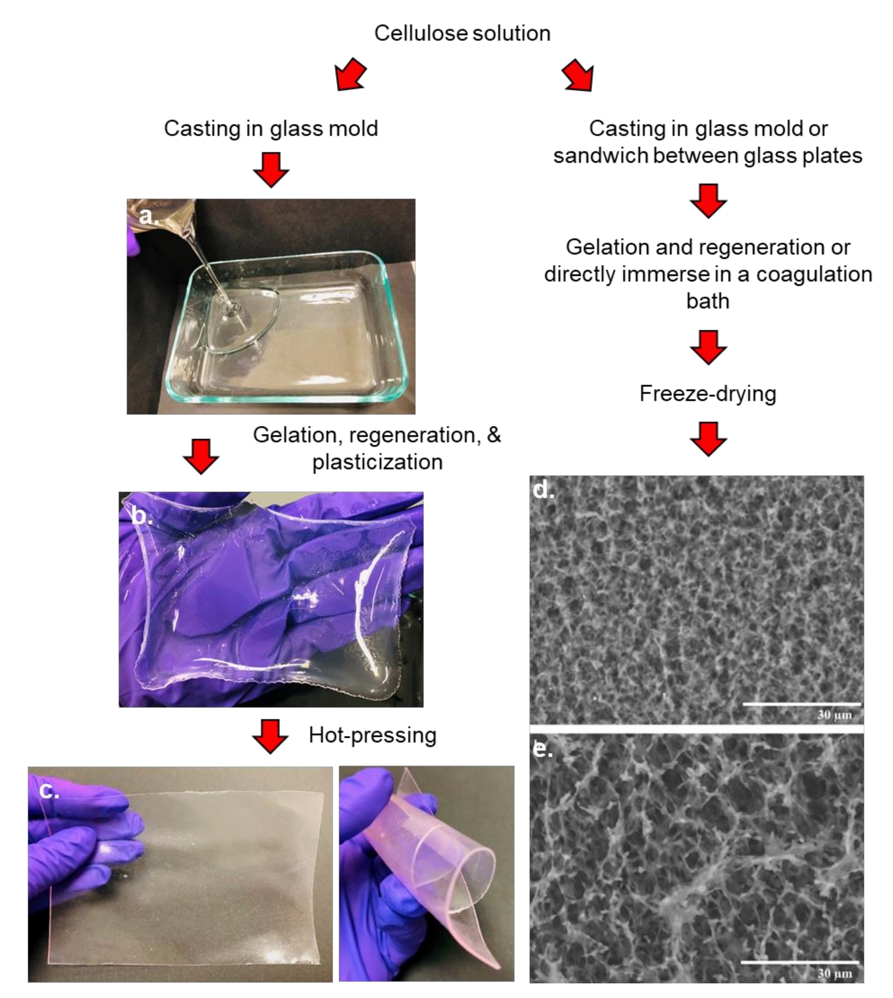
3.2.1. Porous Cellulose Products

Cellulosic materials that hold air within their porous network represent an important class of cellulose bioproducts. Often called cellulose aerogels, they not only possess the fascinating properties of cellulose, but also integrate other important properties of porous materials, such as high porosity, high surface area, and low density. The abundance of hydroxyl groups over a large surface area allows introducing several functional groups onto cellulose chains which renders these porous bioproducts useful for targeted applications (e.g., provides more active sites for heterogeneous catalytic reactions) [113]. Cellulose aerogels include natural cellulose aerogels, regenerated cellulose aerogels, and aerogels of cellulose derivatives of both cellulose I and II crystalline structures [114]. Natural cellulose aerogels are produced by direct drying of cellulose dispersions, whereas regenerated cellulose aerogels are developed through dissolution of the polymer in a proper solvent system, regeneration in anti-solvent, and drying. Cellulose derivative aerogels are obtained through similar preparation methods, but because cellulose derivatives are soluble in volatile organic solvents (VOCs), regeneration in anti-solvent/solvent-exchange steps are often omitted [114].
Cellulose hydrogels are highly porous materials that can absorb, retain, and release water in a reversible manner [115]. Cryogels are prepared by solvent exchange followed by freeze drying of cellulose gels and known to possess high surface area (190–213 m2/g) and porosity characteristics [116]. Drying technique remains a principal factor determining the porosity and pore characteristics of porous cellulose products, however casting process, nature of solvents and antisolvents, and inclusion of additives, such as surfactants, inorganic salts, metal oxides, etc. can affect their surface characteristics.

3.2.2. Cellulose-Based Hydrogels

Cellulose hydrogels are usually prepared through casting cellulose solutions into a mold followed by gelation and regeneration in water or another anti-solvent. Hydrogels range from physical gels (made by polymer chain self-entanglement) to chemically cross-linked networks. The main common attribute of hydrogels is a gel-like network containing both a liquid phase (usually water) and a solid phase. For hydrogels preparation, typically, cellulose solution is cast into a suitable mold, and subsequently gelated under different temperatures. Hydrogel thickness is controlled by the mass of the film-forming solution poured into a mold [117]. Thus, Chang et al. successfully prepared cellulose-based hydrogels using molding, by dissolving carboxymethylcellulose (CMC)/cellulose in NaOH/urea aqueous system containing epichlorohydrin (ECH) cross-linker, followed by keeping the solution at different temperatures (i.e., heating at 60 °C for 12 h, 50 °C for 20 h or freezing at −20 °C for 20 h) [118,119]. Another technique is casting, in which a solution is placed onto a glass plate and spread with a roller or casting knife on a glass substrate and subsequently gelated at ambient temperature [120] or directly immersed into a coagulating bath [116,121]. Two important categories of regenerated cellulose gels, namely hydrogels and alcogels, can be prepared by regenerating the gel in an aqueous or an alcohol medium [121]. Hydrogels and alcogels are dried as needed using an appropriate drying technique, such as reduced pressure/vacuum drying [120,122] and freeze drying [116,121].
It has been reported that the surface area and capillarity, crucial for most of the targeted applications, depend on the hydrogel microstructure [123]. For example, cellulose-based hydrogels loaded with bovine serum albumin (BSA) exhibited a swelling/reswelling ratio of 1000, controlled release of embedded BSA, and smart swelling and shrinking in NaCl or CaCl2 aqueous solution [118] (Figure 4). Owing to their hydrophilicity, biocompatibility, stimuli-sensitivity, and exceptional adsorption capability, cellulose-based hydrogels have found promising applications in a diverse range of fields, including biomedical, pharmaceutical, water purification, and wastewater treatment [115].
There have been many reports on the surface modification of cellulose-based hydrogels for different applications. Thus, Isobe et al. used several aqueous and alcoholic coagulants to prepare gels that were found to have different internal surface polarity, which in turn affected the absorbent behavior of the hydrogels [121]. The authors reported that hydrogels coagulated in alcohols showed better affinity for a Congo red azo-dye than hydrogels that were coagulated in water.
Kim and Kuga grafted polyallylamine onto commercial-grade cellulose gels to increase the ion-exchange capacity targeting chromatographic applications [124]. To introduce additional surface functionalities onto cellulose hydrogels, several researchers have used functionalized cellulose polymer as the starting material. For example, RodrÍguez et al. produced cationic cellulose hydrogels by crosslinking two cationic hydroxyethycelluloses [125]. The resulting cationic hydrogels showed significant drug loading capacity and pH-/ion-sensitive release of the anionic amphiphilic drug, diclofenac sodium. Way et al. showed that surface modified cellulose nanocrystals (CNCs) could be employed to introduce additional functionalities to the corresponding suspensions, gels, and composites, imparting properties such as sensitivity to external stimuli (e.g., pH sensitivity) [126]. Ali et al. reported that the irradiation of carboxymethyl cellulose (CMC)-based hydrogels during crosslinking and gelation steps increased their swelling behavior and induced pH-dependent drug release, possibly due to the chain scission of the polymer network [127].

3.2.3. Aerocellulose Monoliths

Monoliths are single-piece solid cellulosic materials with highly interconnected pores, high surface area, low density, and low thermal conductivity [128]. In addition to unique characteristics of porous materials discussed above, monoliths are mechanically robust, have controllable pore size, and do not have a void volume [129]. Moreover, the convection mode of mass transport adds a significant advantage to the monoliths. Monoliths exhibit higher hydrodynamic porosity than the packed particle beds, implying that the pore structures of monoliths are readily available for the fluid flow than the packed particles where the flow is restricted to interstitial spaces [130]. They are commonly prepared using the sol–gel process, regeneration, and drying [38] (Figure 5). In this process, cellulose solution is poured into a glass mold, gelated at 50 °C for 2 h, regenerated in water, solvent-exchanged with acetone, and subsequently dried using supercritical drying technique [37,38,39], during which the overall volume and network structure of the gel remain largely unchanged. Owing to high porosity and high specific surface area (100 to 400 m2/g [38,39,131,132]), ultra-lightweight aerocellulose monoliths with tunable morphological properties (e.g., pore characteristics, shape, size), and inherent beneficial properties of cellulose, are attractive candidates for various applications. Specifically, cellulose monoliths can be shaped into different forms, including microfluidic channels, micropipette tips, and capillary columns [133].
Cellulose monoliths allow easy functionalization for important applications, including chromatographic and separation technologies [129]. For example, Xin et al. introduced different functional groups on the cellulose backbone, targeting molecular immobilization and separation [134]. The epoxy group was introduced via reaction with epichlorohydrin in NaOH solution followed by conversion into amine- and aldehydes-functionalized aerogels by reacting with 1,2-bis(2-aminoethoxy)ethane (BAE) and glutaraldehyde, respectively. Similarly, Pan et al. introduced anion exchange and pseudo affinity (sulfated cellulose) functionalities onto cellulose monoliths to separate influenza virus (H1N1) [135]. The authors reported that the monolith with pseudo-affinity demonstrated better virus separation efficiency and less DNA and protein retention compared to that of non-modified monoliths. In another study, CVD technique was used to coat the aerogel with fluorinated silane (1H,1H,2H,2H-perfluorodecyltrichlorosilane, PFOTS) [128]. The authors reported that the modified aerogel had high oleophilic properties as indicated by the contact angles of castor oil and hexadecane. Similar to other porous cellulose materials, functionalized aerocellulose monoliths offered a promising approach to wastewater treatment and water purification [39,136]. Hu et al. functionalized cellulose monoliths with 3-chloro-2hydroxypropyl) trimethylammonium chloride to introduce positively charged sites for removal of dye molecules from wastewater [39]. The authors reported that the presence of numerous quaternary ammonium cations in the functionalized samples made them highly efficient in immobilizing negatively charged dye compared to that of control samples.

3.2.4. Cellulose Beads

Cellulose beads (CBs) are smaller-sized particles prepared through the usual route of dissolution of cellulose or cellulose derivatives followed by shaping into a spherical form, sol–gel transition, drying, and fine-tuning of pore characteristics [137]. Particularly, shaping is the most crucial step in CBs preparation, which is usually achieved either by dispersion techniques or dropping the cellulose solution into a coagulating bath using droplet forming machines such as atomizers and jet cutting machines [138]. In the dropping technique, beads are formed by the solidification of cellulose solution by adding it drop-wise into an aqueous coagulating medium (Figure 6). During this process, the combined force of gravity and pressure contribute to the formation of small droplets of cellulose solution pressurized through a narrow orifice or perforated material [139]. The shape and size of cellulose beads could be affected by several factors, including falling height, diameter of the orifice, ejection speed, solution concentration, and coagulation bath [140]. In the dispersion technique, the cellulose solution is dispersed into an immiscible solvent of opposite polarity [141]. Generally, the beads obtained by the dispersion technique are smaller than those obtained by dropping and are in the range of 10–100 µm. However, the mixing speed, viscosity of the cellulose solution, dispersion medium, and the ratio of hydrophobic to hydrophilic solvent affect the size of beads [137].
CBs can be either porous or nonporous. Porous CBs are characterized by high specific surface area and low density. Often named as microspheres, pellets, and beaded cellulose or pearl cellulose, they range from several microns to few millimeters in size [137]. Since CBs reduce the backpressure under flow conditions, they are more advantageous than films or other irregularly shaped particles [142].
There has been extensive research work carried out on the surface functionalization of CBs for various types of applications. Thus, CBs and surface modified CBs have found numerous applications in chromatography, drug loading and control release, protein immobilization, and wastewater treatment [137,143]. Xiong et al. imparted hydrophobicity through the reaction of cellulose with trimethylsilyl chloride or hexamethyldisilazane for size exclusion chromatography (SEC) applications [144]. Similarly, Peksa et al. performed carboxymethylation of cellulose beads with chloroacetic acid for ion-exchange applications [145]. Zhang et al. modified porous cellulose beads with amino functional groups to adsorb and separate hyperin and 2’-O-galloylhyperin from the pyrola extract [146]. The authors reported that the modified cellulose beads had three times higher adsorption capacities compared to that of commercial resins. Ruan et al. prepared chitosan crosslinked 2,3-dialdehyde cellulose beads for chemical waste treatments [142]. The authors obtained a fast and efficient Congo red dye adsorption and desorption at pH 2 and 12, respectively, indicating a great potential for chemical waste treatment.

3.2.5. Films and Membranes

Cellulose films and membranes are sheet-like thin and flexible materials regenerated from cellulose solutions through casting, followed by gelation, regeneration, and dehydration. Usually prepared in either porous or non-porous forms, cellulose films and membranes represent an important class of cellulose bioproducts. Importantly, the ease of preparation, the excellent mechanical properties, and the easy functionalization make them high-value cellulosic materials with widespread applications. Owing to excellent chemical stability, high tenacity in the wet state, biocompatibility, and biodegradability, cellulose films and membranes have found a wide range of applications ranging from simple packaging material, ultrafiltration devices, smart materials to membrane separation (e.g., ultrafiltration, dialysis, and fractionation of polymer mixtures) [147].
In general, films and membranes are developed by casting a thin layer of cellulose solution onto a suitable substrate [16,28,35] and subsequently gelating it in ambient temperature conditions. In addition, a thin layer of cellulose solution can be deposited on glass substrates by slowly dipping glass slides in the solution [148] or by spin-coating [149]. Instead of gelation, cellulose layer deposited on a glass substrate can also be air dried [148], vacuum dried [149], or directly immersed into a protic solvent [150,151,152,153,154,155,156] and regenerated in water to make hydrogels, which are later dried using an appropriate technique (i.e., air, vacuum or freeze drying). Many parameters, including solvents and antisolvent, membrane thickness, additives such as surfactants, and temperature of the coagulation bath affect the properties of the membranes [152,153,155,157]. Cellulose films with homogenous porous structures and mesh networks have been also developed by coagulating cellulose solutions in different coagulating baths (e.g., H2SO4, H2SO4/Na2SO4, Na2SO4, HOAc, and (NH4)2SO4) [152,158]. Recent studies also reported that heat-pressing of hydrogels between two metal plates results in transparent, uniform, and strong films [36,159]. While hot pressing generates non-porous and smooth films, the freeze-drying process preserves the membrane pore structures (Figure 7). Plasticization (with e.g., glycerol) improves elongation and flexibility of cellulose-based films [17,159].
Several studies reported on the preparation of porous cellulose membranes for different applications. For example, Khare et al. prepared porous cellulose films by casting the cellulose solution onto a glass plate and regenerating in acetone followed by solvent exchange [151]. Acharya et al. prepared porous cellulose films by gelating, regenerating, and freeze-drying a cellulose solution sandwiched between glass slides [16].
Similar to other cellulose-based products, the manipulation of the physical and chemical properties is crucial for films and membranes in tailoring their properties for various applications. Fernandes et al. introduced antimicrobial properties by grafting aminoalkyl groups on porous cellulose films [160]. The authors reported that the antimicrobial cellulose membranes prepared by grafting aminoalkyl groups using 3-aminopropyltrimethoxysilane (APS) had excellent antimicrobial activity against S. aureus and E. coli and good biocompatibility, which made them suitable for tissue engineering, wound healing, and as tissue implants. Functionalized cellulose membranes have been used for heavy-metal ion adsorption from aqueous solutions. Wei et al. introduced polyhydroxy functional groups on the regenerated cellulose membrane using poly(glycidyl methacrylate) and N-methylglucamine to remove boron from aqueous solutions. The authors reported maximum boron adsorption of 0.75 mmol/g under neutral pH [161]. Wittmar and Ulbricht prepared catalytically active porous TiO2-cellulose self-cleaning nanocomposite films with potential applications in water purification [162]. Bedane et al. recommended surface coating or chemical modifications of cellulose films for improving films’ moisture barrier properties, especially in highly humid environmental conditions [156] because surface hydrophilicity of films increases due to glycerol plasticization. Rumi et al. showed that hydrophobicity can be imparted into glycerol plasticized and hot-pressed cellulose films via plasma induced surface grafting of oleic acid [159].

3.3. Surface Modification of Cellulose Nanomaterials

Nanocellulose/cellulose nanomaterials (CNMs), nanosized cellulose materials derived often from wood pulp, are also an important class of cellulose with remarkable mechanical, and film forming properties. These materials possess at least one dimension in nanometer range [163]. Owing to the highly ordered organization of cellulose polymer chains into fibrils during biosynthesis, nanoparticles of cellulose can be subsequently extracted [164]. Different from conventional cellulose materials, CNMs possess unique characteristics, such as nano-scale size, special morphology and geometrical shapes, high aspect ratio, crystallinity, high specific surface area, impressive stiffness and strength, dimensional stability, liquid crystalline behavior, alignment and orientation, low coefficient of thermal expansion, and surface chemistry tunability [165,166]. These features combined with abundance and renewability of cellulose make CNMs an important class of futuristic materials with several potential commercial applications including nanocomposite materials, biomedical products, cements, food packaging, antimicrobial materials, energy storage (e.g., supercapacitors), and electronics devices [8,167,168,169,170,171,172].
There are three major categories of CNMs: 1) cellulose nanofibrils (CNFs) or nanofibrillated cellulose (NFC), 2) cellulose nanocrystals (CNCs) or cellulose nanowhiskers (CNWs), and 3) bacterial nanocellulose (BC) (Figure 8) [173,174]. CNFs are entangled individual or bundled fibrils released after mechanical breakdown of already purified cellulose substrate. CNFs possess both crystalline and amorphous domains and are 4–20 nm wide and 500–2000 nm long [8]. CNCs are highly crystalline, elongated, and rod-like (or needle-like) nanoparticles extracted by preferential removal of amorphous domains typically by acid hydrolysis treatment. However, they may also contain residual amorphous domains [175]. CNCs extracted from plant-based cellulose sources are 3–5 nm wide and 50–500 nm long [8]. While CNCs and CNFs are obtained by chemical and mechanical breakdown of the native cellulose structure, bacterial cellulose (BC) is produced by some species of bacteria such as Komagataeibacter xylinus in the form of microfibrils of rectangular cross-sections (6–10 by 30–50 nm). Further aggregation of these microfibrils leads to entangled ultrafine ribbons (nanofibers) of 20–100 nm in width and micrometers in length, ultimately generating a three-dimensional web-shaped network structure [8,176]. BC has unique valuable properties, including highly porous network structure, high specific surface area, and high water holding capacity [177].
In addition to the presence of reactive hydroxyl groups, nanocellulose possesses a large surface area (i.e., several hundred m2/g) and a high ratio of surface area to volume [176]. These attributes impart high reactivity and thus can be modified and functionalized with relative ease. The surface of nanocellulose can be modified by means of oxidation, etherification, esterification, and grafting [178,179,180]. The modifications not only help enhance the processability (e.g., dispersion in solvents) and compatibility (e.g., when mixing with other polymer matrices) of nanocellulose [15,165,181], but can also impart beneficial functionalities such as antimicrobial properties [170].

3.3.1. Cellulose Nanofibrils (CNFs)

CNFs are produced when cellulose fibers are cleaved in the transverse direction as a result of mechanical shearing, leading to the destruction of native hierarchical structure and subsequent release of long and flexible individual or bundle of fibrils [8]. Typically, for the isolation of CNFs, a dilute suspension of cellulose fibers (cellulose slurry) is subjected to high shear forces and various mechanical processes, such as high pressure homogenization (HPH), microfluidization, grinding, cryocrushing, and high-intensity ultrasonication [184]. These mechanical processes are energy intensive. For example, multiple cycles of shearing at high pressure (up to 150 MPa) in the case of HPH and microfludization are required to obtain CNFs of high homogeneity [185,186]. Therefore, usually, various chemically mediated oxidation (e.g., 2,2,6,6-tetramethyl-1-piperidinyloxy (TEMPO) and formic acid hydrolysis) and enzymatic pretreatments (e.g., endoglucanases) are applied prior to mechanical defibrillation for producing CNFs [179,187]. These pretreatments help in loosening the fibrillar structure and facilitate swelling of cellulose, ultimately easing mechanical defibrillation and reducing the energy consumption during CNFs preparation [179]. Specific properties of CNFs such as dimensions, aspect ratio, and surface charge are largely governed by the preparation methods employed with a little effect of cellulose source [174]. “Greener” and more efficient pathways for producing CNFs have also been reported. In a recent study, Ramakrishnan and coworkers reported the preparation of CNFs from cotton cellulose by oil-bath heating cellulose in glycerol at ~180–200 °C [168]. The authors reported an impressive CNFs yield of 71%.
Surface-modified CNFs are currently receiving considerable attention. Mou et al. performed selective oxidation of CNFs at C2–C3 position using periodate oxidation method (NaIO4) to obtain 2,3-dialdehyde nanocellulose [188]. The authors reported that the produced dialdehyde nanocellulose showed good inhibition activity against Staphylococcus aureus and more obnoxious methicillin-resistant Staphylococcus aureus (MRSA) while showing biocompatibility with mammalian cells. Another interesting study showed the importance of the surface modification of nanocellulose comprised of simple oxidation. Weishaupt et al. employed TEMPO-mediated oxidation for preparing surface modified CNFs with negatively charged carboxylic groups [189], and adsorbed positively charged nisin peptide, known for its bioactivity, onto the negatively charged surface modified CNF. The CNFs-nisin composite exhibited antimicrobial activity against Staphylococcus aureus. The authors reported that complete inhibition was achieved at two times lower dose of CNFs-nisin composite compared to that of neat nisin solution. In a recent study, the antimicrobial potential of chemically modified CNFs was investigated by Hassanpour and coworkers [190]. The investigators first modified the surface of the CNFs by covalent attachment of (3-trimethoxysilylpropyl)phenanthridinium iodide (TMSPhI), a quaternary ammonium salt, at different weight percentages (10–100 wt% TMSPhI with respect to CNFs). The modified CNFs containing 100 wt% of TMSPhI (CNFs-TMSPHI) showed strong antibacterial activities against S. aureus and E. coli. The antibacterial effect of the modified nanocellulose was more pronounced against E. coli than against S. aureus. The authors also reported that the modified nanocellulose (CNFs-TMSPHI) was not cytotoxic at low concentrations towards normal cells, indicating potential suitability in biomedical applications such as wound healing bandages.
Surface modifications of CNFs have been performed for other applications, such as adsorption. He et al. prepared modified CNFs by covalently grafting quaternary ammonium functional groups using 2,3-epoxypropyltrimethylammonium chloride [136]. The functionalized CNFs were later used in the preparation of aerogels for heavy-metal ion removal. The authors reported that the aerogels prepared from functionalized CNFs were highly effective in removal of Cr(VI) from water. Only one gram of aerogel adsorbent prepared from modified CNFs was sufficient in removing 99% of Cr(VI) from 1 L solution at a concentration of 1 mg/L in less than 1 h, while the adsorption efficiency of the aerogel prepared from unmodified CNFs was only 84% even after 3 h.

3.3.2. Cellulose Nanocrystals (CNCs)

Cellulose nanocrystals are typically prepared by selective removal of amorphous domains of cellulose using acid hydrolysis of purified cellulose materials (e.g., scoured and bleached wood pulp and cotton fibers). Sulfuric acid is widely used for preparing CNCs [191]. Although slight variations in hydrolysis conditions are common [192,193,194], CNCs are typically prepared by hydrolyzing the cellulose substrate with ~64 wt% sulfuric acid for 1 h at 45–50 °C [175], although the time of the hydrolysis reaction can vary based on the temperature [164]. When native cellulose fibers are subjected to sulfuric acid hydrolysis, cellulose chains in amorphous regions of microfibrils are selectively hydrolyzed as the chains in the amorphous regions are readily accessed by the acid. Consequently, this process releases individual CNCs [195]. Although sulfuric acid hydrolysis remains as the method of choice for the preparation of CNCs, several other inorganic as well as organic acids such as hydrochloric acid, nitric acid, hydrobromic acid, phosphoric acid, oxalic acid, maleic acid, citric acid, and mixture of acids have been successfully employed to prepare CNCs [196]. In addition to acid hydrolysis, studies have shown that other chemical methods such as TEMPO-mediated oxidation [197], periodate oxidation [198], and oxidation with other common oxidizing agents (e.g., ammonium persulfate and potassium permanganate) [199,200] can be exploited to prepare CNCs from cellulose substrates. Moreover, CNCs can also be prepared using certain ionic liquids such as 1-butyl-3-methylimidazolium hydrogen sulfate and 1-ethyl-3-methylimidazolium acetate [201,202]. Apart from various chemical methods, researchers have successfully prepared CNCs by environmentally friendly enzymatic hydrolysis of cellulose using endo-β-1,4-glucanase (endoglucanase, EG) [203,204]. However, chemical methods are more efficient than biological ones in terms of time and yield [196]. Unlike CNFs, whose properties mostly depend on the preparation method with little effect of the cellulose source, properties of CNCs such as morphology and other functional properties (e.g., thermal and colloidal stability) are affected by both the cellulose source and the method of preparation [205,206]. For instance, CNCs derived from tunicates have a length of several micrometers with aspect ratio of approximately 70, while CNCs obtained from lignocellulosic biomass possess lower aspect ratios of 10–30 and are spherical or shorter rod shaped [207]. In general, CNCs prepared by hydrolysis with sulfuric acid under shorter reaction time are larger in size and exhibit enhanced thermal stability but have reduced colloidal stability and vice-versa.
Because of their impressive mechanical properties, CNCs have a great potential as reinforcement fillers for composite materials and have been widely studied accordingly. However, the hydrophilic nature and the high surface charge of CNCs pose challenges in their dispersion in nonpolar solvents and polymer matrices, including water-based polymers [208]. Modifications to impart hydrophobic characteristics help overcome these challenges. Kaboorani and Riedl carried out surface modification of CNCs by physically adsorbing hexadecyltrimethylammonium bromide (HDTMA), a cationic surfactant, and imparted hydrophobicity to the modified CNCs [209]. CNCs modified with higher concentration of HDTMA under longer reaction time showed a good dispersion in tetrahydrofuran. In another study, modified CNCs were prepared by grafting octadecyl isocynate. The grafted CNCs were later used as fillers in nanocomposites of poly (butylene adipate-co-terephthalate) (PBAT) matrices [210]. The authors reported that while the unmodified CNCs were agglomerated and poorly dispersed in PBAT, the CNCs-containing nanocomposite were uniformly dispersed in PBAT (confirmed by ~ 30% increase in elongation at break as compared to the neat PBAT). The authors also observed increased biodegradation of nanocomposites as compared to the neat BPAT. Ogunsona et al. prepared modified CNCs by surface grafting of acrylonitrile butadiene rubber and investigated their applications in nanocomposites with polylactic acid (PLA) [211]. The authors reported that acrylonitrile butadiene rubber grafted CNCs exhibited improved dispersion in PLA, which translated into 25% higher tensile strength of the nanocomposite containing modified CNCs as compared to neat PLA, whereas nanocomposite of PLA containing unmodified CNCs had only 7% increase in tensile strength.
Apart from well-established applications such as reinforcement fillers as discussed earlier, CNCs are emerging as efficient stabilizers of Pickering emulsion systems [212]. Tang et al. modified the surface of CNCs by grafting Poly [2-(dimethylamino)ethyl methacrylate] using free radical polymerization and investigated oil-water emulsion stabilization efficiency of the modified CNCs [66]. The authors observed improved stabilization by modified CNCs as compared to unmodified CNCs. The modified CNCs in the emulsion system were pH and thermal responsive, making them suitable for cosmetic and pharmaceutical applications.

3.3.3. Bacterial Nanocellulose (BC)

The methods of preparing bacterial nanocellulose are fundamentally different from the preparation of CNCs and CNFs. CNFs and CNCs are prepared by using a “top-down” approach, where cellulose fibers of higher dimensions are broken down to nanosized particles using mechanical, chemical, or enzymatic methods. Conversely, BC is prepared by a “bottom-up approach”, where glucose molecules are metabolized by certain bacterial strains to construct a 3D network structure of cellulose microfibrils as their extracellular matrix during fermentation [174,213]. Bacterial strains capable of producing nanocellulose belong to several genera, including Komagateibacter, Alcaligens, Agrobacterium, Rhizobium, Xanthococcus, Pseudomonas, Azotobacter, and Aerobacter. However, owing to high yield, Komagataeibacter xylinus is the most widely used species for BC production [213,214]. Typically, BC in the form of pellicle is obtained by incubating a suitable bacterial strain in glucose-rich culture medium for several days. Numerous factors such as bacterial strains, culture medium, culture conditions (e.g., pH of the medium, static or agitated cultivation), and the duration of cultivation affect the yield and specific properties of BC [215,216]. The fibrous network of BC can also be used as a substrate for the production of bacterial cellulose nanocrystals by acid hydrolysis [205,217] or other measures [218].
Despite the high production cost and lower production rate, adoption of BC may be feasible in specialized applications such as in biomedical and pharmacological fields [219,220], because of desirable properties of BC such as high mechanical strength, highly porous network structure, and high purity. Badshah et al. investigated controlled drug release properties of acetylated surface-modified BC matrices [221]. The authors showed that the drug-loaded acetylated freeze-dried BC matrices showed improved controlled drug release properties under in vitro drug settings compared to those of unmodified BC matrices. In another production method of functionalized BC, Gao et al. demonstrated in situ functionalization of BC during biosynthesis [222]. The authors were able to obtain BC with unnatural characteristic fluorescence by culturing Komagataeibacter sucrofermentans in a medium containing chemically modified glucose (6-carboxyfluorescein-modified glucose). The bacteria metabolized modified glucose units and ultimately imparted fluorescence to the BC.

4. Conclusions and Future Perspectives

Cellulose-based materials can be prepared from a variety of sources: native cellulose (including bacterial cellulose), cellulose composites with other (bio)polymers, cellulose derivatives (e.g., cellulose acetate, methyl cellulose), and cellulose-organic/inorganic hybrids, and their potential applications are countless. Nonetheless, in a highly competitive global market, it is high-value applications that most significantly affect market growth.
In this regard, high-value biotechnological applications that take advantage of cellulose biocompatibility and biodegradability are of interest. Namely, cellulose is used for manufacturing of delivery vehicles for drugs, proteins, and antibodies, scaffolds, sutures, wound healing dressings, etc. For instance, cellulose-based drug excipients, which are able to modify the solubility of drugs and induce different mechanisms of drug release, are used as sustained drug delivery systems [223]. Nanofibrous cellulose derivatives are used as skin substitutes for wound treatment applications [224,225,226]. Antimicrobial nanoparticles/cellulose composites (in form of membranes) have been prepared for slow-release of actives from antibacterial wound dressing [227,228]. Recently, proteins were immobilized into cellulosic aerogel through a water-based self-assembly and used in pH-controlled release [227]. Cellulose-based hydrogels are immensely important for tissue engineering [228], because nanostructured cellulosic materials allow for cellular adhesion [229]. Cellulose templates have been used as artificial blood vessels for modeling tumor invasion in vitro (e.g., cellulose-based microtubes using a chitosan sacrificial template [230]).
There are countless examples, and the value of surface modification of cellulose through either covalent or ionic functionalization cannot be overstated: such tailoring of surface properties of the manufactured materials allows integration of unique material characteristics without affecting the beneficial properties of the bulk cellulose (renewability, biodegradability, biocompatibility, and strength). This approach stimulates adoption of sustainable technologies and allows for wise utilization of resources, resulting in adaptation of sustainable practices.

Author Contributions

All authors contributed equally. All authors have read and agreed to the published version of the manuscript.

Funding

This research received no external funding.

Institutional Review Board Statement

Not required.

Informed Consent Statement

Not applicable.

Conflicts of Interest

The authors declare no conflict of interest.

References

  1. Geyer, R.; Jambeck, J.R.; Law, K.L. Production, use, and fate of all plastics ever made. Sci. Adv. 2017, 3, e1700782. [Google Scholar] [CrossRef] [Green Version]
  2. Klemm, D.; Heublein, B.; Fink, H.-P.; Bohn, A. Cellulose: Fascinating biopolymer and sustainable raw material. Angew. Chem. Int. Ed. 2005, 44, 3358–3393. [Google Scholar] [CrossRef]
  3. Xie, H.; Du, H.; Yang, X.; Si, C. Recent strategies in preparation of cellulose nanocrystals and cellulose nanofibrils derived from raw cellulose materials. Int. J. Polym. Sci. 2018, 7923068. [Google Scholar] [CrossRef]
  4. Shende, P.; Pathan, N. Potential of carbohydrate-conjugated graphene assemblies in biomedical applications. Carbohydr. Polym. 2021, 255, 117385. [Google Scholar] [CrossRef]
  5. Nobles, D.R.; Brown, R.M. Many paths up the mountain: Tracking the evolution of cellulose byosynthesis. In Cellulose: Molecular and Structural Biology; Brown, R.M., Saxena, I.M., Eds.; Springer: Dordrecht, The Netherlands, 2007; pp. 1–16. [Google Scholar]
  6. Sun, Y.; Cheng, J. Hydrolysis of lignocellulosic materials for ethanol production: A review. Bioresour. Technol. 2002, 83, 1–11. [Google Scholar] [CrossRef]
  7. Chang, X.X.; Mubarak, N.M.; Mazari, S.A.; Jatoi, A.S.; Ahmad, A.; Khalid, M.; Walvekar, R.; Abdullah, E.; Karri, R.R.; Siddiqui, M.; et al. A review on the properties and applications of chitosan, cellulose and deep eutectic solvent in green chemistry. J. Ind. Eng. Chem. 2021, in press. [Google Scholar]
  8. Moon, R.J.; Martini, A.; Nairn, J.; Simonsen, J.; Youngblood, J. Cellulose nanomaterials review: Structure, properties and nanocomposites. Chem. Soc. Rev. 2011, 40, 3941–3994. [Google Scholar] [CrossRef] [PubMed]
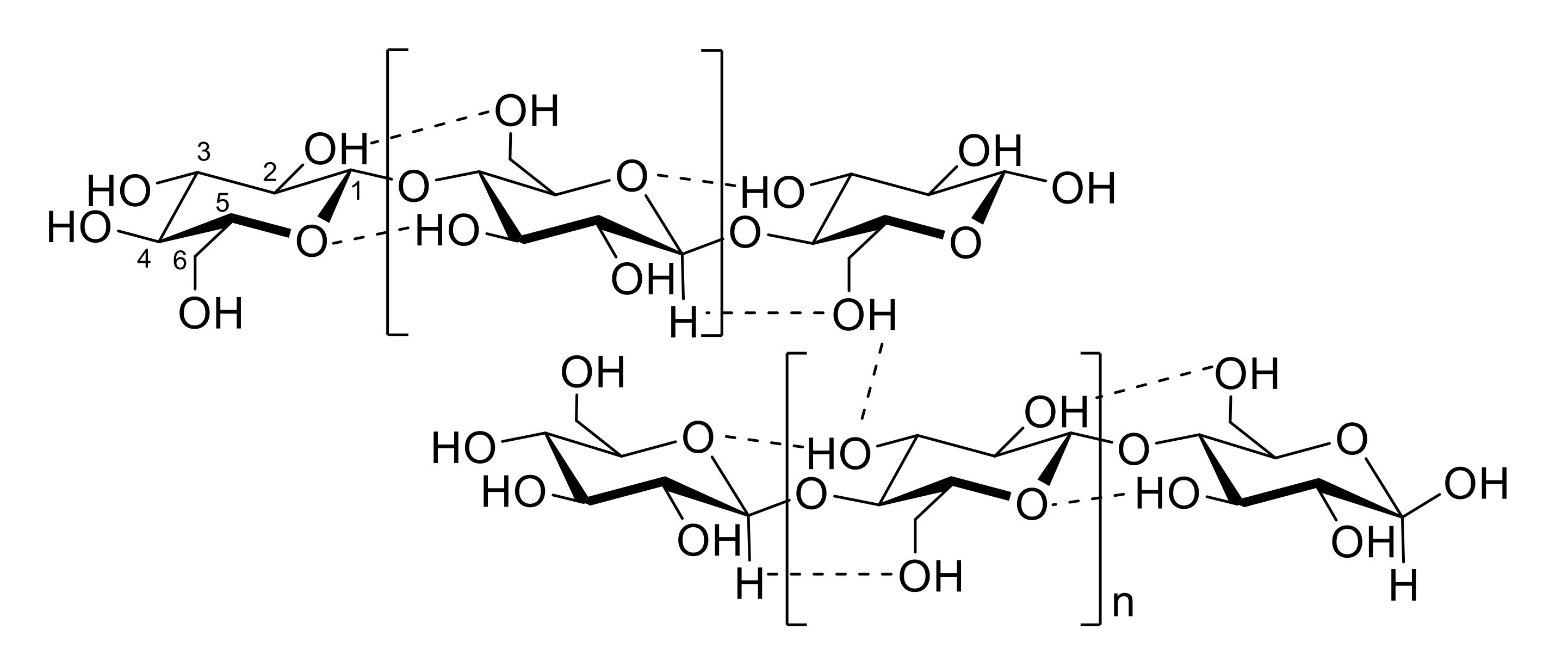
  9. Parthasarathi, R.; Bellesia, G.; Chundawat, S.P.S.; Dale, B.E.; Langan, P.; Gnanakaran, S. Insights into hydrogen bonding and stacking interactions in cellulose. J. Phys. Chem. A 2011, 115, 14191–14202. [Google Scholar] [CrossRef]
  10. Ding, S.-Y.; Himmel, M.E. The maize primary cell wall microfibril: A new model derived from direct visualization. J. Agric. Food Chem. 2006, 54, 597–606. [Google Scholar] [CrossRef]
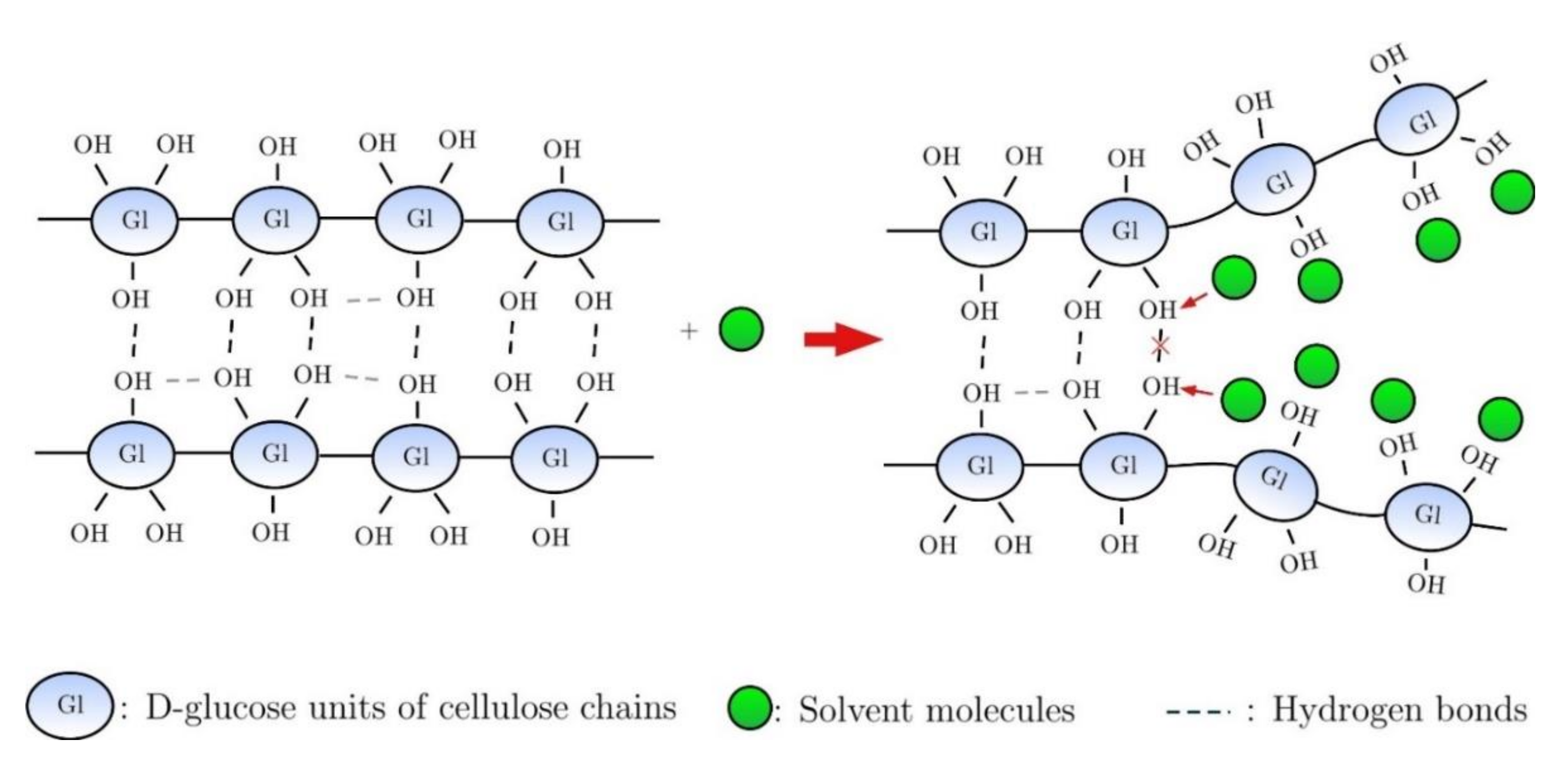
  11. Parviainen, H.; Parviainen, A.; Virtanen, T.; Kilpeläinen, I.; Ahvenainen, P.; Serimaa, R.; Grönqvist, S.; Maloney, T.; Maunu, S.L. Dissolution enthalpies of cellulose in ionic liquids. Carbohydr. Polym. 2014, 113, 67–76. [Google Scholar] [CrossRef]
  12. Medronho, B.; Lindman, B. Competing forces during cellulose dissolution: From solvents to mechanisms. Curr. Opin. Colloid Interface Sci. 2014, 19, 32–40. [Google Scholar] [CrossRef]
  13. Atalla, R.H.; Vanderhart, D.L. Native cellulose: A composite of two distinct crystalline forms. Science 1984, 223, 283–285. [Google Scholar] [CrossRef]
  14. George, J.; Sabapathi, S. Cellulose nanocrystals: Synthesis, functional properties, and applications. Nanotechnol. Sci. Appl. 2015, 8, 45–54. [Google Scholar] [CrossRef] [Green Version]
  15. Wang, S.; Lu, A.; Zhang, L. Recent advances in regenerated cellulose materials. Prog. Polym. Sci. 2016, 53, 169–206. [Google Scholar] [CrossRef]
  16. Acharya, S.; Hu, Y.; Abidi, N. Mild condition dissolution of high molecular weight cotton cellulose in 1-butyl-3-methylimidazolium acetate/N,N-dimethylacetamide solvent system. J. Appl. Polym. Sci. 2016, 135, 45928–45935. [Google Scholar] [CrossRef]
  17. Zhang, L.; Ruan, D.; Gao, S. Dissolution and regeneration of cellulose in NaOH/thiourea aqueous solution. J. Polym. Sci. Part B Polym. Phys. 2002, 40, 1521–1529. [Google Scholar] [CrossRef]
  18. Rogers, R.D.; Seddon, K.R. Chemistry: Ionic liquids—Solvents of the future? Science 2003, 302, 792–793. [Google Scholar] [CrossRef] [PubMed]
  19. Li, Y.; Liu, X.; Zhang, Y.; Jiang, K.; Wang, J.; Zhang, S. Why only ionic liquids with unsaturated heterocyclic cations can dissolve cellulose: A simulation study. ACS Sustain. Chem. Eng. 2017, 5, 3417–3428. [Google Scholar] [CrossRef]
  20. Dissanayake, N.; Thalangamaarachchige, V.D.; Thakurathi, M.; Knight, M.; Quitevis, E.L.; Abidi, N. Dissolution of cotton cellulose in 1:1 mixtures of 1-butyl-3-methylimidazolium methylphosphonate and 1-alkylimidazole co-solvents. Carbohydr. Polym. 2019, 221, 63–72. [Google Scholar] [CrossRef]
  21. Andanson, J.-M.; Bordes, E.; Devémy, J.; Leroux, F.; Padua, A.; Gomes, M.C. Understanding the role of co-solvents in the dissolution of cellulose in ionic liquids. Green Chem. 2014, 16, 2528–2538. [Google Scholar] [CrossRef] [Green Version]
  22. Velioğlu, S.; Yao, X.; Devémy, J.; Ahunbay, M.G.; Tantekin-Ersolmaz, S.B.; Dequidt, A.; Gomes, M.F.C.; Pádua, A.A.H. Solvation of a cellulose microfibril in imidazolium acetate ionic liquids: Effect of a cosolvent. J. Phys. Chem. B 2014, 118, 14860–14869. [Google Scholar] [CrossRef]
  23. Dissanayake, N.; Thalangamaarachchige, V.D.; Troxell, S.; Quitevis, E.L.; Abidi, N. Substituent effects on cellulose dissolution in imidazolium-based ionic liquids. Cellulose 2018, 25, 6887–6900. [Google Scholar] [CrossRef]
  24. Li, Y.; Wang, J.; Liu, X.; Zhang, S. Towards a molecular understanding of cellulose dissolution in ionic liquids: Anion/cation effect, synergistic mechanism and physicochemical aspects. Chem. Sci. 2018, 9, 4027–4043. [Google Scholar] [CrossRef] [Green Version]
  25. Medronho, B.; Lindman, B. Brief overview on cellulose dissolution/regeneration interactions and mechanisms. Adv. Colloid Interface Sci. 2014, 222, 502–508. [Google Scholar] [CrossRef]
  26. Liyanage, S.; Abidi, N. Molecular weight and organization of cellulose at different stages of cotton fiber development. Text. Res. J. 2019, 89, 726–738. [Google Scholar] [CrossRef]
  27. Abidi, N.; Manike, M. X-ray diffraction and FTIR investigations of cellulose deposition during cotton fiber development. Text. Res. J. 2017, 88, 719–730. [Google Scholar] [CrossRef]
  28. Acharya, S.; Hu, Y.; Moussa, H.; Abidi, N. Preparation and characterization of transparent cellulose films using an improved cellulose dissolution process. J. Appl. Polym. Sci. 2017, 134, 44871. [Google Scholar] [CrossRef]
  29. Kosan, B.; Michels, C.; Meister, F. Dissolution and forming of cellulose with ionic liquids. Cellulose 2008, 15, 59–66. [Google Scholar] [CrossRef]
  30. Hauru, L.; Hummel, M.; Michud, A.; Sixta, H. Dry jet-wet spinning of strong cellulose filaments from ionic liquid solution. Cellulose 2014, 21, 4471–4481. [Google Scholar] [CrossRef]
  31. Zhang, H.; Wang, Z.G.; Zhang, Z.N.; Wu, J.; Zhang, J.; He, J.S. Regenerated-cellulose/multiwalled- carbon-nanotube composite fibers with enhanced mechanical properties prepared with the ionic liquid 1-allyl-3-methylimidazolium chloride. Adv. Mater. 2007, 19, 698–704. [Google Scholar] [CrossRef]
  32. Frenot, A.; Henriksson, M.W.; Walkenström, P. Electrospinning of cellulose-based nanofibers. J. Appl. Polym. Sci. 2007, 103, 1473–1482. [Google Scholar] [CrossRef]
  33. Son, W.K.; Youk, J.H.; Lee, T.S.; Park, W.H. Electrospinning of ultrafine cellulose acetate fibers: Studies of a new solvent system and deacetylation of ultrafine cellulose acetate fibers. J. Polym. Sci. Part B Polym. Phys. 2004, 42, 5–11. [Google Scholar] [CrossRef]
  34. Quan, S.-L.; Kang, S.-G.; Chin, I.-J. Characterization of cellulose fibers electrospun using ionic liquid. Cellulose 2010, 17, 223–230. [Google Scholar] [CrossRef]
  35. Ma, H.; Zhou, B.; Li, H.-S.; Li, Y.-Q.; Ou, S.-Y. Green composite films composed of nanocrystalline cellulose and a cellulose matrix regenerated from functionalized ionic liquid solution. Carbohydr. Polym. 2010, 84, 383–389. [Google Scholar] [CrossRef]
  36. Wang, Q.; Cai, J.; Zhang, L.; Xu, M.; Cheng, H.; Han, C.C.; Kuga, S.; Xiao, J.; Xiao, R. A bioplastic with high strength constructed from a cellulose hydrogel by changing the aggregated structure. J. Mater. Chem. A 2013, 1, 6678–6686. [Google Scholar] [CrossRef]
  37. Tan, C.; Fung, B.M.; Newman, J.K.; Vu, C. Organic aerogels with very high impact strength. Adv. Mater. 2001, 13, 644–646. [Google Scholar] [CrossRef]
  38. Parajuli, P.; Acharya, S.; Hu, Y.; Abidi, N. Cellulose-based monoliths with enhanced surface area and porosity. J. Appl. Polym. Sci. 2020, 137, 48975. [Google Scholar] [CrossRef]
  39. Hu, Y.; Li, S.; Jackson, T.; Moussa, H.; Abidi, N. Preparation, characterization, and cationic functionalization of cellulose-based aerogels for wastewater clarification. J. Mater. 2016, 3186589. [Google Scholar] [CrossRef] [Green Version]
  40. Chen, S.; Song, Y.; Xu, F. Highly transparent and hazy cellulose nanopaper simultaneously with a self-cleaning superhydrophobic surface. ACS Sustain. Chem. Eng. 2018, 6, 5173–5181. [Google Scholar] [CrossRef]
  41. Lin, X.; Ma, W.; Chen, L.; Huang, L.; Wu, H.; Takahara, A. Self-healing cellulose nanocrystal-stabilized droplets for water collection under oil. Soft Matter 2018, 14, 9308–9311. [Google Scholar] [CrossRef]
  42. Rasouli, S.; Rezaei, N.; Hamedi, H.; Zendehboudi, S.; Duan, X. Superhydrophobic and superoleophilic membranes for oil-water separation application: A comprehensive review. Mater. Des. 2021, 204, 109599. [Google Scholar] [CrossRef]
  43. Zhan, Y.; Hao, X.; Wang, L.; Jiang, X.; Cheng, Y.; Wang, C.; Meng, Y.; Xia, H.; Chen, Z. Superhydrophobic and flexible silver nanowire-coated cellulose filter papers with sputter-deposited nickel nanoparticles for ultrahigh electromagnetic interference shielding. ACS Appl. Mater. Interfaces 2021, 13, 14623–14633. [Google Scholar] [CrossRef]
  44. Wei, D.W.; Wei, H.; Gauthier, A.C.; Song, J.; Jin, Y.; Xiao, H. Superhydrophobic modification of cellulose and cotton textiles: Methodologies and applications. J. Bioresour. Bioprod. 2020, 5, 1–15. [Google Scholar] [CrossRef]
  45. Lee, H.-J.; Lee, H.-S.; Seo, J.; Kang, Y.-H.; Kim, W.; Kang, T.H.-K. State-of-the-art of cellulose nanocrystals and optimal method for their dispersion for construction-related applications. Appl. Sci. 2019, 9, 426. [Google Scholar] [CrossRef] [Green Version]
  46. Rauscher, H.; Perucca, M.; Buyle, G. (Eds.) Part I. Introduction to plasma technology for surface functionalization. In Plasma Technology for Hyperfunctional Surfaces. Food, Biomedical and Textile Applications; Wiley-VCH: Weinheim, Germany, 2010; pp. 3–32. [Google Scholar]
  47. Denes, F.; Manolache, S. Macromolecular plasma-chemistry: An emerging field of polymer science. Prog. Polym. Sci. 2004, 29, 815–885. [Google Scholar] [CrossRef]
  48. Desmet, T.; Morent, R.; De Geyter, N.; Leys, C.; Schacht, E.; Dubruel, P. Nonthermal plasma technology as a versatile strategy for polymeric biomaterials surface modification: A review. Biomacromolecules 2009, 10, 2351–2378. [Google Scholar] [CrossRef] [Green Version]
  49. Morent, R.; De Geyter, N.; Desmet, T.; Dubruel, P.; Leys, C. Plasma surface modification of biodegradable polymers: A review. Plasma Process. Polym. 2011, 8, 171–190. [Google Scholar] [CrossRef]
  50. Abidi, N.; Hequet, E. Cotton fabric graft copolymerization using microwave plasma. I. Universal attenuated total reflectance-FTIR study. J. Appl. Polym. Sci. 2004, 93, 145–154. [Google Scholar] [CrossRef]
  51. Mather, R.R. Surface modification of textiles by plasma treatments. In Surface Modification of Textiles; Wei, Q., Ed.; Woodhead Publishing: Sawston, UK, 2009; pp. 296–317. [Google Scholar]
  52. Bhat, N.; Benjamin, Y. Surface resistivity behavior of plasma treated and plasma grafted cotton and polyester fabrics. Text. Res. J. 1999, 69, 38–42. [Google Scholar] [CrossRef]
  53. Ryu, G.H.; Yang, W.-S.; Roh, H.-W.; Lee, I.-S.; Kim, J.K.; Lee, G.H.; Lee, D.H.; Park, B.J.; Lee, M.S.; Park, J.-C. Plasma surface modification of poly (d,l-lactic-co-glycolic acid) (65/35) film for tissue engineering. Surf. Coatings Technol. 2005, 193, 60–64. [Google Scholar] [CrossRef]
  54. Lee, S.-D.; Hsiue, G.-H.; Kao, C.-Y.; Chang, P.C.-T. Artificial cornea: Surface modification of silicone rubber membrane by graft polymerization of pHEMA via glow discharge. Biomaterials 1996, 17, 587–595. [Google Scholar] [CrossRef]
  55. Lin, R.; Li, A.; Zheng, T.; Lu, L.; Cao, Y. Hydrophobic and flexible cellulose aerogel as an efficient, green and reusable oil sorbent. RSC Adv. 2015, 5, 82027–82033. [Google Scholar] [CrossRef]
  56. Alanis, A.; Valdés, J.H.; Guadalupe, N.-V.M.; Lopez, R.; Mendoza, R.; Mathew, A.P.; de León, R.D.; Valencia, L. Plasma surface-modification of cellulose nanocrystals: A green alternative towards mechanical reinforcement of ABS. RSC Adv. 2019, 9, 17417–17424. [Google Scholar] [CrossRef] [Green Version]
  57. Rauscher, H.; Perucca, M.; Buyle, G. (Eds.) Part II. Hyperfunctional surfaces for textiles, food and biomedical applications plasma technology for hyperfunctional surfaces. In Plasma Technology for Hyperfunctional Surfaces. Food, Biomedical and Textile Applications; Wiley-VCH: Weinheim, Germany, 2010; pp. 33–65. [Google Scholar]
  58. Roy, D.; Semsarilar, M.; Guthrie, J.T.; Perrier, S. Cellulose modification by polymer grafting: A review. Chem. Soc. Rev. 2009, 38, 2046–2064. [Google Scholar] [CrossRef] [PubMed]
  59. Joubert, F.; Musa, O.M.; Hodgson, D.R.W.; Cameron, N.R. The preparation of graft copolymers of cellulose and cellulose derivatives using ATRP under homogeneous reaction conditions. Chem. Soc. Rev. 2014, 43, 7217–7235. [Google Scholar] [CrossRef] [Green Version]
  60. Abidi, N. Surface grafting of textiles. In Surface Modification of Textiles; Wei, Q., Ed.; Woodhead Publishing: Sawston, UK, 2009; pp. 91–107. [Google Scholar]
  61. Kumar, R.; Sharma, R.K.; Singh, A.P. Grafted cellulose: A bio-based polymer for durable applications. Polym. Bull. 2018, 75, 2213–2242. [Google Scholar] [CrossRef]
  62. Shipp, D. Reversible-deactivation radical polymerizations. Polym. Rev. 2011, 51, 99–103. [Google Scholar] [CrossRef]
  63. Glasing, J.; Champagne, P.; Cunningham, M. Graft modification of chitosan, cellulose and alginate using reversible deactivation radical polymerization (RDRP). Curr. Opin. Green Sustain. Chem. 2016, 2, 15–21. [Google Scholar] [CrossRef]
  64. Marić, M. History of nitroxide mediated polymerization in Canada. Can. J. Chem. Eng. 2021, 99, 832–852. [Google Scholar] [CrossRef]
  65. Littunen, K.; Hippi, U.; Johansson, L.-S.; Österberg, M.; Tammelin, T.; Laine, J.; Seppälä, J. Free radical graft copolymerization of nanofibrillated cellulose with acrylic monomers. Carbohydr. Polym. 2011, 84, 1039–1047. [Google Scholar] [CrossRef]
  66. Tang, J.; Lee, M.F.X.; Zhang, W.; Zhao, B.; Berry, R.M.; Tam, K.C. Dual responsive pickering emulsion stabilized by poly[2-(dimethylamino)ethyl methacrylate] grafted cellulose nanocrystals. Biomacromolecules 2014, 15, 3052–3060. [Google Scholar] [CrossRef]
  67. Abidi, N.; Hequet, E. Cotton fabric graft copolymerization using microwave plasma. II. Physical properties. J. Appl. Polym. Sci. 2005, 98, 896–902. [Google Scholar] [CrossRef]
  68. Zaborniak, I.; Chmielarz, P.; Matyjaszewski, K. Modification of wood-based materials by atom transfer radical polymerization methods. Eur. Polym. J. 2019, 120, 109253. [Google Scholar] [CrossRef]
  69. Chmielarz, P. Cellulose-based graft copolymers prepared by simplified electrochemically mediated ATRP. Express Polym. Lett. 2017, 11, 140–151. [Google Scholar] [CrossRef]
  70. Wang, Z.; Zhang, Y.; Jiang, F.; Fang, H.; Wang, Z. Synthesis and characterization of designed cellulose-graft-polyisoprene copolymers. Polym. Chem. 2014, 5, 3379–3388. [Google Scholar] [CrossRef]
  71. Moreira, G.; Fedeli, E.; Ziarelli, F.; Capitani, D.; Mannina, L.; Charles, L.; Viel, S.; Gigmes, D.; Lefay, C. Synthesis of polystyrene-grafted cellulose acetate copolymers via nitroxide-mediated polymerization. Polym. Chem. 2015, 6, 5244–5253. [Google Scholar] [CrossRef] [Green Version]
  72. Kodama, Y.; Barsbay, M.; Güven, O. Radiation-induced and RAFT-mediated grafting of poly(hydroxyethyl methacrylate) (PHEMA) from cellulose surfaces. Radiat. Phys. Chem. 2014, 94, 98–104. [Google Scholar] [CrossRef]
  73. Lönnberg, H.; Zhou, Q.; Brumer, H.; Teeri, T.T.; Malmström, E.; Hult, A. Grafting of cellulose fibers with poly(ε-caprolactone) and poly(l-lactic acid) via ring-opening polymerization. Biomacromolecules 2006, 7, 2178–2185. [Google Scholar] [CrossRef] [PubMed]
  74. Wohlhauser, S.; Delepierre, G.; Labet, M.; Morandi, G.; Thielemans, W.; Weder, C.; Zoppe, J.O. Grafting polymers from cellulose nanocrystals: Synthesis, properties, and applications. Macromolecules 2018, 51, 6157–6189. [Google Scholar] [CrossRef] [Green Version]
  75. Gates, S.M. Surface chemistry in the chemical vapor deposition of electronic materials. Chem. Rev. 1996, 96, 1519–1532. [Google Scholar] [CrossRef]
  76. Coclite, A.M.; Howden, R.M.; Borrelli, D.C.; Petruczok, C.D.; Yang, R.; Yagüe, J.L.; Ugur, A.; Chen, N.; Lee, S.; Jo, W.J.; et al. 25th anniversary article: CVD polymers: A new paradigm for surface modification and device fabrication. Adv. Mater. 2013, 25, 5392–5423. [Google Scholar] [CrossRef]
  77. Chen, N.; Kim, D.H.; Kovacik, P.; Sojoudi, H.; Wang, M.; Gleason, K.K. polymer thin films and surface modification by chemical vapor deposition: Recent progress. Annu. Rev. Chem. Biomol. Eng. 2016, 7, 373–393. [Google Scholar] [CrossRef] [PubMed]
  78. Kumar, A.; Nanda, D. Methods and fabrication techniques of superhydrophobic surfaces. In Superhydrophobic Polymer Coatings Fundamentals, Design, Fabrication, and Applications; Samal, S.K., Mohanty, S., Nayak, S.K., Eds.; Elsevier: Amsterdam, The Netherlands, 2019; pp. 43–75. [Google Scholar]
  79. Yagüe, J.L.; Coclite, A.M.; Petruczok, C.; Gleason, K.K. Chemical vapor deposition for solvent-free polymerization at surfaces. Macromol. Chem. Phys. 2013, 214, 302–312. [Google Scholar] [CrossRef]
  80. Şakalak, H.; Yılmaz, K.; Gürsoy, M.; Karaman, M. Roll-to roll initiated chemical vapor deposition of super hydrophobic thin films on large-scale flexible substrates. Chem. Eng. Sci. 2020, 215, 115466. [Google Scholar] [CrossRef]
  81. Leal, S.; Cristelo, C.; Silvestre, S.; Fortunato, E.; Sousa, A.; Alves, A.; Correia, D.M.; Lanceros-Mendez, S.; Gama, M. Hydrophobic modification of bacterial cellulose using oxygen plasma treatment and chemical vapor deposition. Cellulose 2020, 27, 10733–10746. [Google Scholar] [CrossRef]
  82. Kettunen, M.; Silvennoinen, R.J.; Houbenov, N.; Nykänen, A.; Ruokolainen, J.; Sainio, J.; Pore, V.; Kemell, M.; Ankerfors, M.; Lindström, T.; et al. Photoswitchable superabsorbency based on nanocellulose aerogels. Adv. Funct. Mater. 2011, 21, 510–517. [Google Scholar] [CrossRef]
  83. Bashir, T.; Skrifvars, M.; Persson, N.-K. Production of highly conductive textile viscose yarns by chemical vapor deposition technique: A route to continuous process. Polym. Adv. Technol. 2011, 22, 2214–2221. [Google Scholar] [CrossRef]
  84. Leskela, M.; Niinisto, J.; Ritala, M. Atomic layer deposition. In Handbook of Thin Films, 4th ed.; Seshan, K., Schrepis, D., Eds.; Academic Press: Cambridge, MA, USA, 2002; pp. 103–159. [Google Scholar]
  85. Johnson, R.W.; Hultqvist, A.; Bent, S.F. A brief review of atomic layer deposition: From fundamentals to applications. Mater. Today 2014, 17, 236–246. [Google Scholar] [CrossRef]
  86. Ritala, M.; Niinistö, J. Industrial applications of atomic layer deposition. ECS Trans. 2019, 25, 641–652. [Google Scholar] [CrossRef]
  87. Hyde, G.K.; McCullen, S.D.; Jeon, S.; Stewart, S.M.; Jeon, H.; Loboa, E.G.; Parsons, G.N. Atomic layer deposition and biocompatibility of titanium nitride nano-coatings on cellulose fiber substrates. Biomed. Mater. 2009, 4, 025001. [Google Scholar] [CrossRef]
  88. Popescu, M.; Ungureanu, C.; Buse, E.; Nastase, F.; Tucureanu, V.; Suchea, M.P.; Draga, S. Antibacterial efficiency of cellulose-based fibers covered with ZnO and Al2O3 by atomic layer deposition. Appl. Surf. Sci. 2019, 481, 1287–1298. [Google Scholar] [CrossRef]
  89. Putkonen, M.; Sippola, P.; Svärd, L.; Sajavaara, T.; Vartiainen, J.; Buchanan, I.; Forsström, U.; Simell, P.; Tammelin, T. Low-temperature atomic layer deposition of SiO2/Al2O3 multilayer structures constructed on self-standing films of cellulose nanofibrils. Philos. Trans. R. Soc A Math. Phys. Eng. Sci. 2018, 376, 20170037. [Google Scholar] [CrossRef] [PubMed] [Green Version]
  90. Yilmaz, N.D. (Ed.) Introduction to smart nanotextiles. In Smart Textiles: Wearable Nanotechnology; John Wiley & Sons: Hoboken, NJ, USA, 2018; pp. 1–37. [Google Scholar]
  91. Pakdel, E.; Jian, F.; Sun, L.; Wang, X. Nanocoatings for smart textiles. In Smart Textiles: Wearable Nanotechnology; Yilmaz, N.D., Ed.; John Wiley & Sons: Hoboken, NJ, USA, 2018; pp. 247–300. [Google Scholar]
  92. Pulit-Prociak, J.; Chwastowski, J.; Kucharski, A.; Banach, M. Functionalization of textiles with silver and zinc oxide nanoparticles. Appl. Surf. Sci. 2016, 385, 543–553. [Google Scholar] [CrossRef]
  93. Riaz, S.; Ashraf, M.; Hussain, T.; Hussain, M.T.; Younus, A. Fabrication of Robust Multifaceted Textiles by Application of Functionalized TiO2 nanoparticles. Colloids Surfaces A Physicochem. Eng. Asp. 2019, 581, 123799. [Google Scholar] [CrossRef]
  94. Mohamed, A.L.; El-Naggar, M.E.; Shaheen, T.I.; Hassabo, A.G. Laminating of chemically modified silan based nanosols for advanced functionalization of cotton textiles. Int. J. Biol. Macromol. 2017, 95, 429–437. [Google Scholar] [CrossRef] [PubMed]
  95. Abidi, N.; Hequet, E.; Tarimala, S.; Dai, L.L. Cotton fabric surface modification for improved UV radiation protection using sol-gel process. J. Appl. Polym. Sci. 2007, 104, 111–117. [Google Scholar] [CrossRef]
  96. Daoud, W.A.; Xin, J.H. Low temperature sol-gel processed photocatalytic titania coating. J. Sol-Gel Sci. Technol. 2004, 29, 25–29. [Google Scholar] [CrossRef]
  97. Wang, P.; Dong, Y.; Li, B.; Li, Z.; Bian, L. A sustainable and cost effective surface functionalization of cotton fabric using TiO2 hydrosol produced in a pilot scale: Condition optimization, sunlight-driven photocatalytic activity and practical applications. Ind. Crop. Prod. 2018, 123, 197–207. [Google Scholar] [CrossRef]
  98. Abidi, N.; Kikens, P. Chemical functionalisation of cotton fabric to impart multifunctional properties. Tekstilec 2016, 59, 156–161. [Google Scholar] [CrossRef]
  99. Abidi, N.; Cabrales, L.; Hequet, E. Functionalization of a cotton fabric surface with titania nanosols: Applications for self-cleaning and UV-protection properties. ACS Appl. Mater. Interfaces 2009, 1, 2141–2146. [Google Scholar] [CrossRef]
  100. Yuranova, T.; Mosteo, R.; Bandara, J.; Laub, D.; Kiwi, J. Self-cleaning cotton textiles surfaces modified by photoactive SiO2/TiO2 coating. J. Mol. Catal. A Chem. 2006, 244, 160–167. [Google Scholar] [CrossRef]
  101. Moiz, A.; Vijayan, A.; Padhye, R.; Wang, X. Chemical and water protective surface on cotton fabric by pad-knife-pad coating of WPU-PDMS-TMS. Cellulose 2016, 23, 3377–3388. [Google Scholar] [CrossRef]
  102. Abidi, N.; Aminayi, P.; Cabrales, L.; Fiber, E.H. Super-hydrophobic cotton fabric prepared using nanoparticles and molecular vapor deposition methods. In Functional Materials from Renewable Sources; Liebner, F., Rosenau, T., Eds.; American Chemical Society: Washington, DC, USA, 2012; Volume 1107, pp. 149–165. [Google Scholar]
  103. Aminayi, P.; Abidi, N. Imparting super hydro/oleophobic properties to cotton fabric by means of molecular and nanoparticles vapor deposition methods. Appl. Surf. Sci. 2013, 287, 223–231. [Google Scholar] [CrossRef]
  104. Cabrales, L.; Abidi, N. Microwave plasma induced grafting of oleic acid on cotton fabric surfaces. Appl. Surf. Sci. 2012, 258, 4636–4641. [Google Scholar] [CrossRef]
  105. Airoudj, A.; Gall, F.B.-L.; Roucoules, V. Textile with durable janus wetting properties produced by plasma polymerization. J. Phys. Chem. C 2016, 120, 29162–29172. [Google Scholar] [CrossRef]
  106. Qin, H.; Li, X.; Zhang, X.; Guo, Z. Preparation and performance testing of superhydrophobic flame retardant cotton fabric. N. J. Chem. 2019, 43, 5839–5848. [Google Scholar] [CrossRef]
  107. Nie, S.; Jin, D.; Yang, J.-N.; Dai, G.; Luo, Y. Fabrication of environmentally-benign flame retardant cotton fabrics with hydrophobicity by a facile chemical modification. Cellulose 2019, 26, 5147–5158. [Google Scholar] [CrossRef]
  108. Song, L.; Sun, L.; Zhao, J.; Wang, X.; Yin, J.; Luan, S.; Ming, W. Synergistic superhydrophobic and photodynamic cotton textiles with remarkable antibacterial activities. ACS Appl. Bio-Mater. 2019, 2, 2756–2765. [Google Scholar] [CrossRef]
  109. Montaser, A.; Rehan, M.; El-Senousy, W.; Zaghloul, S. Designing strategy for coating cotton gauze fabrics and its application in wound healing. Carbohydr. Polym. 2020, 244, 116479. [Google Scholar] [CrossRef]
  110. Kianfar, P.; Abate, M.; Trovato, V.; Rosace, G.; Ferri, A.; Bongiovanni, R.; Vitale, A. Surface functionalization of cotton fabrics by photo-grafting for pH sensing applications. Front. Mater. 2020, 7, 39. [Google Scholar] [CrossRef] [Green Version]
  111. Guido, E.; Colleoni, C.; De Clerck, K.; Plutino, M.R.; Rosace, G. Influence of catalyst in the synthesis of a cellulose-based sensor: Kinetic study of 3-glycidoxypropyltrimethoxysilane epoxy ring opening by Lewis acid. Sens. Actuators B Chem. 2014, 203, 213–222. [Google Scholar] [CrossRef]
  112. Rosace, G.; Guido, E.; Colleoni, C.; Brucale, M.; Piperopoulos, E.; Milone, C.; Plutino, M.R. Halochromic resorufin-GPTMS hybrid sol-gel: Chemical-physical properties and use as pH sensor fabric coating. Sens. Actuators B Chem. 2017, 241, 85–95. [Google Scholar] [CrossRef]
  113. Hoang-Van, C.; Zegaoui, O.; Pichat, P. Vanadia–titania aerogel deNOx catalysts. J. Non. Cryst. Solids 1998, 225, 157–162. [Google Scholar] [CrossRef]
  114. Long, L.-Y.; Weng, Y.-X.; Wang, Y.-Z. Cellulose aerogels: Synthesis, applications, and prospects. Polymers 2018, 10, 623. [Google Scholar] [CrossRef] [Green Version]
  115. Sannino, A.; Demitri, C.; Madaghiele, M. Biodegradable cellulose-based hydrogels: Design and applications. Materials 2009, 2, 353–373. [Google Scholar] [CrossRef]
  116. Wang, Z.; Liu, S.; Matsumoto, Y.; Kuga, S. Cellulose gel and aerogel from LiCl/DMSO solution. Cellulose 2012, 19, 393–399. [Google Scholar] [CrossRef]
  117. de Moraes, J.O.; Scheibe, A.S.; Sereno, A.; Laurindo, J.B. Scale-up of the production of cassava starch based films using tape-casting. J. Food Eng. 2013, 119, 800–808. [Google Scholar] [CrossRef] [Green Version]
  118. Chang, C.; Duan, B.; Cai, J.; Zhang, L. Superabsorbent hydrogels based on cellulose for smart swelling and controllable delivery. Eur. Polym. J. 2010, 46, 92–100. [Google Scholar] [CrossRef]
  119. Chang, C.; Zhang, L.; Zhou, J.; Zhang, L.; Kennedy, J.F. Structure and properties of hydrogels prepared from cellulose in NaOH/urea aqueous solutions. Carbohydr. Polym. 2010, 82, 122–127. [Google Scholar] [CrossRef]
  120. Kadokawa, J.-I.; Murakami, M.-A.; Kaneko, Y. A facile preparation of gel materials from a solution of cellulose in ionic liquid. Carbohydr. Res. 2008, 343, 769–772. [Google Scholar] [CrossRef] [PubMed]
  121. Isobe, N.; Kim, U.-J.; Kimura, S.; Wada, M.; Kuga, S. Internal surface polarity of regenerated cellulose gel depends on the species used as coagulant. J. Colloid Interface Sci. 2011, 359, 194–201. [Google Scholar] [CrossRef] [PubMed]
  122. Zhou, J.; Chang, C.; Zhang, R.; Zhang, L. Hydrogels prepared from unsubstituted cellulose in NaOH/Urea aqueous solution. Macromol. Biosci. 2007, 7, 804–809. [Google Scholar] [CrossRef] [PubMed]
  123. Bao, Y.; Ma, J.; Li, N. Synthesis and swelling behaviors of sodium carboxymethyl cellulose-g-poly(AA-co-AM-co-AMPS)/MMT superabsorbent hydrogel. Carbohydr. Polym. 2011, 84, 76–82. [Google Scholar] [CrossRef]
  124. Kim, U.-J.; Kuga, S. Polyallylamine-grafted cellulose gel as high-capacity anion-exchanger. J. Chromatogr. A 2002, 946, 283–289. [Google Scholar] [CrossRef]
  125. RodrÍguez, R.; Alvarez-Lorenzo, C.; Concheiro, A. Cationic cellulose hydrogels: Kinetics of the cross-linking process and characterization as pH-/ion-sensitive drug delivery systems. J. Control. Release 2003, 86, 253–265. [Google Scholar] [CrossRef]
  126. Way, A.E.; Hsu, L.; Shanmuganathan, K.; Weder, C.; Rowan, S.J. pH-responsive cellulose nanocrystal gels and nanocomposites. ACS Macro Lett. 2012, 1, 1001–1006. [Google Scholar] [CrossRef]
  127. Ali, A.E.-H.; El-Rehim, H.A.A.; Kamal, H.; Hegazy, D.E.A. Synthesis of carboxymethyl cellulose based drug carrier hydrogel using ionizing radiation for possible use as site specific delivery system. J. Macromol. Sci. Part A 2008, 45, 628–634. [Google Scholar] [CrossRef]
  128. Aulin, C.; Netrval, J.; Wågberg, L.; Lindström, T. Aerogels from nanofibrillated cellulose with tunable oleophobicity. Soft Matter 2010, 6, 3298–3305. [Google Scholar] [CrossRef]
  129. Gustavsson, P.-E.; Larsson, P.-O. Monolithic Polysaccharide materials. In Monolithic Materials: Preparation, Properties and Applications; Svec, F., Tennikova, T.B., Deyl, Z., Eds.; Elsevier: Amsterdam, The Netherlands, 2003; Volume 67, pp. 121–141. [Google Scholar]
  130. Potter, O.G.; Hilder, E.F. Porous polymer monoliths for extraction: Diverse applications and platforms. J. Sep. Sci. 2008, 31, 1881–1906. [Google Scholar] [CrossRef]
  131. Schestakow, M.; Karadagli, I.; Ratke, L. Cellulose aerogels prepared from an aqueous zinc chloride salt hydrate melt. Carbohydr. Polym. 2016, 137, 642–649. [Google Scholar] [CrossRef]
  132. Innerlohinger, J.; Weber, H.K.; Kraft, G. Aerocellulose: Aerogels and aerogel-like materials made from cellulose. Macromol. Symp. 2006, 244, 126–135. [Google Scholar] [CrossRef]
  133. Strong, E.B.; Schultz, S.A.; Martinez, A.W.; Martinez, N.W. Fabrication of miniaturized paper-based microfluidic devices (MicroPADs). Sci. Rep. 2019, 9, 7. [Google Scholar] [CrossRef] [Green Version]
  134. Xin, Y.; Xiong, Q.; Bai, Q.; Miyamoto, M.; Li, C.; Shen, Y.; Uyama, H. A hierarchically porous cellulose monolith: A template-free fabricated, morphology-tunable, and easily functionalizable platform. Carbohydr. Polym. 2017, 157, 429–437. [Google Scholar] [CrossRef]
  135. Pan, X.; Jinka, S.; Singh, V.K.; Ramkumar, S.; Karim, M.N. Cellulose monolith with tunable functionality for immobilization of influenza virus. J. Bioeng. Biomed. Sci. 2018, 8, 1000246. [Google Scholar] [CrossRef]
  136. He, X.; Cheng, L.; Wang, Y.; Zhao, J.; Zhang, W.; Lu, C. Aerogels from quaternary ammonium-functionalized cellulose nanofibers for rapid removal of Cr(VI) from water. Carbohydr. Polym. 2014, 111, 683–687. [Google Scholar] [CrossRef]
  137. Gericke, M.; Trygg, J.; Fardim, P. Functional cellulose beads: Preparation, characterization, and applications. Chem. Rev. 2013, 113, 4812–4836. [Google Scholar] [CrossRef]
  138. Voon, L.K.; Pang, S.C.; Chin, S.F. Porous cellulose beads fabricated from regenerated cellulose as potential drug delivery carriers. J. Chem. 2017, 2017, 1943432. [Google Scholar] [CrossRef] [Green Version]
  139. Qi, H. Novel Functional Materials Based on Cellulose, 1st ed.; Springer Briefs in Applied Sciences and Technology; Springer International Publishing AG: Cham, Switzerland, 2017; pp. 25–43. [Google Scholar]
  140. Trygg, J.; Fardim, P.; Gericke, M.; Mäkilä, E.; Salonen, J. Physicochemical design of the morphology and ultrastructure of cellulose beads. Carbohydr. Polym. 2013, 93, 291–299. [Google Scholar] [CrossRef]
  141. Karbstein, H.P.; Schubert, H. Developments in the continuous mechanical production of oil-in-water macro-emulsions. Chem. Eng. Process. Process. Intensif. 1995, 34, 205–211. [Google Scholar] [CrossRef]
  142. Ruan, C.-Q.; Strømme, M.; Lindh, J. Preparation of porous 2,3-dialdehyde cellulose beads crosslinked with chitosan and their application in adsorption of Congo red dye. Carbohydr. Polym. 2018, 181, 200–207. [Google Scholar] [CrossRef] [PubMed]
  143. Benhalima, T.; Ferfera-Harrar, H.; Lerari, D. Optimization of carboxymethyl cellulose hydrogels beads generated by an anionic surfactant micelle templating for cationic dye uptake: Swelling, sorption and reusability studies. Int. J. Biol. Macromol. 2017, 105, 1025–1042. [Google Scholar] [CrossRef]
  144. Xiong, X.; Zhang, L.; Wang, Y. Polymer fractionation using chromatographic column packed with novel regenerated cellulose beads modified with silane. J. Chromatogr. A 2005, 1063, 71–77. [Google Scholar] [CrossRef] [PubMed]
  145. Ishimura, D.; Morimoto, Y.; Saito, H. Influences of chemical modifications on the mechanical strength of cellulose beads. Cellulose 1998, 5, 135–151. [Google Scholar] [CrossRef]
  146. Zhang, D.-Y.; Zhang, N.; Song, P.; Hao, J.-Y.; Wan, Y.; Yao, X.-H.; Chen, T.; Li, L. Functionalized cellulose beads with three dimensional porous structure for rapid adsorption of active constituents from Pyrola incarnata. Carbohydr. Polym. 2018, 181, 560–569. [Google Scholar] [CrossRef] [PubMed]
  147. Thakur, V.K.; Voicu, S.I. Recent advances in cellulose and chitosan based membranes for water purification: A concise review. Carbohydr. Polym. 2016, 146, 148–165. [Google Scholar] [CrossRef] [PubMed]
  148. Garnier, G.; Wright, J.; Godbout, L.; Yu, L. Wetting mechanism of alkyl ketene dimers on cellulose films. Colloids Surfaces A Physicochem. Eng. Asp. 1998, 145, 153–165. [Google Scholar] [CrossRef]
  149. Da Róz, A.; Leite, F.; Pereiro, L.; Nascente, P.; Zucolotto, V.; Oliveira, O.; Carvalho, A. Adsorption of chitosan on spin-coated cellulose films. Carbohydr. Polym. 2010, 80, 65–70. [Google Scholar] [CrossRef]
  150. Turner, M.B.; Spear, S.K.; Holbrey, J.; Rogers, R.D. Production of bioactive cellulose films reconstituted from ionic liquids. Biomacromolecules 2004, 5, 1379–1384. [Google Scholar] [CrossRef] [PubMed]
  151. Khare, V.P.; Greenberg, A.R.; Kelley, S.S.; Pilath, H.; Roh, I.J.; Tyber, J. Synthesis and characterization of dense and porous cellulose films. J. Appl. Polym. Sci. 2007, 105, 1228–1236. [Google Scholar] [CrossRef]
  152. Cai, J.; Wang, L.; Zhang, L. Influence of coagulation temperature on pore size and properties of cellulose membranes prepared from NaOH–urea aqueous solution. Cellulose 2007, 14, 205–215. [Google Scholar] [CrossRef]
  153. Yang, Q.; Fukuzumi, H.; Saito, T.; Isogai, A.; Zhang, L. Transparent cellulose films with high gas barrier properties fabricated from aqueous alkali/urea solutions. Biomacromolecules 2011, 12, 2766–2771. [Google Scholar] [CrossRef] [PubMed]
  154. Jia, B.; Mei, Y.; Cheng, L.; Zhou, J.; Zhang, L. Preparation of copper nanoparticles coated cellulose films with antibacterial properties through one-step reduction. ACS Appl. Mater. Interfaces 2012, 4, 2897–2902. [Google Scholar] [CrossRef] [PubMed]
  155. Li, R.; Zhang, L.; Xu, M. Novel regenerated cellulose films prepared by coagulating with water: Structure and properties. Carbohydr. Polym. 2012, 87, 95–100. [Google Scholar] [CrossRef]
  156. Bedane, A.H.; Eić, M.; Farmahini-Farahani, M.; Xiao, H. Water vapor transport properties of regenerated cellulose and nanofibrillated cellulose films. J. Membr. Sci. 2014, 493, 46–57. [Google Scholar] [CrossRef]
  157. Voicu, S.I.; Muhulet, A.; Antoniac, I.V.; Corobea, M.S. Cellulose derivatives based membranes for biomedical applications. Key Eng. Mater. 2015, 638, 27–30. [Google Scholar] [CrossRef]
  158. Zhang, L.; Mao, Y.; Zhou, J.; Cai, J. Effects of coagulation conditions on the properties of regenerated cellulose films prepared in NaOH/urea aqueous solution. Ind. Eng. Chem. Res. 2005, 44, 522–529. [Google Scholar] [CrossRef]
  159. Rumi, S.S.; Liyanage, S.; Abidi, N. Conversion of low-quality cotton to bioplastics. Cellulose 2021, 28, 2021–2038. [Google Scholar] [CrossRef]
  160. Fernandes, S.C.; Sadocco, P.; Alonso-Varona, A.; Palomares, T.; Eceiza, A.; Silvestre, A.; Mondragon, I.; Freire, C. Bioinspired antimicrobial and biocompatible bacterial cellulose membranes obtained by surface functionalization with aminoalkyl groups. ACS Appl. Mater. Interfaces 2013, 5, 3290–3297. [Google Scholar] [CrossRef]
  161. Wei, Y.-T.; Zheng, Y.-M.; Chen, J.P. Functionalization of regenerated cellulose membrane via surface initiated atom transfer radical polymerization for boron removal from aqueous solution. Langmuir 2011, 27, 6018–6025. [Google Scholar] [CrossRef]
  162. Wittmar, A.S.M.; Ulbricht, M. Ionic liquid-based route for the preparation of catalytically active cellulose—TiO2 porous films and spheres. Ind. Eng. Chem. Res. 2017, 56, 2967–2975. [Google Scholar] [CrossRef]
  163. Foster, E.J.; Moon, R.J.; Agarwal, U.P.; Bortner, M.J.; Bras, J.; Camarero-Espinosa, S.; Chan, K.J.; Clift, M.J.D.; Cranston, E.D.; Eichhorn, S.J.; et al. Current characterization methods for cellulose nanomaterials. Chem. Soc. Rev. 2018, 47, 2609–2679. [Google Scholar] [CrossRef] [Green Version]
  164. Habibi, Y.; Lucia, L.A.; Rojas, O.J. Cellulose nanocrystals: Chemistry, self-assembly, and applications. Chem. Rev. 2010, 110, 3479–3500. [Google Scholar] [CrossRef]
  165. Eichhorn, S.J.; Rahatekar, S.S.; Vignolini, S.; Windle, A.H. New horizons for cellulose nanotechnology. Philos. Trans. R. Soc. A Math. Phys. Eng. Sci. 2018, 376, 20170200. [Google Scholar] [CrossRef] [Green Version]
  166. Koshani, R.; Madadlou, A. A viewpoint on the gastrointestinal fate of cellulose nanocrystals. Trends Food Sci. Technol. 2018, 71, 268–273. [Google Scholar] [CrossRef]
  167. Lin, N.; Dufresne, A. Nanocellulose in biomedicine: Current status and future prospect. Eur. Polym. J. 2014, 59, 302–325. [Google Scholar] [CrossRef] [Green Version]
  168. Ramakrishnan, A.; Ravishankar, K.; Dhamodharan, R. Preparation of nanofibrillated cellulose and nanocrystalline cellulose from surgical cotton and cellulose pulp in hot-glycerol medium. Cellulose 2019, 26, 3127–3141. [Google Scholar] [CrossRef]
  169. Kumar, V.; Elfving, A.; Koivula, H.M.; Bousfield, D.W.; Toivakka, M. Roll-to-roll processed cellulose nanofiber coatings. Ind. Eng. Chem. Res. 2016, 55, 3603–3613. [Google Scholar] [CrossRef]
  170. Li, J.; Cha, R.; Mou, K.; Zhao, X.; Long, K.; Luo, H.; Zhou, F.; Jiang, X. Nanocellulose-based antibacterial materials. Adv. Health Mater. 2018, 7, e1800334. [Google Scholar] [CrossRef] [PubMed]
  171. Jose, J.; Thomas, V.; Vinod, V.; Abraham, R.; Abraham, S. Nanocellulose based functional materials for supercapacitor applications. J. Sci. Adv. Mater. Devices 2019, 4, 333–340. [Google Scholar] [CrossRef]
  172. Agate, S.; Joyce, M.; Lucia, L.; Pal, L. Cellulose and nanocellulose-based flexible-hybrid printed electronics and conductive composites—A review. Carbohydr. Polym. 2018, 198, 249–260. [Google Scholar] [CrossRef]
  173. Abitbol, T.; Rivkin, A.; Cao, Y.; Nevo, Y.; Abraham, E.; Ben-Shalom, T.; Lapidot, S.; Shoseyov, O. Nanocellulose, a tiny fiber with huge applications. Curr. Opin. Biotechnol. 2016, 39, 76–88. [Google Scholar] [CrossRef]
  174. Thomas, B.; Raj, M.C.; Athira, B.K.; Rubiya, H.M.; Joy, J.; Moores, A.; Drisko, G.L.; Sanchez, C. Nanocellulose, a versatile green platform: From biosources to materials and their applications. Chem. Rev. 2018, 118, 11575–11625. [Google Scholar] [CrossRef] [PubMed]
  175. Hu, Y.; Abidi, N. Distinct chiral nematic self-assembling behavior caused by different size-unified cellulose nanocrystals via a multistage separation. Langmuir 2016, 32, 9863–9872. [Google Scholar] [CrossRef]
  176. Trache, D.; Tarchoun, A.F.; Derradji, M.; Hamidon, T.S.; Masruchin, N.; Brosse, N.; Hussin, M.H. Nanocellulose: From fundamentals to advanced applications. Front. Chem. 2020, 8, 392. [Google Scholar] [CrossRef] [PubMed]
  177. Khan, F.; Ahmad, S.R. Polysaccharides and their derivatives for versatile tissue engineering application. Macromol. Biosci. 2013, 13, 395–421. [Google Scholar] [CrossRef] [PubMed]
  178. Eyley, S.; Thielemans, W. Surface modification of cellulose nanocrystals. Nanoscale 2014, 6, 7764–7779. [Google Scholar] [CrossRef] [PubMed] [Green Version]
  179. Missoum, K.; Belgacem, M.N.; Bras, J. Nanofibrillated cellulose surface modification: A review. Materials 2013, 6, 1745–1766. [Google Scholar] [CrossRef] [PubMed] [Green Version]
  180. Habibi, Y. Key advances in the chemical modification of nanocelluloses. Chem. Soc. Rev. 2014, 43, 1519–1542. [Google Scholar] [CrossRef]
  181. Gupta, P.; Singh, B.; Agrawal, A.; Maji, P.K. Low density and high strength nanofibrillated cellulose aerogel for thermal insulation application. Mater. Des. 2018, 158, 224–236. [Google Scholar] [CrossRef]
  182. Beyene, D.; Chae, M.; Dai, J.; Danumah, C.; Tosto, F.; DeMesa, A.G.; Bressler, D.C. Characterization of cellulase-treated fibers and resulting cellulose nanocrystals generated through acid hydrolysis. Materials 2018, 11, 1272. [Google Scholar] [CrossRef] [Green Version]
  183. Niamsap, T.; Lam, N.T.; Sukyai, P. Production of hydroxyapatite-bacterial nanocellulose scaffold with assist of cellulose nanocrystals. Carbohydr. Polym. 2019, 205, 159–166. [Google Scholar] [CrossRef]
  184. Khalil, H.A.; Davoudpour, Y.; Islam, N.; Mustapha, A.; Sudesh, K.; Dungani, R.; Jawaid, M. Production and modification of nanofibrillated cellulose using various mechanical processes: A review. Carbohydr. Polym. 2014, 99, 649–665. [Google Scholar] [CrossRef] [PubMed]
  185. Habibi, Y.; Mahrouz, M.; Vignon, M.R. Microfibrillated cellulose from the peel of prickly pear fruits. Food Chem. 2009, 115, 423–429. [Google Scholar] [CrossRef]
  186. Zimmermann, T.; Bordeanu, N.; Strub, E. Properties of nanofibrillated cellulose from different raw materials and its reinforcement potential. Carbohydr. Polym. 2010, 79, 1086–1093. [Google Scholar] [CrossRef]
  187. Wang, Q.; Du, H.; Zhang, F.; Zhang, Y.; Wu, M.; Yu, G.; Liu, C.; Li, B.; Peng, H. Flexible cellulose nanopaper with high wet tensile strength, high toughness and tunable ultraviolet blocking ability fabricated from tobacco stalk via a sustainable method. J. Mater. Chem. A 2018, 6, 13021–13030. [Google Scholar] [CrossRef]
  188. Mou, K.; Li, J.; Wang, Y.; Cha, R.; Jiang, X. 2,3-Dialdehyde nanofibrillated cellulose as a potential material for the treatment of MRSA infection. J. Mater. Chem. B 2017, 5, 7876–7884. [Google Scholar] [CrossRef] [PubMed]
  189. Weishaupt, R.; Heuberger, L.; Siqueira, G.; Gutt, B.; Zimmermann, T.; Maniura-Weber, K.; Salentinig, S.; Faccio, G. Enhanced antimicrobial activity and structural transitions of a nanofibrillated cellulose–nisin biocomposite suspension. ACS Appl. Mater. Interfaces 2018, 10, 20170–20181. [Google Scholar] [CrossRef] [PubMed]
  190. Hassanpour, A.; Asghari, S.; Lakouraj, M.M.; Mohseni, M. Nanofibrillated cellulose surface modification: A review. Int. J. Biol. Macromol. 2018, 115, 528–539. [Google Scholar] [CrossRef]
  191. Vanderfleet, O.M.; Osorio, D.A.; Cranston, E.D. Optimization of cellulose nanocrystal length and surface charge density through phosphoric acid hydrolysis. Philos. Trans. R. Soc. A 2018, 376, 20170041. [Google Scholar] [CrossRef] [Green Version]
  192. Jordan, J.H.; Easson, M.W.; Dien, B.; Thompson, S.; Condon, B.D. Extraction and characterization of nanocellulose crystals from cotton gin motes and cotton gin waste. Cellulose 2019, 26, 5959–5979. [Google Scholar] [CrossRef]
  193. Pirich, C.L.; Picheth, G.F.; Machado, J.P.E.; Sakakibara, C.N.; Martin, A.A.; de Freitas, R.A.; Sierakowski, M.R. Influence of mechanical pretreatment to isolate cellulose nanocrystals by sulfuric acid hydrolysis. Int. J. Biol. Macromol. 2019, 130, 622–626. [Google Scholar] [CrossRef]
  194. Wang, Q.; Zhu, J.Y.; Reiner, R.S.; Verrill, S.P.; Baxa, U.; McNeil, S.E. Approaching zero cellulose loss in cellulose nanocrystal (CNC) production: Recovery and characterization of cellulosic solid residues (CSR) and CNC. Cellulose 2012, 19, 2033–2047. [Google Scholar] [CrossRef]
  195. Brinchi, L.; Cotana, F.; Fortunati, E.; Kenny, J.M. Production of nanocrystalline cellulose from lignocellulosic biomass: Technology and applications. Carbohydr. Polym. 2013, 94, 154–169. [Google Scholar] [CrossRef]
  196. Acharya, S.; Rumi, S.S.; Parajuli, P.; Abidi, N. Cellulose nanocrystals-sources, preparation, and applications: Research Advances. In Cellulose Nanocrystals: Advances in Research and Applications; Croteau, O., Ed.; Nova Science Publishers: Hauppauge, NY, USA, 2019; pp. 1–54. [Google Scholar]
  197. Peyre, J.; Pääkkönen, T.; Reza, M.; Kontturi, E. Simultaneous preparation of cellulose nanocrystals and micron-sized porous colloidal particles of cellulose by TEMPO-mediated oxidation. Green Chem. 2015, 17, 808–811. [Google Scholar] [CrossRef]
  198. Chen, D.; van de Ven, T.G.M. Morphological changes of sterically stabilized nanocrystalline cellulose after periodate oxidation. Cellulose 2016, 23, 1051–1059. [Google Scholar] [CrossRef]
  199. Bashar, M.M.; Zhu, H.; Yamamoto, S.; Mitsuishi, M. Highly carboxylated and crystalline cellulose nanocrystals from jute fiber by facile ammonium persulfate oxidation. Cellulose 2019, 26, 3671–3684. [Google Scholar] [CrossRef]
  200. Zhou, L.; Li, N.; Shu, J.; Liu, Y.; Wang, K.; Cui, X.; Yuan, Y.; Ding, B.; Geng, Y.; Wang, Z.; et al. One-pot preparation of carboxylated cellulose nanocrystals and their liquid crystalline behaviors. ACS Sustain. Chem. Eng. 2018, 6, 12403–12410. [Google Scholar] [CrossRef]
  201. Tan, X.Y.; Hamid, S.B.A.; Lai, C.W. Preparation of high crystallinity cellulose nanocrystals (CNCs) by ionic liquid solvolysis. Biomass.-Bioenergy 2015, 81, 584–591. [Google Scholar] [CrossRef]
  202. Abushammala, H.; Krossing, I.; Laborie, M.-P. Ionic liquid-mediated technology to produce cellulose nanocrystals directly from wood. Carbohydr. Polym. 2015, 134, 609–616. [Google Scholar] [CrossRef]
  203. Filson, P.B.; Dawson-Andoh, B.E.; Schwegler-Berry, D. Enzymatic-mediated production of cellulose nanocrystals from recycled pulp. Green Chem. 2009, 11, 1808–1814. [Google Scholar] [CrossRef]
  204. Juárez-Luna, G.N.; Favela-Torres, E.; Quevedo, I.; Batina, N. Enzymatically assisted isolation of high-quality cellulose nanoparticles from water hyacinth stems. Carbohydr. Polym. 2019, 220, 110–117. [Google Scholar] [CrossRef]
  205. Sacui, I.A.; Nieuwendaal, R.; Burnett, D.J.; Stranick, S.J.; Jorfi, M.; Weder, C.; Foster, J.; Olsson, R.T.; Gilman, J.W. Comparison of the properties of cellulose nanocrystals and cellulose nanofibrils isolated from bacteria, tunicate, and wood processed using acid, enzymatic, mechanical, and oxidative methods. ACS Appl. Mater. Interfaces 2014, 6, 6127–6138. [Google Scholar] [CrossRef] [PubMed]
  206. Beck-Candanedo, S.; Roman, M.; Gray, D.G. Effect of reaction conditions on the properties and behavior of wood cellulose nanocrystal suspensions. Biomacromolecules 2005, 6, 1048–1054. [Google Scholar] [CrossRef] [PubMed]
  207. Trache, D.; Hussin, M.H.; Haafiz, M.K.M.; Thakur, V.K. Recent progress in cellulose nanocrystals: Sources and production. Nanoscale 2017, 9, 1763–1786. [Google Scholar] [CrossRef] [Green Version]
  208. Kaboorani, A.; Riedl, B.; Blanchet, P.; Fellin, M.; Hosseinaei, O.; Wang, S. Nanocrystalline cellulose (NCC): A renewable nano-material for polyvinyl acetate (PVA) adhesive. Eur. Polym. J. 2012, 48, 1829–1837. [Google Scholar] [CrossRef]
  209. Kaboorani, A.; Riedl, B. Surface modification of cellulose nanocrystals (CNC) by a cationic surfactant. Ind. Crop. Prod. 2015, 65, 45–55. [Google Scholar] [CrossRef]
  210. Pinheiro, I.; Ferreira, F.; Souza, D.D.H.S.; Gouveia, R.; Lona, L.; Morales, A.R.; Mei, L.H.I. Mechanical, rheological and degradation properties of PBAT nanocomposites reinforced by functionalized cellulose nanocrystals. Eur. Polym. J. 2017, 97, 356–365. [Google Scholar] [CrossRef]
  211. Ogunsona, E.O.; Panchal, P.; Mekonnen, T.H. Surface grafting of acrylonitrile butadiene rubber onto cellulose nanocrystals for nanocomposite applications. Compos. Sci. Technol. 2019, 184, 107884. [Google Scholar] [CrossRef]
  212. Grishkewich, N.; Mohammed, N.; Tang, J.; Tam, M.K. Recent advances in the application of cellulose nanocrystals. Curr. Opin. Colloid Interface Sci. 2017, 29, 32–45. [Google Scholar] [CrossRef]
  213. Abol-Fotouh, D.; Hassan, M.A.; Shokry, H.; Roig, A.; Azab, M.S.; Kashyout, A.E.-H.B. Bacterial nanocellulose from agro-industrial wastes: Low-cost and enhanced production by Komagataeibacter saccharivorans MD1. Sci. Rep. 2020, 10, 879. [Google Scholar] [CrossRef] [Green Version]
  214. Saichana, N.; Matsushita, K.; Adachi, O.; Frébort, I.; Frebortova, J. Acetic acid bacteria: A group of bacteria with versatile biotechnological applications. Biotechnol. Adv. 2015, 33, 1260–1271. [Google Scholar] [CrossRef]
  215. Azeredo, H.M.C.; Barud, H.; Farinas, C.S.; Vasconcellos, V.M.; Claro, A.M. Bacterial cellulose as a raw material for food and food packaging applications. Front. Sustain. Food Syst. 2019, 3, 7. [Google Scholar] [CrossRef] [Green Version]
  216. Luo, H.; Zhang, J.; Xiong, G.; Wan, Y. Evolution of morphology of bacterial cellulose scaffolds during early culture. Carbohydr. Polym. 2014, 111, 722–728. [Google Scholar] [CrossRef] [PubMed]
  217. George, J.; Siddaramaiah, J. High performance edible nanocomposite films containing bacterial cellulose nanocrystals. Carbohydr. Polym. 2012, 87, 2031–2037. [Google Scholar] [CrossRef]
  218. Rovera, C.; Ghaani, M.; Santo, N.; Trabattoni, S.; Olsson, R.T.; Romano, D.; Farris, S. Enzymatic hydrolysis in the green production of bacterial cellulose nanocrystals. ACS Sustain. Chem. Eng. 2018, 6, 7725–7734. [Google Scholar] [CrossRef]
  219. Gatenholm, P.; Klemm, D. Bacterial nanocellulose as a renewable material for biomedical applications. MRS Bull. 2010, 35, 208–213. [Google Scholar] [CrossRef]
  220. Singhsa, P.; Narain, R.; Manuspiya, H. Bacterial cellulose nanocrystals (BCNC) preparation and characterization from three bacterial cellulose sources and development of functionalized BCNCs as nucleic acid delivery systems. ACS Appl. Nano Mater. 2017, 1, 209–221. [Google Scholar] [CrossRef]
  221. Badshah, M.; Ullah, H.; Khan, A.R.; Khan, S.; Park, J.K.; Khan, T. Surface modification and evaluation of bacterial cellulose for drug delivery. Int. J. Biol. Macromol. 2018, 113, 526–533. [Google Scholar] [CrossRef]
  222. Gao, M.; Li, J.; Bao, Z.; Hu, M.; Nian, R.; Feng, D.; An, D.; Li, X.; Xian, M.; Zhang, H. A natural in situ fabrication method of functional bacterial cellulose using a microorganism. Nat. Commun. 2019, 10, 437. [Google Scholar] [CrossRef] [Green Version]
  223. Sun, B.; Zhang, M.; Shen, J.; He, Z.; Fatehi, P.; Ni, Y. Applications of cellulose-based materials in sustained drug delivery systems. Curr. Med. Chem. 2019, 26, 2485–2501. [Google Scholar] [CrossRef]
  224. Vatankhah, E.; Prabhakaran, M.P.; Jin, G.; Mobarakeh, L.G.; Ramakrishna, S. Development of nanofibrous cellulose acetate/gelatin skin substitutes for variety wound treatment applications. J. Biomater. Appl. 2014, 28, 909–921. [Google Scholar] [CrossRef] [PubMed]
  225. Wu, J.; Zheng, Y.; Wen, X.; Lin, Q.; Chen, X.; Wu, Z. Silver nanoparticle/bacterial cellulose gel membranes for antibacterial wound dressing: Investigation in vitro and in vivo. Biomed. Mater. 2014, 9, 035005. [Google Scholar] [CrossRef] [PubMed]
  226. Wu, J.; Zheng, Y.; Song, W.; Luan, J.; Wen, X.; Wu, Z.; Chen, X.; Wang, Q.; Guo, S. In situ synthesis of silver-nanoparticles/bacterial cellulose composites for slow-released antimicrobial wound dressing. Carbohydr. Polym. 2014, 102, 762–771. [Google Scholar] [CrossRef] [PubMed]
  227. Rostami, J.; Gordeyeva, K.; Benselfelt, T.; Lahchaichi, E.; Hall, S.A.; Riazanova, A.V.; Larsson, P.A.; Ciftci, G.C.; Wågberg, L. Hierarchical build-up of bio-based nanofibrous materials with tunable metal–organic framework biofunctionality. Mater. Today 2021. [Google Scholar] [CrossRef]
  228. Rendon, J.G.T.; Femmer, T.; De Laporte, L.; Tigges, T.; Rahimi, K.; Gremse, F.; Zafarnia, S.; Lederle, W.; Ifuku, S.; Wessling, M.; et al. Bioactive gyroid scaffolds formed by sacrificial templating of nanocellulose and nanochitin hydrogels as instructive platforms for biomimetic tissue engineering. Adv. Mater. 2015, 27, 2989–2995. [Google Scholar] [CrossRef]
  229. Wickström, S.A.; Niessen, C.M. Cell adhesion and mechanics as drivers of tissue organization and differentiation: Local cues for large scale organization. Curr. Opin. Cell Biol. 2018, 54, 89–97. [Google Scholar] [CrossRef] [Green Version]
  230. Wang, X.-Y.; Pei, Y.; Xie, M.; Jin, Z.-H.; Xiao, Y.-S.; Wang, Y.; Zhang, L.-N.; Li, Y.; Huang, W.-H. An artificial blood vessel implanted three-dimensional microsystem for modeling transvascular migration of tumor cells. Lab Chip 2015, 15, 1178–1187. [Google Scholar] [CrossRef] [PubMed]
Figure 1. Intra-chain and inter-chain hydrogen bonding in cellulose.
Polymers 13 03433 g001
Figure 2. Schematic of cellulose dissolution.
Polymers 13 03433 g002
Figure 3. pH sensitive cotton fabrics prepared by covalent immobilization of Nitrazine Yellow (NY) containing hybrid matrix on the textile surface. Reprinted from ref. [111] © 2021 with permission from Elsevier.
Polymers 13 03433 g003
Figure 4. Photographs of superabsorbent cellulose/carboxymethylcellulose (CMC) hydrogels: (a) original hydrogel, (b) swollen hydrogel, (c) dried hydrogel, and (d) hydrogel after swelling in NaCl solution for a week. Reprinted from ref. [118] © 2021 with permission from Elsevier.
Polymers 13 03433 g004
Figure 5. Preparation of cellulose aerogel monoliths. (a) cellulose solution in glass molds, (b) gelated cellulose solution, (c) regenerated cellulose hydrogel, (d) supercritical dried cellulose aerogel monoliths (aerocellulose monoliths), (e) surface modified and non-modified cellulose aerogels in dye solutions (the non-modified samples have not adsorbed dye from the solution and the aerogels have retained their original color. The surface modified samples have adsorbed dye from the solution, and the solution has become clear, and aerogels have become dark blue due to dye adsorption), and (f) SEM micrographs of an aerocellulose monolith cross-section.
Polymers 13 03433 g005
Figure 6. Cellulose beads (CBs) prepared by dropping technique. (a) CBs in a coagulation bath, (b) SEM micrograph of a CB, and (c) SEM micrograph of a CB cross-section.
Polymers 13 03433 g006
Figure 7. Production of non-porous and porous cellulose films via casting approach. (a) Casting cellulose solution in a glass mold, (b) regenerated and plasticized cellulose hydrogel, (c) hot-pressed flexible cellulose films, and porous cellulose films (d) free surface and (e) fracture surface (Figure 7d,e were reprinted from Ref. [16] with permission from wileyonlinelibrary.com).
Polymers 13 03433 g007
Figure 8. Different types of nanomaterials: (a) cellulose nanofibrils (CNFs) obtained from mechanical disintegration of pinewood cellulose, (b) cellulose nanocrystals (CNCs) obtained from sulfuric acid hydrolysis of filter paper, and (c) bacterial cellulose obtained from culture of Komagataeibacter xylinus. Reprinted, (a) from ref. [182] © 2021 with permission from Elsevier, (b) from ref. [181] as distributed by Creative Common CC BY license, (c) from ref. [183] © 2021 with permission from Elsevier.
Polymers 13 03433 g008
Publisher’s Note: MDPI stays neutral with regard to jurisdictional claims in published maps and institutional affiliations.

