
Succinylation of Polyallylamine: Influence on Biological Efficacy and the Formation of Electrospun Fibers


 ,
,  ,
,  and
and
Abstract
1. Introduction
2. Materials and Methods
2.1. Materials
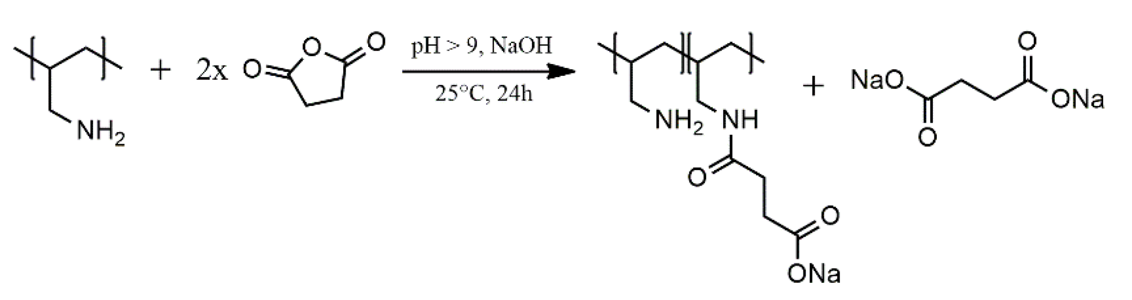
2.2. Succinylation of Polyallylamine
2.3. Attenuated Total Infrared Reflectance Infrared Spectroscopy (ATR-IR)
2.4. Nuclear Magnetic Resonance (NMR)
2.5. Potentiometric and Polyelectrolyte Charge Titration
2.6. XRD Measurements and Optical Microscopy
2.7. Evaluation of Biological Efficacy
2.7.1. Biocompatibility Testing
2.7.2. Antimicrobial Testing
2.8. Electrospinning of Fibers
2.8.1. Preparation of Solutions
2.8.2. Production and Characterization of Fibers
3. Results and Discussion
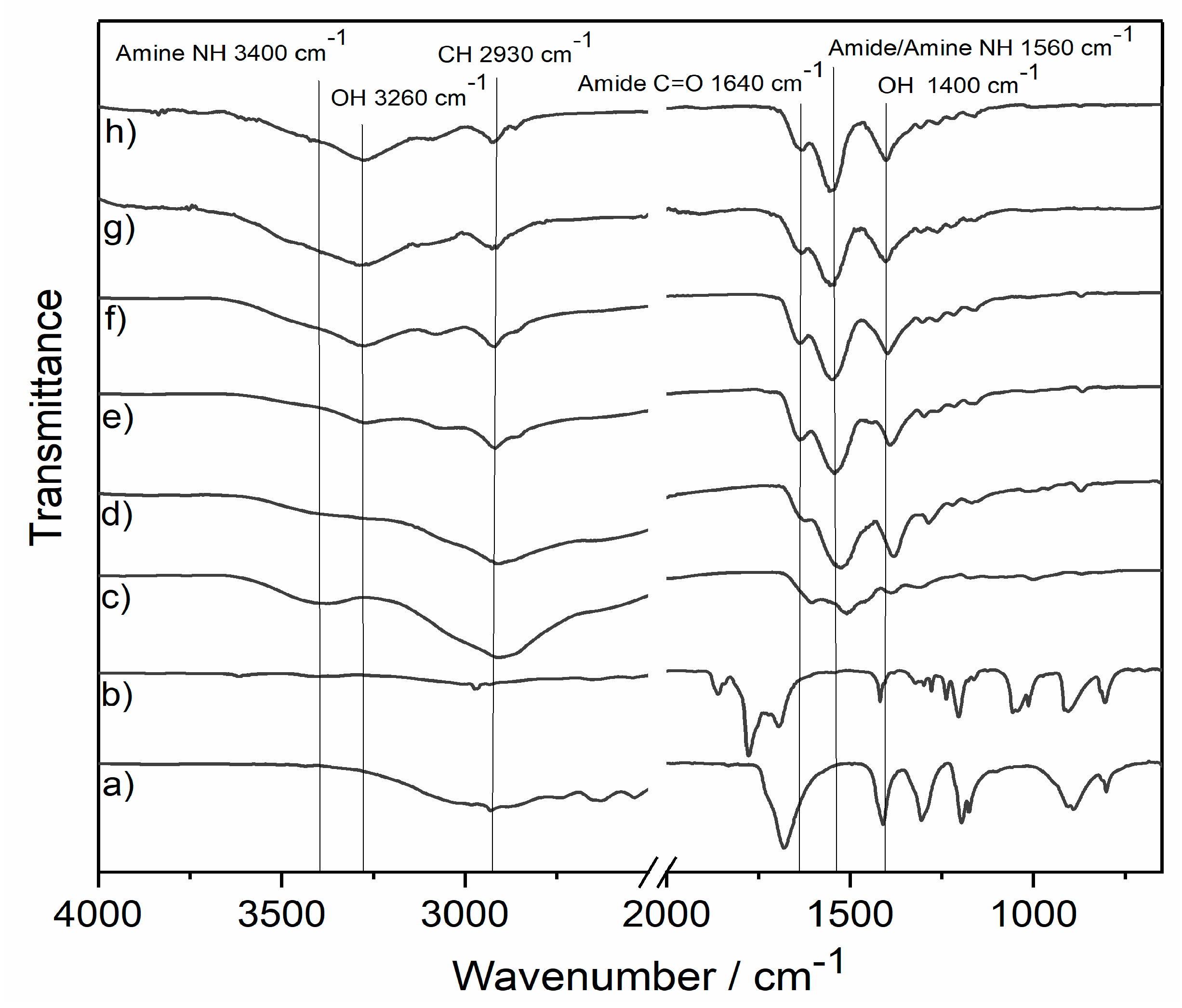
3.1. ATR-IR
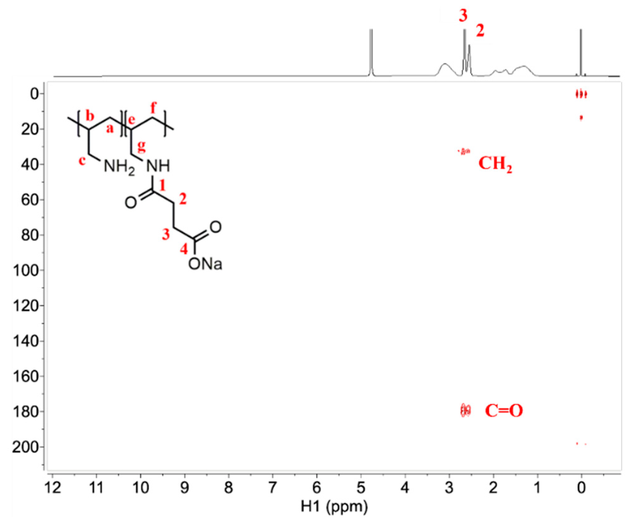
3.2. NMR
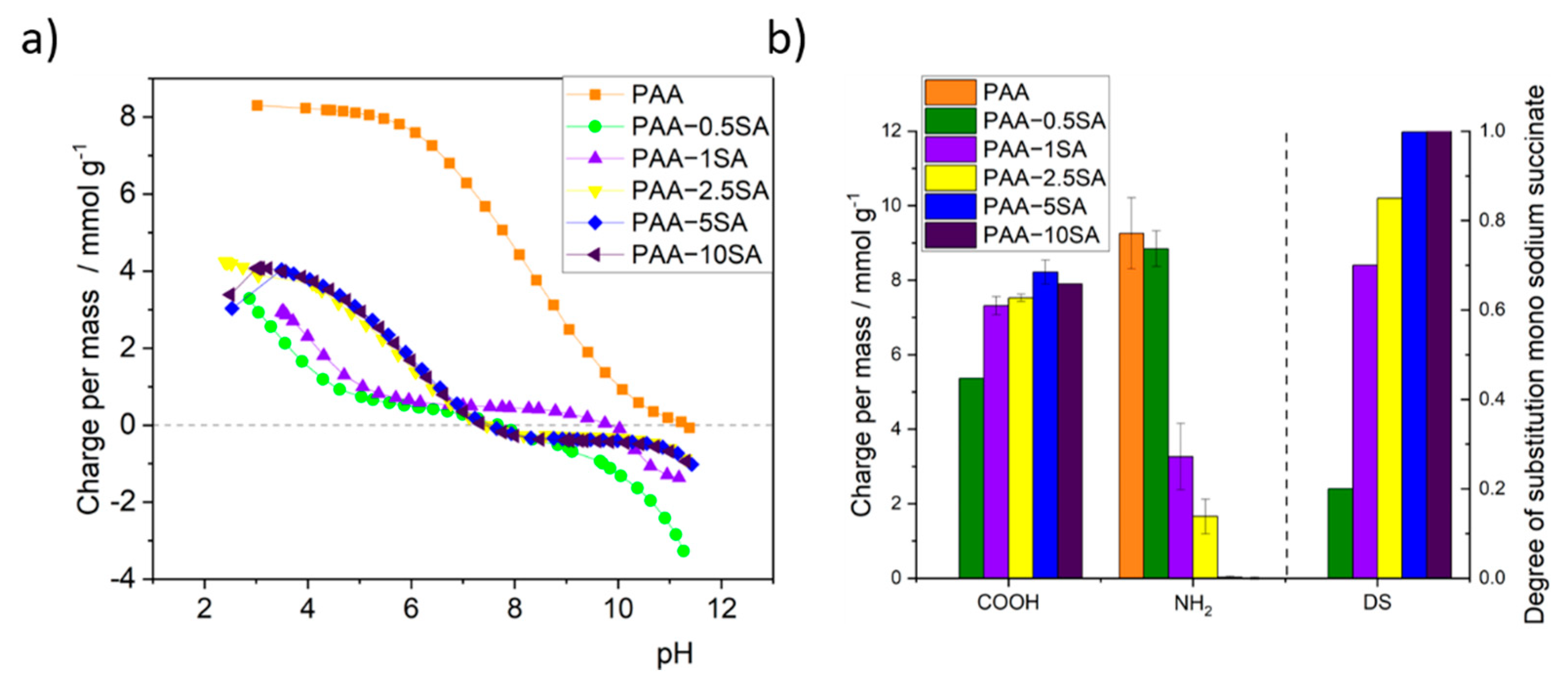
3.3. Titration, DS, and PAA Recovery
3.4. Biological Activity of PAA and the PAA-xSA Samples
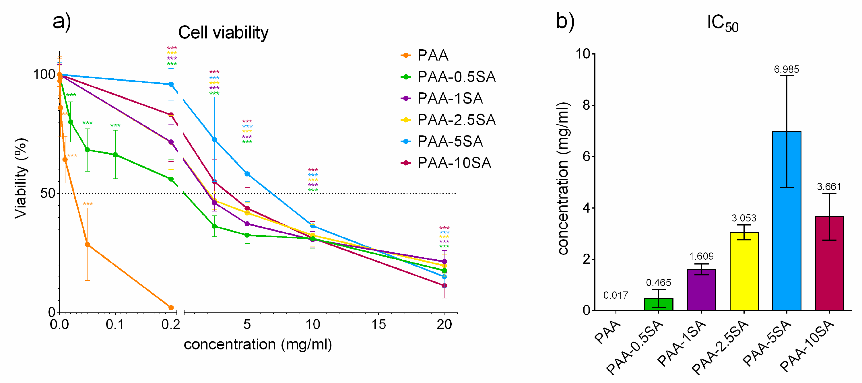
3.4.1. Biocompatibility Testing
3.4.2. Antimicrobial Testing
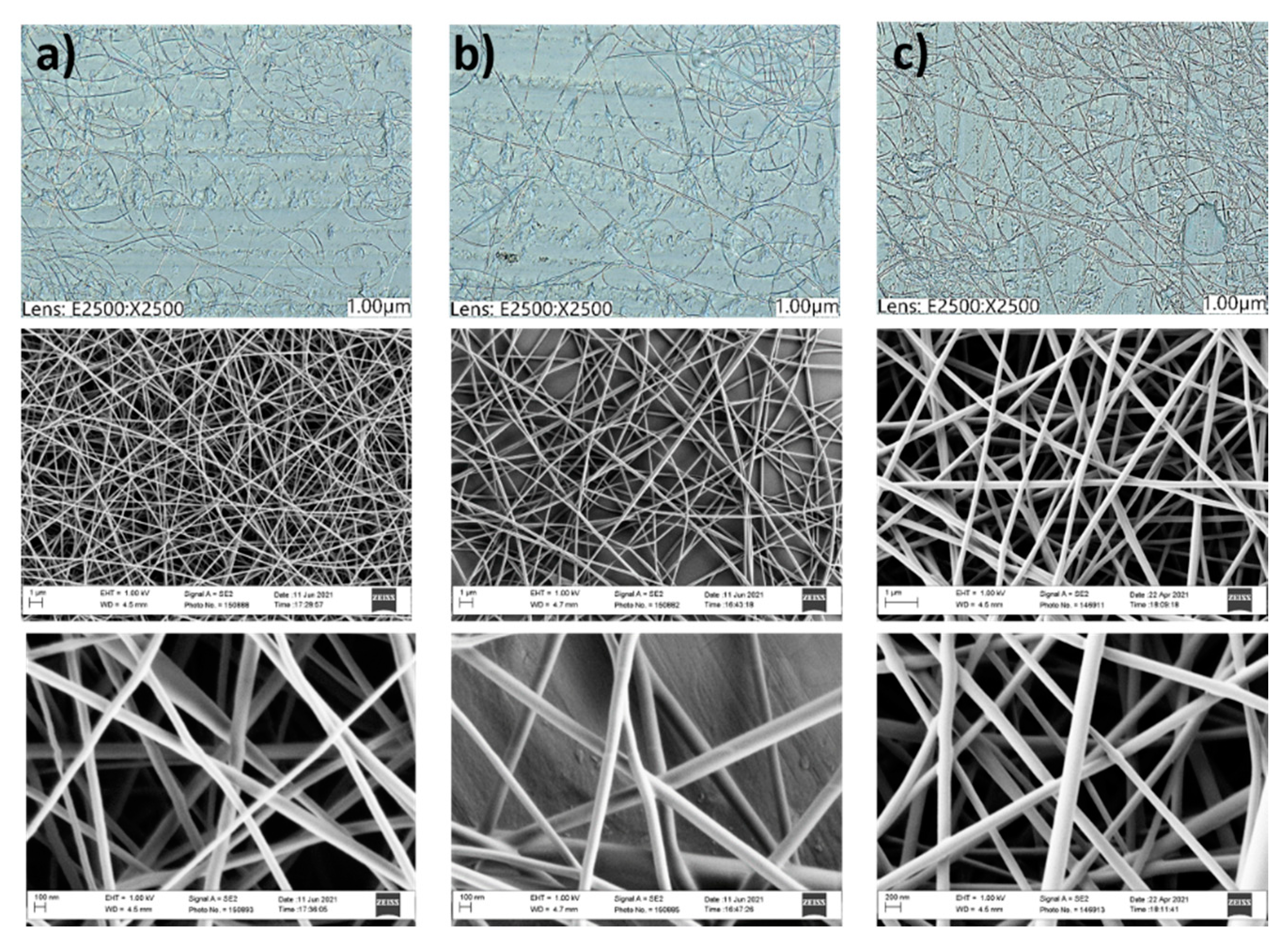
3.5. Production of PVA/PAA-xSA Fibers
4. Conclusions
Supplementary Materials
Author Contributions
Funding
Institutional Review Board Statement
Informed Consent Statement
Data Availability Statement
Acknowledgments
Conflicts of Interest
References
- Nimesh, S.; Kumar, R.; Chandra, R. Novel polyallylamine-dextran sulfate-DNA nanoplexes: Highly efficient non-viral vector for gene delivery. Int. J. Pharm. 2006, 320, 143–149. [Google Scholar] [CrossRef] [PubMed]
- Elahi, M.F.; Guan, G.; Wang, L.; King, M.W. Influence of layer-by-layer polyelectrolyte deposition and EDC/NHS activated heparin immobilization onto silk fibroin fabric. Materials 2014, 7, 2956–2977. [Google Scholar] [CrossRef]
- Lewis, S.R.; Datta, S.; Gui, M.; Coker, E.L.; Huggins, F.E.; Daunert, S.; Bachas, L.; Bhattacharyya, D. Reactive nanostructured membranes for water purification. Proc. Natl. Acad. Sci. USA 2011, 108, 8577–8582. [Google Scholar] [CrossRef] [PubMed]
- Kim, U.J.; Kuga, S. Ion-exchange separation of proteins by polyallylamine-grafted cellulose gel. J. Chromatogr. A 2002, 955, 191–196. [Google Scholar] [CrossRef]
- Lu, Z.; Yu, J.; Zeng, H.; Liu, Q. Polyamine-modified magnetic graphene oxide nanocomposite for enhanced selenium removal. Sep. Purif. Technol. 2017, 183, 249–257. [Google Scholar] [CrossRef]
- Wytrwal, M.; Koczurkiewicz, P.; Wõjcik, K.; Michalik, M.; Kozik, B.; Zylewski, M.; Nowakowska, M.; Kepczynski, M. Synthesis of strong polycations with improved biological properties. J. Biomed. Mater. Res. Part A 2014, 102, 721–731. [Google Scholar] [CrossRef]
- Youssef, E.; El-Moneim, M.A.; Fathalla, W.; Nafie, M.S. Design, synthesis and antiproliferative activity of new amine, amino acid and dipeptide-coupled benzamides as potential sigma-1 receptor. J. Iran. Chem. Soc. 2020, 17, 2515–2532. [Google Scholar] [CrossRef]
- Pan, Y.; Xia, Q.; Xiao, H. Cationic polymers with tailored structures for rendering polysaccharide-based materials antimicrobial: An overview. Polymers 2019, 11, 1283. [Google Scholar] [CrossRef]
- Simões, D.; Miguel, S.P.; Ribeiro, M.P.; Coutinho, P.; Mendonça, A.G.; Correia, I.J. Recent advances on antimicrobial wound dressing: A review. Eur. J. Pharm. Biopharm. 2018, 127, 130–141. [Google Scholar] [CrossRef]
- Alfei, S.; Schito, A.M. Positively charged polymers as promising devices against multidrug resistant gram-negative bacteria: A Review. Polymers 2020, 12, 1195. [Google Scholar] [CrossRef]
- Singh, P.; Samorì, C.; Toma, F.M.; Bussy, C.; Nunes, A.; Al-Jamal, K.T.; Ménard-Moyon, C.; Prato, M.; Kostarelos, K.; Bianco, A. Polyamine functionalized carbon nanotubes: Synthesis, characterization, cytotoxicity and siRNA binding. J. Mater. Chem. 2011, 21, 4850–4860. [Google Scholar] [CrossRef]
- Gevorgyan, S.; Rossi, E.; Cappelluti, M.A.; Tocchio, A.; Martello, F.; Gerges, I.; Lenardi, C.; Milani, P.; Argentiere, S. Photocrosslinked poly(amidoamine) nanoparticles for central nervous system targeting. Colloids Surfaces B Biointerfaces 2017, 151, 197–205. [Google Scholar] [CrossRef]
- Zhang, X.; Zhang, Y.W.; Zhang, H.; Yang, Q.; Wang, H.; Zhang, G. Preparation, characterization and antibacterial activity of octenyl succinic anhydride modified inulin. Int. J. Biol. Macromol. 2015, 78, 79–86. [Google Scholar] [CrossRef]
- Lee, J.S.; Nah, H.; Moon, H.J.; Lee, S.J.; Heo, D.N.; Kwon, I.K. Controllable delivery system: A temperature and pH-responsive injectable hydrogel from succinylated chitosan. Appl. Surf. Sci. 2020, 528, 146812. [Google Scholar] [CrossRef]
- Sun, G.; Feng, C.; Kong, M.; Cheng, X.; Bing, J.; Xia, G.; Bao, Z.; Park, H.; Chen, X. Development of part-dissolvable chitosan fibers with surface N-succinylation for wound care dressing. Front. Mater. Sci. 2015, 9, 272–281. [Google Scholar] [CrossRef]
- Sreedhar, A.; Wiese, E.K.; Hitosugi, T. Enzymatic and metabolic regulation of lysine succinylation. Genes Dis. 2020, 7, 166–171. [Google Scholar] [CrossRef] [PubMed]
- Awad, S.; Dharmavaram, S.; Wearn, C.S.; Dube, M.G.; Lobo, D.N. Effects of an intraoperative infusion of 4 succinylated gelatine (Gelofusine®) and 6 hydroxyethyl starch (Voluven®) on blood volume. Br. J. Anaesth. 2012, 109, 168–176. [Google Scholar] [CrossRef]
- Jing, Y.; Liu, Z.; Tian, G.; Bao, X.; Ishibashi, T.; Li, X.D. Site-Specific Installation of Succinyl Lysine Analog into Histones Reveals the Effect of H2BK34 Succinylation on Nucleosome Dynamics. Cell Chem. Biol. 2018, 25, 166–174.e7. [Google Scholar] [CrossRef]
- Söyler, Z.; Onwukamike, K.N.; Grelier, S.; Grau, E.; Cramail, H.; Meier, M.A.R. Sustainable succinylation of cellulose in a CO2-based switchable solvent and subsequent Passerini 3-CR and Ugi 4-CR modification. Green Chem. 2018, 20, 214–224. [Google Scholar] [CrossRef]
- Liu, H.; Yu, L.; Simon, G.; Zhang, X.; Dean, K.; Chen, L. Effect of annealing and pressure on microstructure of cornstarches with different amylose/amylopectin ratios. Carbohydr. Res. 2009, 344, 350–354. [Google Scholar] [CrossRef]
- Qing, X.; He, G.; Liu, Z.; Yin, Y.; Cai, W.; Fan, L.; Fardim, P. Preparation and properties of polyvinyl alcohol/N–succinyl chitosan/lincomycin composite antibacterial hydrogels for wound dressing. Carbohydr. Polym. 2021, 261, 117875. [Google Scholar] [CrossRef]
- Fras-Zemljič, L.; Kokol, V.; Čakara, D. Antimicrobial and antioxidant properties of chitosan-based viscose fibres enzymatically functionalized with flavonoids. Text. Res. J. 2011, 81, 1532–1540. [Google Scholar] [CrossRef]
- Zemljič, L.F.; Čakara, D.; Michaelis, N.; Heinze, T.; Kleinschek, K.S. Protonation behavior of 6-deoxy-6-(2-aminoethyl)amino cellulose: A potentiometric titration study. Cellulose 2011, 18, 33–43. [Google Scholar] [CrossRef]
- Zhao, H.; Wu, X.; Tian, W.; Ren, S. Synthesis and thermal property of poly(allylamine hydrochloride). Adv. Mater. Res. 2011, 150–151, 1480–1483. [Google Scholar] [CrossRef]
- Ando, I. Some aspects of the NMR chemical shift/structure correlation in the structural characterization of polymers and biopolymers. Polym. J. 2012, 44, 734–747. [Google Scholar] [CrossRef][Green Version]
- Petrov, A.I.; Antipov, A.A.; Sukhorukov, G.B. Base-acid equilibria in polyelectrolyte systems: From weak polyelectrolytes to interpolyelectrolyte complexes and multilayered polyelectrolyte shells. Macromolecules 2003, 36, 10079–10086. [Google Scholar] [CrossRef]
- Saallah, S.; Roslan, J.; Julius, F.S.; Saallah, S.; Mohamad Razali, U.H.; Pindi, W.; Sulaiman, M.R.; Pa’ee, K.F.; Mustapa Kamal, S.M. Comparative study of the yield and physicochemical properties of collagen from sea cucumber (Holothuria scabra), obtained through dialysis and the ultrafiltration membrane. Molecules 2021, 26, 2564. [Google Scholar] [CrossRef]
- Fröhlich, E. The role of surface charge in cellular uptake and cytotoxicity of medical nanoparticles. Int. J. Nanomed. 2012, 7, 5577–5591. [Google Scholar] [CrossRef]
- Warriner, L.W.; Duke, J.R.; Pack, D.W.; Derouchey, J.E. Succinylated Polyethylenimine Derivatives Greatly Enhance Polyplex Serum Stability and Gene Delivery in Vitro. Biomacromolecules 2018, 19, 4348–4357. [Google Scholar] [CrossRef]
- Gabrielson, N.P.; Pack, D.W. Acetylation of Polyethylenimine Enhances Gene Delivery via Weakened Polymer/DNA Interactions. Biomacromolecules 2006, 7, 2427–2435. [Google Scholar] [CrossRef]
- Triyana, K.; Mu’Min, M.S.; Faizah, K.; Yusuf, Y.; Kusumaatmaja, A. Harsojo Electrospun nanofibers based on polyvinyl Alcohol/chitosan and its stability in KOH solution. Mater. Sci. Forum 2015, 827, 321–325. [Google Scholar] [CrossRef]


| S. Aureus | P. Aeruginosa | |||
|---|---|---|---|---|
| MIC | MBC | MIC | MBC | |
| PAA | 0.08 mg/mL | 0.16 mg/mL | 0.08 mg/mL | 0.16 mg/mL |
| PAA-0.5SA | Insoluble in culture | Insoluble in culture | Insoluble in culture | Insoluble in culture |
| PAA-1SA | >20 mg/mL | n.d. | >20 mg/mL | n.d. |
| PAA-2.5SA | >20 mg/mL | n.d. | >20 mg/mL | n.d. |
| PAA-5SA | >20 mg/mL | n.d. | >20 mg/mL | n.d. |
| PAA-10SA | >20 mg/mL | n.d. | >20 mg/mL | n.d. |
Publisher’s Note: MDPI stays neutral with regard to jurisdictional claims in published maps and institutional affiliations. |
© 2021 by the authors. Licensee MDPI, Basel, Switzerland. This article is an open access article distributed under the terms and conditions of the Creative Commons Attribution (CC BY) license (https://creativecommons.org/licenses/by/4.0/).
Share and Cite
Jurko, L.; Bračič, M.; Hribernik, S.; Makuc, D.; Plavec, J.; Jerenec, F.; Žabkar, S.; Gubeljak, N.; Štern, A.; Kargl, R. Succinylation of Polyallylamine: Influence on Biological Efficacy and the Formation of Electrospun Fibers. Polymers 2021, 13, 2840. https://doi.org/10.3390/polym13172840
Jurko L, Bračič M, Hribernik S, Makuc D, Plavec J, Jerenec F, Žabkar S, Gubeljak N, Štern A, Kargl R. Succinylation of Polyallylamine: Influence on Biological Efficacy and the Formation of Electrospun Fibers. Polymers. 2021; 13(17):2840. https://doi.org/10.3390/polym13172840
Chicago/Turabian StyleJurko, Lucija, Matej Bračič, Silvo Hribernik, Damjan Makuc, Janez Plavec, Filip Jerenec, Sonja Žabkar, Nenad Gubeljak, Alja Štern, and Rupert Kargl. 2021. "Succinylation of Polyallylamine: Influence on Biological Efficacy and the Formation of Electrospun Fibers" Polymers 13, no. 17: 2840. https://doi.org/10.3390/polym13172840
APA StyleJurko, L., Bračič, M., Hribernik, S., Makuc, D., Plavec, J., Jerenec, F., Žabkar, S., Gubeljak, N., Štern, A., & Kargl, R. (2021). Succinylation of Polyallylamine: Influence on Biological Efficacy and the Formation of Electrospun Fibers. Polymers, 13(17), 2840. https://doi.org/10.3390/polym13172840