Article

Anion-Dominated Copper Salicyaldimine Complexes—Structures, Coordination Mode of Nitrate and Decolorization Properties toward Acid Orange 7 Dye

Department of Applied Chemistry, National Chi Nan University, Nantou 545, Taiwan
*
Author to whom correspondence should be addressed.
Polymers 2020, 12(9), 1910; https://doi.org/10.3390/polym12091910
Submission received: 24 July 2020 / Revised: 21 August 2020 / Accepted: 22 August 2020 / Published: 24 August 2020
(This article belongs to the Special Issue Coordination Polymer II)

Abstract

:
A salicyaldimine ligand, 3-tert-butyl-4-hydroxy-5-(((pyridin-2-ylmethyl)imino)methyl)benzoic acid (H2Lsalpyca) and two Cu(II)−salicylaldimine complexes, [Cu(HLsalpyca)Cl] (1) and [Cu(HLsalpyca)(NO3)]n (2), have been synthesized. Complex 1 has a discrete mononuclear structure, in which the Cu(II) center is in a distorted square-planar geometry made up of one HLsalpyca monoanion in an NNO tris-chelating mode and one Cl anion. Complex 2 adopts a neutral one-dimensional zigzag chain structure propagating along the crystallographic [010] direction, where the Cu(II) center suits a distorted square pyramidal geometry with a τ value of 0.134, consisted of one HLsalpyca monoanion as an NNO tris-chelator and two NO3 anions. When the Cu∙∙∙O semi coordination is taken into consideration, the nitrato ligand bridges two Cu(II) centers in an unsymmetrical bridging-tridentate with a μ, κ4O,O′:O′,O″ coordination. Clearly, anion herein plays a critical role in dominating the formation of discrete and polymeric structures of copper salicyaldimine complexes. Noteworthy, complex 2 is insoluble but highly stable in H2O and various organic solvents (CH3OH, CH3CN, acetone, CH2Cl2 and THF). Moreover, complex 2 shows good photocatalytic degradation activity and recyclability to accelerate the decolorization rate and enhance the decolorization performance of acid orange 7 (AO7) dye by hydrogen peroxide (H2O2) under daylight.

Graphical Abstract

1. Introduction

Salicyaldimine derivatives with the NO donor set of azomethine nitrogen and phenolato oxygen have been extensively studied in coordination chemistry due to their preparational accessibilities and their versatility in terms of steric and electronic modifications [1,2,3,4,5]. Metal–salicyaldimine complexes so far have achieved a remarkable success in structure variation including traditional coordination complexes [2,3,4,6,7] and metallosupramolecular architectures such as discrete macrocycles [8,9], helicates [10,11] and coordination polymers [12,13,14,15]. Moreover, metal–salicyaldimine complexes have wide applications in fields such as catalysis [5,15,16], molecular magnetism [7,17] and fluorescent chemosensors [18,19].
On the other hand, the presence of organic pollutants, such as azo dyes, the most diverse group of synthetic dyes used in food, textile and printing industries, in water streams is becoming a seriously environmental problem due to the fact of their color, toxicity, potential carcinogenicity and non-biodegradable nature [20,21,22,23,24]. Therefore, the treatment of dye-containing wastewater is a matter of great important issue for environmental protection and has attracted worldwide attention. Nowadays, adsorptive removal (non-destructive process) [25,26] and degradation (destructive process) [27,28] are the two most promising techniques widely utilized in the decolorization of dye-contaminated water owing to their advantages of mild operation conditions, convenience, high efficiency and relative low costs [20,22,23,24,29,30,31].
In recent years, our group has reported several transition metal complexes of salicyaldimine Schiff base ligands with NO and N2O donor sets, which have represented mononuclear molecular structures and one- and two-dimensional periodical structures (1D chain and 2D layer) with interesting structural and photophysical properties [14,32,33]. Further, our group has also contributed efforts in the decolorization of aqueous solutions of organic dyes through the adsorptive removal process [34,35,36,37]. As a continuation, we report herein the synthesis and structures of two new Cu(II)−salicylaldimine complexes, [Cu(HLsalpyca)Cl] (1, H2Lsalpyca = 3-tert-butyl-4-hydroxy-5-(((pyridin-2-ylmethyl)imino)methyl)benzoic acid) and [Cu(HLsalpyca)(NO3)]n (2). Complex 1 shows a mononuclear structure while complex 2 has a 1D zigzag chain structure. Such structural diversities are tentatively ascribed to the influence of anion. It is also noted that the nitrato in 2 suits a bridging-tridentate ligand with a coordination mode of μ, κ4O, O′:O′,O″ to bridge two Cu(II) ions to form the extended structure. Moreover, complex 2 is very stable in H2O and would cooperate with H2O2 to show remarkable photocatalytic activity toward degradation of acid orange 7 (AO7) under daylight.

2. Experimental Section

2.1. Materials and Methods

Chemical reagents were of reagent grade quality obtained from commercial sources (Alfa Aesar, Heysham, UK; ACROS, Pittsburgh, PA, USA; SHOWA, Saitama, Japan) and used as received without further purification. 5-tert-Butyl-3-formyl-4-hydroxybenzoic acid [38,39] were synthesized according to the reported literature. Proton nuclear magnetic resonance (1H NMR)1H NMR spectra were collected at room temperature by using a Bruker AMX-300 Solution-NMR spectrometer (Bruker, New Taipei, Taiwan) operating at 300 MHz. Chemical shifts are reported in parts per million (ppm) with reference to the residual protons of the deuterated solvent and coupling constants are given in hertz (Hz). Mass spectra were recorded by using a Bruker Daltonics flexAnalysis matrix-assisted laser desorption/ionization time of flight (MALDI-TOF) mass spectrometer (Bruker, New Taipei, Taiwan). Thermogravimetric (TG) analyses were carried out under a flux of nitrogen by using a Thermo Cahn VersaTherm HS TG analyzer (Thermo, Newington, NH, USA) at a heating rate of 5 °C per minute from 25 to 900 °C. X-ray powder diffraction (XRPD) patterns were acquired on a Shimadzu XRD-7000 diffractometer (Shimadzu, Kyoto, Japan) with a graphite monochromatized Cu Kα radiation (λ = 1.5406 Å; 40 kV, 30 mA) at a scan speed of 1.2° per minute. Infrared (IR) spectra were collected on a Perkin-Elmer Frontier Fourier transform infrared spectrometer (Perkin-Elmer, Taipei, Taiwan) in the 4000–500 cm−1 region using attenuated total reflection (ATR) technique. Microanalyses were carried out by using an Elementar Vario EL III analytical instrument (Elementar, Langenselbold, Germany). UV-vis absorption spectra were recorded on a JASCO V-750 UV/VIS spectrophotometer (JASCO, Tokyo, Japan). Fluorescence spectra were recorded at room temperature on a Hitachi F7000 fluorescence spectrophotometer (Hitachi, Tokyo, Japan). Inductively coupled plasma optical emission spectrometry (ICP-OES) analyses were conducted on an Agilent 5100 ICP-OES instrument (Agilent, Santa Clara, CA, US).

2.2. Synthesis of 3-Tert-butyl-4-hydroxy-5-(((pyridin-2-ylmethyl)imino)methyl)benzoic Acid (H2Lsalpyca)

To a methanolic solution (2 mL) of 2-(aminomethyl)pyridine (0.10 g, 1.0 mmol), a methanolic solution (6 mL) of 5-tert-butyl-3-formyl-4-hydroxybenzoic acid (0.22 g, 1.0 mmol) was added under nitrogen atmosphere. The solution was stirred for 2 h at room temperature. After the solvent was removed under reduced pressure, yellow powdered product of H2Lsalpyca was obtained in a yield of 54% (0.17 g, 0.54 mmol). 1H NMR (300 MHz, DMSO-d6, ppm): δ 8.81 (s, 1H), 8.56 (d, J = 4.8 Hz, 1H), 7.96 (s, 1H), 7.86–7.80 (m, 2H), 7.44 (d, J = 7.8 Hz, 1H), 7.36–7.31 (m, 1H), 4.92 (s, 2H), 1.34 (s, 9H) (Figure S1). MS (MALDI-TOF): m/z 312.878 [M + H]+(calcd for C18H20N2O3: m/z 312.18) (Figure S2). IR (ATR, cm−1): 3425, 1961, 2924, 2862, 1956, 1677, 1632, 1602, 1477, 1392, 1364, 4334, 1307, 1279, 1239, 1202, 1179, 1120, 1014, 946, 879, 839, 804, 767, 705, 674, 602. Anal. Calcd for C18H20N2O3: C, 69.21; H, 6.45; N, 8.97%. Found: C, 69.14; H, 6.29; N, 8.68%.

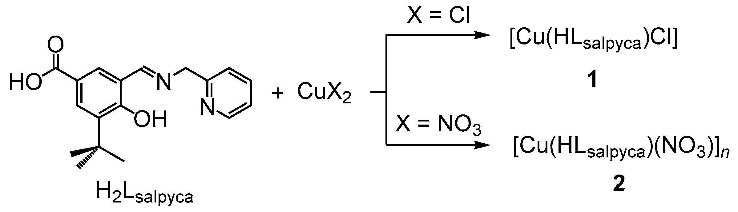
2.3. Synthesis of [Cu(HLsalpyca)Cl] (1)

A methanolic solution (1 mL) of H2Lsalpyca (31.2 mg, 0. 10 mmol) and an aqueous solution (1 mL) of CuCl2 (13.4 mg, 0.10 mmol) were sealed in a Teflon-lined stainless steel container. The container was heated at 80 °C for 12 h and then cooled to 30 °C. Green needle-shaped crystals of 1 were obtained in a yield of 39% (16.3 mg, 3.9 × 10² mmol). MS (MALDI-TOF): m/z 410.806 [M]+(calcd for C18H19N2O3ClCu: m/z 410.34) (Figure S3). IR (ATR, cm−1): 3065, 2961, 2908, 2869, 2568, 1672, 1596, 1536, 1484, 1407, 1355, 1263, 1258, 1230, 1173, 1052, 996, 921, 824, 795, 761, 697, 658. TGA: 200 °C (decomp.). Anal. Calcd for C18H19N2O3ClCu: C, 52.68; H, 4.67; N, 6.83%. Found: C, 52.88; H, 4.39; N, 6.89%.

2.4. Synthesis of [Cu(HLsalpyca)(NO3)]n (2)

A methanolic solution (0.5 mL) of H2Lsalpyca (31.2 mg, 0. 10 mmol) and an aqueous solution (1.5 mL) of Cu(NO3)2∙2.5H2O (23.2 mg, 0.10 mmol) were sealed in a Teflon-lined stainless steel container. The container was heated at 80 °C for 12 h and then cooled to 30 °C. Green needle-shaped crystals of 2 were obtained in a yield of 28% (12.2 mg, 2.8 × 10² mmol). IR (ATR, cm−1): 3431, 2928, 2590, 2003, 1682, 1629, 1600, 1573, 1532, 1488, 1475, 1440, 1413, 1394, 1354, 1331, 1274, 1244, 1216, 1191, 1065, 1054, 1029, 1004, 948. TGA: 215 °C (decomp.). Anal. Calcd for C18H19N3O6Cu: C, 49.48; H, 4.38; N, 9.62%. Found: C, 49.45; H, 4.66; N, 9.47%.

2.5. X-Ray Data Collection and Structure Refinement

Quality crystals of 1 and 2 were obtained for single-crystal structure determination. Data collections were performed on an Oxford Diffraction Gemini S diffractometer for 1 and on a Bruker D8 Venture diffractometer for 2. Both diffractometers are equipped with a graphite monochromated Mo Kα radiation (λ = 0.71073 Å). Starting models for structure refinement were found using direct methods with SHELXS-97 program [40] and the structural data were refined by full-matrix least-squares methods against F2 using the SHELXL-2014/7 [41], incorporated in WINGX-v2014.1 [42] crystallographic collective package. All non-hydrogen atoms were found from the different Fourier maps and refined anisotropically. Carbon-bound hydrogen atoms were placed by geometrical calculation and refined as riding mode. Oxygen-bound hydrogen atoms were located in a difference Fourier map and the positional parameters were refined, with the restraint O–H = 0.82(1) Å. Isotropic displacement parameters of all hydrogen atoms were derived from the parent atoms. ORTEP plots and crystal structure drawings for 1 and 2 were drawn using the Diamond software [43]. CCDC 1955728–1955729 contain the supplementary crystallographic data for this paper. These data can be obtained free of charge via http://www.ccdc.cam.ac.uk/conts/retrieving.html or from the Cambridge Crystallographic Data Centre, 12 Union Road, Cambridge CB2 1EZ, UK; fax: (+44) 1223-336-033; or e-mail: [email protected]. Detailed crystallographic data are summarized in Table 1. Selected bond lengths and angles are listed in Table 2 and important hydrogen-bonding parameters are shown in Table 3.

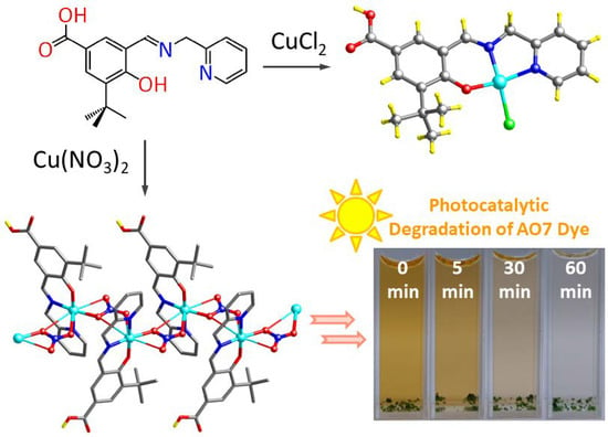
3. Results and Discussion

3.1. Syntheses and Characterization

Complexes 1 and 2 were synthesized from the reactions of copper(II) salts (CuCl2 and Cu(NO3)2∙2.5H2O) and H2Lsalpyca under hydro(solvo)thermal conditions at 80 °C for 12 h (Scheme 1). Their molecular structures have been determined by the single-crystal X-ray structure analyses and their bulky samples have been characterized to be a single crystalline phase through X-ray powder diffraction (XRPD) measurements (Figure S4).

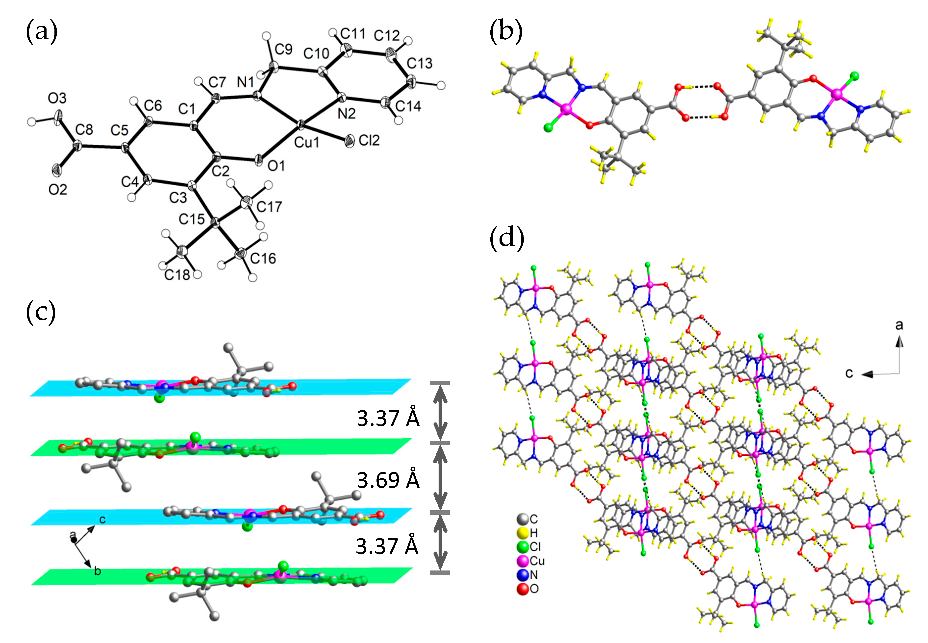
3.2. Crystal Structural Description of [Cu(HLsalpyca)Cl] (1)

Complex 1 crystallizes in the triclinic space group P 1 ¯ and the asymmetric unit consists of one Cu(II) center, one HLsalpyca monoanion and one Cl anion. The Cu(II) center is tris-chelated by one HLsalpyca monoanion through its phenolato, imino and pyridine groups and further coordinated by one Cl anion, furnishing a distorted square-planar geometry (Figure 1a), with the Cu and the Cl away from the plane 0.24 and 1.11 Å, respectively. The carboxyl group of the HLsalpyca ligand is protonated and non-coordinated. The Cu–Npyridine, Cu–Nimino, Cu–Ophenolato and Cu–Cl bond lengths are 1.9971(17), 1.9376(17), 1.8857(14) and 2.2299(6) Å, respectively and the Npyridine–Cu–Nimino and Nimino–Cu–Ophenolato bond angles are 82.98(7) and 92.00(6)°, respectively. Two [Cu(HLsalpyca)Cl] molecules form a hydrogen-bonded dimer through intermolecular hydrogen bonds (O3–H3∙∙∙O2#1, d(D∙∙∙A) = 2.637(2) Å, ∠(D–H∙∙∙A) = 168(3)°, #1 = −1−x, −y, −z) between two carboxyl groups of two HLsalpyca ligands, with a graph set of R 2 2 ( 8 ) (Figure 1b) [44]. In addition, there are also intermolecular π∙∙∙π interactions (plane∙∙∙plane: 3.37 and 3.69 Å) between any two neighboring [Cu(HLsalpyca)Cl] molecules that stack in a column manner along the crystallographic [010] direction, that is, b axis (Figure 1c). Moreover, there are also weak but significant intermolecular C–H∙∙∙Cl interactions (C9–H9B∙∙∙Cl2#2, d(D∙∙∙A) = 3.567(3) Å, ∠(D–H∙∙∙A) = 150°, #1 = −1 + x, y, z) between the chloro ligands and the methylene moieties of the HLsalpyca ligands (Figure 1d).

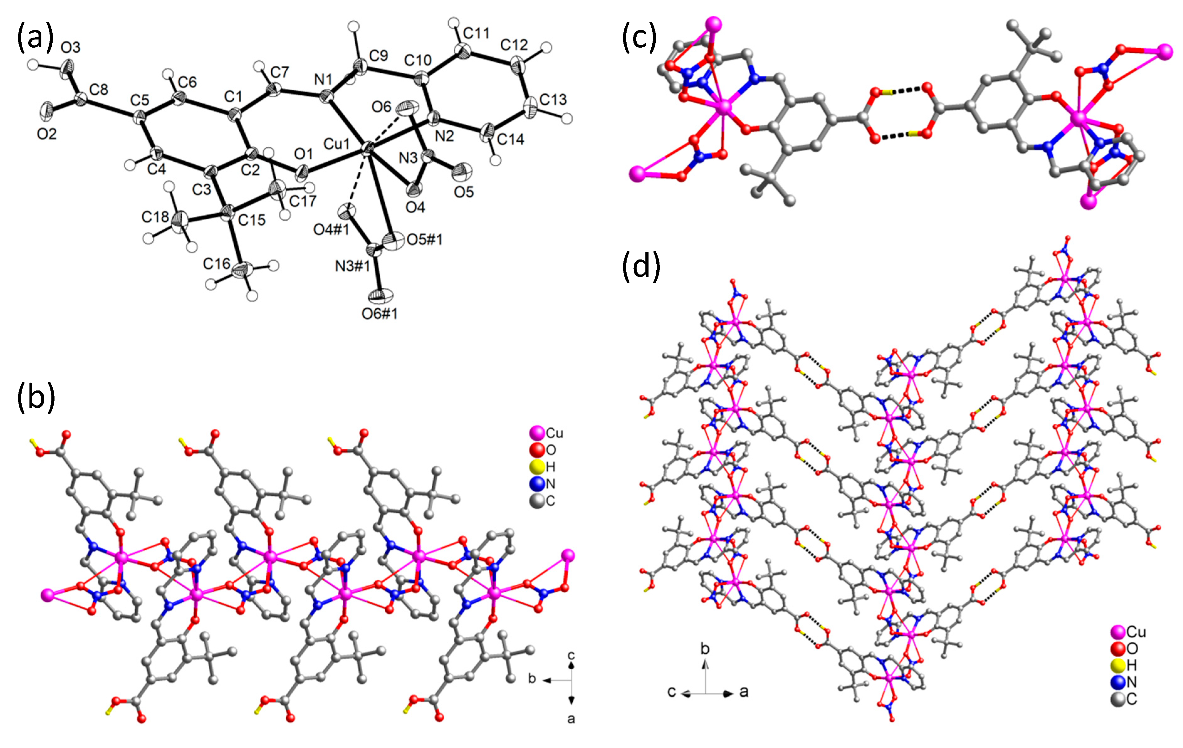
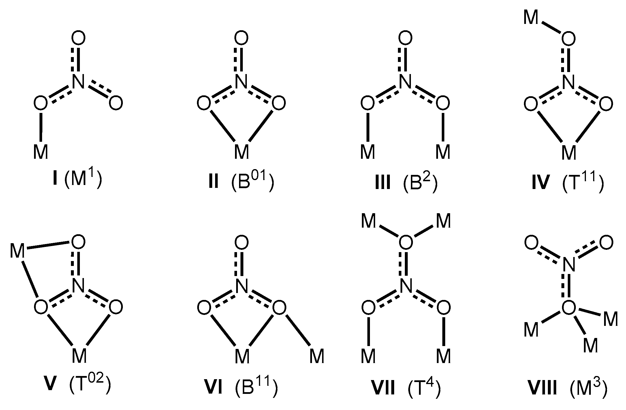
3.3. Crystal Structural Description of [Cu(HLsalpyca)(NO3)]n (2)

Complex 2 crystallizes in the monoclinic space group P21/c and has an infinite zigzag chain structure. The asymmetric unit consists of one Cu(II) center, one HLsalpyca monoanion and one NO3 anion. The Cu(II) center is coordinated by one HLsalpyca monoanion as a tris-chelator through its phenolato, imino and pyridine groups and further coordinated to two NO3 anion (Figure 2a), furnishing a distorted square pyramidal geometry, with a τ value of 0.134 [45]. The carboxyl group of the HLsalpyca ligand is protonated and non-coordinated. The Cu–Npyridine, Cu–Nimino, Cu–Ophenolato and Cu–Onitrato bond lengths are 1.994(3), 1.931(2), 1.892(2) and 2.019(2)/2.437(3) Å, respectively and the Npyridine–Cu–Nimino and Nimino–Cu–Ophenolato bond angles are 83.45(10) and 93.00(9)°, respectively. Basically, the nitrato ligand adopts a bridging-bidentate with a semi-coordination (μ, κ2O, O′). However, when the Cu∙∙∙O semi-coordination (Cu1∙∙∙O6 = 2.73 Å, Cu1∙∙∙O4#1 = 3.01, #1 = −x, y + 1/2, −z + 1/2) is taken into consideration, the nitrato ligand suits an unsymmetrical bridging-tridentate with a μ, κ4O, O′:O′,O″ coordination, with a Cu∙∙∙Cu separation of 4.97 Å, among various possible coordination modes of nitrato ligand [46,47,48,49,50,51]. Morozov and coworkers have shown that nitrato ligand exhibits 28 coordination modes that are classified in terms of the number of oxygen atoms used by the nitrato ligand for binding to complexing metal centers [52]. Based on the definition, M, B and T denote one, two and three nitrato O atoms used for binding, respectively and the superscripts mean the number of metal atoms connected to one nitrato O atom (the first superscript) or two nitrato O atoms (the second superscript). Accordingly, the coordination mode of nitrato ligand in complex 2 is classified to be T02 (Scheme 2, mode V).
Each nitrato ligand bridges two [Cu(HLsalpyca)]+cationic motifs to generate a neutral 1D zigzag chain structure that propagates along the crystallographic [010] direction, that is, the b axis (Figure 2b). There are chain-to-chain hydrogen bonds (O3–H3∙∙∙O2#2, d(D∙∙∙A) = 2.633(3) Å, ∠(D–H∙∙∙A) = 173°, #2 = 1 − x, 2 − y, −z) between two carboxyl groups of two HLsalpyca ligands, with a graph set of R 2 2 ( 8 ) [44], in adjacent chains (Figure 2c). As a result, a hydrogen-bonded 2D wavy sheet is formed (Figure 2d). This structure is compared to a zigzag chain structure of the Cu(II)−salicylaldimine complex {[Cu(salpyca)]∙3H2O}n (where H2salpyca = 4-hydroxy-3-(((pyridin-2-yl)methylimino)methyl)benzoic acid) [33], prepared from a solvent-mediated cation-exchange of [Zn(salpyca)(H2O)]n and Cu(NO3)2∙6H2O in aqueous solution, where the salpyca2− displays not only the NNO tris-chelating mode from its pyridine, imino and phenolato groups but also bridging behavior though its monodentately-carboxylato group.

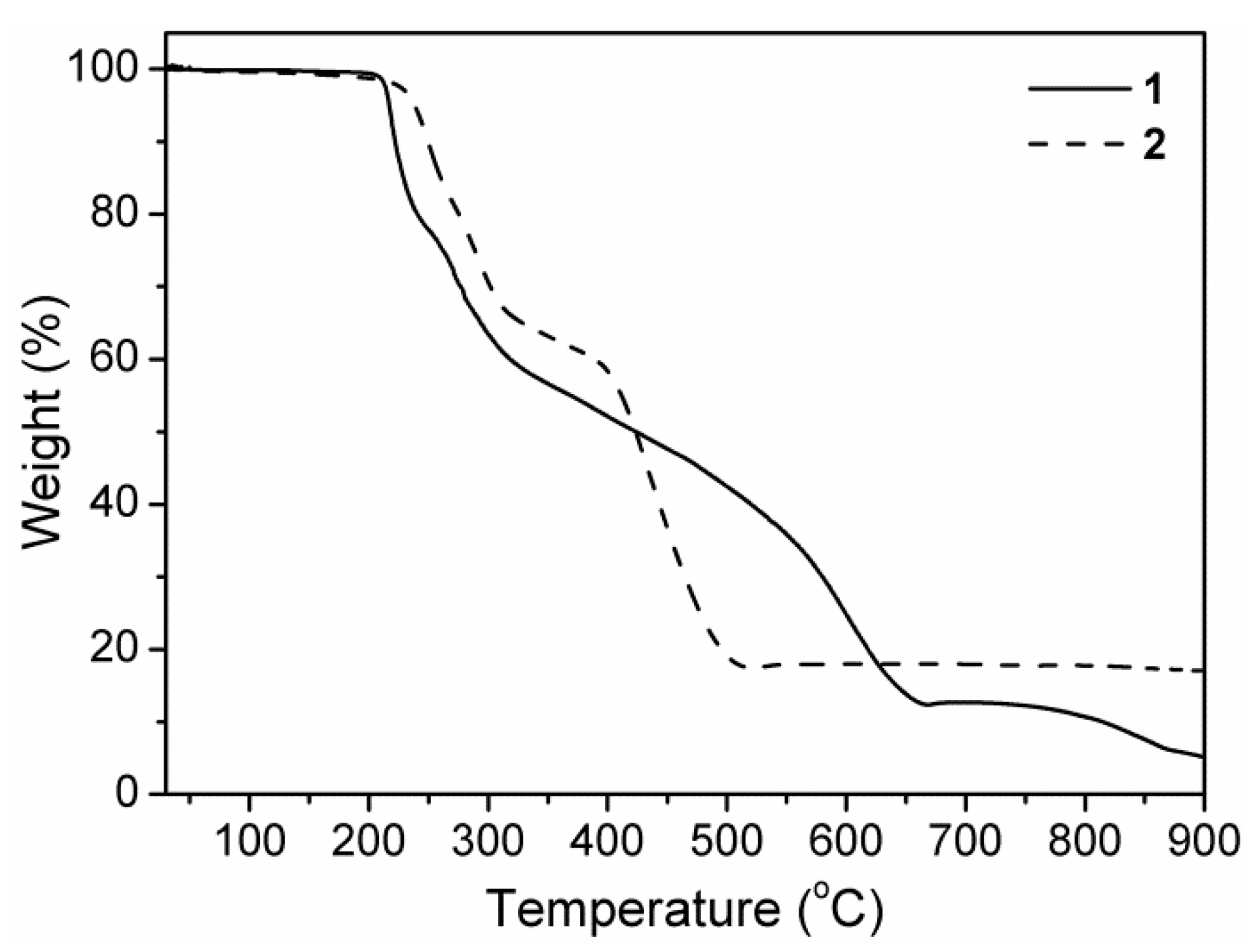
3.4. Thermogravimetric (TG) Analysis
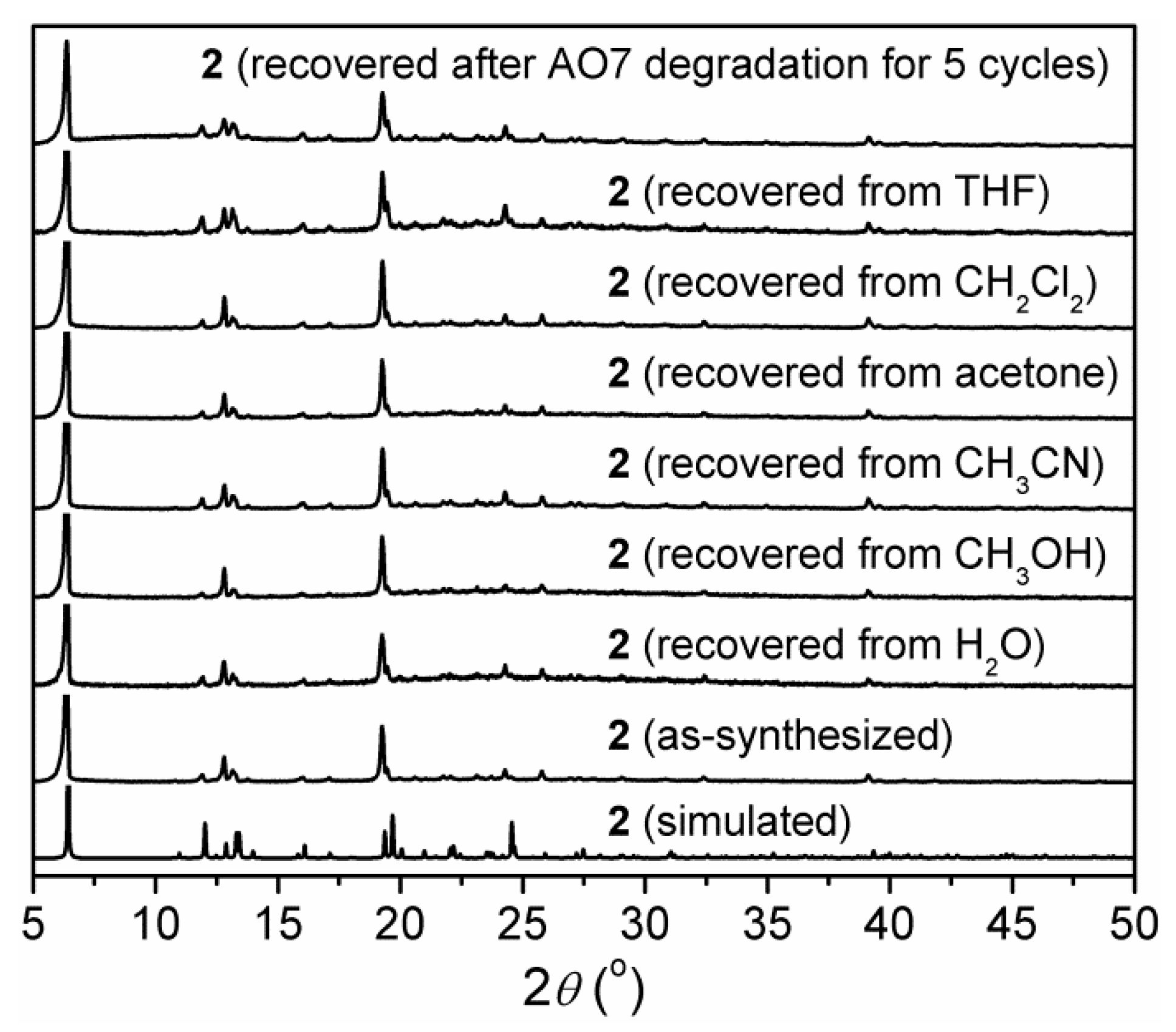
The thermogravimetric (TG) traces of 1 and 2 both showed a long plateau before a decomposition process occurred at temperature approaching 200 and 215 °C, respectively (Figure 3). For 2, after a two-step decomposition of the framework ended at a temperature approaching 509 °C, a weight of 17.8% of the total sample was left which is reasonably assigned to CuO (calcd. 18.2%).

3.5. Stability of 2

The chemical stability of zigzag chain 2 in crystalline phase has been examined. As observation, 2 displayed high chemical stability to maintain the framework integrity and crystallinity after immersing in H2O, methanol (CH3OH), acetonitrile (CH3CN), acetone, dichloromethane (CH2Cl2) and tetrahydrofuran (THF), respectively, for 1 day. This is supported by the checked XRPD profiles which are almost identical with that of as-synthesized crystalline samples (Figure 4).

3.6. Photophysical Properties

In solid state, the salicyaldimine ligand, H2Lsalpyca, shows an unstructured fluorescence centered at 502 nm (Figure S5), upon excitation at 360 nm, which is tentatively assigned to intraligand transition [32]. Comparably, the Cu(II) complexes 1 and 2 are fluorescence silent due to the paramagnetic perturbation and/or energy/charge transfer [2,53,54].

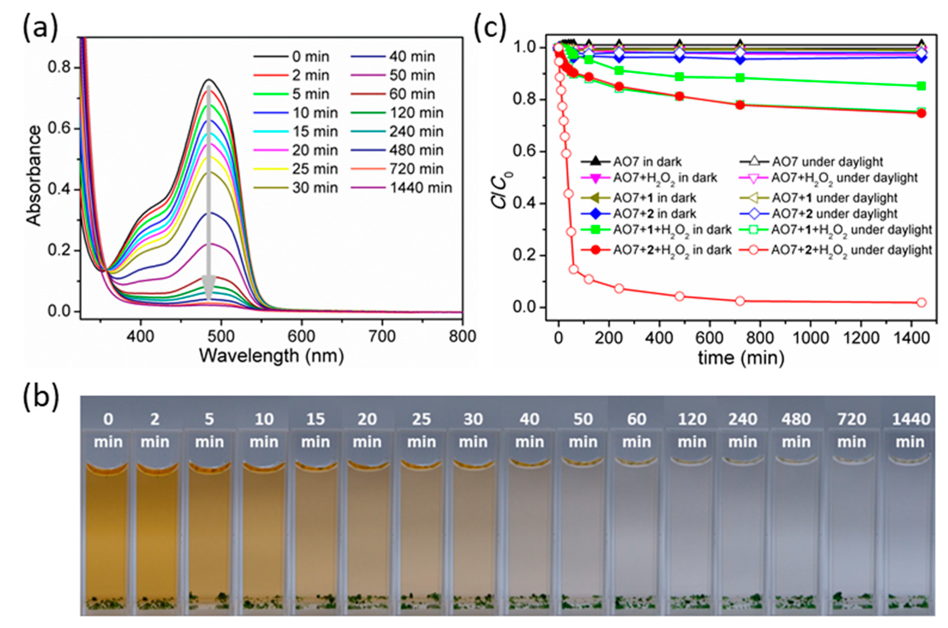
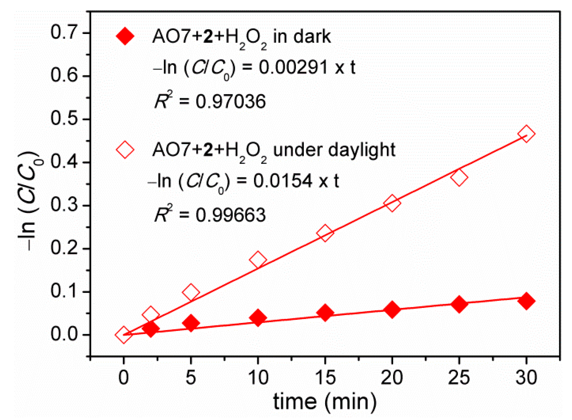
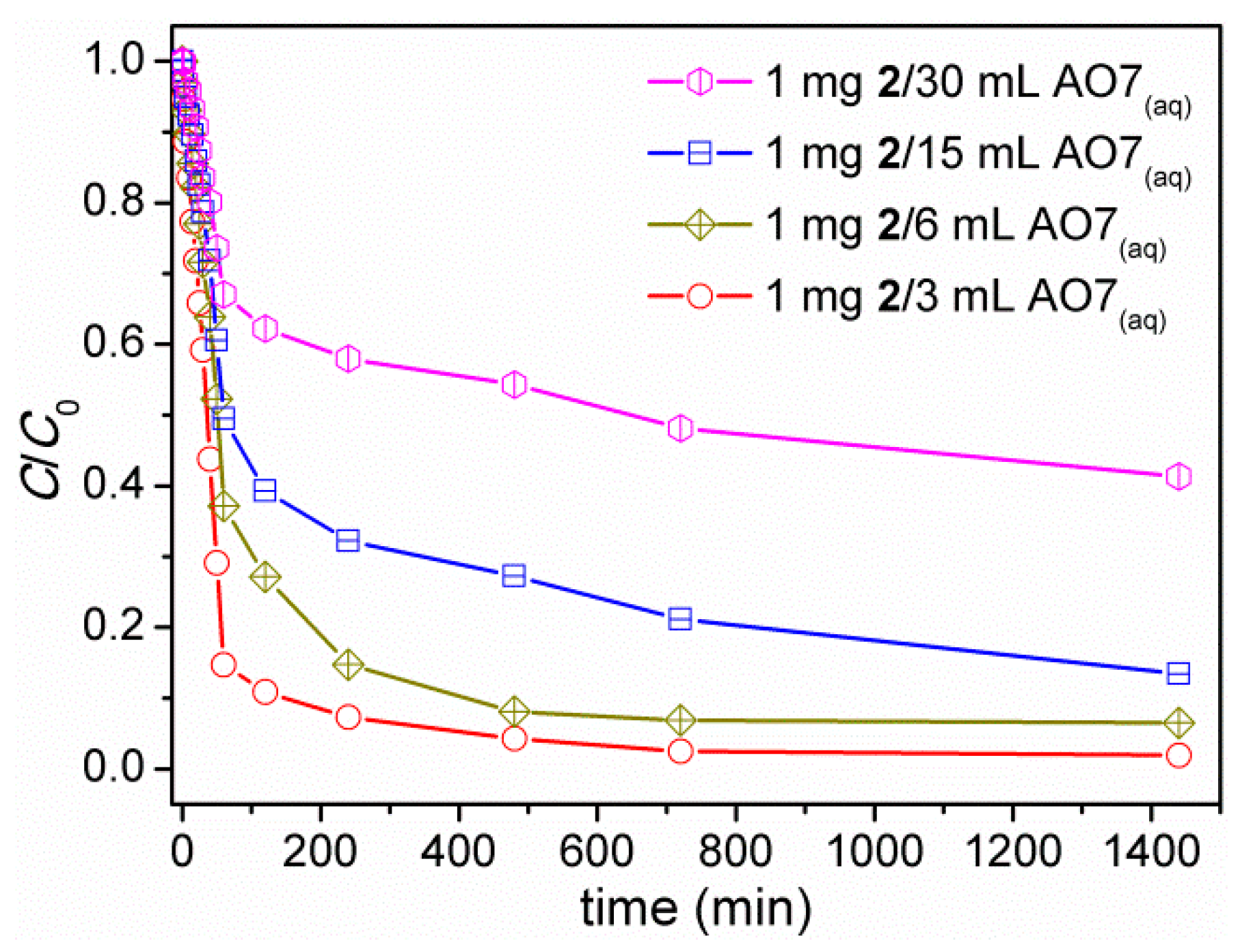
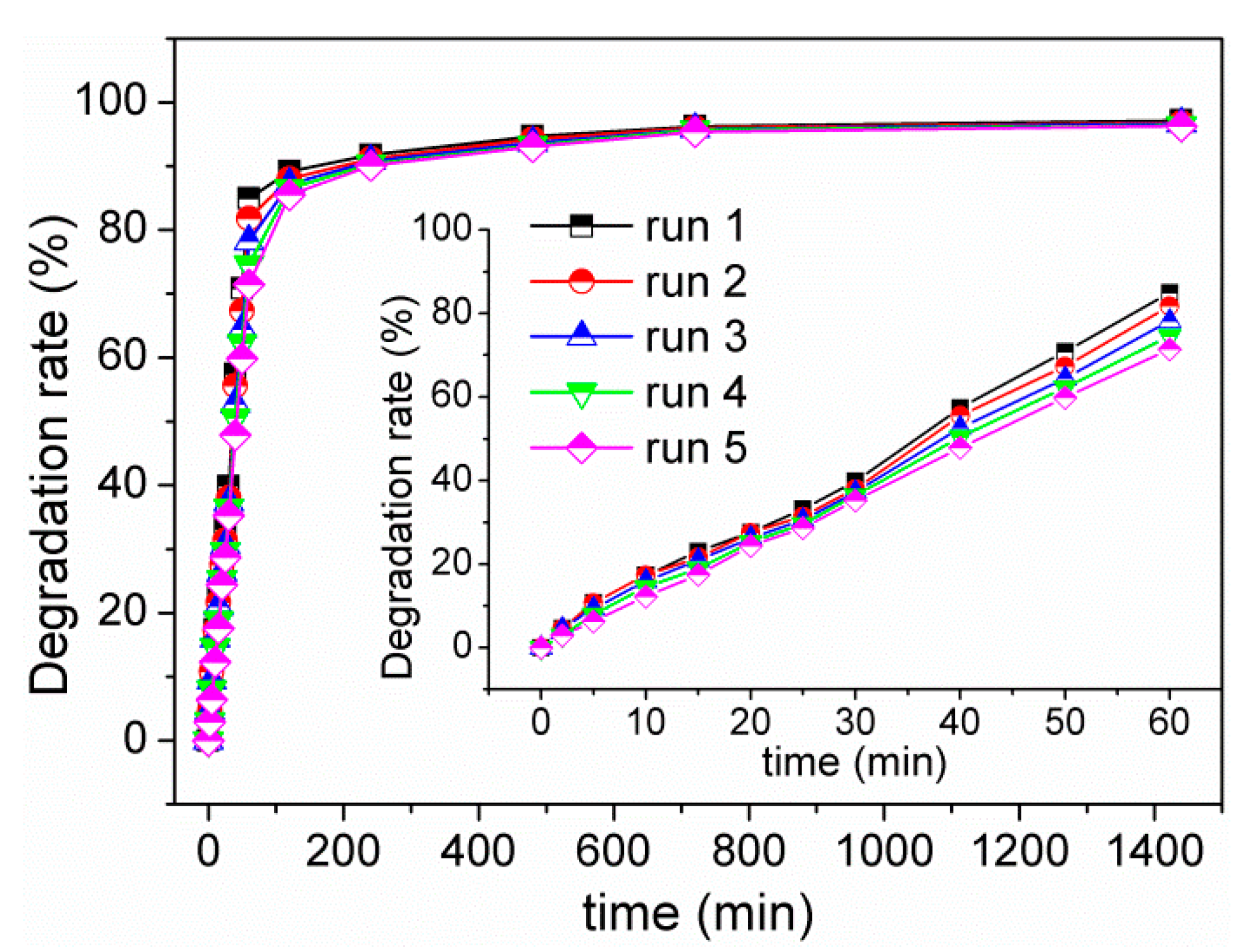
3.7. Decolorization of Acid Orange 7 (AO7) Dye

Cu(II) complexes have shown the ability to various oxidation reactions including degradation of organic dyes [24,55,56,57,58,59]. Thus the decolorization properties of Cu(II) complexes 1 and 2 were evaluated via photocatalytic degradation of acid orange 7 (AO7) dye, a common azo dye, under natural conditions. One milligram of Cu(II) complex (1 or 2) was dipped into to 3 mL of aqueous solutions of AO7 (20 mg L−1), which was magnetically stirring in the dark for 30 min. Then, an amount of 30% hydrogen peroxide (H2O2, 1 mL) was added under daylight. The UV/vis spectra of the reaction solution were measured at specific intervals with a UV/vis spectrophotometer and the AO7 concentrations were determined by the maximum absorbances at 485 nm. For comparison, the same experiments were also carried out in dark conditions. In addition, several AO7 decolorization experiments examined by bare (only AO7 in H2O without H2O2, 1 or 2), H2O2-only, 1-only and 2-only were also conducted in dark conditions and under daylight. As observations, the absorbance of the major absorption band of AO7 in aqueous solutions remained almost constant in these checked decolorization experiments by bare, H2O2-only, 1-only and 2-only in dark conditions and under daylight (Figure S6a–h). The results imply that AO7 cannot be adsorbed by 1 and 2 and is very difficultly photodecomposed by daylight irradiation or oxidative/photocatalytic degradation by H2O2 only or Cu(II) complex (1 or 2) only in dark conditions and even under daylight irradiation. However, the degradation of AO7 was occurred in cases of simultaneous existence of H2O2 and Cu(II) complex (1 or 2) in dark conditions and under daylight, as supported by time-dependent UV/vis spectra of AO7 after degradation (Figure 5a and Figure S6i–k). In case of the simultaneous existence of 2 and H2O2 under daylight, the aqueous solutions of AO7 showed remarkable color change from orange at initial to very light after 60-min degradation and to colorless after about 480-min degradation under daylight (Figure 5b). The findings suggest an oxidative degradation process. Of particular note, the degradation rate and the degradation performance of AO7 by 1/H2O2 and 2/H2O2 are greatly improved and enhanced under daylight in compared to that in dark conditions, implying photocatalytic degradation of AO7 by 1/H2O2 and 2/H2O2. Moreover, the oxidative degradation ability of 2 is better than that of 1. As a representative, the decolorization performances after 60-min degradation are about 3% and 10% for 1/H2O2 and 2/H2O2, respectively, in dark conditions and about 10% and 85% for 1/H2O2 and 2/H2O2, respectively, under daylight (Figure 5c). The decolorization performances would reach a maximum of approximately 15% for 1/H2O2 and 25% for 2/H2O2 in dark conditions and 25% for 1/H2O2 and 98% for 2/H2O2 under daylight. This is comparable with other cases of AO7 degradation reported in the literature [21,59,60,61,62,63,64,65].
To gain a better understanding of the degradation kinetics of AO7 catalyzed by 2/H2O2, the experimental data were fitted by a first-order model as expressed by the following equation—ln (C/C0) = kt—where C0 and C are the initial and apparent concentration of AO7, respectively, k is the kinetic rate constant and t is time. As a result, the k values are determined to be 0.00291 min−1 and 0.0154 min−1 for degradation of AO7 in dark conditions and under daylight (Figure 6), respectively, within 30 min. Clearly, the rate constant of AO7 degradation under daylight is about five times larger than that in dark conditions, supporting again a photocatalytic degradation of AO7. Further, there exists the first-order exponential decay relationships between the concentration of AO7 (ppm) and degradation time (min), with the formulae of [AO7] = 3.97 × exp(−t/219.85) + 15.36 (R2 = 0.93874) in dark conditions and [AO7] = 19.52 × exp(−t/48.37) + 0.94 (R2 = 0.98367) under daylight (Figure S7).
In terms of controls, several different concentrations of 2 have been conducted to check the required concentration that shows significant photocatalytic degradation activity (Figure S8). As decreasing concentration of 2, the degradation performance decreased but still in significant extents (Figure 7). For all tested concentrations (1 mg 2 dipped into 3, 6, 15, 30 mL AO7(aq)), the photocatalytic degradation performances of AO7 are quickly increased to 33–85% within 60-min irradiation and then gradually reach to approximately 59–98% as increasing irradiation time to 1440 min. These results clearly indicated that 2 exhibits good photocatalytic activity to degrade AO7 in the presence of H2O2 under daylight, even in a concentration as low as 1 mg 2/30 mL AO7(aq).
The possible reaction mechanisms are proposed. The preliminary experiments carried out in different conditions have clearly indicated that the degradation of AO7 only taken place when Cu(II) complex (1 or 2) and H2O2 were collaborated. In addition, the degradation performance of AO7 by 1/H2O2 and 2/H2O2 would be significantly improved (acceleration and enhancement) under daylight in compared to that in dark conditions. These observations imply that advanced oxidative processes (AOPs) [22], which is worked by the in situ generated active hydroxyl radical (OH) as oxidizing agent to oxidatively degrade dyes, have been applied to the degradation of AO7 dye and there might be multiple pathways to dominate the AOPs. In general, the water-insoluble Cu(II) complexes can react with H2O2 to produce OH radicals via homolytic cleavage or OH and HO2 radicals via Cu redox cycles for degrading AO7 dye both in dark and under daylight [66,67,68]. However, daylight irradiation might result in Cu-based Fenton processes that would cause the generation of photoexcited holes (h+) and electrons (e) [66,67,68]. The former might react with OH to produce OH whereas the latter might react with H2O2 to produce OH or O2 to form O2 for oxidative degradation of AO7 dye. The formation of more active OH, HO2 and O2 radicals accelerates the whole photodegradation process and also enhances the photodegradation performances.
From the viewpoint of practices, the recycling performance and stability of photocatalysts are of particular importance during photocatalytic reactions [67]. Owing to the high decolorization performances, the recyclability and stability to leaching of 2 has been examined. Recovered powdered samples of 2 were simply washed with deionized water and MeOH several times, which were then used directly for next photocatalytic degradation experiment of AO7 (Figure S9). As observation, the photocatalytic activity of 2 toward AO7 degradation retained. The decolorization performance of 2 after 60-min degradation under daylight after five sequential experiments was slightly reduced by about 16%, giving rise to about 84% degradation efficiency of that in the first time (Figure 8, inset). However, when the irradiation time was extended over 120 min, the decolorization performances were almost unchanged (Figure 8). These results indicate that complex 2 possesses excellent long-term activity and high reusability. Noteworthy, the XRPD patterns of 2 recovered from five sequential photocatalytic degradation experiments of AO7 were in good agreement with that of as-synthesized samples (Figure 4), suggesting high stability in maintaining pristine crystalline phase. On the other hand, inductively coupled plasma optical emission spectrometry (ICP-OES) analyses indicated that there were 0.0620 ± 0.0042 mg/L of Cu(II) ions in the supernatants of AO7 aqueous solutions after 1440-min decolorization by 2 under daylight, which corresponds to 0.128 ± 0.009% Cu(II) ions leached from the solid samples of 2. The very low ratios of Cu(II) ions in the supernatants suggests that 2 demonstrates high stability to leaching during AO7 degradation in the presence of H2O2 under daylight. As a result, 2 is a good photocatalytic material toward recyclable AO7 degradation.

4. Conclusions

In summary, two Cu(II)–salicyaldimine complexes have been successfully synthesized and characterized. Chloro-based complex 1 has the mononuclear structure while nitrato-containing complex 2 adopts a zigzag chain structure. The salicyaldimine ligand in the two complexes is partially deprotonated to be a mono-charged anion and acts as a NNO tris-chelator to bind a Cu(II) center through its pyridine, imino and phenolato groups and leave the neutral carboxyl group free to coordination. Noteworthy, nitrato ligand in 2 suits a bridging-tridentate with a μ, κ4O, O′:O′,O″ coordination mode to bridge two Cu(II) ions, resulted in the formation of extended zigzag chain structure. The results significantly show the critical role of anions in the formation of discrete and polymeric structures of Cu(II)–salicyaldimine complexes. Moreover, 2 would be an excellent photocatalytic material to show remarkable water stability and recyclable photocatalytic degradation activity that is capable of acceleration and enhancement of AO7 decolorization by H2O2 under daylight.

Supplementary Materials

The following are available online at https://www.mdpi.com/2073-4360/12/9/1910/s1, Figure S1: 1H NMR spectrum of H2Lsalpyca in DMSO-d6 at room temperature, Figure S2. MALDI-TOF mass spectrum of H2Lsalpyca, Figure S3. MALDI-TOF mass spectrum of [Cu(HLsalpyca)Cl] (1), Figure S4. XRPD patterns of 1 and 2, Figure S5. Solid-state excitation and emission spectra of H2Lsalpyca (λem = 505 nm, λex = 360 nm), Figure S6. Time-dependent UV/vis spectra of AO7 aqueous solutions under various degradation conditions: (a) AO7 only in dark; (b) AO7 only under daylight, (c) AO7 + H2O2 in dark; (d) AO7 + H2O2 under daylight; (e) AO7 + 1 in dark; (f) AO7 + 1 under daylight; (g) AO7 + 2 in dark; (h) AO7 + 2 under daylight; (i) AO7 + 1 + H2O2 in dark; (j) AO7 + 1 + H2O2 under daylight; (k) AO7 + 2 + H2O2 in dark, Figure S7. Concentrations of AO7 after degradation by 2/H2O2 versus degradation time in dark conditions and under daylight. Solid lines show the first-order exponential decay, Figure S8. Time-dependent UV/vis spectra of AO7 aqueous solution after the photocatalytic degradation by 2/H2O2 under daylight in different concentrations of 2: (a) 1 mg 2/6 mL AO7(aq); (b) 1 mg 2/15 mL AO7(aq); (c) 1 mg 2/30 mL AO7(aq), Figure S9. Time-dependent UV/vis spectra of AO7 aqueous solutions for the recycling experiments with the simultaneous existence of 2 and H2O2 under daylight.

Author Contributions

J.-Y.W. conceived and designed the experiments; M.-J.T., C.-J.T. and K.L. performed the experiments; M.-J.T. and J.-Y.W. analyzed the data; J.-Y.W. contributed reagents/materials/analysis tools; J.-Y.W. wrote the paper. All authors have read and agreed to the published version of the manuscript.

Funding

This research was funded by the National Chi Nan University and the Ministry of Science and Technology of Taiwan, grant number NSC 100-2113-M-260-003-MY2, NSC 102-2113-M-260-004-MY2, MOST 104-2113-M-260-005-, MOST 107-2113-M-260-001- and MOST 108-2113-M-260-002-.

Conflicts of Interest

The authors declare no conflict of interest.

References

  1. Yamada, S. Advancement in stereochemical aspects of Schiff base metal complexes. Chem. Rev. 1999, 190–192, 537–555. [Google Scholar] [CrossRef]
  2. Majumder, A.; Rosair, G.M.; Mallick, A.; Chattopadhyay, N.; Mitra, S. Synthesis, structures and fluorescence of nickel, zinc and cadmium complexes with the N,N,O-tridentate Schiff base N-2-pyridylmethylidene-2-hydroxy-phenylamine. Polyhedron 2006, 25, 1753–1762. [Google Scholar] [CrossRef]
  3. Basak, S.; Sen, S.; Marschner, C.; Baumgartner, J.; Batten, S.R.; Turner, D.R.; Mitra, S. Synthesis, crystal structures and fluorescence properties of two new di- and polynuclear Cd(II) complexes with N2O donor set of a tridentate Schiff base ligand. Polyhedron 2008, 27, 1193–1200. [Google Scholar] [CrossRef]
  4. Li, S.-N.; Zhai, Q.-G.; Hu, M.-C.; Jiang, Y.-C. Synthesis, crystal structures and characterization of three novel complexes with N-[2-(2-hydroxybenzylideneamino)ethyl]-4-methyl-benzene-sulfonamide as ligand. Inorg. Chim. Acta 2009, 362, 2217–2221. [Google Scholar] [CrossRef]
  5. Gupta, K.C.; Sutar, A.K. Catalytic activities of Schiff base transition metal complexes. Coord. Chem. Rev. 2008, 252, 1420–1450. [Google Scholar] [CrossRef]
  6. Zhou, X.-X.; Fang, H.-C.; Ge, Y.-Y.; Zhou, Z.-Y.; Gu, Z.-G.; Gong, X.; Zhao, G.; Zhan, Q.-G.; Zeng, R.-H.; Cai, Y.-P. Assembly of a Series of Trinuclear Zinc(II) Compounds with N2O2 Donor Tetradentate Symmetrical Schiff Base Ligand. Cryst. Growth Des. 2010, 10, 4014–4022. [Google Scholar] [CrossRef]
  7. Lu, Z.; Yuan, M.; Pan, F.; Gao, S.; Zhang, D.; Zhu, D. Syntheses, Crystal Structures, and Magnetic Characterization of Five New Dimeric Manganese(III) Tetradentate Schiff Base Complexes Exhibiting Single-Molecule-Magnet Behavior. Inorg. Chem. 2006, 45, 3538–3548. [Google Scholar] [CrossRef]
  8. Whiteoak, C.J.; Salassa, G.; Kleij, A.W. Recent advances with π-conjugated salen systems. Chem. Soc. Rev. 2012, 41, 622–631. [Google Scholar] [CrossRef]
  9. Escudero-Adán, E.C.; Benet-Buchholz, J.; Kleij, A.W. Expedient Method for the Transmetalation of Zn(II)-Centered Salphen Complexes. Inorg. Chem. 2007, 46, 7265–7267. [Google Scholar] [CrossRef]
  10. Houjou, H.; Iwasaki, A.; Ogihara, T.; Kanesato, M.; Akabori, S.; Hiratani, K. The architecture of dinuclear Ni and Cu complexes: Twisted and parallel forms controlled by the self-assembly of Schiff bas ligands. New J. Chem. 2003, 27, 886–889. [Google Scholar] [CrossRef]
  11. Yoshida, N.; Oshio, H.; Ito, T. Slef-assembling tetranuclear copper(II) complex of a bis-bidentate Schiff base: Double-helical structure induced by aromatic π∙∙∙π and CH∙∙∙π interactions. Chem. Commun. 1998, 63–64. [Google Scholar] [CrossRef]
  12. Kitaura, R.; Onoyama, G.; Sakamoto, H.; Matsuda, R.; Noro, S.-I.; Kitagawa, S. Immobilization of a Metallo Schiff Base into a Microporous Coordination Polymer. Angew. Chem. Int. Ed. 2004, 43, 2684–2687. [Google Scholar] [CrossRef] [PubMed]
  13. Zhu, C.; Xuan, W.; Cui, Y. Luminescent microporous metal–metallosalen frameworks with the primitive cubic net. Dalton Trans. 2012, 41, 3928–3932. [Google Scholar] [CrossRef]
  14. Wu, J.-Y.; Chang, C.-Y.; Tsai, C.-J.; Lee, J.-J. Reversible Single-Crystal to Single-Crystal Transformations of a Zn(II)–Salicyaldimine Coordination Polymer Accompanying Changes in Coordination Sphere and Network Dimensionality upon Dehydration and Rehydration. Inorg. Chem. 2015, 54, 10918–10924. [Google Scholar] [CrossRef] [PubMed]
  15. Cho, S.-H.; Ma, B.; Nguyen, S.T.; Hupp, J.T.; Albrecht-Schmitt, T.E. A metal–organic framework material that functions as an enantioselective catalyst for olefin epoxidation. Chem. Commun. 2006, 2563–2565. [Google Scholar] [CrossRef] [PubMed]
  16. Jacobsen, E.N. Asymmetric Catalysis of Epoxide Ring-Opening Reactions. Acc. Chem. Res. 2000, 33, 421–431. [Google Scholar] [CrossRef]
  17. Bagai, R.; Abboud, K.A.; Christou, G. Ligand-induced distortion of a tetranuclear manganese butterfly complex. Dalton Trans. 2006, 3306–3312. [Google Scholar] [CrossRef]
  18. Pandey, R.; Kumar, P.; Singh, A.K.; Shahid, M.; Li, P.-Z.; Singh, S.K.; Xu, Q.; Misra, A.; Pandey, D.S. Fluorescent Zinc(II) Complex Exhibiting “On-Off-On” Switching Toward Cu2+and Ag+Ions. Inorg. Chem. 2011, 50, 3189–3197. [Google Scholar] [CrossRef]
  19. Anbu, S.; Ravishankaran, R.; da Silva, M.F.C.G.; Karande, A.A.; Pombeiro, A.J.L. Differentially Selective Chemosensor with Fluorescence Off–On Responses on Cu2+and Zn2+Ions in Aqueous Media and Applications in Pyrophosphate Sensing, Live Cell Imaging, and Cytotoxicity. Inorg. Chem. 2014, 53, 6655–6664. [Google Scholar] [CrossRef]
  20. Iervolino, G.; Vaiano, V.; Pepe, G.; Campiglia, P.; Palma, V. Degradation of Acid Orange 7 Azo Dye in Aqueous Solution by a Catalytic-Assisted, Non-Thermal Plasma Process. Catalysts 2020, 10, 888. [Google Scholar] [CrossRef]
  21. Coughlin, M.F.; Kinkle, B.K.; Bishop, P.L. Degradation of acid orange 7 in an aerobic biofilm. Chemosphere 2002, 46, 11–19. [Google Scholar] [CrossRef]
  22. Zheng, J.; Gao, Z.; He, H.; Yang, S.; Sun, C. Efficient degradation of Acid Orange 7 in aqueous solution by iron ore tailing Fenton-like process. Chemosphere 2016, 150, 40–48. [Google Scholar] [CrossRef] [PubMed]
  23. Guo, J.; Dong, C.; Zhang, J.; Lan, Y. Biogenic synthetic schwertmannite photocatalytic degradation of acid orange 7 (AO7) assisted by citric acid. Sep. Purif. Technol. 2015, 143, 27–31. [Google Scholar] [CrossRef]
  24. Liu, C.-X.; Zhang, W.-H.; Wang, N.; Guo, P.; Muhler, M.; Wang, Y.; Lin, S.; Chen, Z.; Yang, G. High;y efficient phoyocatalytic degradation of dyes bt a novel copper–triazolate metal–organic framework. Chem. Eur. J. 2018, 24, 16804–16813. [Google Scholar] [CrossRef]
  25. Hasan, Z.; Jhung, S.H. Removal of hazardous organics from water using metal-organic frameworks (MOFs): Plausible mechanisms for selective adsorptions. J. Hazard. Mater. 2015, 283, 329–339. [Google Scholar] [CrossRef]
  26. Dias, E.M.; Petit, C. Towards the use of metal–organic frameworks for water reuse: A review of the recent advances in the field of organic pollutants removal and degradation and the next steps in the field. J. Mater. Chem. A 2015, 3, 22484–22506. [Google Scholar] [CrossRef] [Green Version]
  27. Chen, C.; Ma, W.; Zhao, J. Semiconductor-mediated photodegradation of pollutants under visible-light irradiation. Chem. Soc. Rev. 2010, 39, 4206–4219. [Google Scholar] [CrossRef]
  28. Wang, C.-C.; Li, J.-R.; Lv, X.-L.; Zhang, Y.-Q.; Guo, G. Photocatalytic organic pollutants degradation in metal–organic frameworks. Energy Environ. Sci. 2014, 7, 2831–2867. [Google Scholar] [CrossRef]
  29. Du, P.-Y.; Li, H.; Fu, X.; Gu, W.; Liu, X. A 1D anionic lanthanide coordination polymer as an adsorbent material for the selective uptake of cationic dyes from aqueous solutions. Dalton Trans. 2015, 44, 13752–13759. [Google Scholar] [CrossRef]
  30. Yang, J.-M.; Ying, R.-J.; Han, C.-X.; Hu, Q.-T.; Xu, H.-M.; Li, J.-H.; Wang, Q.; Zhang, W. Adsorptive removal of organic dyes from aqueous solution by a Zr-based metal–organic framework: Effects of Ce(iii) doping. Dalton Trans. 2018, 47, 3913–3920. [Google Scholar] [CrossRef]
  31. Huang, L.; He, M.; Chen, B.; Hu, B. Magnetic Zr-MOFs nanocomposites for rapid removal of heavy metal ions and dyes from water. Chemosphere 2018, 199, 435–444. [Google Scholar] [CrossRef] [PubMed]
  32. Chiang, H.-W.; Su, Y.-T.; Wu, J.-Y. Ligand dissociation/recoordination in fluorescent ionic zinc–salicylideneimine compounds: Synthesis, characterization, photophysical properties, and 1H NMR studies. Dalton Trans. 2013, 42, 15169–15182. [Google Scholar] [CrossRef] [PubMed]
  33. Wu, J.-Y.; Tsai, C.-J.; Chang, C.-Y.; Wu, Y.-Y. Metal-ion exchange induced structural transformation as a way of forming novel Ni(II)− and Cu(II)−salicylaldimine structures. J. Solid State Chem. 2017, 246, 23–28. [Google Scholar] [CrossRef]
  34. Tsai, M.-J.; Wu, J.-Y. Insight into the influence of framework metal ion of analogous metal–organic frameworks on the adsorptive removal performances of dyes from water. J. Taiwan Inst. Chem. Eng. 2019, 102, 73–84. [Google Scholar] [CrossRef]
  35. Tsai, M.-J.; Luo, J.-H.; Wu, J.-Y. Two-fold 2D + 2D → 2D interweaved rhombus (4,4) grid: Synthesis, structure, and dye removal properties in darkness and in daylight. Dalton Trans. 2019, 48, 1095–1107. [Google Scholar] [CrossRef]
  36. Tsai, M.-J.; Wu, J.-Y. Synthesis, Structure, and Dye Adsorption Properties of a Nickel(II) Coordination Layer Built from d-Camphorate and Bispyridyl Ligands. Polymers 2017, 9, 661. [Google Scholar] [CrossRef] [Green Version]
  37. Tsai, M.-J.; Wu, J.-Y. Synthesis, characterization, and dye capture of a 3D Cd(II)–carboxylate pcu network. Polyhedron 2017, 122, 124–130. [Google Scholar] [CrossRef]
  38. Williams, A.B.; Weiser, P.T.; Hanson, R.N.; Gunther, J.R.; Katzenellenbogen, J.A. Synthesis of Biphenyl Proteomimetics as Estrogen Receptor-α Coactivator Binding Inhibitors. Org. Lett. 2009, 11, 5370–5373. [Google Scholar] [CrossRef] [Green Version]
  39. Song, F.; Wang, C.; Falkowski, J.M.; Ma, L.; Lin, W. Isoreticular Chiral Metal–Organic Frameworks for Asymmetric Alkene Epoxidation: Tuning Catalytic Activity by Controlling Framework Catenation and Varying Open Channel Sizes. J. Am. Chem. Soc. 2010, 132, 15390–15398. [Google Scholar] [CrossRef]
  40. Sheldrick, G.M. A short history of SHELX. Acta Crystallogr. Sect. A 2008, 64, 112–122. [Google Scholar] [CrossRef] [Green Version]
  41. Sheldrick, G.M. Crystal structure refinement with SHELXL. Acta Crystallogr. Sect. C 2015, 71, 3–8. [Google Scholar] [CrossRef] [PubMed]
  42. Farrugia, L.J. WinGX and ORTEP for Windows: An update. J. Appl. Crystallogr. 2012, 45, 849–854. [Google Scholar] [CrossRef]
  43. Brandenburg, K.; Putz, H. Diamond; Crystal Impact GbR: Bonn, Germany, 1999. [Google Scholar]
  44. Etter, M.C. Encoding and decoding hydrogen-bond patterns of organic compounds. Acc. Chem. Res. 1990, 23, 120–126. [Google Scholar] [CrossRef]
  45. Addison, A.W.; Rao, T.N.; Reedijk, J.; van Rijn, J.; Verschoor, G.C. Synthesis, structure, and spectroscopic properties of copper(II) compounds containing nitrogen–sulphur donor ligands; the crystal and molecular structure of aqua[1,7-bis(N-methylbenzimidazol-2′-yl)-2,6-dithiaheptane]copper(II) perchlorate. J. Chem. Soc. Dalton Trans. 1984, 1349–1356. [Google Scholar] [CrossRef]
  46. Addison, C.C.; Logan, N.; Wallwork, S.C.; Garner, C.D. Structural Aspects of Co-ordinated Nitrate Groups. Q. Rev. Chem. Soc. 1971, 25, 289–322. [Google Scholar] [CrossRef]
  47. Jianmin, L.; Huaqianga, Z.; Yugenb, Z.; Jinhua, C.; Yanxiong, K.; Quanming, W.; Xintao, W. The Crystal Structure of [La(NO3)6{Cu(2,2′-bipy)2}2][La(NO3)6Cu(2,2′-bipy)2]·CH3CN with the Most Profuse Modes of Nitrate Coordination. Cryst. Res. Technol. 1999, 34, 925–928. [Google Scholar] [CrossRef]
  48. Banu, K.S.; Ghosh, T.; Guha, A.; Chattopadhyay, T.; Das, D.; Zangrando, E. Structure and luminescence of a nitrate-bridged heterotrinuclear Cu2-Pr complex with compartmental Schiff base ligand. J. Coord. Chem. 2010, 63, 3714–3723. [Google Scholar] [CrossRef]
  49. Ling, J.; Ozga, M.; Stoffer, M.; Burns, P.C. Uranyl peroxide pyrophosphate cage clusters with oxalate and nitrate bridges. Dalton Trans. 2012, 41, 7278–7284. [Google Scholar] [CrossRef]
  50. Chaudhari, A.K.; Joarder, B.; Rivière, E.; Rogez, G.; Ghosh, S.K. Nitrate-Bridged “Pseudo-Double-Propeller”-Type Lanthanide(III)−Copper(II) Heterometallic Clusters: Syntheses, Structures, and Magnetic Properties. Inorg. Chem. 2012, 51, 9159–9161. [Google Scholar] [CrossRef]
  51. She, S.; Chen, Y.; Zaworotko, M.J.; Liu, W.; Cao, Y.; Wu, J.; Li, Y. Synthesis, structures and magnetic properties of a family of nitrate-bridged octanuclear [Na2Ln6] (Ln = Dy, Tb, Gd, Sm) complexes. Dalton Trans. 2013, 42, 10433–10438. [Google Scholar] [CrossRef]
  52. Morozov, I.V.; Serezhkin, V.N.; Troyanov, S.I. Modes of coordination and stereochemistry of the NO3 anions in inorganic nitrates. Russ. Chem. Bull. Int. Ed. 2008, 57, 439–450. [Google Scholar] [CrossRef]
  53. Senthilkumar, S.; Goswami, R.; Smith, V.J.; Bajaj, H.C.; Neogi, S. Pore Wall-Functionalized Luminescent Cd(II) Framework for Selective CO2 Adsorption, Highly Specific 2,4,6-Trinitrophenol Detection, and Colorimetric Sensing of Cu2+Ions. ACS Sustain. Chem. Eng. 2018, 6, 10295–10306. [Google Scholar] [CrossRef]
  54. Udhayakumari, D.; Velmathi, S.; Sung, Y.-M.; Wu, S.-P. Highly fluorescent probe for copper (II) ion based on commercially available compounds and live cell imaging. Sens. Actuators B Chem. 2014, 198, 285–293. [Google Scholar] [CrossRef]
  55. Yang, H.; He, X.-W.; Wang, F.; Kang, Y.; Zhang, J. Doping copper into ZIF-67 for enhancing gas uptake capacity and visible-light-driven photocatalytic degradation of organic dye. J. Mater. Chem. 2012, 22, 21849–21851. [Google Scholar] [CrossRef]
  56. Wen, T.; Zhang, D.-X.; Zhang, J. Two-Dimensional Copper(I) Coordination Polymer Materials as Photocatalysts for the Degradation of Organic Dyes. Inorg. Chem. 2013, 52, 12–14. [Google Scholar] [CrossRef]
  57. Sun, X.; Zhang, J.; Fu, Z. Polyoxometalate Cluster Sensitized with Copper-Viologen Framework for Efficient Degradation of Organic Dye in Ultraviolet, Visible, and Near-Infrared Light. ACS Appl. Mater. Interfaces 2018, 10, 35671–35675. [Google Scholar] [CrossRef]
  58. Peng, Y.-F.; Qian, L.-L.; Ding, J.-G.; Zheng, T.-R.; Zhang, Y.-Q.; Li, B.-L. Syntheses, structures and photocatalytic degradation of organic dyes for two isostructural copper coordination polymers involving in situ hydroxylation reaction. J. Coord. Chem. 2018, 71, 1392–1402. [Google Scholar] [CrossRef]
  59. Hou, Y.-L.; Sun, R.W.-Y.; Zhou, X.-P.; Wang, J.-H.; Li, D. A copper(I)/copper(II)–salen coordination polymer as a bimetallic catalyst for three-component Strecker reactions and degradation of organic dyes. Chem. Commun. 2014, 50, 2295–2297. [Google Scholar] [CrossRef]
  60. Wu, J.; Zhang, H.; Qiu, J. Degradation of Acid Orange 7 in aqueous solution by a novel electro/Fe2+/peroxydisulfate process. J. Hazard. Mater. 2012, 215–216, 138–145. [Google Scholar] [CrossRef]
  61. Piumetti, M.; Freyria, F.S.; Armandi, M.; Saracco, G.; Garrone, E.; Gonzalez, G.E.; Bonelli, B. Catalytic degradation of Acid Orange 7 by H2O2 as promoted by either bare or V-loaded titania under UV light, in dark conditions, and after incubating the catalysts in ascorbic acid. Catal. Struct. React. 2015, 1, 183–191. [Google Scholar] [CrossRef] [Green Version]
  62. Ji, P.; Zhang, J.; Chen, F.; Anpo, M. Study of adsorption and degradation of Acid Orange 7 on the surface of CeO2 under visible light irradiation. Appl. Catal. B 2009, 85, 148–154. [Google Scholar] [CrossRef]
  63. Elías, V.; Vaschetto, E.; Sapag, K.; Oliva, M.; Casuscelli, S.; Eimer, G. MCM-41-based materials for the photo-catalytic degradation of Acid Orange 7. Catal. Today 2011, 172, 58–65. [Google Scholar] [CrossRef]
  64. Yin, C.; Zhao, C.; Xu, X.-J. Crystal structure and photocatalytic degradation properties of a new two-dimensional zinc coordination polymer based on 4,4′-oxy-bis(benzoic acid). Z. Naturforsch. 2019, 74, 861–864. [Google Scholar] [CrossRef] [Green Version]
  65. Wang, F.; Li, F.-L.; Xu, M.-M.; Yu, H.; Zhang, J.-G.; Xia, H.-T.; Lang, J.-P. Facile synthesis of a Ag(I)-doped coordination polymer with enhanced catalytic performance in the photodegradation of azo dyes in water. J. Mater. Chem. A 2015, 3, 5908–5916. [Google Scholar] [CrossRef]
  66. Li, J.; Pham, A.N.; Dai, R.; Wang, Z.; Waite, T.D. Recent advances in Cu-Fenton systems for the treatment of industrial wastewaters: Role of Cu complexes and Cu composites. J. Hazard. Mater. 2020, 392, 122261. [Google Scholar] [CrossRef]
  67. Wang, D.; Zhao, P.; Yang, J.; Xu, G.; Yang, H.; Shi, Z.; Hu, Q.; Dong, B.; Guo, Z. Photocatalytic degradation of organic dye and phytohormone by a Cu(II) complex powder catalyst with added H2O2. Colloids Surf. A 2020, 603, 125147. [Google Scholar] [CrossRef]
  68. Carvalho, S.S.F.; Rodrigues, A.C.C.; Lima, J.F.; Carvalho, N.M.F. Photocatalytic degradation of dyes by mononuclear copper(II) complexes from bis-(2-pyridylmethyl)amine NNN-derivative ligands. Inorg. Chim. Acta 2020, 512, 119924. [Google Scholar] [CrossRef]
Scheme 1. Synthesis of complexes 1 and 2.
Polymers 12 01910 sch001
Figure 1. (a) ORTEP plot of 1 (30% probability ellipsoid) (b) A hydrogen-bonded dimer of [Cu(HLsalpyca)Cl] generated from the R 2 2 ( 8 ) hydrogen-bonding pattern. (c) Representation of intermolecular π∙∙∙π interactions (plane∙∙∙plane: 3.37 and 3.69 Å) between any two neighboring [Cu(HLsalpyca)Cl] molecules in 1. (d) Packing diagram of 1, showing Ocarboxyl–H∙∙∙Ocarboxyl and C–H∙∙∙Cl hydrogen-bonding interactions (dashed lines).
Polymers 12 01910 g001
Figure 2. Crystal structure of 2: (a) ORTEP plot of the coordination environment around the Cu(II) center (30% probability ellipsoid). Weak Cu∙∙∙O semi-coordinations are shown as dashed lines. Symmetry code: #1, −x, y + 1/2, −z + 1/2. (b) View of the 1D zigzag chain structure. (c) Highlight the R 2 2 ( 8 ) hydrogen-bonding pattern between two carboxyl groups of two HLsalpyca ligands in two neighboring chains. (d) A 2D supramolecular wavy sheet supported by Ocarboxyl–H∙∙∙Ocarboxyl hydrogen bonds (dashed lines).
Polymers 12 01910 g002
Scheme 2. Some possible coordination modes of nitrato ligands. The terms shown in parentheses are the coordination modes described by Morozov notation. Adapted from [52], with permission from Springer Nature, 2009.
Polymers 12 01910 sch002
Figure 3. The thermogravimetric (TG) diagrams of 1 and 2.
Polymers 12 01910 g003
Figure 4. X-ray powder diffraction (XRPD) patterns of 2: simulated, as-synthesized, after immersing in various solvents (H2O, CH3OH, CH3CN, acetone, CH2Cl2 and THF) for 1 day and after AO7 degradation experiments by H2O2 for 5 cycles under daylight.
Polymers 12 01910 g004
Figure 5. (a) Time-dependent UV/vis spectra of AO7 aqueous solution after the photocatalytic degradation by 2 and H2O2 under daylight. (b) Time-dependent photographs of AO7 aqueous solutions with the simultaneous existence of 2 and H2O2 under daylight. (c) Relative concentration of AO7 versus time under various degradation conditions: AO7 only in dark (▲); AO7 only under daylight (△); AO7 + H2O2 in dark (▼); AO7 + H2O2 under daylight (▽); AO7 + 1 in dark (◀); AO7 + 1 under daylight (◁); AO7 + 2 in dark (◆); AO7 + 2 under daylight (◇); AO7 + 1 + H2O2 in dark (■); AO7 + 1 + H2O2 under daylight (□); AO7 + 2 + H2O2 in dark (●); AO7 + 2 + H2O2 under daylight (○).
Polymers 12 01910 g005
Figure 6. Plots of first-order kinetics model for AO7 degradation by 2/H2O2 in dark conditions and under daylight.
Polymers 12 01910 g006
Figure 7. Relative concentration of AO7 versus time after the photocatalytic degradation by 2 and H2O2 in different concentrations of 2 under daylight. Conditions: initial concentration of AO7 = 20 mg L−1, H2O2 = 1 mL (30%).
Polymers 12 01910 g007
Figure 8. Photocatalytic degradation rates for multiple cycles for AO7 dye by 2/H2O2 under daylight. Inset: Highlight the photocatalytic degradation rates within 60 min.
Polymers 12 01910 g008
Table 1. Crystallographic data for 1 and 2.
Complex1 2
Empirical formula C18H19ClCuN2O3 C18H19CuN3O6
Mw410.34 436.90
Crystal system Triclinic Monoclinic
Space group P 1 ¯ P21/c
a (Å) 8.2742 (7) 13.8487 (7)
b (Å) 8.5837 (7) 8.2653 (4)
c (Å) 12.6658 (9) 16.2531 (8)
α (°) 99.124 (6)90
β (°) 90.116 (6) 97.386 (2)
γ (°) 102.508 (7)90
V3) 866.49 (12) 1844.95 (16)
Z2 4
T (K) 150(2) 150 (2)
λ (Å) 0.71073 0.71073
Dcalc (g cm−3) 1.573 1.573
F000422 900
μ (mm−1) 1.434 1.226
θmin,θmax (deg) 2.952, 29.356 2.966, 26.431
R1a (I > 2σ (I)) 0.0327 0.0411
wR2b (I > 2σ (I)) 0.0738 0.1061
R1a (all data) 0.0398 0.0563
wR2b (all data) 0.0780 0.1250
GOF on F2 1.046 1.169
a R1 = Σ||Fo| − |Fc||/Σ|Fo|. b wR2 = [Σw(Fo2Fc2)2w(Fo2)2]1/2. w = 1/[σ2(Fo2) + (ap)2 + (bp)], p = (Fo2 + 2Fc2)/3. For 1, a = 0.0270, b = 0.4203; For 2, a = 0.0581, b = 2.4290.
Table 2. Selected bond lengths (Å) and angles (°) in 1 and 2 a.
1
Cu1–Cl2 2.2299 (6)Cu1–O11.8857 (14)
Cu1–N11.9376 (17)Cu1–N21.9971 (17)
O1–Cu1–N192.00 (6)O1–Cu1–N2165.35 (7)
N1–Cu1–N282.99 (7)O1–Cu1–Cl290.81 (5)
N1–Cu1–Cl2164.08 (6)N2–Cu1–Cl297.79 (5)
2
Cu1–O1 1.892 (2)Cu1–N11.931 (2)
Cu1–N21.994 (3)Cu1–O42.019 (2)
Cu1–O5#12.437 (3)
O1–Cu1–N193.00 (9)O1–Cu1–N2176.25 (9)
N1–Cu1–N283.45 (10)O1–Cu1–O489.81 (9)
N1–Cu1–O4168.19 (10)N2–Cu1–O493.43 (10)
O1–Cu1–O5#195.63 (9)N1–Cu1–O5#1120.20 (9)
N2–Cu1–O5#187.21 (10)O4–Cu1–O5#170.84 (8)
aSymmetry code: For 2, #1, −x, y + 1/2, −z + 1/2.
Table 3. Hydrogen bonding parameters in 1 and 2 (D, Donor atom; A, Acceptor atom) a.
D–H∙∙∙Ad(D–H) (Å) d(H∙∙∙A) (Å)d(D∙∙∙A) (Å)∠(D–H∙∙∙A) (°)
1
O3–H3∙∙∙O2#10.81 (2)1.84 (2)2.637 (2)168 (3)
C9–H9B∙∙∙Cl2#20.972.703.567 (3)150
2
O3–H3∙∙∙O2#2 0.82 (3)1.81 (3)2.633 (3)174 (4)
aSymmetry code: 1, #1, −1 − x, −y, −z; #2, −1 + x, y, z. 2, #1, 1 − x, 2 − y, −z.

