1. Introduction
Persistently high glucose concentrations (hyperglycemia) can cause severe complications through multiple pathways. Acute effects include diabetic ketoacidosis and hyperosmolar hyperglycemic state, both life-threatening conditions requiring immediate intervention [
1]. Chronic hyperglycemia induces oxidative stress and advanced glycation end-products, leading to microvascular damage (retinopathy, nephropathy) and macrovascular diseases (atherosclerosis, stroke) [
2].
Glucose detection is critical for diabetes management and clinical diagnostics, with well-established methods primarily relying on enzymatic and electrochemical techniques. Enzymatic glucose sensors, particularly those using glucose oxidase (GOx), dominate the market due to their high specificity and reliability [
3]. These sensors operate by catalyzing glucose oxidation, producing measurable electrical signals proportional to glucose concentration. However, they suffer from drawbacks such as enzyme instability under extreme pH or temperature, limited shelf life, and interference from oxygen fluctuations [
4]. Additionally, the cost of enzyme-based sensors remains high, restricting accessibility in low-resource settings.
Non-enzymatic electrochemical sensors have emerged as promising alternatives, leveraging direct glucose oxidation on catalytic materials like noble metals (e.g., Pt, Au) or transition metal oxides [
5]. These materials circumvent enzyme-related limitations, but often require complex synthesis or suffer from poor selectivity against interferents (e.g., uric acid, ascorbic acid) [
6]. Compared to electrochemical glucose sensors based on enzyme electrodes, there are several advantages of non-enzymatic glucose sensors. They have shown excellent stability, simplicity, and reproducibility, and they are also free from oxygen limitation [
7]. Thus, numerous metals, alloys, metal oxides, and carbon-based nanostructures were investigated in glucose sensing. Among them, platinum and gold electrodes were the foremost materials [
8,
9]. Early researchers have reported the kinetics and mechanism of electrochemical oxidation on platinum electrodes [
10,
11]. Many gold nanomaterial electrodes were prepared as glucose sensing electrodes [
12,
13]. When the particle size ranged from 3 to 6 nm, nanogold represented good catalytic activity. Meanwhile, their performance quickly declined because of the aggregation of particles.
Compared with nano-size particles, nanoporous materials with self-standing ligament and pore structures improved the catalyst stability. Recent advances in dealloying techniques have enabled the fabrication of nanoporous materials with tunable compositions for diverse applications. For biomedical implants, dealloyed Ti-Ag alloys exhibit enhanced antibacterial properties while maintaining biocompatibility [
14]. In energy storage, nanoporous Au-Pt alloys demonstrate exceptional catalytic activity for fuel cells when dealloyed in HNO
3 [
15]. The nanoporous gold electrodes represented highly active performance and good stability, which benefits from the large surface roughness and specific surface area [
16]. As noble metals, they have unique excellent optical, physical, and catalytic properties. Unfortunately, their drawbacks of poisoning by chloride ions and significant cost hinder wide practical uses of noble metal electrodes [
17]. Therefore, there is still an important challenge to exploit nanoporous materials with superior catalytic performance, outstanding stability, and lower economic cost.
Recently, it has been proved that transition metals, bimetallic alloys, and their oxides, like Ni and Cu, are expected to reach the requirements for non-enzymatic glucose sensing electrodes [
18,
19,
20,
21]. These electrodes are commonly carried out in alkaline solutions for electrochemical glucose oxidation. Tian et al. reported the Ni(II)/Ni(III) redox couple could account for the catalytic process. The sensitivity was 576.7 μA Mm
−1cm
−2 and the detection limit is 50 μM [
22]. Many different kinds of copper structures have been investigated for glucose sensing [
23,
24,
25]. The catalytic process was similar to Ni-based electrodes. With low cost and easy fabrication, transition metals with equivalently high catalytic activity for non-enzymatic glucose sensors are promising alternative electrodes.
Most previous studies focused on binary alloys (e.g., Cu-Zn, Ni-Mn) for glucose sensing, and recent work has demonstrated the advantages of ternary precursors. Specifically, Liu et al. [
26] reported that Al-Cu-Mg ternary alloys produce more uniform nanoporous copper (NPC) after dealloying, with 30% higher surface area than binary-derived NPC. The ternary system offers three key benefits for glucose detection: (1) finer pore size distribution (20–80 nm vs 50–200 nm in binaries), enabling faster mass transport [
27]; (2) residual Al
2O
3 stabilizes the NPC structure [
28]; and (3) tunable electronic structure enhances glucose oxidation kinetics [
29]. In this paper, we report on the dealloying behavior of single-phase ternary MnCuAl ribbons. The microstructure evolution of the nanoporous structure is described, and, as a potential material, the electrocatalytic activity of dealloyed MnCuAl alloys for glucose oxidation is preliminarily examined.
2. Experimental
Ternary alloy with nominal composition of Mn48Cu28Al24 (at.%) was prepared by induction melting pure Mn (99.97 wt.%), pure Cu (99.98 wt.%), and pure Al (99.99 wt.%). When the vacuity was better than 2 × 10⁻³ Pa, high-purity Ar was charged as protective atmosphere. The induction melting (high-frequency induction melting furnace, TEGP-60, Zhuzhou, China) of stoichiometric Mn-Cu-Al charges was conducted in a water-cooled copper crucible. Three melting cycles were performed to ensure homogeneity. Then, the ingot was cut into small cuboid blocks. Following induction melting, the alloy was rapidly solidified using the melt-spinning technique (single-roll rotary quenching and spinning machine, XC-500, Shenyang, China). The molten alloy was ejected through a quartz nozzle, and spun onto a rotating copper roller at 2000 rpm. Mn-Cu-Al ribbons with a thickness of 30~50 μm and a width of 3 mm were obtained. The dealloying process was carried out for different times at 50 °C in 0.2 M HCl solution, which was prepared with de-ionized water and de-aerated by high-purity Ar purging. Then, the dealloyed specimens were rinsed with de-ionized water and dried in air.
The phase constitutions of precursor alloys and dealloyed samples were analyzed by a Bruker-AXS D8 Advance X-ray diffraction instrument (XRD, Karlsruhe, Germany) using Cu-Kα source radiation at 40 kV and 40 mA. The microstructure and composition of nanoporous copper were characterized by an FEI Quanta 250 FEG field emission scanning electron microscope (SEM, FEI Company, USA) equipped with an energy dispersive spectrometer (EDS, Oxford, UK). The pore size and ligament size were measured by Nano Measurer 1.2.5 software. The average pore size and ligament size were measured more than 100 sites by using the single chord length method, and then summarized statistically. Dealloying behavior was tested by using an electrochemical work-station with a three-electrode system (Zahner-IM6, Germany). A platinum plate was used as the counter electrode, Mn-Cu-Al ribbon as the working electrode, saturated calomel electrode (SCE) as the reference electrode, and 0.2 M HCl solution as electrolyte.
The electrocatalytic properties of the nanoporous copper/glassy carbon electrode (NPC/GCE) for glucose oxidation were examined with a platinum plate as the counter electrode and SCE as the reference electrode. The catalyst suspensions were made by mechanically mixing 4 mg of nanoporous copper, 4 mg of carbon powder, 300 μL of ethanol, and 100 μL of Nafion solution (0.5 wt.%). All mixtures were sonicated for 10 min to form a uniform suspension. A volume of 4 μL of Catalyst ink was placed on a polished 2 mm diameter GCE as the working electrode for electrochemical tests. A 0.2 M NaOH electrolyte solution was deoxygenated by purging high-purity Ar for 20 min before testing. The glucose sensing performance of NPC/GCE was detected in 0.2 M NaOH + 50 mM glucose by using cyclic voltammetry. All tests were carried out at 25 °C. After each experiment, the GCE was polished to a mirror-like surface with 0.05 μm Al2O3, and then sonicated in water for 3 min.
The chemicals used above, including HCl, NaOH, and glucose (C6H12O6·H2O), were analytical purity and supplied by Chengdu Kelong Chemicals Co., Ltd. (China). The 5% Nafion solution was supplied by Shanghai Aladdin Biochemical Technology Co., Ltd. (China).
3. Results and Discussion
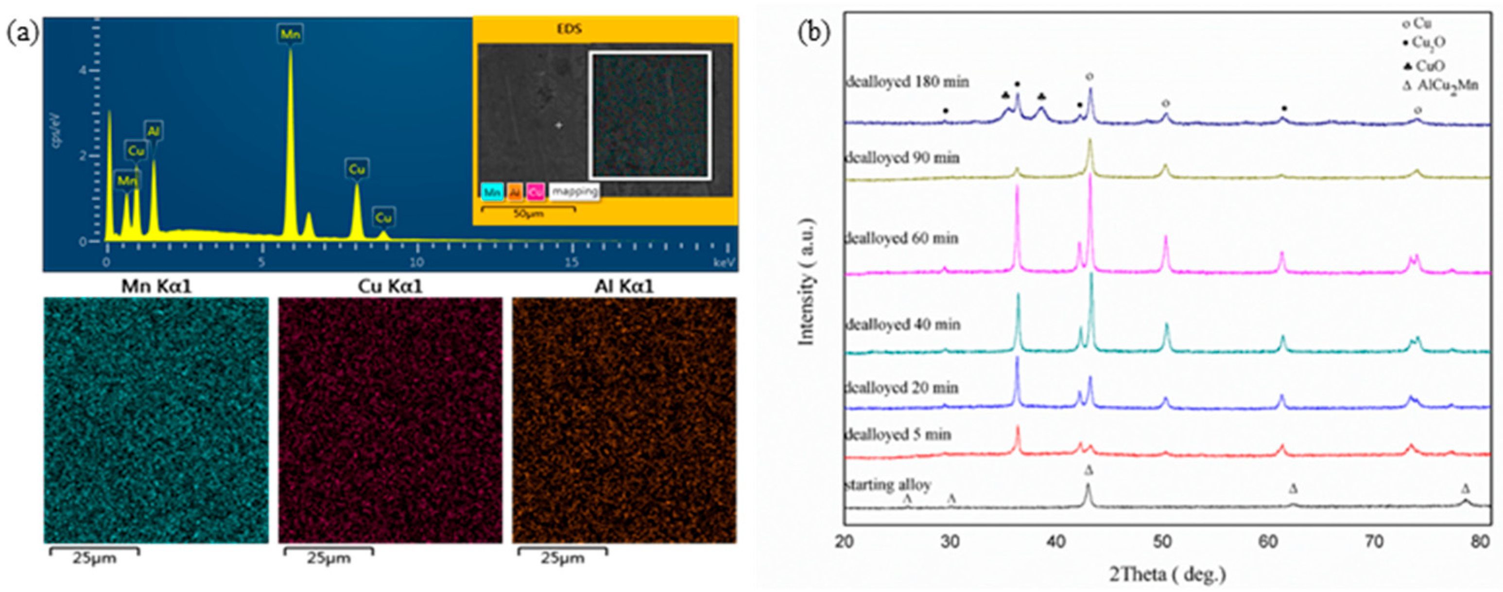
Before dealloying, the composition of the starting alloy was determined to be Mn
47.32Cu
26.86Al
25.82 (at.%) by EDS (showed in
Figure 1a), which is close to the nominal composition Mn
48Cu
28Al
24 (at.%). The EDS mapping of the precursor presented the uniform distribution of the three elements, and no component segregation was observed.
Figure 1b shows XRD results of starting alloy and dealloyed samples. For the starting alloy, the diffraction peaks at 25.9º, 30.1º, 42.9º, 62.3º, and 78.58º can be assigned to the (111), (200), (220), (400), and (422) diffractions of the face-centered cubic structure, respectively, according to Powder Diffraction File (PDF) card No. 65-6606. It is a single-phase AlCu
2Mn structure. During dealloying in 0.2 M HCl solution for 5 min at 50 °C, the AlCu
2Mn is decomposed and the dealloyed sample is composed of Cu and Cu
2O (PDF card No. 65-3288). With increasing dealloying time, the peak intensity enhancement of Cu is observed, until 60 min, and the peak intensity was max. After that, there was a reduction in intensity. In the case of 180 min, another copper oxide (CuO) was identified by the diffraction peaks at 35.5º and 38.7º, in accordance with PDF card No. 78-0428.
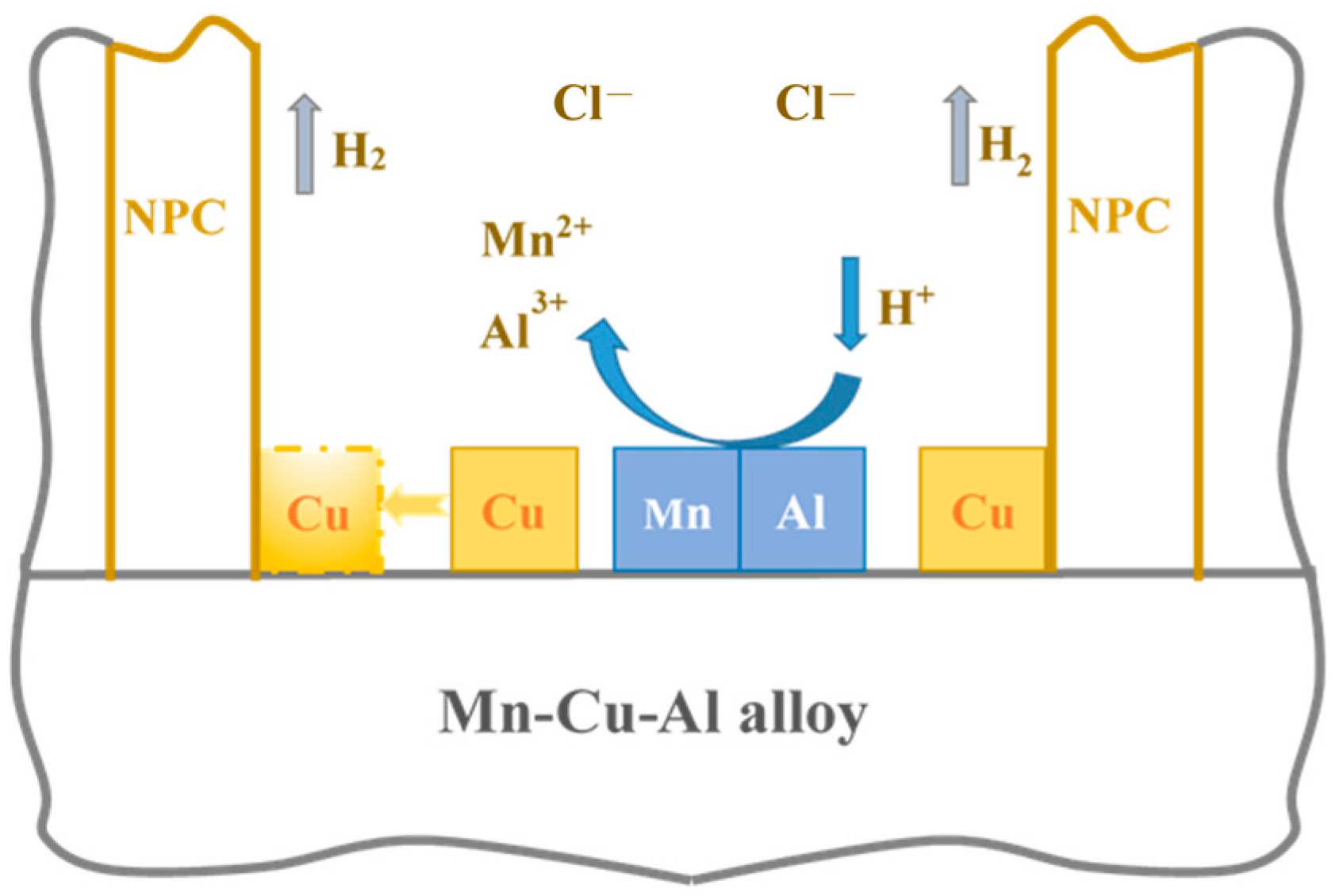
Schematic illustration of dealloying process of single-phase Mn-Cu-Al precursor in HCl solution is shown in
Figure 2. The standard reversible potential of Mn and Al is −1.182 V and −1.662 V, respectively (vs. standard hydrogen electrode (SHE)), whereas that of Cu is 0.337 V (vs. SHE). Due to its active chemical property, Mn-based alloys are easily etched in acid solutions. Immersed in 0.2 M HCl solution, the less noble element (Mn, Al) is etched away, while the more noble element (Cu) is retained and gathered to form a nanoporous copper structure. Meanwhile, hydrogen is generated from the reacted solution [
30,
31]. The nanoporous structure is a result from the competition behavior between dissolution (Mn, Al) and diffusion (Cu).
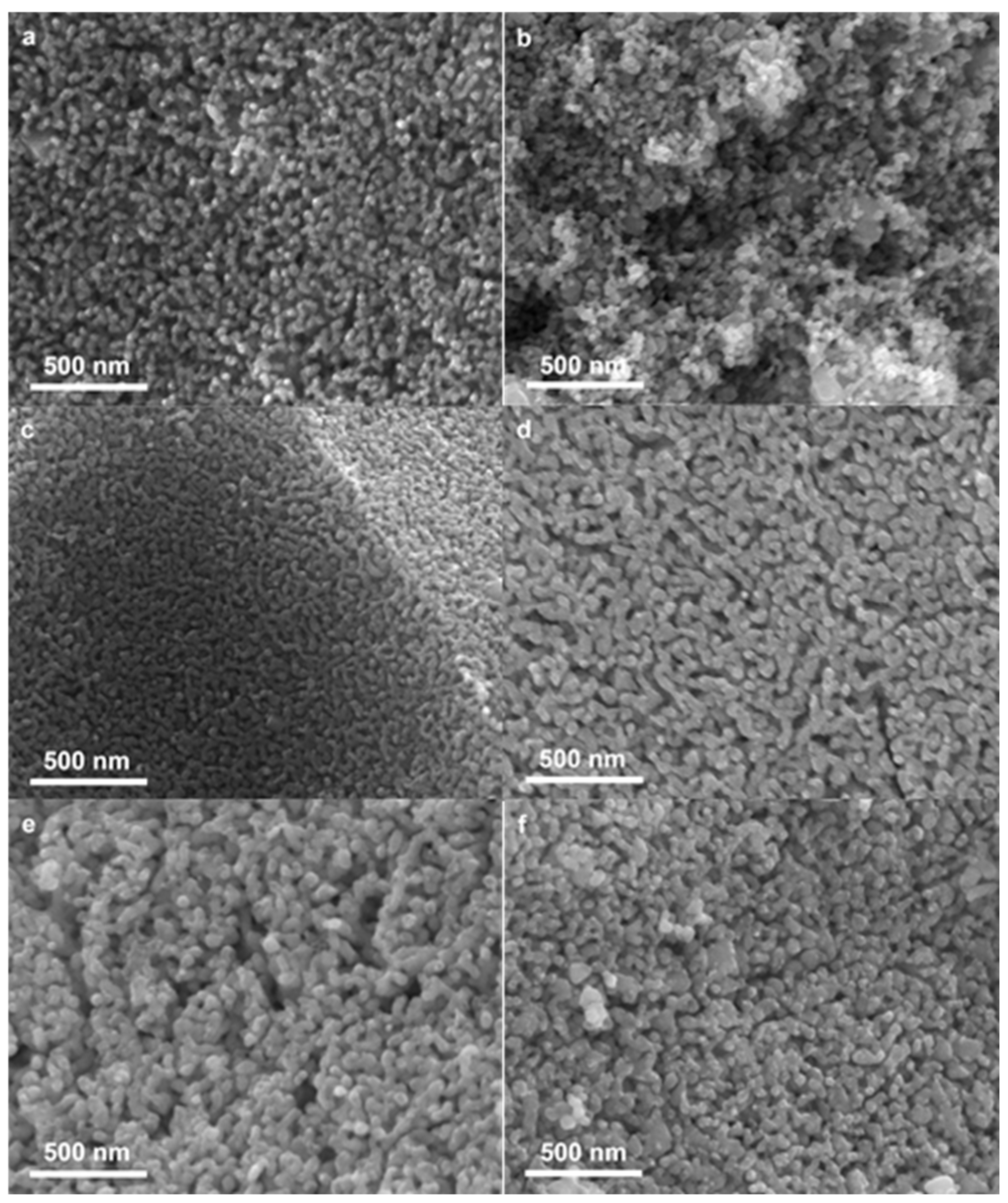
After dealloyed for 5 min, 20 min, 40 min, 60 min, 90 min, and 180 min at 50 °C, the nanoporous copper exhibits an open, bicontinuous ligament–pore structure (
Figure 3a–f), with an average pore size of 34~84 nm and average ligament size of 50~100 nm, shown in
Figure 4. In the case of dealloying for 5 min (
Figure 3a), selective dissolution of precursor ribbons began, and there was still a small amount of remaining unreacted areas. Larger pore size and no difference of ligament size were observed after dealloying for 20 min (
Figure 3b). After that, in the case of dealloying 40 min (
Figure 3c) and 60 min (
Figure 3d), the average ligament size and average pore size increased at almost the same pace. With the increase of dealloying time to 90 min (
Figure 3e) and 180 min (
Figure 3f), pore size was dramatically decreased. That is because the ligaments started to coarsen.
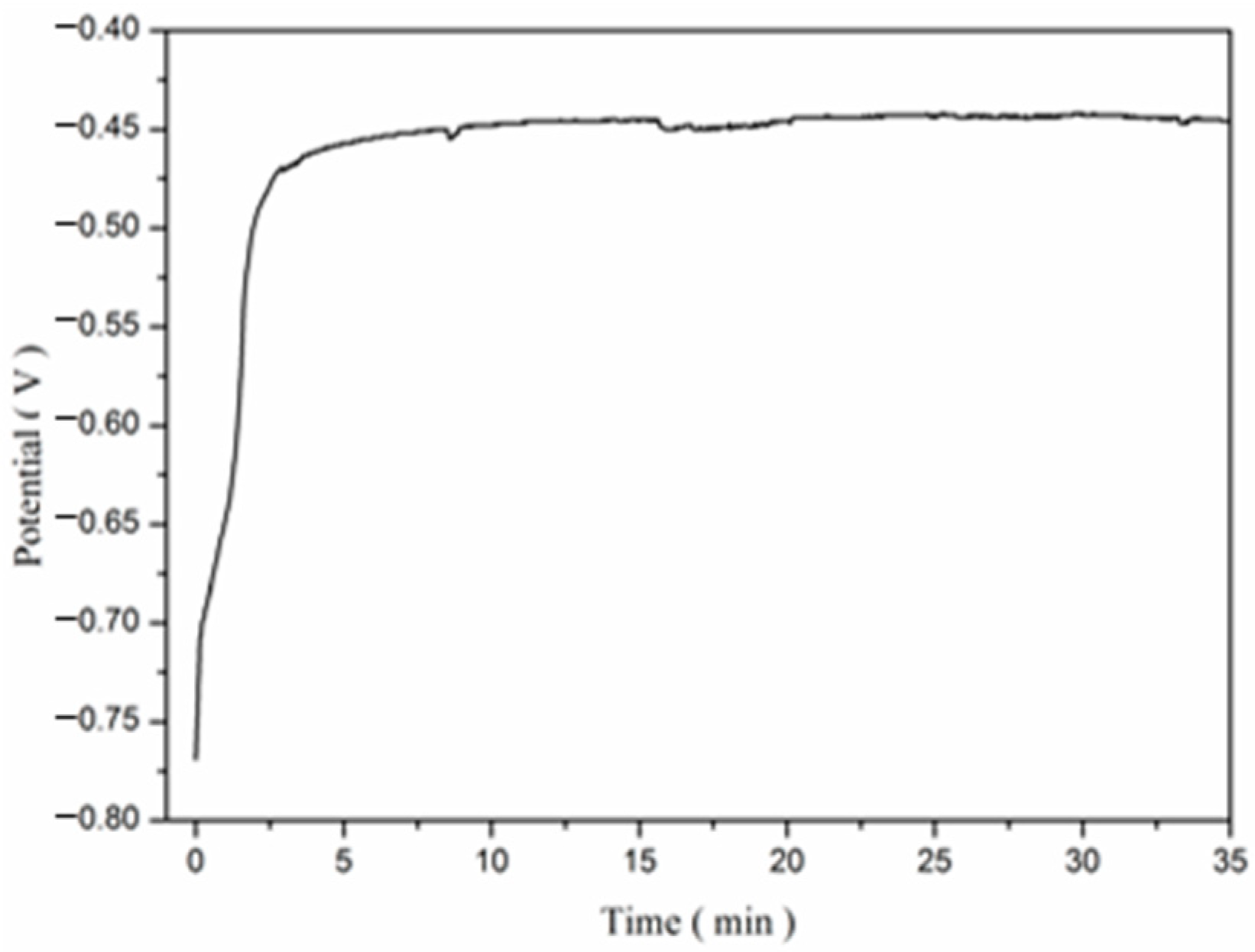
The dealloying process of Mn-Cu-Al alloy includes two steps: dissolution of Mn and Al elements, and diffusion of Cu elements. It is well explained by the ligament size to pore size ratio, shown in
Figure 5. To further determine the start time of the stable dealloying process, the MnCuAl precursor was tested by electrochemical open-circuit potential. The open circuit potential variation curve of the MnCuAl ribbon immersed in 0.2 M HCl solution is presented in
Figure 6. With the immersion time increased to 4 min, the open-circuit potential was up to −0.45 V vs. SCE, and maintained. This means that the dealloying corrosion reached a stable state.
Based on the above experimental results, the evolution process of MnCuAl to nanoporous copper can be divided into three stages. First, the dissolution of Mn and Al is so significant that formation of pores is dominant, so the ratio of ligament size to pore size decreased. Stage two is the rapid growth of both ligament size and pore size. The ratio is nearly unchanged. The last stage is ligament coarsening. The dealloying process mainly consists of the corrosion dissolution of the active components (Mn and Al) and the diffusion and rearrangement of the inert metal components (Cu). When MnCuAl is immersed in the HCl solution, the active atoms (Mn and Al) are susceptible to corrosion and are removed from the surface position of the precursor, resulting in many vacancies. At the same time, the noble component atoms (Cu) tend to diffuse along the exposed surface and are constantly agglomerating to form clusters. With the continuous dealloying, the corrosion channels of Mn and Al form nanoporous pore sizes, and the diffusion and aggregation of Cu clusters form nanoporous ligaments. The diffusion movement of copper atoms is much slower compared to the dissolution rate of the active elements, so the formation of pores in the nanoporous structure predominates in the early stage of dealloying, which leads to a sharp decrease in the ligament/pore (L/P) ratio in the first stage. With the dealloying, Cu atoms were rearranged to form more clusters after aggregation, while Mn and Al continued to dissolve, and the size of the nanoporous ligaments and pore sizes increased rapidly. As a result, the ratio of L/P remains almost constant. With the continuous corrosion and dissolution of the active components, Mn and Al atoms, the Cu atoms are constantly exposed, and more and more Cu atoms are involved in the formation of ligaments. On the other hand, the dissolution of Mn and Al was inhibited. As a result, the gap between the ligaments is reduced. At this stage, the diffusion rearrangement of Cu atoms predominates. As a result, the ligament thickening becomes larger and the aperture decreases accordingly. In the last stage, the ligament continues to thicken and even appears in size of several hundred nanometers, and the L/P ratio rises sharply.
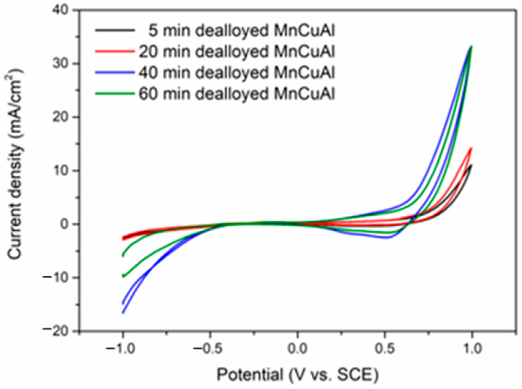
The electrocatalytic activity for glucose oxidation of different NPC/GCE are examined with a platinum plate as the counter electrode and SCE as the reference electrode.
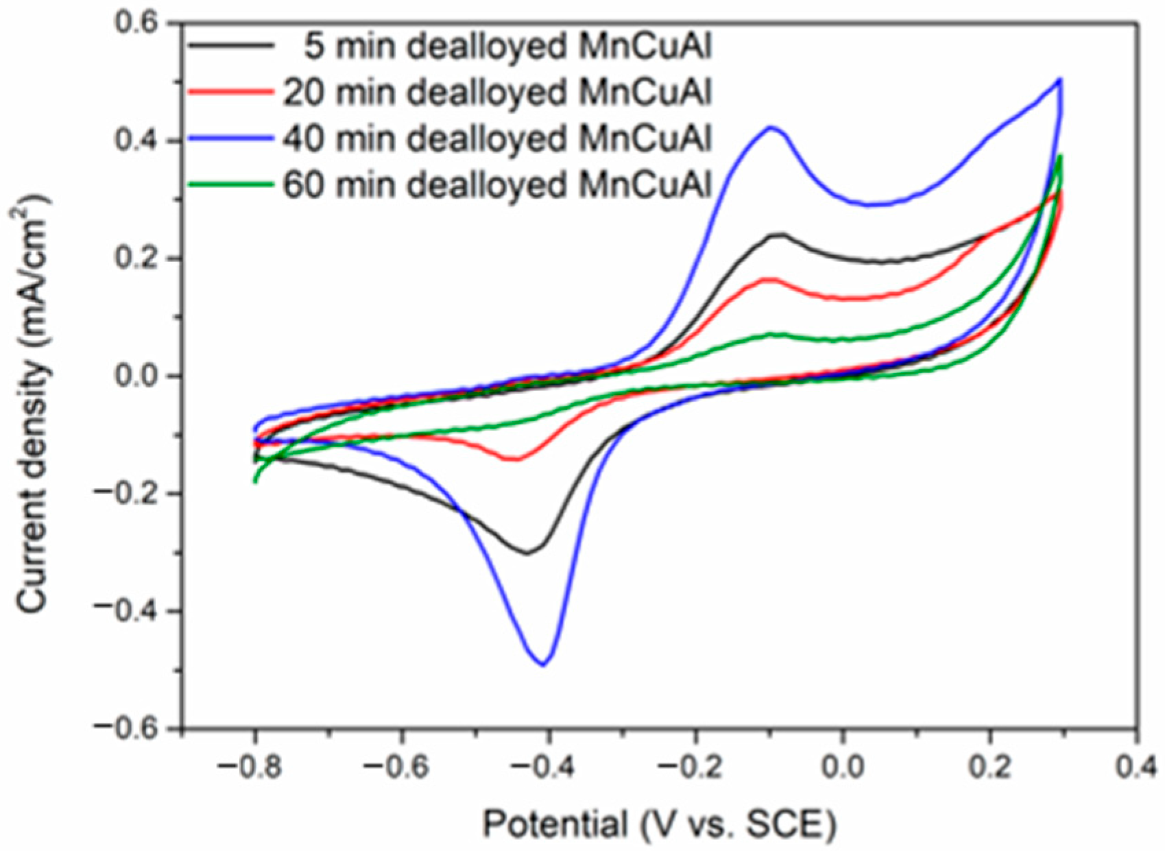
Figure 7 and
Figure 8 show the cyclic voltammograms with the absence and presence of 50 mM glucose in 0.2 M NaOH aqueous solution at a scan rate of 50 mV·s
−1, respectively. The different NPC/GCE electrodes have no redox peaks at the potential range of −1 V to 0.5 V in
Figure 7. Whereas the presence of 50 mM glucose leads to notable redox peaks in
Figure 8. It can be seen that four different samples have quite similar electrochemical behavior. The 40 min dealloyed MnCuAl ribbon exhibited the highest sensitivity and relatively maximum current density. According to
Figure 4, the average pore and ligament size of this sample was 53 nm and 58 nm, respectively. It was in the second stage of porosity microstructure evolution, which is the rapid growth of both ligament and pore. The well-distributed nanoporous structure (
Figure 3c) of the 40 min dealloyed sample can also account for the excellent glucose sensing performance. The detection potential is −0.25 V, which is much less positive potential compared to other similar studies [
32,
33,
34].
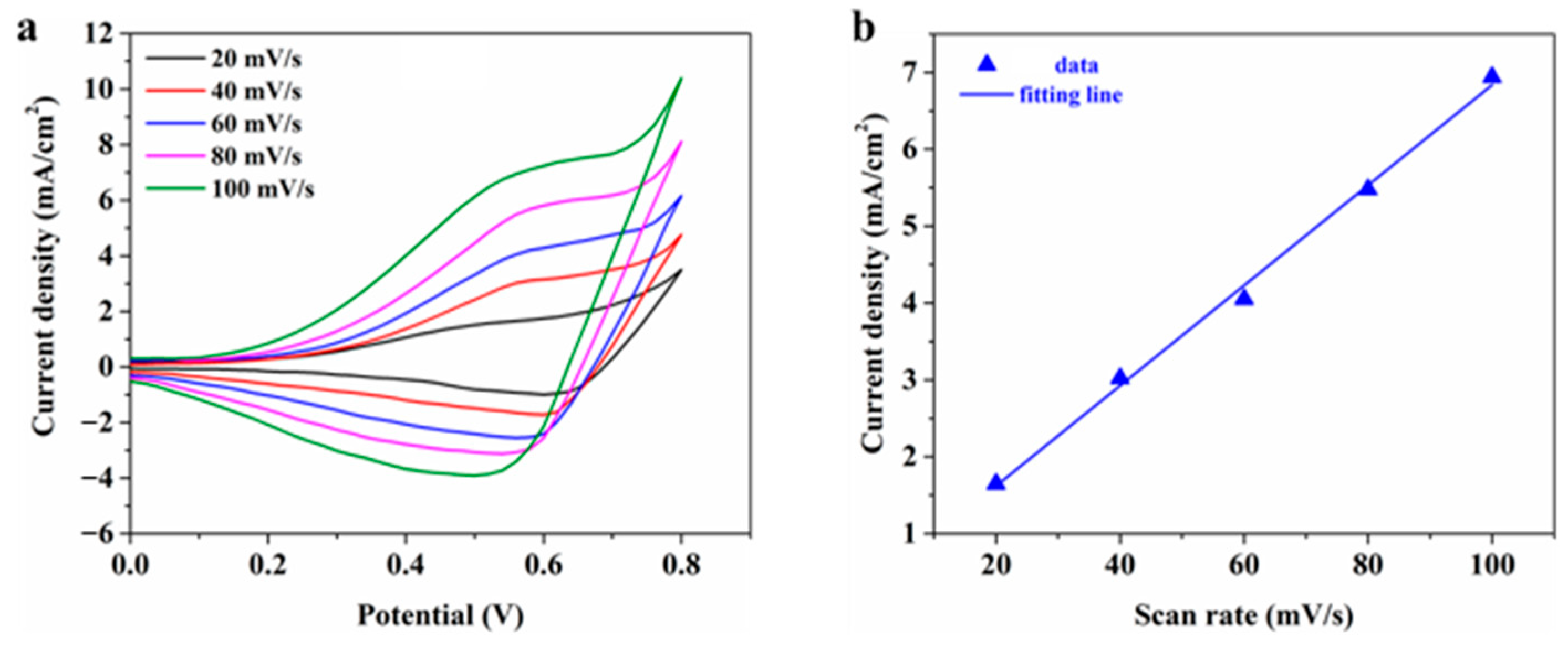
To further elucidate the electrochemical kinetics of glucose oxidation on the NPC electrodes, cyclic voltammetry (CV) measurements were conducted at various scan rates ranging from 20 to 100 mV/s (20, 40, 60, 80, and 100 mV/s) in 0.1 M NaOH solution, as shown in
Figure 9. The linear relationship between peak current density (
I) and scan rate (
v) can be expressed as:
with a correlation coefficient of R
2 = 0.996. This strong linear dependence indicates that the electrocatalytic oxidation of glucose on NPC electrodes follows a surface-controlled electrochemical process. The observed behavior suggests that the reaction kinetics are dominated by the adsorption/desorption of glucose molecules on the electrode surface rather than diffusion-limited processes.
The electrocatalytic performance of nanoporous copper (NPC) electrodes toward glucose oxidation was systematically investigated using chronoamperometry. Based on cyclic voltammetry results (
Figure 7), an optimal working potential of 0.58 V (vs. reference electrode) was selected in 0.1 M NaOH electrolyte solution. After stabilization of the baseline current, successive additions of glucose solutions with varying concentrations were introduced into the electrochemical cell.
Figure 10a illustrates the time-dependent current density response of the NPC electrode upon each glucose addition, while the insert in
Figure 10a specifically focuses on the low concentration range. The NPC electrode exhibited distinct stepwise current responses corresponding to each glucose concentration increment. The calibration curve depicting the relationship between glucose concentration and steady-state current density is presented in
Figure 10b.
Linear regression analysis yielded the equation shown in
Figure 10b, where the slope represents the sensor sensitivity. The electrode demonstrated a linear detection range from 0.1 mM to 2 mM with a sensitivity of 0.727 mA·mM
−1·cm
−2, indicating significant electrocatalytic activity toward glucose detection.
The limit of detection (LOD) represents the minimum glucose concentration that can be reliably detected by the sensor and serves as one of the key indicators for evaluating the sensitivity of glucose sensors. In this study, the LOD was determined using the signal-to-noise ratio (S/N) method. Multiple blank tests were performed to calculate the relative standard deviation (N) of the current density. The detection limit for glucose was obtained when the signal-to-noise ratio (S/N) reached 3. Based on these calculations, the fabricated electrode demonstrated an excellent detection limit of 1.62 μM for non-enzymatic glucose sensing.
A possible mechanism was given to explain the glucose electrochemical oxidation behavior. The Cu(III)/(II) redox couple played a crucial role during the oxidation process of glucose to gluconolactone, shown in following reactions:


