Article

Influence of Homogenization Heat Treatments on the Mechanical, Structural, Biodegradation, and Cavitation Behavior of Some Alloys in the ZnMg(Fe) System

by
Brandușa Ghiban
1,*,
Ilare Bordeasu
2,
Aurora Antoniac
1,
Iulian Antoniac
1,
Cristina Maria Gheorghe
1,
Dorin Bordeasu
3,
Lavinia Madalina Micu
4,*,
Cristian Ghera
2,*,
Laura Cornelia Salcianu
5,
Bogdan Florea
6,
Daniel Ostoia
2 and
Anca Maria Fratila
7
1
Department of Metallic Materials Science, Faculty of Materials Science and Engineering, National University of Science and Technology Politehnica Bucharest, 060042 Bucharest, Romania
2
Department of Mechanical Machinery, Equipment and Transport, Politehnica University of Timisoara, 1 Mihai Viteazu Boulevard, 300222 Timisoara, Romania
3
Department of Automation and Applied Informatics, Politehnica University of Timisoara, 2 Vasile Parvan Boulevard, 300223 Timisoara, Romania
4
Department of Agricultural Technologies-Department I, King Mihai I University of Life Sciences, 300645 Timișoara, Romania
5
Department of Mechatronics, Politehnica University of Timisoara, 1 Mihai Viteazu Boulevard, 300222 Timisoara, Romania
6
Department of Engineering and Management of Metallic Materials, Faculty of Materials Science and Engineering, National University of Science and Technology Politehnica Bucharest, 060042 Bucharest, Romania
7
Department of Dental Medicine and Nursing, Faculty of Medicine, Lucian Blaga University of Sibiu, 550169 Sibiu, Romania
*
Authors to whom correspondence should be addressed.
Crystals 2025, 15(5), 458; https://doi.org/10.3390/cryst15050458
Submission received: 5 March 2025 / Revised: 25 April 2025 / Accepted: 27 April 2025 / Published: 14 May 2025
(This article belongs to the Special Issue Metallurgy-Processing-Properties Relationship of Metallic Materials)

Abstract

:
This paper presents the biodegradation and cavitational erosion behavior of new zinc alloys in the ZnMgFe system. The alloys were heat-treated through homogenization at 300 °C and 400 °C, with maintenance times of 5 and 10 h each. The experimental research consisted of characterizing the structure and mechanical properties of the newly made alloys in different structural states, as well as determining their biodegradation and cavitation behavior. Biodegradability was achieved using laboratory tests in SBF, with different immersion durations (3, 7, 14, 21, or 35 days). The cavitation behavior was assessed by performing tests on a piezoceramic crystal vibrator in compliance with ASTM G32-2016, thus constructing the curves of the erosion velocity MDER(t) and the cumulative average erosion depth MDE(t). The analyses performed on the mechanical properties, microscopic images, and the cavitation parameters MDER and MDEmax (results at the end of the cavitation attack) showed the effect of the heat treatments on the structure and structural resistance to cyclic loadings of the cavitation. The double alloying of zinc with magnesium and iron may increase either the mechanical properties or the corrosion resistance to cavitation and can control the biodegradability of the resulting ZnMgFe alloy. The best heat treatment for improving these properties is homogenization at 400 °C/10 h, which may increase the cavitation erosion of zinc by up to seven times. The experimental results demonstrate that the new alloys from the ZnMgFe system are a good option for manufacturing biodegradable implants with functional in vitro properties.

1. Introduction

Metallic biomaterials are used in multiple biomedical applications, including joint replacements, fracture fixations, cardiovascular stents, and bone remodeling, due to their high mechanical strength and corrosion resistance [1,2,3,4,5,6]. These materials have undergone significant development for these applications. In recent years, this development has trended towards the achievement of metallic biomaterials with a temporary stability, i.e., biodegradable materials [7,8]. There are three categories of biodegradable metallic biomaterials: magnesium-based alloys [9,10], zinc-based alloys [11,12,13,14], and ferrous alloys [12]. The biomedical applications of zinc-based alloys are based on the spectacular advantages offered by alloying zinc with different alloying elements, and the most intensely studied influence of binary alloying with zinc is that offered by magnesium. The alloying of zinc with magnesium improves the mechanical characteristics; therefore, such alloys can be used for bone -fixation applications [11,12,13,14,15]. On the other hand, magnesium is also used in biodegradation-based applications, i.e., temporal implants [16,17,18,19]. Therefore, the synergetic effect conferred by magnesium can lead to the achievement of a particularly successful combination for temporal medical applications, with mechanical properties resulting from the hardening of the solid solution through the precipitation of the interstitial compound Mg2Zn11. The alloying of zinc with iron improves the mechanical characteristics due to the formation of the compound FeZn13, following a peritectic reaction [20]. Complex alloys of zinc with both magnesium and iron have been studied very little in the literature; as such, this was a major objective of the present work. ZnMgFe alloys were studied in [21,22,23,24], but their fine structure was not detailed. Such an alloy would contain the compounds Mg2Zn11 and FeZn13, which are hard and brittle compounds with hardening effects on solid solutions.
On the other hand, when considering some applications with temporary effects such as cardiovascular stents, the cavitation phenomenon is an issue with these biodegradable zinc alloys. While cavitation has been extensively studied for various material classes, such as stainless steels, nickel superalloys, and aluminum alloys, it has only recently been investigated in zinc alloys [25,26,27,28].
The present work aimed to study new experimental zinc alloys, specifically the alloying of magnesium and iron with zinc (the ZnMg(Fe) system). This was achieved through investigations into the influence of different homogenization heat treatments on the structure, mechanical properties, biodegradation, and cavitation behavior of these alloys.

2. Materials and Experimental Procedures

The experimental zinc alloys were developed in a classical furnace, cast, and prepared for the experimental investigations. The elaboration of the experimental alloys was carried out inside an induction furnace at a temperature generally between 650 °C and 750 °C in a vacuum; the protective atmosphere was argon. The chemical compositions of the experimental alloys, determined using the Spectro test CCDTXCO1, are shown in Table 1.
Homogenization heat treatments were performed on the experimental specimens in the cast state at 300 °C and 400 °C with a maintenance time of 5 h and 10 h at each temperature, either for pure zinc or for the experimental zinc alloys (the ZnMg alloy or the ZnMgFe alloy). The heat treatments were carried out in a Nabertherm oven type 3L Muffle Furnace, L3/11/B510, 1100 °C. For each type of heat treatment, six tests were carried out to determine the following mechanical properties: tensile strength, yield strength, elongation, and hardness. The mechanical characteristics of the test samples subjected to various aging heat treatments were determined using a Walter + Bai AD Switzerland universal testing machine, model LFV 300.
The structural investigations were performed using an OLYMPUS SZX stereomicroscope equipped with the QuickMicroPhoto 2.2 software (for stereomacrostructural analyses) or using an OLYMPUS microscope equipped with image-processing software that quantitatively determined the phases and constituents (for microstructural analyses). The fractographic analyses were performed using an electronic microscope (Thermophischer Quattro S model). We used well-known EDS methods to determine the elemental composition, in %wt., of each alloying element.
The biodegradation behavior of the experimental zinc alloys was investigated using immersion tests performed in a simulated physiological fluid (SBF) solution. The SBF solution, with an ionic composition similar to that of human blood plasma, was prepared in the laboratory according to the protocol proposed by Kokuboo (SBF chemical composition: 8.035 g/L of NaCl; 0.355 g/L of NaHCO3; 0.225 g/L of KCI; 0.231 g/L of K2HPO4·3H2O; 0.311 g/L of MgCI2-6H2O; 39 mL of 1M HCl; 0.292 g/L of CaCI2; 0.072 g/L of Na2SO4; and 6.118 g/L of Tris-NH2C(CH2OH)3). The immersion tests made it possible to determine the mass loss and pH variation for the investigated samples. The initial pH of the SBF was 7.4. To assess the mass loss, we used five parallelepiped samples of each type of alloy with dimensions of 15 × 15 × 5 mm (length × width × height). The test was performed at a temperature of 37 ± 0.5 °C over a period of 3, 7, 14, 21, 28, or 35 days, with each sample being placed in 50 mL of test medium. Simulated body fluid (SBF) was used as the test medium.
The cavitation resistance testing was performed on a vibrating apparatus with piezoceramic crystals, in compliance with ASTM G32-2016 [25,26,27,28]. The total duration of cavitation was 165 min, with 12 intermediate periods used to track the evolution of the destruction in the cavity surface area. Before starting the test, the surface was polished to a roughness of Ra = 0.02…1.6 μm [29,30,31]. To ensure the accuracy of the experiment, three samples were tested for each state. The assessment of the cavitation behavior was carried out by constructing the curves of the erosion velocity, vmax, and the mean depth erosion at the end of the cavitation attack, MDEmax.

3. Experimental Results and Discussion and Interpretation of Results

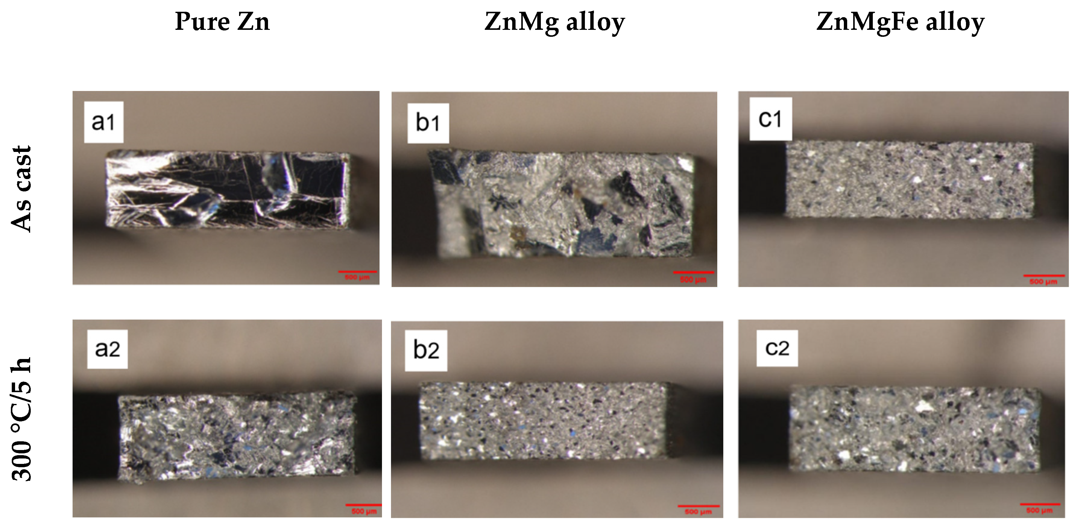
3.1. Microstructural Characterization of the Experimental Alloys

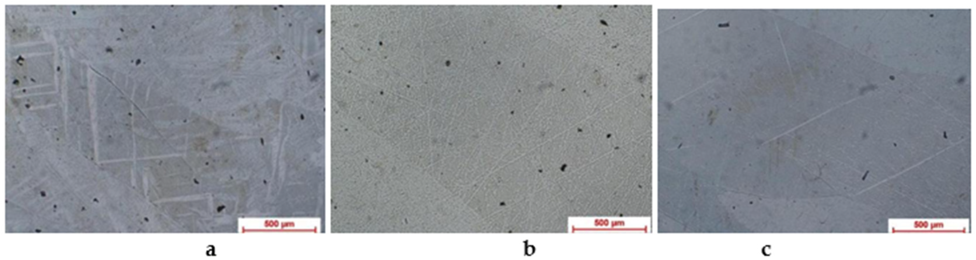
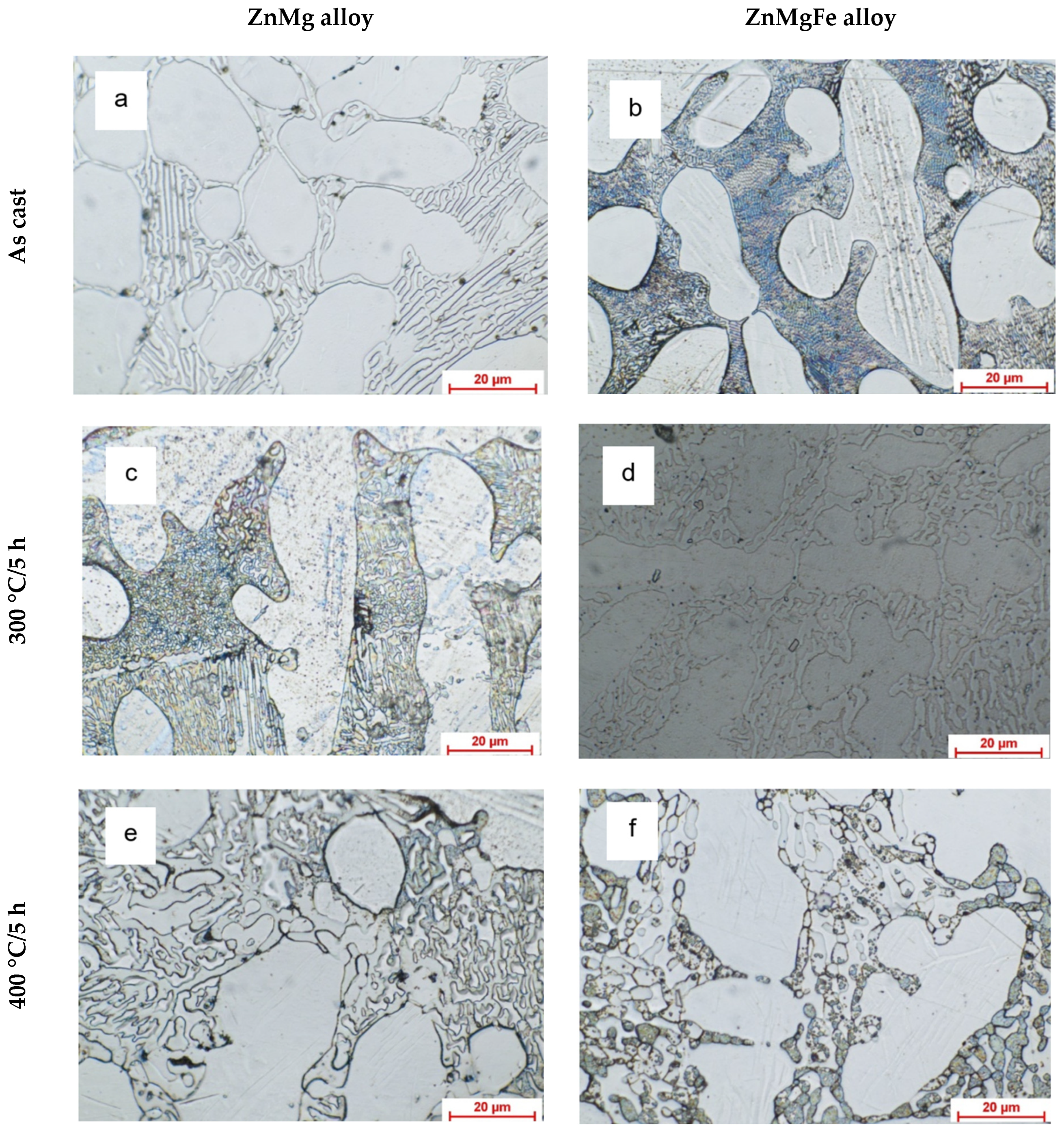
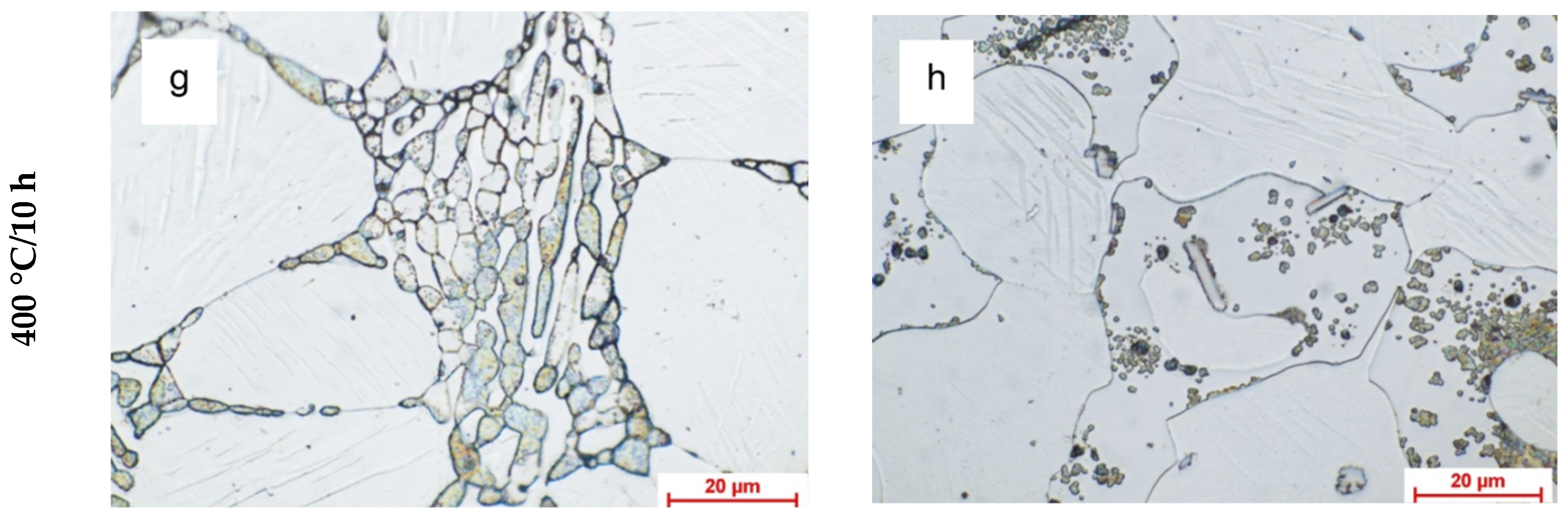
3.1.1. Optic Metallographic Analysis

The results of the structural analysis of the experimental alloys performed under the metallographic light microscope are shown in Figure 1 for pure zinc, Figure 2 for the ZnMg alloy (a,c,e,g), and Figure 2 for the ZnMgFe alloy (b,d,f,g). Figure 1 shows the structural aspects of pure zinc in a cast state, either untreated or after various homogenization heat treatments. Obviously, the structure in the cast state was made of pure zinc, with a coarse grain and dendritic segregation. When applying homogenization at 300 °C, depending on the holding time, both the homogenization of the structure (at a maintenance time of 5 h) and the beginning of the annealing process (at a maintenance time of 10 h) took place. The application of homogenization at 400 °C led to the homogenization of the structure and an obvious grinding with the formation of annealing poppies, regardless of the maintenance time (Figure 1b, for a duration of 10 h).
The structural aspects of the binary alloy ZnMg are rendered for the cast state in Figure 2a,c,e,g. The analysis of the sample in the cast state of the ZnMg alloy revealed a specific structure of an inhomogeneous solid solution, with a dendritic appearance (Figure 2a). The dendrites had long arms of 100–300 μm and arm thicknesses of 15–20 μm. The eutectic mixture had interdendritic precipitation and a quite fine digital lamellar appearance. The interdendritic eutectic mixture had a fingerprint appearance, with distances of about 1–3 μm between the slides.
When applying homogenization to the ZnMg alloy at 300 °C/5 h, Figure 2c shows that there was an increase in the proportion of eutectic, fine, lamellar dendrites; however, they had lengths of less than 100–200 μm. There was also a slight tendency toward macella detected inside the dendritic arms. The structural appearance of the ZnMg alloy that was cast and homogenized at 400 °C/5 h is shown in Figure 2e. It was noted that the dendritic structure was still maintained, comprising thick-looking dendrites with rounded arms; these had a lamellar eutectic structure, but with short and thick lamellae. The structural appearance of the ZnMg alloy that was cast and homogenized at 400 °C/10 h is shown in Figure 2g. It was noted that the dendritic appearance almost disappeared completely, and the eutectic structure appeared to be intergranular with a well-defined rectangular/globular appearance. The structural appearance of the ZnMgFe alloy in the cast state is shown in Figure 2b,d,f,g. There was a specific aspect of a disordered, inhomogeneous solid solution that displayed a dendritic structure with axes of variable lengths (100–150 μm), arm diameters of about 20–40 μm (Figure 2b), and fine, lamellar, interdendritic, and eutectic properties. For homogenization at 300 °C/5 h, the same dendritic structure was highlighted, with the tendency to homogenize via the spheroidization of the dendritic arms (Figure 2d). In addition, the presence of iron-based compounds with polygonal–rectangular shapes, pink in color, in a laced eutectic structure was noted. Increasing the homogenization temperature to 400 °C/5 h (Figure 2f) did not completely remove the dendritic structure, with the dendrites being 200–300 μm long and having thicknesses of 20–30 μm. The interdendritic eutectic mixture had a polyhedral laced appearance, with a tendency to globulize. When increasing the homogenization time to 400 °C/10 h, it was noted that the dendrites almost disappeared and showed an insular appearance (Figure 2g), in which a tendency toward an intergranular twin boundary appeared. The eutectic structure was very fine and globular, and the FeZn11 compounds appeared rectangular–polyhedral.

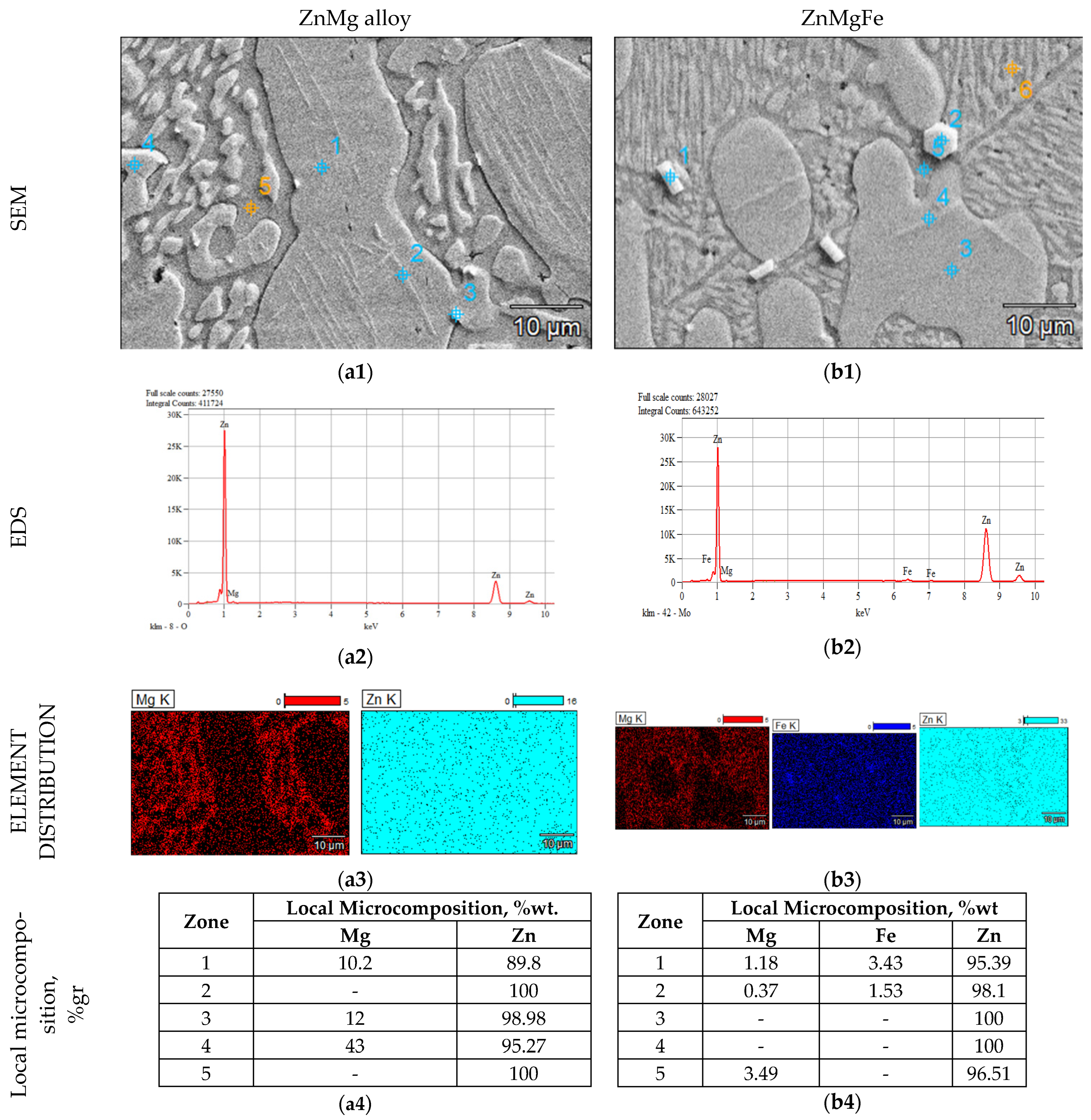
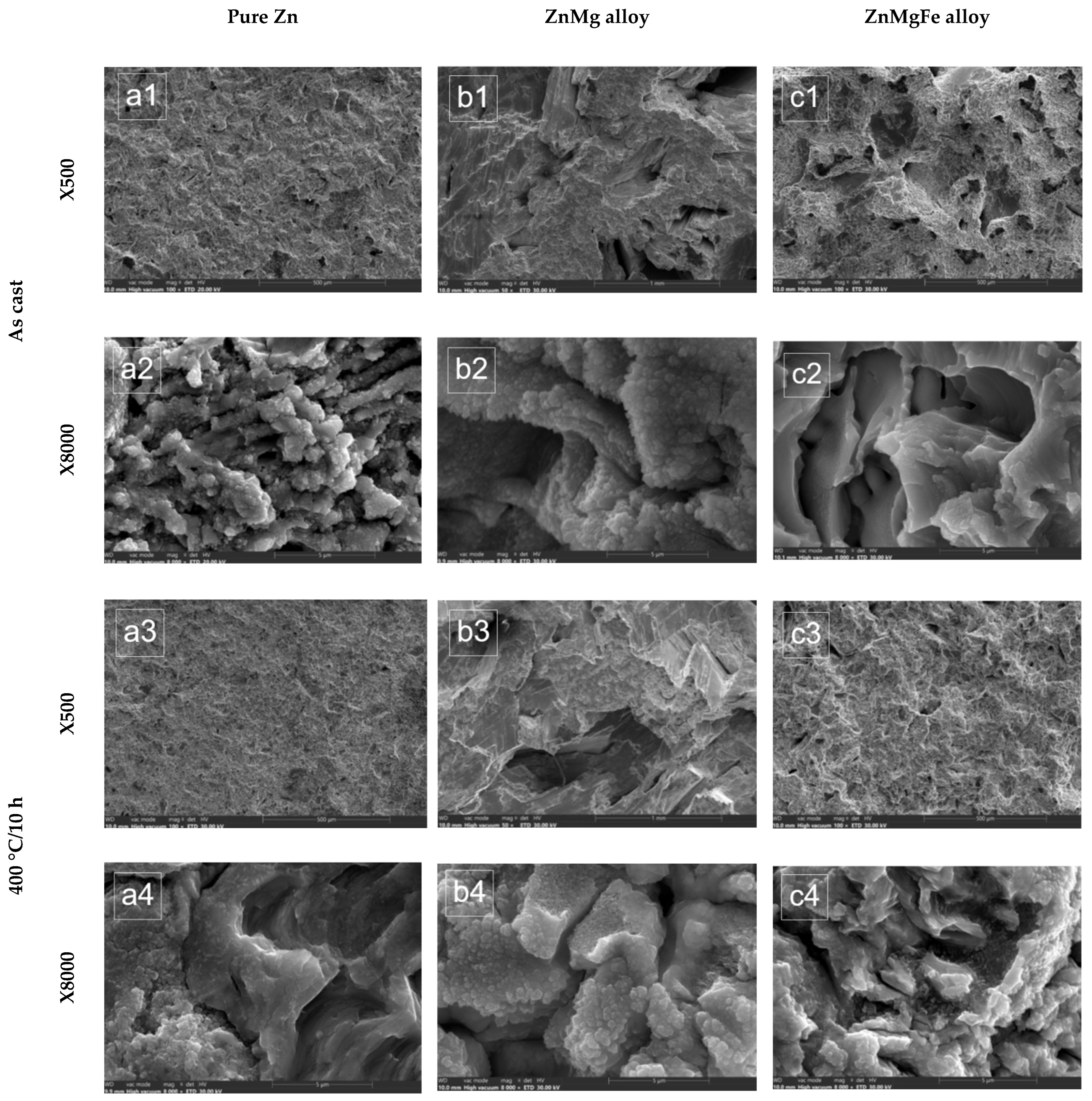
3.1.2. SEM Analysis of Alloys from the ZnMg(Fe) System

The images obtained using a scanning electron microscope to examine the ZnMg alloy are shown in Figure 3(a1–a4). These images provide a detailed representation of the structure of the alloy. Figure 3(a1) shows a large-grained structure containing intermetallic compounds with intragranular precipitation, either insular, dendritic, or needle. In the detailed image, the presence of needle compounds can be seen, either with discontinuous precipitation inside the matrix or at the grain limit. Thus, the presence of magnesium in the alloy was confirmed by the local composition over the entire surface (Figure 3(a3)), and the distribution of the elements on the surface, which was determined by the creation of the maps, indicates the nature of the zinc-based solid solution. There was magnesium in the zinc-based solid solution, according to the local chemical microcomposition table in Figure 3(a4).
A structure from the zinc-based solid solution was created by the complex alloy of zinc with magnesium iron. This structure included intermetallic compounds with various shapes and distributions, as well as a lamellar eutectic structure in the shape of a fish bone (Figure 3(b1)). The metal matrix, which consisted of a solid zinc-based solution, contained polyhedral compounds. The structural elements were revealed by the detailed analysis of the structural constituents, which is shown in Figure 3b. Therefore, the alloy’s chemical composition included the essential elements magnesium and iron as well as intermetallic compounds. The maps in Figure 3(b3) show the distribution of the alloying elements.

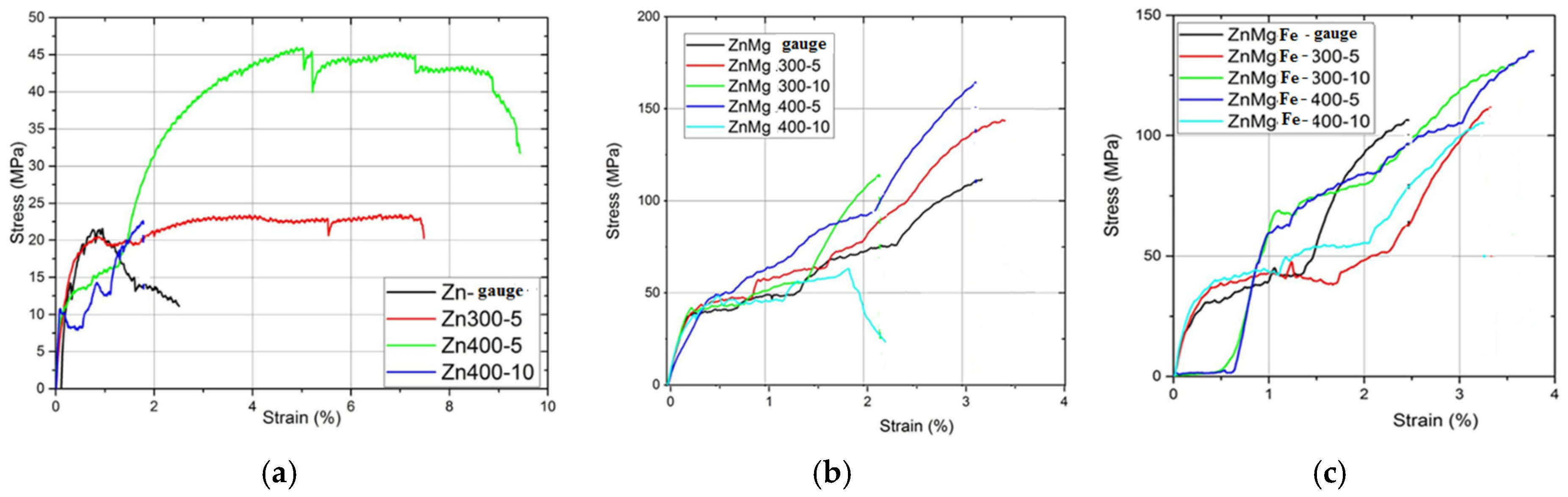
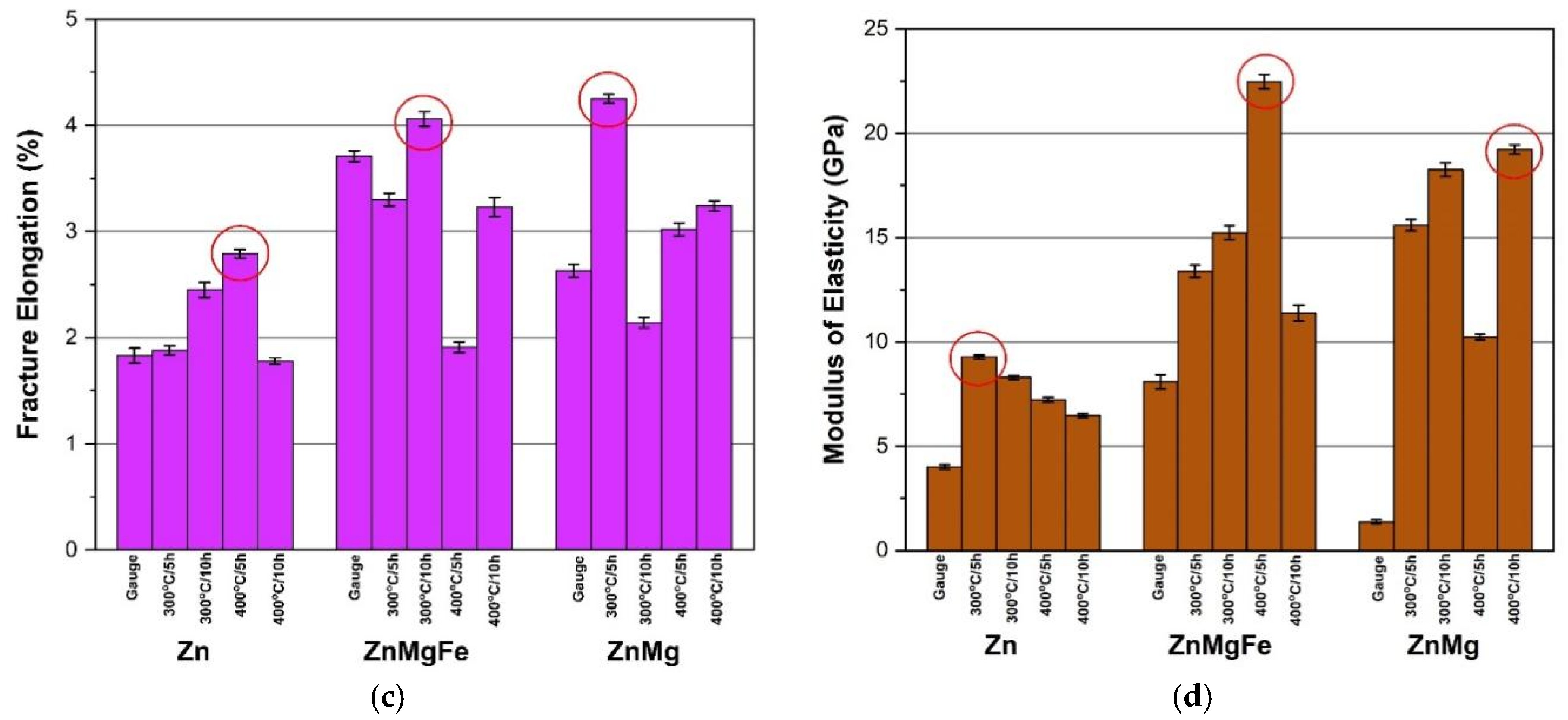
3.2. Physical–Mechanical Characterization of the Experimental Alloys

The stress–strain variation curves of the experimental alloys are shown in Figure 4. From the analysis of these stress–strain curves, it was evident that the homogenization treatment applied to pure zinc at 400 °C/5 h resulted in the best toughness, since the area under this curve was the largest compared to the other results. Similar results in terms of the toughness were observed for the other experimental alloys. Thus, the same homogenization treatment at 400 °C/5 h resulted in the maximum toughness for the ZnMg alloy and the ZnMgFe alloy.
For a complete analysis of the mechanical behavior, histograms of each mechanical characteristic were created based on the structural condition (Figure 5).
The comparative analysis of the mechanical behavior of the experimental zinc alloys, presented in the form of the histograms in Figure 5, allowed the following conclusions to be drawn. The simple alloy of zinc with magnesium led to only a moderate increase in the mechanical strength, from 130 MPa (for ZnMg) to 160 MPa (for ZnMgFe). The other mechanical characteristics were lower in the ZnMg alloy than in the ZnMgFe alloy, as shown in Figure 5b, Figure 5c (elongation at break), and Figure 5d (modulus of elasticity). The double alloy of zinc with magnesium and iron led to significant increases in the mechanical characteristics, namely in the mechanical strength, from 45 MPa (for zinc) to 130 MPa (ZnMgFe) (Figure 5a); in the yield strength, from 15 MPa (for zinc) to 67 MPa (for ZnMgFe) (Figure 5b); in the elongation at break, from 2.8% (for zinc) to 4.13% (for ZnMgFe) (Figure 5c); and in the modulus of elasticity, from 9.3 MPa (for zinc) to 22.60 GPa (for ZnMgFe) (Figure 5d). These represent increases by two, three, or four times the values for zinc. By applying different homogenization heat treatments to both pure zinc and the ZnMg(Fe) alloys, their mechanical characteristics were improved as follows.: The mechanical strength increased by up to more than three times (from 45 MPa for zinc homogenized at 400 °C/5 h to 130 MPa for the ZnMgFe alloy homogenized at 300 °C/10 h, and up to 160 MPa for the ZnMg alloy homogenized at 400 °C/10 h). The yield strength increased by up to more than four times (from 15 MPa for zinc homogenized at 400 °C/10 h to 50 MPa for the ZnMg alloy homogenized at 400 °C/5 h, and up to 67 MPa for the ZnMgFe alloy homogenized at 300 °C/10 h). The elongation increased by almost twofold (from 2.8% for zinc homogenized at 400 °C/5 h to 4.13% for the ZnMgFe alloy homogenized at 300 °C/10 h, and up to 4.18% for the ZnMg alloy homogenized at 300 °C/5 h). The modulus of elasticity increased by up to three times (from 9.3 MPa for homogenized zinc at 300 °C/5 h to 19.32 MPa for the ZnMg alloy homogenized at 400 °C/10 h, and up to 22.6 MPa for the ZnMgFe alloy homogenized at 400 °C/5 h).
Therefore, it can be concluded that a simple alloy of zinc with magnesium led to a moderate increase in the mechanical characteristics, while the double alloying of zinc with magnesium and iron led to a significant increase in the mechanical characteristics (breaking strength, yield strength, and elasticity modulus). At the same time, the most appropriate homogenization treatment applied to the ZnMgFe alloy was homogenization at 400 °C/5 h, resulting in a homogenized, but also hardened, structure. The phenomenon of hardening by precipitation can be explained by the simultaneous precipitation of intermetallic compounds in a basic solid solution of zinc.
The macrofractographic analysis of the tensile specimens, performed under the stereomicroscope using both longitudinal and cross-sectional sections, allowed the fracture surfaces to be evaluated after testing the mechanical characteristics. It also enabled a critical analysis of the fracture mode of the experimental zinc alloys compared to pure zinc in different structural states. The stereomicroscope analysis is shown in Figure 6. The detailed analysis included an initial observation for the zinc and experimental zinc alloys in the casting state. The various homogenization heat treatments resulted in brittleness, with a bright, transcrystalline, transgranular appearance (cross-section) and the zig-zagged propagation of the rupture front (longitudinal section). Thus, the zinc breakdown, regardless of the homogenization treatment applied, was transcrystalline, with a shiny appearance. When the granulation in the simple, poured state was particularly large (over 400 µm) with solidification twin boundaries (Figure 6(a1)), the granulation finished at up to 50–100 µm as the homogenization heat treatments were applied (as shown in Figure 6(a2–a5)). The fractographic aspects were similar in the homogenized specimens, differing only in the slightly modified granulation from one treatment to another.
The fractographic aspects of the tensile specimens of the ZnMg alloy are shown in Figure 6(b1–b5). In the control specimen (Figure 6(b1)), the appearance showed a sudden brittle break, with a large grain, solidification, and large differences in relief; in the homogenized specimens, there was a grainy finish and fine transcrystalline breaks, with a shiny appearance. The surfaces were mixed, with areas of maculate grains and fine areas with the abundant presence of intercrystalline compounds (Figure 6(b1,b2)). No significant differences were observed between surfaces that received different homogenization heat treatments. The macrostructural aspects of the tensile fracture surfaces of the ZnMgFe alloy specimens are shown in Figure 6(c1–c5). In this case, the surfaces showed brittle, transgranular, and transcrystalline breaks, with a shiny crystalline appearance, annealing spots, and fine areas with numerous intermetallic compounds. The fractographic aspects were similar, both in the control sample (Figure 6(c1)), with speckled grains and a shiny crystalline appearance, and in the homogenized samples (Figure 6(c2–c5)), for which no significant fractographic changes were recorded.

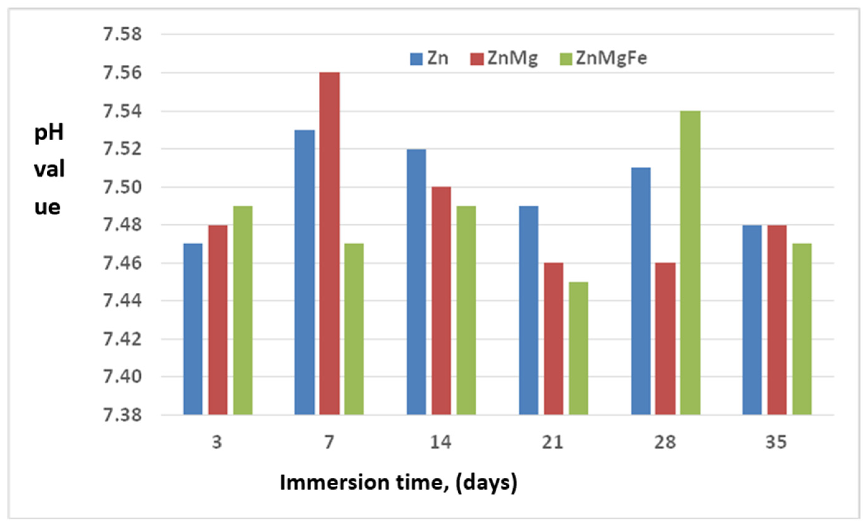
3.3. Characterization of the Biodegradability of the Experimental Alloys

The results regarding the biodegradation behavior of the experimental zinc alloys, compared to pure zinc, in simulated body fluid (SBF) are rendered in Figure 7 and Figure 8 in the form of graphs that show the variation in thickness losses, either intermediate or as an absolute value depending on the immersion duration. We tested all the alloys in the cast state. Zinc showed fairly good biodegradation behavior, with monotonic decreasing losses with low values (below 0.003 mm/year) being registered. There was a certain rate of degradation that started on the 14th day of immersion, with very low speeds (below 0.0015 mm/year). The Mg alloy of zinc demonstrated a considerable increase in the biodegradation rates, up to almost three times in order of magnitude. The behavior of this alloy was similar to that of zinc. The ZnMg alloy had the highest intermediate degradation rates, starting from 7 days, with variations in the intermediate degradation rates, from 0.007 mm/year and a continuous increase until the last test duration, reaching almost 0.002 mm/year. The intermediate degradation rates showed a continuous decreasing evolution. The ZnMgFe alloy started degrading later than pure zinc and the ZnMg alloy, and all the removal values from the seventh day of immersion were much higher than for the two metallic materials (zinc and ZnMg). Regarding the evolution of thickness losses as a function of the immersion duration (Figure 8), the following comments can be made: Zinc showed an upward evolution of thickness loss, even after 7 days of immersion. It reached a thickness loss of 1 mm/year after 35 days of immersion. The ZnMg alloy had the lowest values of absolute thickness losses. The maximum loss was reached after 7 days, and then the values decreased, being slightly higher than 0.002 mm/year after 35 days of immersion. The ZnMgFe alloy had greater thickness losses than pure zinc, reaching a total loss after 35 days of about 1 mm/year, like zinc.
It can be concluded that the simultaneous alloying of zinc with Mg and Fe results in better behavior than that of zinc, but this behavior is inferior to that of the ZnMg alloy, which demonstrated much more significant degradation.
The macrostructural aspects of the degraded surfaces with different immersion durations in SBF, and after the removal of corrosion products in the experimental alloys, are shown in Figure 9. The degradation of zinc began slowly, even after the first removal, at 3 days. The biodegradation process was slow, proceeding with the tearing and dissolving of material in small quantities, so that, after 35 days, the appearance of corrosion points was noticed on the test surfaces. These points were usually initiated on material discontinuities. The entire surface was covered with corrosion spots, but only sporadically, which is a sign of relatively slow biodegradation (Figure 9a). In the ZnMgFe alloy (Figure 9c), the same slow biodegradation process was noted, with the deepening of the degradation zones slowly and continuously until the last removal time of 35 days. In addition, biodegradation initially occurred on the discontinuity areas of exposed surfaces and then became more aggressive until the last immersion duration. The appearance of degraded areas after 35 days, highlighted in Figure 9c, showed the development of areas with localized degradation, with large depths of up to 0.05 mm. The ZnMg alloy showed a behavior similar to that of zinc, as can be seen in Figure 9b. Biodegradation began very slowly, from the first removal at 3 days, and then continued progressively and slowly until the last removal after 35 days. An interconnected network of corrosion points with relatively shallow depths is also noteworthy.
An analysis of the variation in the pH of the SBF simulant solution after the different immersion periods, which is shown in Figure 10, revealed the following.: Zinc resulted in the lowest pH values, between 7.45 and 7.56. The highest pH values were observed for the ternary alloy, ZnMgFe, with values in the range of 7.45–7.54. For the binary alloy, ZnMg, values intermediate to the values of zinc and those of the ternary alloy were obtained, in the range of 7.46 ÷ 7.56. It was noted that the highest pH value was obtained after 7 days of immersion, which was 7.56, and the lowest value was obtained at an extraction of 28 days, which was 7.46.
A comparison of the experimental results of this study with data from the literature confirmed that the biodegradation behavior of the new zinc-based alloys proposed in this study was modified. At the same time, the values obtained were comparable and even unexpected for the complex alloys chosen in this paper. Thus, regardless of the alloying mode, the degradation of either zinc or various zinc-based alloys occurs in human-simulated environments [32]. The simple alloy of zinc with magnesium resulted in the significant biodegradation of the alloy from the seventh day of immersion. The appearance of degraded areas after 35 days showed the development of localized degradation areas, with large depths of up to 0.05 mm. The absolute degradation rate of the alloy reached slightly more than 0.002 mm/year after 35 days of immersion. The simultaneous alloying of zinc with magnesium and iron yielded a behavior similar to that of pure zinc. Biodegradation began very slowly, from the first removal at 3 days, and continued progressively and slowly until the last removal after 35 days. An interconnected network of corrosion points with relatively shallow depths is also noteworthy. The ZnMgFe alloy showed greater thickness losses than pure zinc, reaching a total loss after 35 days of about 1 mm/year, like zinc [33,34].

3.4. Characterization of the Erosion Cavitation Behavior of the Experimental Alloys

As mentioned above, to analyze the cavitational erosion behavior of the experimental alloys in the ZnMg(Fe) system, diagrams were used that contained the experimental values of the three samples (red, green, black, and blue dots), tested for each heat treatment state, and the specific mediation curves, which gave the variation in the average cumulative erosion depth MDE(t) and velocity MDER(t). Therefore, the cavitational erosion behavior was evaluated using the dispersion of the experimental values and the evolution of the specific mediation curves shown in Figure 11, MDE(t) and MDER(t), as recommended by the ASTM G32-2016 international standards. The analytical relationships for the construction of these curves, whose coefficients A and B were statistically determined, were established by Bordeasu [26,29] and have the following forms:
-
For the cumulative average of erosion depth,
MDE (t) = A·t·(1 − e−B·t) or MDE(t) = A·t·(1 + e−B·t)
-
For the average erosion rate,
MDER(t) = A·(1 − e−B·t) + A·B·t·e−B·t
An analysis of the data from the diagrams presented in Figure 11 showed that the investigated zinc alloys had similar behaviors, characteristic of the type of alloy and with differences dictated by its condition (with or without volume heat treatment). The data showed the same erosion mechanisms specific to alloys with a low cavitational erosion behavior, but much differentiated from that of other classes of alloys and composite materials due to the mechanical properties and the way of breaking under static and dynamic stresses [29,35,36,37,38,39]. An analysis of the data in these charts showed the following:
-
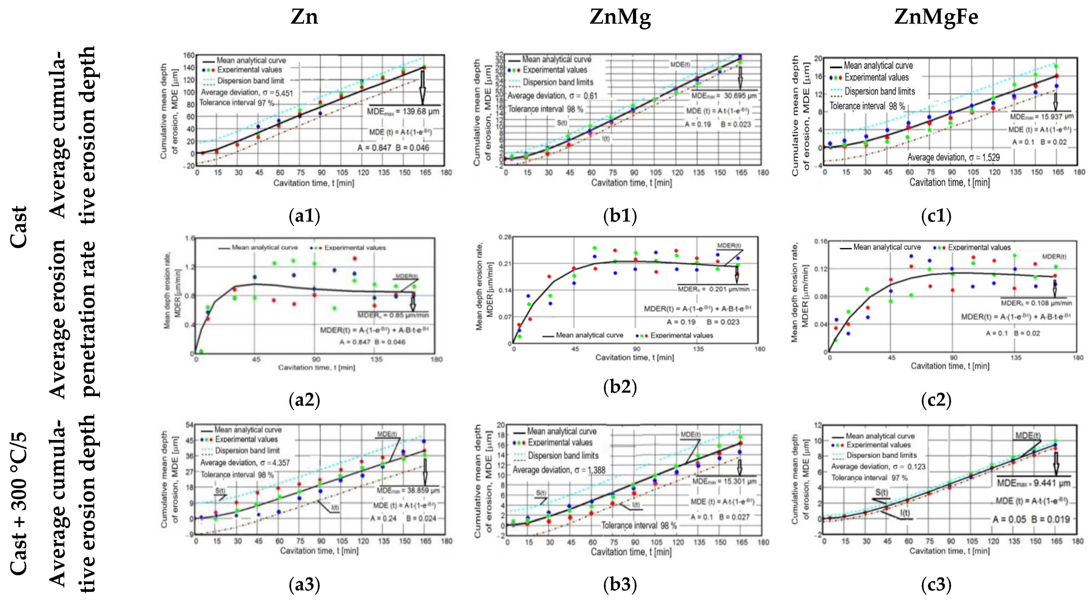
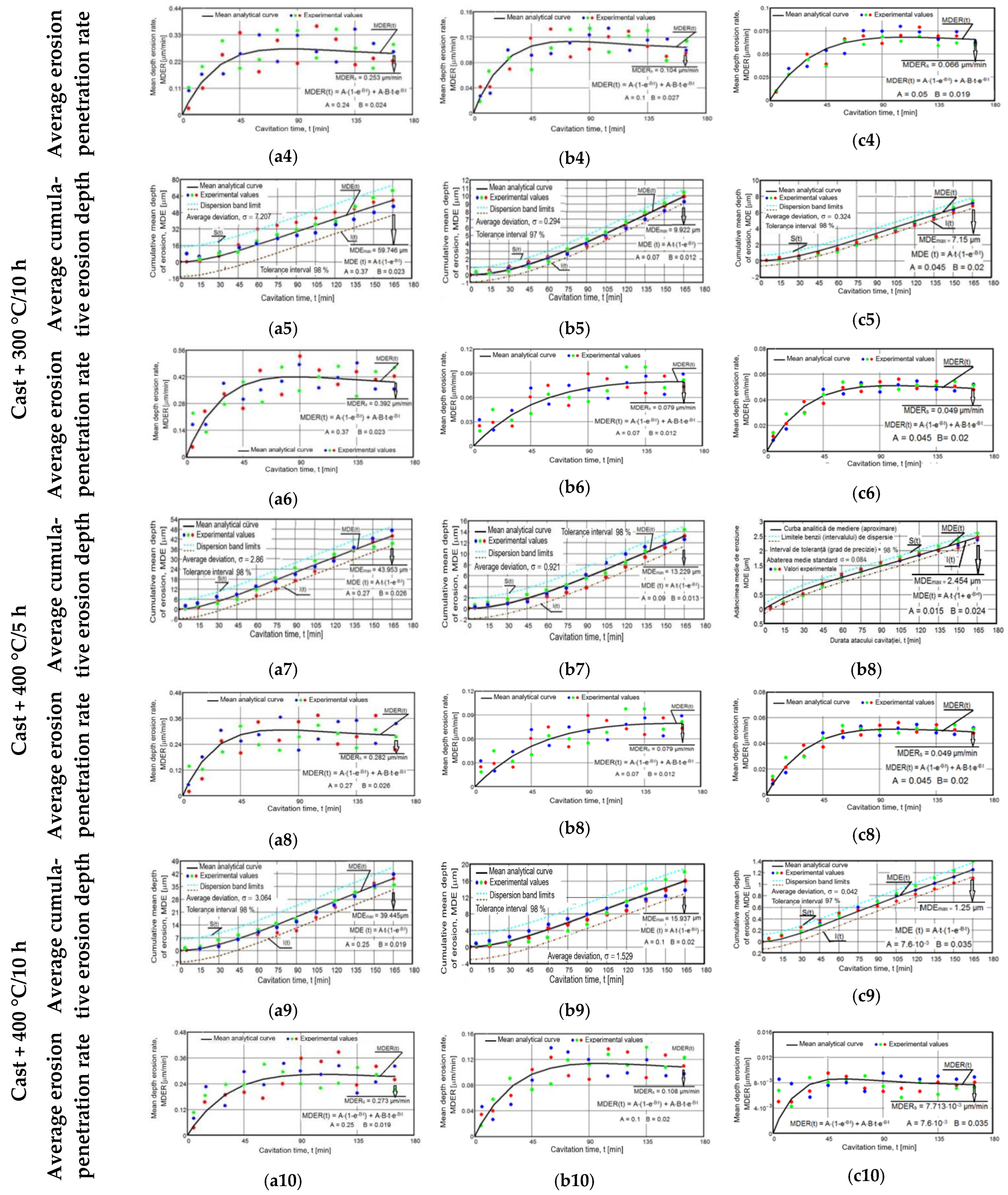
The most significant material losses developed after 45–60 min, and after 120 min, they increased approximately linearly/constantly, with small differences between successive values, as is shown in the erosion depth diagrams (Figure 11(a1,a3,a5,a7,a9,b1,b3,b5,b7,b9,c1,c3,c5,c7,c9)).
-
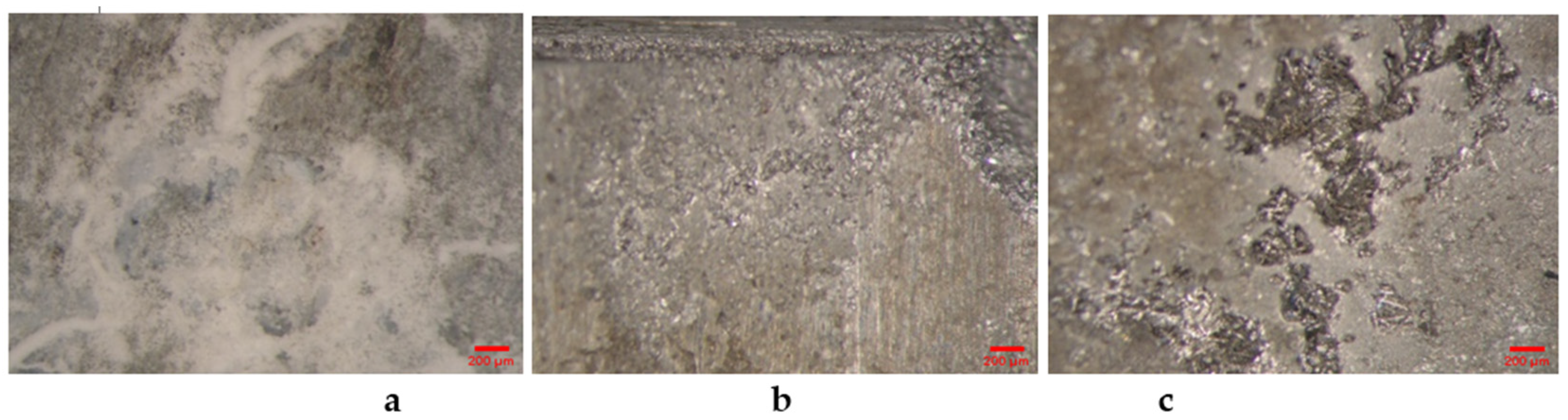
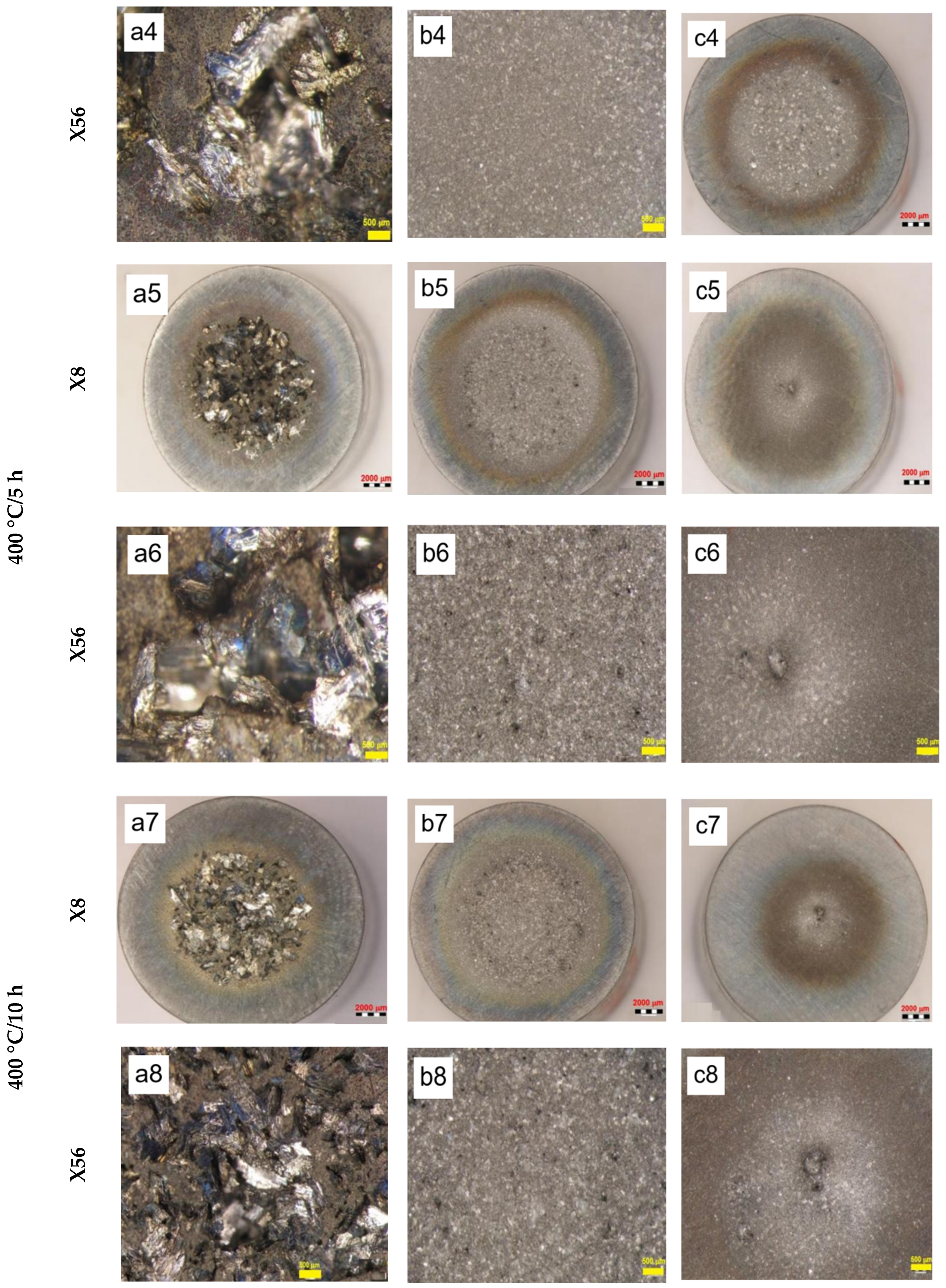
There were large differences between the experimental values of the MDE and MDER parameters obtained for the three samples in the same state and after the same duration of exposure to cavitation attack. In addition, Figure 11(a2,a4,a6,a8,a10,b2,b4,b6,b8,b10,c2,c4,c6,c8,c10) show that there were increases and decreases in the values obtained for the erosion velocities (MDER) during cavitation. These developments led to an irregular dispersion from the averaging curve. These aspects were also clarified by the stereomacroscopic and SEM images in Figure 12; through the dimensions and shapes of the caverns, as well as through the connections between them, these figures show the dimensions and geometries of the ejected grains, as well as the types of breaks generated by the cyclic stress of cavitation fatigue, which was dependent on the type of microstructure and the value of the mechanical properties.
-
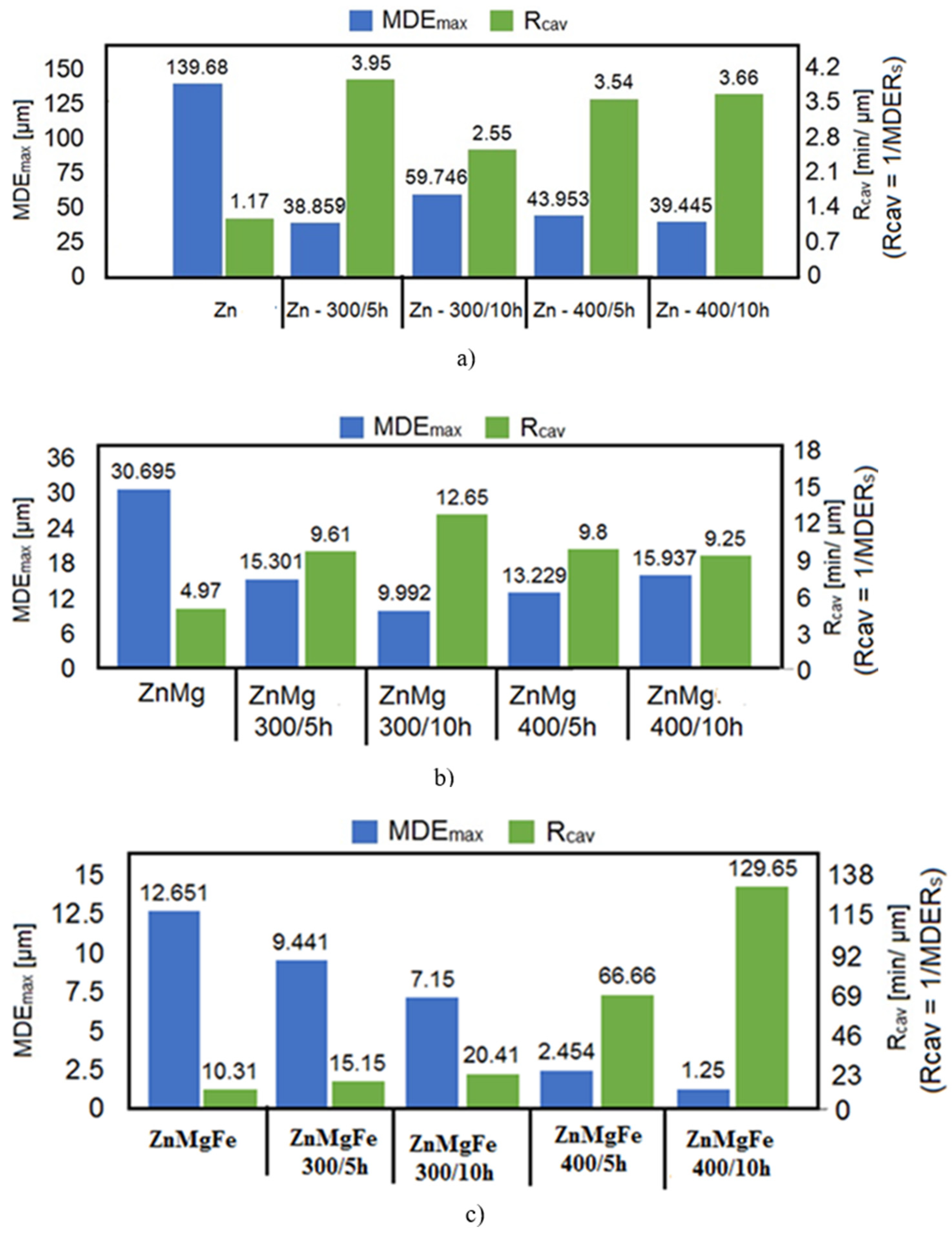
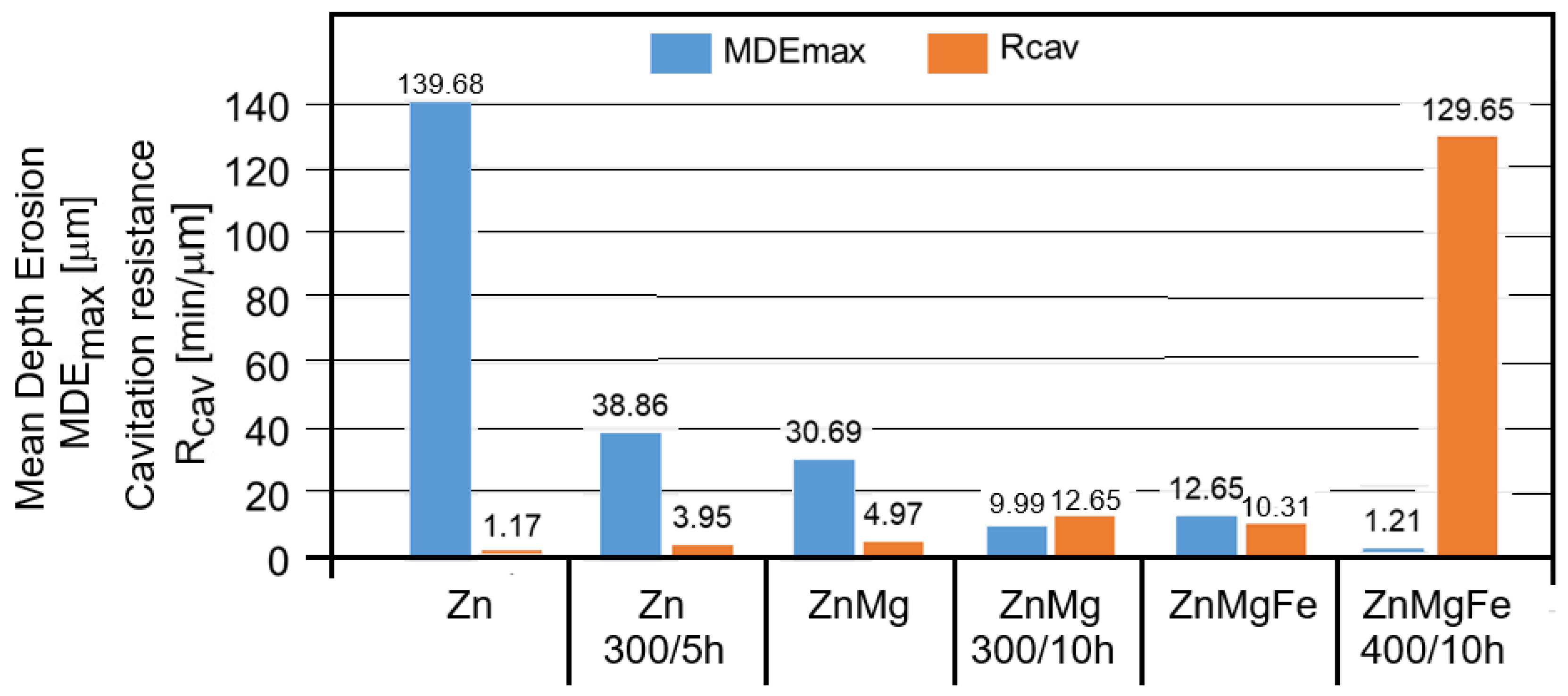
The appearance of the asymptotic evolution of the MDE curves, with linearization for a certain duration of the cavitation attack, was similar in all specimens, with differences in terms of the final values. For the different structural states of zinc, the values of the maximum cumulative cavitational erosion penetration depth were the highest, at 139 μm in the end (for casting) and in the range of 40–60 μm (for homogenized specimens). In the ZnMg alloy, the maximum cavitational erosion depth was 30 μm and it reached about 13–15 μm in the homogenized specimens. In the ternary alloy, ZnMgFe, the maximum cumulative cavitational erosion penetration depth was 15 μm in the cast specimens, and it reached the lowest values in the homogenized specimens, in the range of 2–12 μm. This mode of evolution was specific to the alloy, but the differences in the slope of the linear area are given by the values of the mechanical properties. According to studies in this field [33,35,40,41], the decrease in the slope and the increase in the resistance of the structure to cavitation stresses are specific to the alloy state, with high values for the ultimate strength, yield strength, modulus of elasticity, and Brinell hardness and low values for elongation.
-
The stabilization of the MDER curves began very early in zinc (after 45 min; Figure 11(a2)); in the range of 45–60 min in homogenized zinc (Figure 11(a3–a10)); much later for ZnMg alloys, after 60 min (Figure 11(b3–b10)); and right from minute 90 in the ZnMgFe alloy (Figure 11(c3–c10)). This stabilization shows that the stressed layer was mechanically hardened by impact with shock waves and cavitational microjets, and that the pressure force was dampened by the water and air that penetrated into the formed caverns [33,35,40,41,42].
The macrostructural analysis of the cavitationally eroded surfaces of pure, untreated zinc (Figure 12a,b) revealed a rough surface, with relatively homogeneous material tears and a brittle appearance. The image in Figure 12b highlights the propagation of the transcrystalline rupture front, with a stratified appearance. The application of aging heat treatment caused a slight change in the appearance of the cavitationally eroded surfaces; the rough appearance of these surfaces was preserved, highlighting small cavities evenly distributed on the surface (Figure 12c–h).
The images in Figure 12(b1–b8) show that the alloy of zinc with magnesium (ZnMg alloy) exhibited a modified appearance of the cavitationally eroded surfaces, compared to both that of zinc (Figure 12(a1–a8)) and that of the ZnMgFe alloy (Figure 12(c1–c8)). Thus, the macrostructural images (Figure 12b) showed a mixed appearance, with two areas: an area with terraced surfaces and fine, intergranular cleavage and an area with extensive cleavage over a larger area, generating large and deep cavities. The images in Figure 12(b2–b8) show the effect of the aging treatment applied to the binary alloy ZnMg, with the maintenance of the intergranular cleavage appearance of the affected surfaces by the cyclic stresses of cavitational microjets. Thus, a mixed terraced surface was observed, with large cavities along with cleavage areas with different crystallographic orientations, and with cleavage planes of fine cavitations delimited by fine, intergranular cracks.
The fractographic aspects of the cavitationally eroded surfaces of the biodegradable zinc alloy ZnMgFe are suggestively reproduced in Figure 12(c1–c8). The macroscopic appearance of the surface eroded by cavitation comprised deep cavitations with uneven diameters, from a few microns to 300–400 μm (Figure 12(c1,c2)). The effect of the aging heat treatment on the cavitation erosion behavior of the molded ZnMgFe alloy specimen revealed small punctiform cavities evenly distributed on the surface, strongly carved into the cavity surface structure, resulting from the fine intergranular propagation of the fractured front (Figure 12(c3–c8)).
The fractographic analysis of the experimental biodegradable zinc alloys, in different structural states and after 165 h of immersion, highlighted the erosion of the surfaces and is shown in Figure 13. The results of this analysis allowed for the formulation of the following aspects.
The macrostructural analysis of the cavitationally eroded surfaces of pure zinc (Figure 3(a1,a2)) revealed a rough surface, with relatively homogeneous material tears and a brittle appearance. In the microstructural analysis at different magnification powers, the propagation of the rupture front was highlighted as being transcrystalline, with a stratified appearance, intergranular propagation, and the existence of numerous interconnected cracks with a specific fragile appearance. At the highest magnification power, 8000 times (Figure 13(a2)), the propagation of cracks on the sliding lines was observed, with the existence of fine cracks along the mashed lines.
By applying an aging heat treatment to the cast zinc specimens, a slight change in the appearance of the cavitationally eroded surfaces was noticed, which is illustrated in Figure 13. In the macrostructural analysis, the rough appearance of the cavitationally eroded surface was preserved (Figure 13(a4)), with the highlighting of small cavities evenly distributed on the surface. In the microstructural analysis, it was possible to highlight the propagation of the rupture front, with an intercrystalline, stratified appearance; the rupture front showed massive intergranular propagation with the existence of numerous interconnected cracks, a specific fragile appearance, and the existence of large cavities with different diameters.
The magnesium alloy of zinc, ZnMg, demonstrated a modified appearance of its cavitationally eroded surfaces compared to those of both zinc and the ZnMgFe alloy. In the macrostructural analysis (Figure 13(b1)), the appearance was mixed, with two areas: an area with terraced surfaces and fine, intergranular cleavage and an area with extensive cleavage over a larger area, generating large and deep cavities. The microstructural analysis highlighted the alternation of these two areas—of fine, intergranular cleavage and extended cleavage—with a terraced appearance, delimited by fine secondary fissures. In the detailed image in Figure 13(b2), you can see the walls of the cavities, with a relief appearance, generated by the numerous and finely precipitated compounds. The application of the aging treatment to the binary ZnMg alloy maintained the appearance of intergranular cleavage to cavitationally eroded surfaces. In the macrostructural analysis (Figure 13(b3)), a mixed terraced surface was observed, with large cavities along with cleavage areas with different crystallographic orientations. The detailed analysis of the cleavage zones highlighted the existence on flat cleavage surfaces of fine cavitations delimited by fine, intergranular cracks, which generated fine cavities with different diameters and depths. In the analysis of the fine structure (Figure 13(b4)), the presence of interstitial compounds on the ridges of the cavitations and the secondary intergranular cracks was noted, maintaining the fragile appearance of the fracture.
In summary, the careful observation of these images allowed the following assessments to be made. The attack on the cast zinc started with the formation of visible pinches from the very first minutes, and they deepened according to the attack time. In all the pure zinc specimens, surface erosion had similar appearances, with sharp, faceted, and shiny pinches—a sign of brittle behavior. The magnesium alloy of zinc (the ZnMg samples) displayed the formation of finely dispersed caverns on the surface, and towards the end, the color of the attacked areas began to brown. Simultaneous alloying with magnesium and iron (the ZnMgFe samples) resulted in the formation of the first cavities after a longer duration of exposure to cavitation, after about 60 min; with an increase in the duration, and until the completion of the test, a yellow/blue ring formed on the cavity surface. The final appearance of the cavitationally eroded surfaces depended on the alloying mode, but also on the condition of the material. For pure zinc, the final appearance was rough, with deep caverns on large surfaces; homogenization at 400 °C for either 5 or 10 h reduced the surface affected by the cavitational attack. For the ZnMg alloy, the final appearance was finely rough, with caverns distributed on the attacked surface; homogenization at 300 °C/5 h reduced the surface affected by the cavitational attack. For the ZnMgFe alloy, the final appearance was finely rough, with caverns distributed relatively homogeneously on the attacked surface; homogenization at 400 °C/10 h considerably reduced the surface affected by the cavitational attack.
To evaluate the cavitation resistance, the histograms in Figure 14 were constructed. These diagrams used the parameters MDEmax and Rcav = 1/MDERs (known as the cavitation resistance [26,28]) recommended by the ASTM G32-2016 standards [25].
Figure 14a shows the beneficial effect of the homogenization heat treatment on the cavitation behavior of pure zinc. According to the data, the highest resistance to cavitation stresses was obtained by homogenizing at 300 °C/5 h. The increase was significant compared to the resistance of the pure zinc state (by over 3.3 times); was more than 55% compared to the 300 °C/10 h state; and was insignificant compared to the 400 °C/5 h (by about 12%) and 400 °C/10 h (by about 8%) states. Figure 14b shows the beneficial effect of the homogenization heat treatment on the cavitation behavior of the ZnMg alloy. According to the data, the highest resistance to cavitation stresses was obtained by homogenization at 400 °C/10 h. The increase was significant compared to the casting state (by over 12.5 times), but also compared to the 300 °C/5 h (by over 8.5 times) and 300 °C/10 h (by over 6.3 times) states. An important increase was also observed when comparing to the 400 °C/5 h state, but this increase was substantially lower compared to the other three, by about 94%. Figure 14c shows the effect of the homogenization heat treatment on the cavitation behavior of the ZnMgFe alloy. According to the data, the highest resistance to cavitation stresses was obtained by homogenizing at 300 °C/10 h. The increase was significant, by about 2.5 times, compared to the control sample of the alloy without homogenization. The differences in strength between the structures of the 300 °C/5 h, 400 °C/5 h, and 400 °C/10 h states were very small (2–6%), but they were significantly smaller, by 31–37%, compared to the 300 °C/10 h state.
The comparative analysis of the results regarding the behavior of the experimental ZnMgFe alloy specimens in different structural states to cavitational erosion is shown in Figure 15. The analysis of the data from the histograms presented in Figure 15 showed that, among the molded states without homogenization heat treatments, the ZnMgFe alloy had the highest strength. Its increase compared to pure zinc was by 9–11 times, depending on the parameter being evaluated, and the increase compared to the binary alloy ZnMg was by 2–2.4 times. In addition, among the states that received homogenization heat treatment, the highest resistance was presented by the ZnMgFe alloy homogenized at 400 °C/10 h. Its increase compared to pure zinc homogenized at 300 °C/5 h was by 32–33 times, depending on the parameter being evaluated, and compared to the binary alloy ZnMg homogenized at 300 °C/10 h, the increase was by 8–10 times.

4. Conclusions

This study investigated the modification of the structure and properties of some alloys in the ZnMg(Fe) system by applying different homogenization heat treatments. The structure of these alloys was heavily modified compared to that of the pure form, from grains with twin boundaries (in pure zinc) to the dendritic structure of zinc alloys, with a eutectic mechanical mixture and intermetallic compounds of the Mg2Zn11 type and/or polyhedral FeZn13 compounds. The different homogenization heat treatments applied to both alloys may cause the disappearance of dendrites. The mechanical characteristics of the alloys from the ZnMg(Fe) system were strongly dependent on both the alloying mode and the homogenization treatment applied. The most appropriate homogenization treatment applied to the ZnMgFe alloy was homogenization at 400 °C/5 h, with a homogenized structure while preserving the same hardened behavior. The biodegradability of the ZnMgFe alloys in SBF showed that the simultaneous alloying of zinc with Mg and Fe caused better behavior than that of zinc. However, this behavior was still inferior to that of the ZnMg alloy, which exhibited much more significant degradation. The cavitation behavior of these alloys was better than that of pure zinc. The magnesium alloy resulted in an increase in the cavitation resistance of up to 7 times that of zinc, while the simultaneous alloying with magnesium and iron led to an increase in the cavitation resistance by more than 100 times that of zinc. The obtained experimental results demonstrate that the new alloys from the ZnMgFe system are a good option for manufacturing biodegradable implants from the perspective of functional in vitro properties. Future studies on their biocompatibility and the functional implant properties of manufactured implants from ZnMgFe alloys must be conducted in order to confirm our hypothesis.

Author Contributions

Methodology, A.A., C.M.G. and D.B.; Software, D.B.; Validation, D.B.; Formal analysis, L.M.M., C.G. and B.F.; Investigation, I.B., A.A., I.A., C.M.G., C.G., L.C.S. and A.M.F.; Resources, L.C.S.; Data curation, C.M.G., L.M.M. and D.O.; Writing—original draft, I.B. and I.A.; Writing—review and& editing, B.G.; Supervision, B.G. All authors have read and agreed to the published version of the manuscript.

Funding

This research received no external funding.

Data Availability Statement

The original contributions presented in this study are included in the article. Further inquiries can be directed to the corresponding authors.

Conflicts of Interest

The authors declare no conflicts of interest.

References

  1. Niinomi, M. Recent metallic materials for biomedical applications. Metall. Mater. Trans. 2002, 33, 477–486. [Google Scholar] [CrossRef]
  2. Love, B.J. Biomaterials: A Systems Approach to Engineering Concepts, 1st ed.; Academic Press: London, UK, 2017. [Google Scholar]
  3. Oliveira, N.T.C.; Guastaldi, A.C. Electrochemical stability and corrosion resistance of Ti–Mo alloys for biomedical application. Acta Biomater. 2009, 5, 399–405. [Google Scholar] [CrossRef] [PubMed]
  4. Yoda, K.; Suyalatu; Takaichi, A.; Nomura, N.; Tsutsumi, Y.; Doi, H.; Kurosu, S.; Chiba, A.; Igarashi, Y.; Hanawa, T. Effects of chromium and nitrogen content on the microstructures and mechanical properties of as-cast Co–Cr–Mo alloys for dental applications. Acta Biomater. 2012, 8, 2856–2862. [Google Scholar] [CrossRef] [PubMed]
  5. Manam, N.; Harun, W.; Shri, D.N.A.; Ghani, S.A.C.; Kurniawan, T.; Ismail, M.; Ibrahim, M. Study of corrosion in biocompatible metals for implants: A review. J. Alloys Compd. 2017, 701, 698–715. [Google Scholar] [CrossRef]
  6. Biesiekierski, A.; Wang, J.; Abdel-Hady Gepreel, M.; Wen, C. A new look at biomedical Ti-based shape memory alloys. Acta Biomater. 2012, 8, 1661–1669. [Google Scholar] [CrossRef]
  7. Hermawan, H. Biodegradable Metals: From Concept to Applications; Springer Science & Business Media: Johor, Malaysia, 2012. [Google Scholar]
  8. Toong, D.W.Y.; Ng, J.C.K.; Huang, Y.; Wong, P.E.H.; Leo, H.L.; Venkatraman, S.S.; Ang, H.Y. Bioresorbable metals in cardiovascular stents: Material insights and progress. Materialia 2020, 12, 100727. [Google Scholar] [CrossRef]
  9. Zhang, S.; Zhang, X.; Zhao, C.; Li, J.; Song, Y.; Xie, C.; Tao, H.; Zhang, Y.; He, Y.; Jiang, Y.; et al. Research on an Mg–Zn alloy as a degradable biomaterial. Acta Biomater. 2010, 6, 626–640. [Google Scholar] [CrossRef]
  10. Antoniac, I.; Miculescu, M.; Mănescu, V.; Stere, A.; Quan, P.H.; Păltânea, G.; Robu, A.; Earar, K. Magnesium-Based Alloys Used in Orthopedic Surgery. Materials 2022, 15, 1148. [Google Scholar] [CrossRef]
  11. Dambatta, M.S.; Murni, N.; Izman, S.; Kurniawan, D.; Froemming, G.; Hermawan, H. In vitro degradation and cell viability assessment of Zn–3Mg alloy for biodegradable bone implants. Proc. Inst. Mech. Eng. H J. Eng. Med. 2015, 229, 335–342. [Google Scholar] [CrossRef]
  12. Kafri, A.; Ovadia, S.; Goldman, J.; Drelich, J.; Aghion, E. The suitability of Zn-1.3%Fe alloy as a biodegradable implant material. Metals 2018, 8, 153. [Google Scholar] [CrossRef]
  13. Wang, C.; Yang, H.T.; Li, X.; Zheng, Y.F. In vitro evaluation of the feasibility of commercial Zn alloys as biodegradable metals. J. Mater. Sci. Technol. 2016, 32, 909–918. [Google Scholar] [CrossRef]
  14. Zhang, Y.; Yan, Y.; Xu, X.; Lu, Y.; Chen, L.; Li, D.; Dai, Y.; Kang, Y.; Yu, K. Investigation on the microstructure, mechanical properties, in vitro degradation behavior and biocompatibility of newly developed Zn-0.8%Li-(Mg, Ag) alloys for guided bone regeneration. Mater. Sci. Eng. C 2019, 99, 1021–1034. [Google Scholar] [CrossRef] [PubMed]
  15. Bakhsheshiad, H.R.; Hamzah, E.; Low, H.T.; Cho, M.H.; Kasirisgarani, M.; Farahany, S.; Mostafa, A.; Medraj, M. Thermal characteristics, mechanical properties, in vitro degradation and cytotoxicity of novel biodegradable Zn–Al–Mg and Zn–Al–Mg–xBi alloys. Acta Metall. Sin-Engl. 2017, 30, 201–211. [Google Scholar] [CrossRef]
  16. Venezuela, J.; Dargusch, M.S. The influence of alloying and fabrication techniques on the mechanical properties, biodegradability and biocompatibility of zinc: A comprehensive review. Acta Biomater. 2019, 87, 1–40. [Google Scholar] [CrossRef]
  17. Shuai, C.; Li, S.; Peng, S.; Feng, P.; Lai, Y.; Gao, C. Biodegradable metallic bone implants. Mater. Chem. Front. 2019, 3, 544–562. [Google Scholar] [CrossRef]
  18. Virtanen, S. Biodegradable Mg and Mg alloys: Corrosion and biocompatibility. Mater. Sci. Eng. B 2011, 176, 1600–1608. [Google Scholar] [CrossRef]
  19. Levy, G.; Goldman, J.; Aghion, E. The prospects of zinc as a structural material for biodegradable implants-a review paper. Metals 2017, 7, 402. [Google Scholar] [CrossRef]
  20. Han, K.; Ohnuma, I.; Okuda, K.; Kainuma, R. Experimental determination of phase diagram in the Zn-Fe binary system. J. Alloys Compd. 2018, 737, 490–504. [Google Scholar] [CrossRef]
  21. Yue, R.; Niu, J.; Li, Y.; Ke, G.; Huang, H.; Pei, J.; Ding, W.; Yuan, G. In vitro cytocompatibility, hemocompatibility and antibacterial properties of biodegradable Zn-Cu-Fe alloys for cardiovascular stents applications. Mater. Sci. Eng. C 2020, 113, 111007. [Google Scholar] [CrossRef]
  22. Yue, R.; Huang, H.; Ke, G.; Zhang, H.; Pei, J.; Xue, G.; Yuan, G. Microstructure, mechanical properties and in vitro degradation behavior of novel Zn-Cu-Fe alloys. Mater. Char. 2017, 134, 114–122. [Google Scholar] [CrossRef]
  23. Shi, Z.-Z.; Gao, X.-X.; Chen, H.-T.; Liu, X.-F.; Li, A.; Zhang, H.-J.; Wang, L.-N. Enhance-ment in mechanical and corrosion resistance properties of a biodegradable Zn-Fe alloy through second phase refinement. Mater. Sci. Eng. C 2020, 116, 111197. [Google Scholar] [CrossRef] [PubMed]
  24. Guan, Z.; Linsley, C.S.; Pan, S.; DeBenedetto, C.; Liu, J.; Wu, B.M.; Li, X. Highly ductile Zn-2Fe-WC nanocomposite as biodegradable. Mater. Metall. Mater. Trans. A 2020, 51, 4406–4413. [Google Scholar] [CrossRef] [PubMed]
  25. ASTM, Standard G32; Standard Method of Vibratory Cavitation Erosion Test. ASTM: West Conshohocken, PE, USA, 2016.
  26. Bordeasu, I. Monografia Laboratorului de Cercetare a Eroziunii prin Cavitatie al Universitatii Politehnica Timisoara (1960–2020); Editura Politehnica: Timişoara, Romania, 2020. [Google Scholar]
  27. Bordeaşu, I.; Mitelea, I.; Salcianu, L.; Craciunescu, C.M. Cavitation Erosion Mechanisms of Solution Treated X5CrNi18-10 Stainless Steels. J. Tribol.-Trans. ASM 2016, 138, 031102. [Google Scholar] [CrossRef]
  28. Bordeasu, I.; Ghera, C.; Luca, A.-N.; Manea, A.-S.; Stroita, D.-C.; Salcianu, L.C.; Ghiban, B.; Micu, L.M. Investigation of Cavitation Resistance of Biocompatible Zinc-Based Alloys for Biomedical Applications. In International Conference on Machine and Industrial Design in Mechanical Engineering; Springer Nature: Cham, Switzerland, 2024; pp. 272–281. [Google Scholar]
  29. Bordeaşu, I.; Patrascoiu, C.; Badarau, R.; Sucitu, L.; Popoviciu, M.O.; Balasoiu, V. New contributions to cavitation erosion curves modeling. FME Trans. 2006, 34, 39–43. [Google Scholar]
  30. Ciungu, G.; Micu, L.M.; Bordeasu, I.; Iordache, C.M.; Ghiban, B.; Ghera, C. Research of the Cavitation Resistance of a Biodegradable Alloy Zn-Cu. U.P.B. Sci. Bull. Series B 2024, 86, 247–260. [Google Scholar]
  31. Iordache, C.M.; Luca, A.N.; Bordeasu, I.; Ciungu, G.; Ghiban, B. Cavitational Erosion Behavior of a Biodegradable Alloy from the Zn-Mg System for Biomedical Applications. Tribol. Ind. 2024, 46, 315–323. [Google Scholar] [CrossRef]
  32. Rau, J.V.; Antoniac, I.; Filipescu, M.; Cotrut, C.; Fosca, M.; Nistor, L.C.; Birjega, R.; Dinescu, M. Hydroxyapatite Coatings on Mg-Ca Alloy Prepared by Pulsed Laser Deposition: Properties and Corrosion Resistance in Simulated Body Fluid. Ceram. Int. 2018, 44, 16678–16687. [Google Scholar] [CrossRef]
  33. Iordache (Gheorghe), C.M. Correlation Between Structure-Mechanical Characteristics-Biodegradability- Cavitation of Some Biomedical Alloys form the System ZnMg(Fe). Ph.D. Thesis, National University of Science and Technology Politehnica Bucharest, Bucharest, Romania, 2024. (in Romanian). [Google Scholar]
  34. Ciungu, G. Experimental Studies on the Cavitation and Biodegradation Behaviour of Some Alloys of the System ZnCu(Mg) System. Ph.D. Thesis, University Politehnica Bucharest, Bucharest, Romania, 2024; pp. 247–260. [Google Scholar]
  35. Ghiban, B.; Antoniac, A.; Bordeasu, I.; Antoniac, I.; Petre, G.; Rau, J.; Bordeasu, D.; Micu, L.M. Biodegradability and Cavitation Erosion Behavior of Some Zinc Alloys from the System ZnCuMg. Metals 2025, 15, 161. [Google Scholar] [CrossRef]
  36. Cerbu, C.; Ursache, S.; Botis, M.F.; Hadar, A. Simulation of the Hybrid Carbon-Aramid Composite Materials Based on Mechanical Characterization by Digital Image Correlation Method. Polymers 2021, 13, 4148. [Google Scholar] [CrossRef]
  37. Ursache, S.; Cerbu, C.; Hadar, A. Characteristics of Carbon and Kevlar Fibres, Their Composites and Structural Applications in Civil Engineering-A Review. Polymers 2024, 16, 127. [Google Scholar] [CrossRef]
  38. Gheorghe, A.; Hadar, A.; Apostolescu, Z.; Amza, C.G.; Anton, L. Determination through numerical computation of the designed and functional parameters of ultra acustic systems for ultrasonic welding of intelligent composite materials, 2007. Mat. Plast. 2007, 44, 121–128. [Google Scholar]
  39. Cormos, R.; Petrescu, H.; Hadar, A.; Gheorghiu, H. Finite Element Analysis of the Multilayered Honeycomb Composite Material Subjected to Impact Loading. Mat. Plast. 2017, 54, 180–185. [Google Scholar] [CrossRef]
  40. ASTM STP 408; Experience with a 20—KC Cavitations Erosion Test, Erosion by Cavitations or Impingement. ASTM: Atlantic City, NJ, USA, 1960.
  41. ASTM STP 408; Corelation of Cavitation Damage with Other Material and Fluid Properties, Erosion by Cavitation or Impingement. ASTM: Atlantic City, NJ, USA, 1966.
  42. Franc, J.P.; Kueny, J.L.; Karimi, A.; Fruman, D.H.; Fréchou, D.; Briançon-Marjollet, L.; Yves Billard, J.Y.; Belahadji, B.; Avellan, F.; Michel, J.M. La Cavitation. Mécanismes Physiques et Aspects Industriels; Press Universitaires de Grenoble: Grenoble, France, 1995. [Google Scholar]
Figure 1. Structural aspects of zinc samples in different states: (a)—cast; (b)—after homogenization at 400 °C/5 h/air; (c)—after homogenization at 400 °C/10 h/air.
Crystals 15 00458 g001
Figure 2. Structural aspects of experimental zinc alloys in different states: (a,c,e,g)—ZnMg; (b,d,f,h)—ZnMgFe alloy; (a,b)—cast; (c,d)—after homogenization at 300 °C/5 h/air; (e,f)—after homogenization at 400 °C/5 h/air; (g,h)—after homogenization at 400 °C/10 h/air.
Crystals 15 00458 g002aCrystals 15 00458 g002b
Figure 3. SEM analysis of the experimental zinc alloys: (a1,b1)—SEM image; (a2,b2)—EDS; (a3,b3)—elemental distribution; (a4,b4)—local microcomposition.
Crystals 15 00458 g003
Figure 4. Stress–strain curves of the alloys from the ZnMg(Fe) system in different structural states: (a)—zinc; (b)—ZnMg alloy; (c)—ZnMgFe alloy.
Crystals 15 00458 g004
Figure 5. Comparative variation in the mechanical characteristics of experimental ZnMg(Fe) alloys compared to those of pure zinc: (a)—mechanical strength; (b)—yield strength; (c)—elongation at breakage; (d)—modulus of elasticity.
Crystals 15 00458 g005aCrystals 15 00458 g005b
Figure 6. Macroscopic aspects of the tensile tested samples of the experimental zinc alloys, in transversal cross-sections and in different structural states: (a1a5)—pure zinc; (b1b5)—ZnMg alloy; (c1c5)—ZnMgFe alloy; (a1,b1,c1)—cast state; (a2,b2,c2)—after homogenization at 300 °C/5 h; (a3,b3,c3)—after homogenization at 300 °C/10 h; (a4,b4,c4)—after homogenization at 400 °C/5 h; (a5,b5,c5)—after homogenization at 400 °C/10 h.
Crystals 15 00458 g006aCrystals 15 00458 g006b
Figure 7. Variation in the intermediate degradation rates as a function of the degradation time in SBF of experimental biodegradable zinc alloys in the ZnMg(Fe) system.
Crystals 15 00458 g007
Figure 8. Variation in the absolute degradation rates as a function of the degradation time in SBF of experimental biodegradable zinc alloys in the ZnMg(Fe) system.
Crystals 15 00458 g008
Figure 9. Macrostructural aspects of zinc surfaces degraded in SBF, without corrosion products, after 35 days of immersion; (a)—zinc; (b)—ZnMg alloy; (c)—ZnMgFe alloy.
Crystals 15 00458 g009
Figure 10. Evolution of pH values during immersion of samples in SBF.
Crystals 15 00458 g010
Figure 11. Characteristic diagrams of the cavitation erosion of the experimental zinc alloys in different states: (a1a10)—pure zinc; (b1b10)—ZnMg Alloy; (c1c10)—ZnMgFe alloy; (a1,a3,a5,a7,a9,b1,b3,b5,b7,b9,c1,c3,c5,c7,c9)—average cumulative erosion depth diagram; (a2,a4,a6,a8,a10,b2,b4,b6,b8,b10,c2,c4,c6,c8,c10)—average erosion penetration rate diagram.
Crystals 15 00458 g011aCrystals 15 00458 g011b
Figure 12. Macroscopic aspects of the experimental zinc alloys in different structural states: (a1a8)—pure zinc; (b1b8)—ZnMg alloy; (c1c8)—ZnMgFe alloy; (a1,a2,b1,b2,c1,c2)—cast; (a3,a4,b3,b4,c3,c4)—homogenized at 300 °C/5 h; (a5,a6,b5,b6,c5,c6)—homogenized at 300 °C/10 h; (a7,a8,b7,b8,c7,c8)—homogenized at 400 °C/10 h.
Crystals 15 00458 g012aCrystals 15 00458 g012b
Figure 13. SEM images of the eroded cavitation samples of experimental zinc alloys, after 165 h, in different structural states: (a1a4)—pure zinc; (b1b4)—ZnMg Alloy; (c1c4)—ZnMgFe alloy; (a1,a2,b1,b2,c1,c2)—cast; (a3,a4,b3,b4,c3,c4)—homogenization at 400 °C/10 h/air.
Crystals 15 00458 g013
Figure 14. Cavitation strength assessment histograms: (a)—pure zinc; (b)—ZnMg alloy; (c)—ZnMgFe alloy.
Crystals 15 00458 g014
Figure 15. Comparison of cavitation strengths of alloys in the ZnMg(Fe) system.
Crystals 15 00458 g015
Table 1. Chemical compositions of the experimental zinc alloys.
AlloyChemical Composition, %wt
MgFeSPSiNiZn
Zn---0.0190.450.009Rest
ZnMg3.30-0.360.0191.060.02Rest
ZnMgFe3.611,010.3-0.720.01Rest
Disclaimer/Publisher’s Note: The statements, opinions and data contained in all publications are solely those of the individual author(s) and contributor(s) and not of MDPI and/or the editor(s). MDPI and/or the editor(s) disclaim responsibility for any injury to people or property resulting from any ideas, methods, instructions or products referred to in the content.

