Article

Shape-Engineering and Mechanism Investigation of AgCl Microcrystals

College of Materials Science and Engineering, Nanjing Tech University, No.30 Puzhu South Road, Jiangbei New Area, Nanjing 211816, China
*
Author to whom correspondence should be addressed.
Crystals 2025, 15(5), 451; https://doi.org/10.3390/cryst15050451
Submission received: 17 April 2025 / Revised: 8 May 2025 / Accepted: 9 May 2025 / Published: 10 May 2025
(This article belongs to the Section Crystal Engineering)

Abstract

:
AgCl microcrystals are used in visible light photocatalysis. However, their properties depend strongly on the morphology of the crystals and the degree of exposure of the crystal planes. Despite extensive research conducted on the synthesis of AgCl microcrystals, the majority of existing studies have focused on the stable growth of crystals. The role of Cl ions concentration as a key factor controlling the microcrystals morphology has not been fully explored, which limits the precise tuning of the morphology of AgCl microcrystals. In this study, AgCl microcrystals with controllable morphology are successfully synthesized by a facile solvothermal method. During the preparation process, ethylene glycol (EG) is utilized as a solvent, while polyvinylpyrrolidone (PVP) is employed as a surfactant. We systematically investigate the etching mechanism of AgCl microcrystals by analyzing the effect of sodium chloride (NaCl) concentration on their morphology. This investigation involves the integration of diverse characterization methods, including scanning electron microscopy (SEM), X-ray diffraction (XRD), energy-dispersive X-ray spectroscopy (EDS), and geometrical struc-ture analysis. The results demonstrate that Cl functions as both a surfactant, thereby promoting the nucleation of cubic microcrystals, and as an etchant, selectively etching the crystal surface. The order of selective etching on the crystal surface follows (100) planes > (110) planes > (111) planes. Based on this new mechanism, AgCl microcrystals with various morphologies, such as cube, octopod and dendrite, are successfully prepared, which provides a new idea for the precise design of noble metal halide microcrystals.

1. Introduction

In recent years, inorganic materials have attracted increasing attention due to their unique physical, chemical, optical, magnetic, and catalytic properties [1,2,3,4,5]. It is well known that the crystal structures of inorganic materials can be engineered by altering their composition, morphology, and crystallinity through various methods [6,7,8]. Shape control has become of critical importance for precious metals and other inorganic materials, as crystals with controllable shapes offer great potential for many emerging applications, particularly for photocatalysis [9]. Among numerous inorganic materials, AgCl has high stability and activity as a surface electron photocatalyst [10,11,12,13,14]. AgCl inherits the surface plasmon resonance (SPR) property of the metallic Ag crystal, and thus presents strong absorption signals in a wide range of visible light, which makes it highly valuable for applications in visible photocatalysis [15,16,17,18,19,20,21].
Recently, the structural engineering of AgCl nanocrystals has been explored in order to produce materials with improved photocatalytic activity. In this regard, one-dimensional (1D) AgCl nanotubes [22], two-dimensional (2D) AgCl nanosheets [23], and three-dimensional (3D) AgCl microcrystalline cubes [24] have been successfully synthesized. For instance, Lou’s group [25] prepares hierarchical Cu2O/Ag/AgCl microcubes using Cu2O/Ag microcubes as templates and CuCl2 as oxidant at room temperature. To enhance the performance of photocatalysis, researchers have also explored highly branched 3D crystal structures, such as multiple-pod nanocrystals [26,27,28]. Wu’s group [29] synthesizes Ag/AgCl/ZnO tetrapod complexes by using the ZnO tetrapod as a template for the dense deposition of Ag/AgCl on a 3D ZnO framework under the reducing action of dopamine. Zhang et al. [30] use a specific ionic liquid, poly(diallyldimethylammonium) chloride (PDDA), as both a Cl ion precursor and a morphology stabilizer to form AgCl nanocrystals with high-index (311) and (15 5 2) facets in a solution with appropriate concentrations of AgNO3 and ethylene glycol (EG). Despite the fact that these investigations have significantly advanced the controllable synthesis of Ag/AgCl and AgCl nanocrystals, it is still quite challenging to completely reveal the growth mechanism of these nanocrystals. A thorough understanding of growth mechanisms offers help in designing, synthesizing, and optimizing the use of crystalline materials by providing a strong theoretical foundation and practical guidance [31,32,33].
In addition, traditional synthesis strategies of AgCl usually involve templates, multiple steps, and tedious procedures. For instance, Wang’s group [34] synthesizes hollow octahedra AgCl/TiO2 by adding Ag2O octahedral templates directly into TiCl4 ethanol solution using the template method; The Hanmant Gaikwad’s group [35] reports tuning of Ag/AgCl hollow cubic cage nanostructures using the NaCl template method. It is worth noting that although Wang’s group and Hanmant Gaikwad’s group have made preliminary explorations of the synthesis mechanism, their template synthesis methods are relatively complex and costly, and this limits their wide applications to some extent. Therefore, a straightforward, easily implementable synthesis methodology that enables the shape engineering of AgCl, which can support the comprehensive mechanism investigation, is desperately needed.
In addition to the simple synthesis method, the shape and size of the crystals are equally important for the performance of the crystals in practical applications [36,37]. Wang et al. [38] successfully synthesizes AgCl crystals of various shapes by a one-pot method, which has different morphologies, including octahedral, trapezoidal hexahedral (TPH), 12-pod and hexapods. Harnchana Gatemala’s team [39] has successfully prepared a series of morphologically diverse AgCl microstructures using a selective precipitation technique in a chloride-rich environment. The structures include octopods, octopods with fishbone pods, hexapods, hexapods with 4-blade arrowhead pods, concave octahedra, and octahedra, which exhibit a wide range of morphological features. Although they successfully prepared these AgCl crystals with different shapes, their research is still insufficient in exploring the size characteristics of the crystals with different shapes in depth. However, precise regulation of crystal morphology and size is of particular importance in the synthesis and preparation of crystalline materials, which has become a key strategy to enhance the properties of materials and expand their applications.
Herein, we demonstrate the robust preparation of controllable 3D AgCl microcrystals using solvothermal synthesis. The experiment yields AgCl crystals of varying dimensions. At the same time, we also investigate the direction of anisotropic growth, selective etching, and growth mechanisms for the synthesis of AgCl microcrystals with various shapes. We employ AgNO3 as the precursor and ethylene glycol (EG) as the solvent to manufacture these AgCl microcrystals under the synergistic function of NaCl and PVP. These findings are expected to enhance the understanding of the various morphologies of AgCl crystals and may have potential applications in other face-centered cubic (FCC) nanostructures.

2. Materials and Methods

2.1. Materials

All solvents and reagents are purchased from commercial sources and used as received without further treatment. Silver nitrate (AgNO3, 99.99%) used in this study is purchased from Shanghai Aladdin Science and Biochemistry, (shanghai, China). Polyvinylpyrrolidone (PVP, AR) with an average molecular weight of 8000 and ethylene glycol (EG, ≥99%) are purchased from Nanjing Juyou Scientific Equipment, (Nanjing, China). Sodium chloride (NaCl, AR) is purchased from Shanghai Xianding Biotechnology, (shanghai, China). Ethanol (≥99.7%, AR) and deionised (DI) water are purchased from Nanjing Wanqing Chemical Glass ware & Instrument, (Nanjing, China).

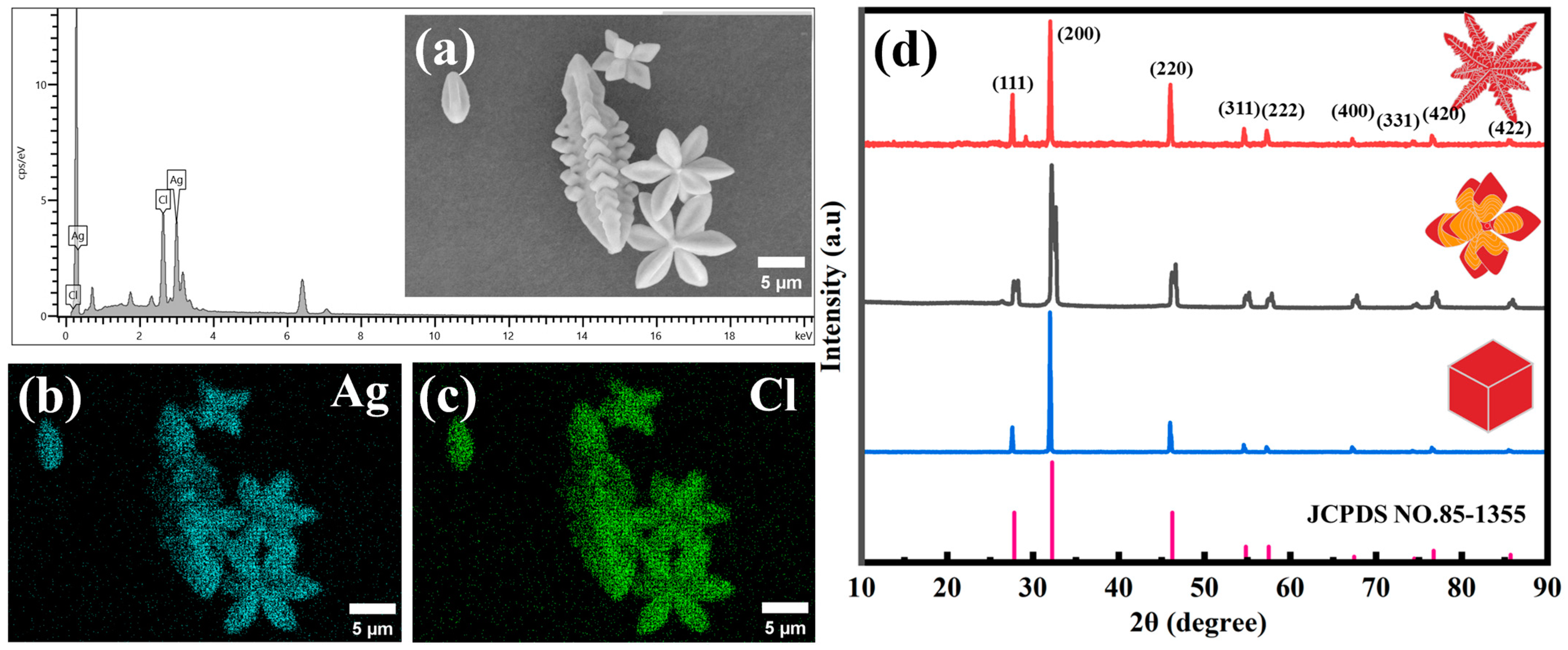
2.2. Characterizations

The high-temperature reaction is carried out using a Kemi Instruments NSG50 reactor, (Hefei China). A Yingtai TG16 centrifuge is used for cleaning and sample collection, (Changsha China). Scanning electron microscope (SEM) images are obtained using Zeiss Supra 55, Sigma 500, and SNE-4500M, (Suwon, Korea). Elemental mapping of the 3D AgCl structures is performed on SEM coupled with energy-dispersive X-ray spectroscopy (SEM/EDS), which is obtained using Hitachi Regulus 8100, (Tokyo, Japan). X-ray diffraction (XRD) analysis is performed on a Bruker D2 phase diffractometer using Cu Kα1 radiation, (Karlsruhe, Germany).

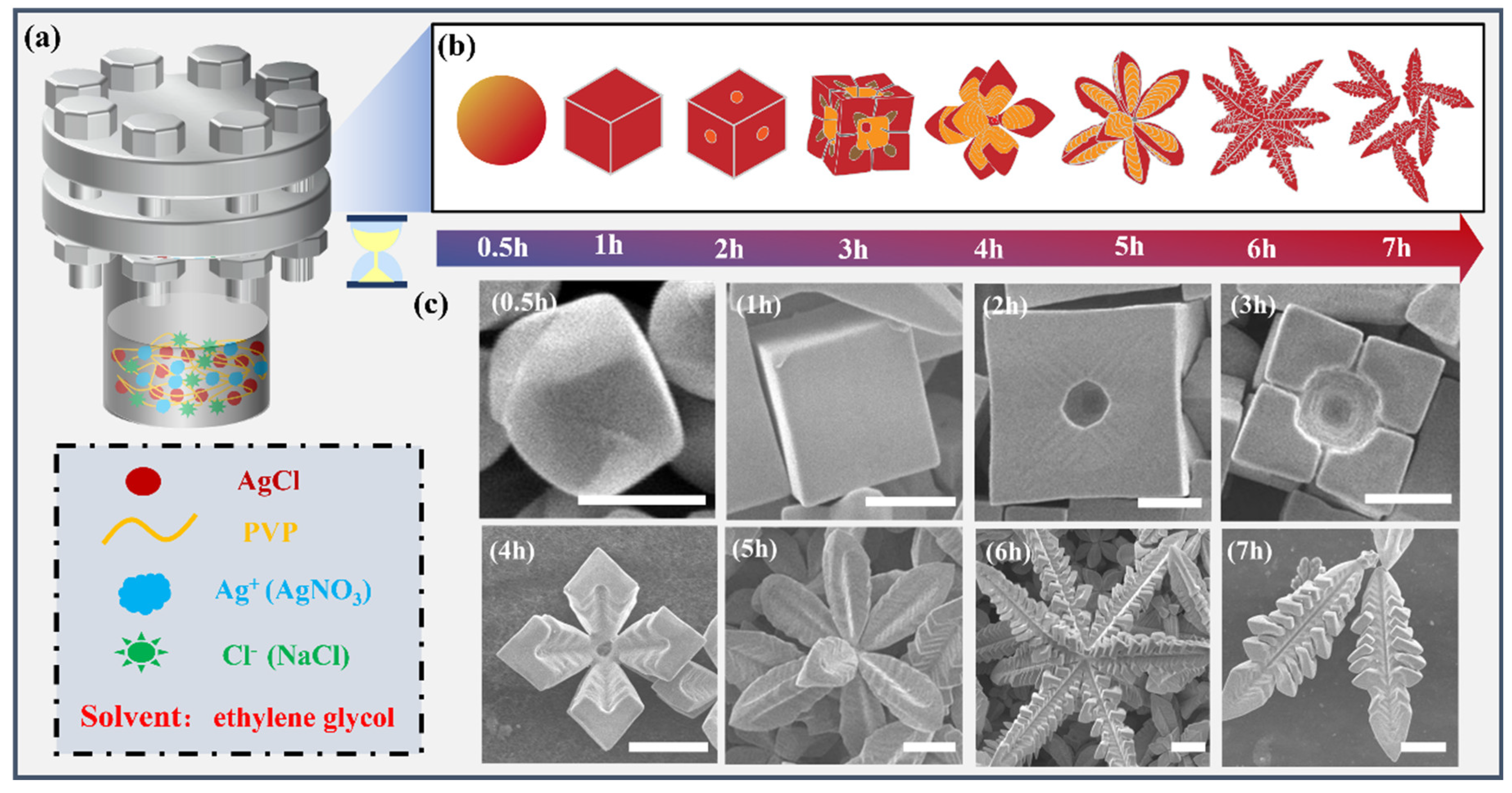
2.3. Preparation of AgCl Microcrystals

A mixture of AgNO3, NaCl, and PVP is used with ethylene glycol (EG) as the solvent to synthesize 3D AgCl crystals under high-temperature conditions. In particular, AgNO3 (0.67 mM, 6 mL), PVP (240 mM, 6 mL), and NaCl (5.5 M, 6 mL) are added into the reaction flask and magnetically stirred at room temperature for 1 min. The mixed solution is poured into a 50 mL high-pressure vessel. Temperature is increased at a rate of 6.5 °C/min to 160 °C. When the reaction is finished, the system is quickly quenched with cool water. The final reaction solution is centrifuged at 8000 rpm for 5 min, followed by alternately washed with water and ethanol 3 times. Finally, the AgCl powder is dried at 90 °C for 30 min.
In order to investigate the effect of specific reaction conditions (e.g., reaction concentrations of NaCl and PVP and reaction time) on the shape of AgCl crystals, we vary only specific parameters, while others are kept constant. The specific parameters of the AgCl microcrystal reaction are shown in Table 1.

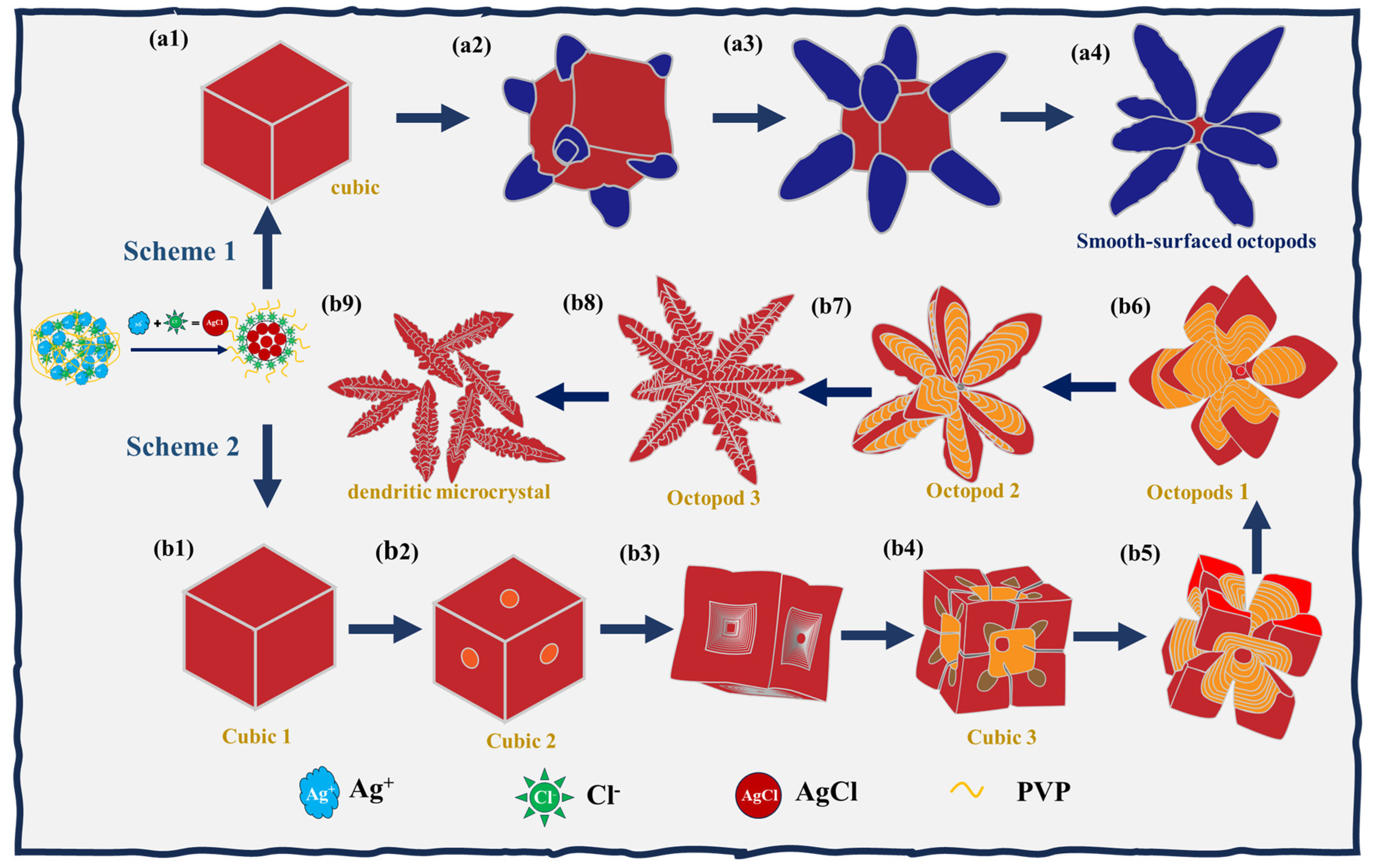
3. Results and Discussion

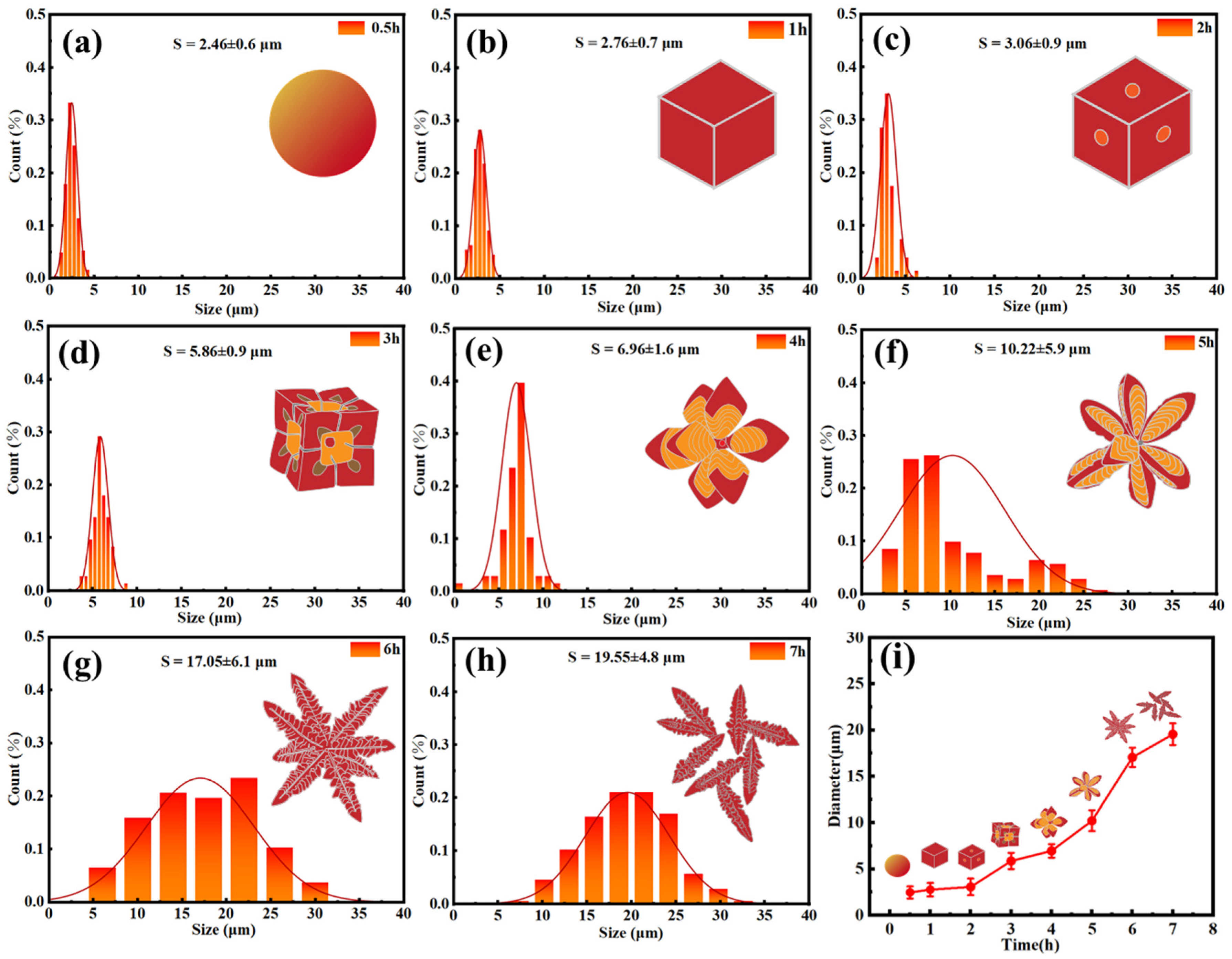
3.1. Effect of Time on the Formation of 3D AgCl Microcrystals

In this work, 3D AgCl microcrystals with controllable shapes are formed by a simple solvothermal synthesis, and the experimental procedure does not require any catalyst or template. The preparation process of samples is illustrated in Figure 1a. As shown, this is realized by using a mixed solution of AgNO3, PVP, and NaCl in a reaction kettle. The microcrystals of 3D AgCl with different shapes are achieved by controlling the reaction time under specific reaction conditions, as schematically shown in Figure 1b. Similarly, we can observe specific experimental data in Table 1 (a-1 to a-8). The corresponding SEM images of crystal shapes at each stage are shown in Figure 1c. At the initial stage of the reaction, we find the crystals grow into particles with irregular shapes, as shown in Figure 1c (t = 0.5 h). When the reaction time increases to 1 h, they develop in to well-defined cubes (cube 1, t = 1 h). As the reaction progresses, identical circular holes appear in the center of each face of the cube, and we define the shape at this stage as cube 2 (t = 2 h). When the reaction time reaches 3 h, the holes at the center of cube planes become progressively larger, and cross lines homogeneously appear on each surface of the cube. We define the shape of crystals at this stage as cube 3 (t = 3 h). With time further extending, the crystals continuously evolve into a mixture of cube-octopod architecture, which presents a clear transition stage from cubic to octopod structure. Since the number of the branches is always eight, we name the crystal at this moment as octopod 1 (t = 4 h). As the reaction time continues, branches of octopod 1 gradually grow longer and thinner, continuously leading to a morphological transformation, and the shape at this stage is named as octopod 2 (t = 5 h). When the reaction time reaches 6 h, distinct second-order branches appear above the primary branches of octopod 2, indicating that the shape of the crystals evolves to a new well-defined morphology, which is named as octopod 3 (t = 6 h). Finally, the first-order branches of octopod 3 begin to break off and gradually detach from the main body of crystals. We define this detached structure as the dendritic microcrystal (t = 7 h).
Additionally, Figure S1 presents more SEM images of these microcrystals captured at time-lapsed stages. By analyzing these images, we can clearly track the changes in the shape, size, and surface features of the microcrystals. At the same time, these figures provide crucial information for understanding the underlying mechanisms governing their growth and transformation, which we will discuss further in the following sections.
In order to better understand the morphology, composition, and crystalline structure of the synthesized microcrystals, we perform energy dispersive X-ray spectroscopy (EDS) and X-ray diffraction (XRD) to investigate cube 1, octopod 1, and octopod 3. The elemental composition of octopod 3 is determined via EDS, and the spectrum is shown in Figure 2a. Only peaks of Ag and Cl are detected in the sample. Figure 2b,c show the distribution and relative intensities of Ag and Cl elements in the selected area. Moreover, the crystal structures of cube 1, octopod 1, and octopod 3 microcrystals are characterized by XRD, as shown in Figure 2d, and the result is compared with the JCPDS NO.85-1355 database. There are nine clear diffraction peaks centered at 27.7, 32.1, 46.1, 54.7, 57.4, 67.3, 74.3, 76.6 and 85.6°, and correspond to (111), (200), (220), (311), (222), (400), (331), (420), and (422) planes of AgCl, respectively, indicating that the microcrystals at each stage are pure AgCl.
To better understand the structure evolution of AgCl at these three stages, we calculate the relative intensities of (111), (220), and (200) and study their relationship among different AgCl microcrystals. As is known, the intensity of diffraction peaks not only reflects the crystalline structure characteristics in XRD analysis, but is also closely related to the degree of crystallinity and structural integrity [40,41]. Despite the fact that the diffraction peak morphology remains essentially unchanged, a notable enhancement in intensity is observed. In this work, the I111/I200 ratios for cube 1, octopod 1, and octopod 3 are 0.18, 0.21, and 0.41, respectively, which shows apparent enhancement along the reaction time. Referring to Figure 1c, one can see that the cube 1 has an integral cubic structure and has not yet started to etch (100) planes. During the transition stage of cube 1 to octopod 1, (100) planes are gradually etched, and correspondingly, the ratio of 111/200 starts to increase. The XRD results support this opinion as the ratio increases to 0.21 for octopod 1. For the situation of octopod 3, (100) planes are heavily etched, and (110) and (111) planes are dramatically enriched. As such, the ratio of 111/200 at the octopod 3 stage is significantly enhanced to 0.41. The same enhancement trend occurs on (220) planes compared to the changes on (111) planes. Specifically, the I220/I200 ratios for cube 1, octopod 1, and octopod 3 are 0.21, 0.32, and 0.49, respectively.
The size evolution of crystals is one of the important parameters, that helps us better understand their growth process. Therefore, we investigate the size of crystals by analyzing low-magnification SEM images of crystals grown at different reactions, as shown in Figure S2. The sizes of particles, cubes, and octopods are defined by their diameter, length of edges, and calculated length of edges, respectively. As the AgCl microcrystal changes from the octopod 1 to an octopod 2, the edge of the cubes begins to disappear. Starting from the octopod 2, we count the edge length of the AgCl microcrystals using simple geometric calculations, as shown in Figure S3. As a statistical result, Figure 3 presents the size distribution of the AgCl microcrystals at different reaction times. Particles of AgCl crystals grow from 2.46 ± 0.6 μm to 2.76 ± 0.7 μm cubes from the reaction time of 0.5 h to 1 h. As the reaction time progresses from 1 h to 2 h, the crystals develop into cube 2 with an average size of 3.06 ± 0.9 μm. Subsequently, extending the time by another hour leads to the growth of cube 2 into cube 3, achieving a size of 5.86 ± 0.9 μm. However, at the reaction time of 4 h, a notable morphological shift occurs as the crystals transform into octopod 1 with a size of 6.96 ± 1.6 μm. This structure evolves from octopod 1 into octopod 2 with dimensions of 10.22 ± 5.9 μm at the time of 5 h. As the reaction continues to 6 h, the size of octopod 2 sharply increases and transforms into octopod 3 with a substantially larger size of 17.05 ± 6.1 μm. Despite the occurrence of branch breakage during this period, the overall crystal size still experiences partial augmentation, culminating in a final size of 19.55 ± 4.8 μm at the time of 7 h.
Figure 3i shows the relationship between the reaction time and the average size of AgCl microcrystals. It can be concluded that the crystal size gradually increases as the reaction time increases. The growth of crystal size is attributed to the dissolution-redeposition process undergone by AgCl crystals. Specifically, the intrinsic surface energy order of AgCl microcrystals is usually (100) < (110) < (111) planes. However, under conditions of high chloride concentration, a significant change in the surface energy ordering of the crystals is observed, with (100) > (110) > (111) planes becoming the predominant order [42]. In order to reduce the total surface energy of the entire crystal, the etching action commences gradually from the crystal’s surface. The Ag+ ions produced by the etching process are deposited onto the tips of the AgCl microcrystals [43]. The process of redeposition leads to an enhancement in the proportion of crystalline planes on the surface (111), thereby enabling an increase in crystal size. This refined description highlights the intricate relationship between reaction duration, morphological changes, and the dynamic nature of crystal growth.

3.2. Effect of NaCl on the Formation of 3D AgCl Microcrystals

As is known, the concentration of Cl is crucial for the growth of AgCl crystals [38,44]. In order to better understand the function of Cl in this work, we synthesize AgCl crystals by varying the concentrations of NaCl and the SEM images of AgCl obtained with different concentrations of NaCl are shown in Figure 4. The specific experimental data can be found in Table 1 (b-1 to b-4, and a-7). In the system without using Cl, we obtain the crystals that mainly have nanowire morphology, as presented in Figure 4a. Increasing the Cl concentration to 2 M promotes the formation of well-defined cubic microcrystals while increasing the yield and structural homogeneity of the crystals, as shown in Figure 4b. Therefore, Cl ions play an important role in the AgCl crystal growth process, which possibly promotes the growth of (110) and (111) planes and eventually forms the regular cube morphology by regulating factors such as growth kinetics and surface energy.
In addition to the above effects, Cl also has an etching effect. It has been widely reported that Cl can produce etching in various ways during the reaction process [45,46,47]. We believe that the synthesized AgCl microcrystals undergo a typical wet etching phenomenon. It is known that AgCl microcrystals are formed during synthesis through reaction (1). This reaction is typically considered reversible, as AgCl becomes unstable at higher Cl concentrations and produces the complex [AgCl2] of AgCl (2) [48].
AgN O 3 + NaCl = AgCl + NaN O 3
AgCl + Cl = Ag Cl 2
As shown in Figure 4c, Cl produces etching of circular holes on (100) planes of the crystal when the concentration of Cl is increased to 3 M. With the addition of 4 M NaCl, we observe the etching area on (100) planes become larger and at the same time the etching on (110) planes start to be apparent, as shown in Figure 4d. We attribute this to the higher concentration of Cl, which allows more ions to be allocated to (110) planes. By raising the concentration to 5.5 M, the Cl ions initiate etching on (110) planes along <111> direction, resulting in the formation of second-order branches on each of the eight primary branches, as illustrated in Figure 4e. More low-magnification SEM images of Figure 4 are provided in Figure S5 for further clarification.
When other variables are controlled for, it is observed that the size of the crystals tends to increase with the gradual enlargement in Cl concentration. This growth is primarily attributed to the fact that the increase in Cl concentration promotes the rise in the etching rate of the crystals. Specifically, the most significant etching effect is observed when the Cl concentration reaches 5.5 M, which leads to the largest crystal size. This phenomenon can be explained by the increased number of dispersed Ag+ resulting from etching in the solution at this concentration. These ions have the capacity to be deposited at the tip sites of the microcrystals, facilitating the crystal growth process.

3.3. Effect of PVP on the Growth and Etching of 3D AgCl Microcrystals

The ultimate structures and shapes of AgCl microcrystals are not only influenced by the concentration of Cl, but also possibly dependent on the concentration of PVP. The concentration of PVP is varied by controlling other conditions while maintaining them constant, and the effect of the PVP concentration on the morphology of AgCl microcrystals is observed. The specific experimental data can be found in Table 1 (c-1, and a-5). Figure 5a illustrates the etching effect of Cl in the absence of PVP. Without the protection of PVP, the etching speed of the crystal is very fast, and the structure of AgCl microcrystals is uneven. When the concentration of PVP reaches 240 mM, we observe a notable reduction in the etching of the crystals, further corroborating the protective role of PVP molecules on the crystal planes. This finding suggests that at higher PVP concentrations, the molecules form a more effective barrier against etching agents, thereby limiting their access to the crystal surface and slowing down the etching process. The enhanced protection provided by PVP at this concentration highlights its ability to modulate the interaction between the crystal surface and the surrounding environment, ultimately influencing the morphology and stability of the AgCl crystals.
Maintaining constant experimental conditions, we analyze the specific effects of the presence or absence of PVP on the size of AgCl microcrystals. As illustrated in Figure 5, the presence of PVP has been demonstrated to exert a pronounced influence on AgCl microcrystal size. In the absence of PVP molecules, AgCl microcrystals lack the necessary protection, resulting in accelerated surface etching. During this process, a significant amount of Ag+ is released, subsequently depositing onto the microcrystal surface and promoting an augmentation in the size of the AgCl microcrystals. Nevertheless, the incorporation of PVP molecules into the solution results in their tight binding to the surface of AgCl microcrystals, thereby providing an efficacious protective layer. This protective effect significantly slows down the etching rate of the crystals, which in turn leads to a decrease in the number of dispersed Ag+ in solution. Due to the reduced deposition of Ag+, AgCl microcrystals are formed with smaller dimensions than those without the addition of PVP molecules.

3.4. Second-Order Etching of 3D AgCl Microcrystals

Due to the instability of AgCl microcrystals under strong electron beam irradiation, it is rather challenging to study AgCl crystals by high-resolution transmission electron microscopy [49,50]. However, the AgCl structure can be analysed by a simple geometrical analysis based on its cubic structure.
It is clear in the SEM image of Figure S5a that the 3D AgCl microcrystals are composed of first-order and secondary branches. As is shown in Figure S5b, we establish a coordinate system with the centre of the body as the origin. First-order branches grow along the <111> direction of the cube. Figure 6a shows that the second-order branches are within (110) planes of octopod 1. This observation provides an explanation for the relatively strong peaks observed in (220) planes of the XRD. The angles between first-order and secondary branches of the etched structure of AgCl for 6 h and 7 h are observed to be 70.3° and 70.4°. (Figure S5a,c). It is also found that the angles of second-order branches obtained in different reaction periods are similar. As illustrated in Figure S5d, the atomic structure model of AgCl reveals that the theoretical angle in the <111> direction is 70.5° [42]. This angle is identical to the one between first-order and secondary branches in the etched microcrystal of 3D AgCl.
Based on Figure 6b, we construct a structural simulation diagram as shown in Figure 6c. We select two branches, oa and ob, from octopod 3 to establish a (110) plane. This facet aob is then shown in Figure 6d at an equal-scale enlargement. As previously discussed, it has been proven that the value of ∠1 (the angle between oa and ob, both in the <111> direction) is approximately equal to ∠2 (the angle between a secondary branch on ob and ob itself). Consequently, the secondary branches on one side of the first-order branch ob are parallel to the first-order branches oa. Therefore, we conclude that the secondary branches originating from the first-order branches ob within the facet aob ((110) plane) are etched along the <111> direction.
We know that the primary branches of 3D AgCl microcrystals have a triple secondary branch (Figure S1g). It is also clearly able to see the triple branch of AgCl microcrystals, as shown in Figure 6e (1). The first-order branch ob has secondary branches within each of the planes aob, boc, and bod. Figure 6e (2–4) show the simulation diagrams of the triple secondary branches of the first-order branch ob within different planes. Consequently, it can be concluded that all second-order branches of AgCl are also etched along the <111> direction.

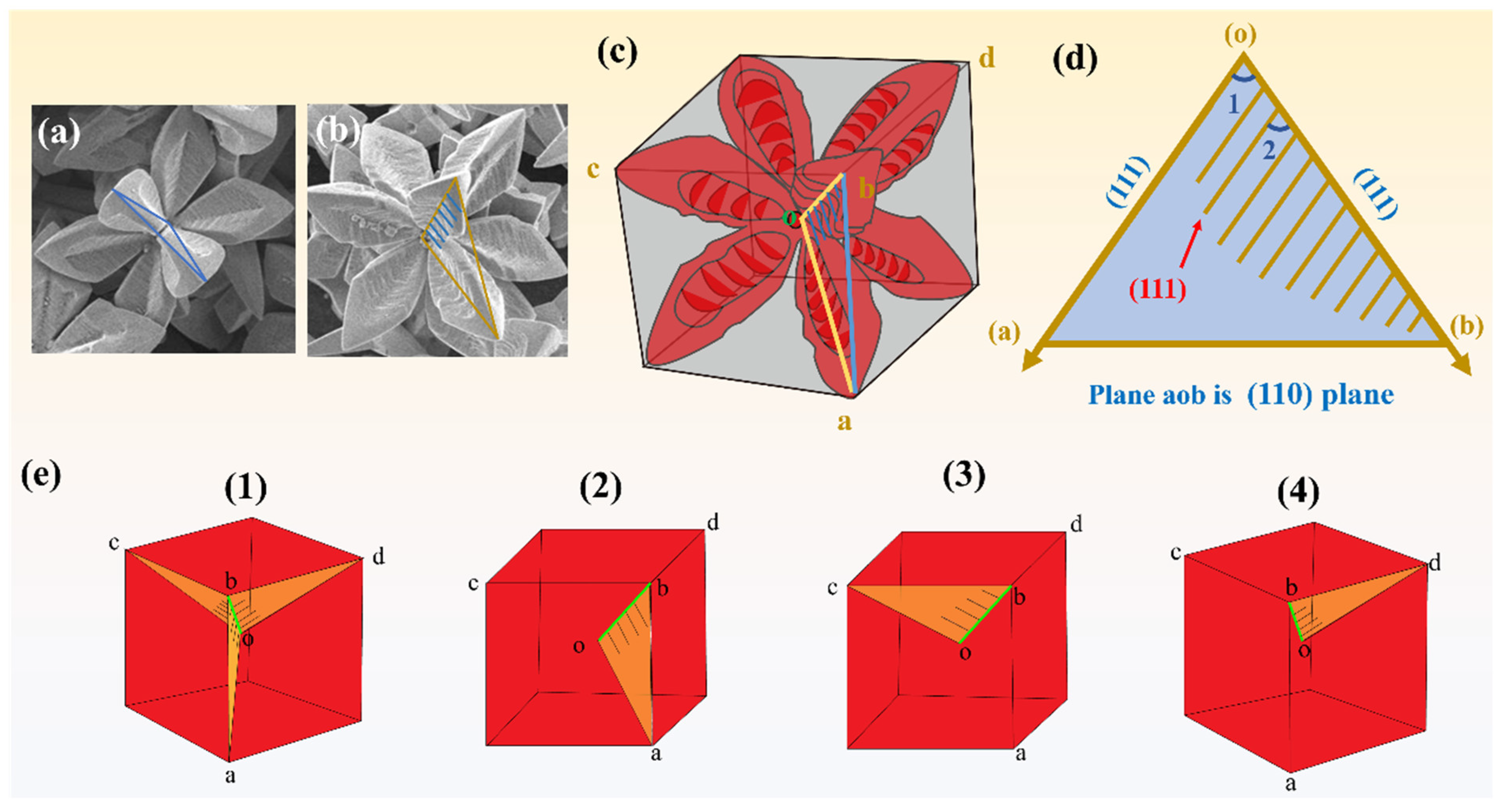
3.5. Growth and Etching Mechanism of 3D AgCl Microcrystals

Figure 7a depicts the crystal structure evolution of overgrown AgCl polypods. Firstly, one of the traditional explanations for the formation of polypods is the addition of capping agents. These capping agents are able to selectively passivate (100) planes of the crystal, thereby reducing the growth rate of (100) planes. As the growth of (100) planes is inhibited, the growth of crystals is preferred in other directions. This change in the growth direction leads to an overgrowth of the crystal along the <111> direction, which in turn forms the structure of the polypod [44]. Another explanation emphasizes the role of Cl ions. This view suggests that Cl ions are able to promote growth on (111) planes by lowering the surface free energy of those planes. The reduction in the surface free energy means that less energy is required for crystal growth on that facet, and therefore, crystals are more inclined to grow in that direction, forming the characteristic structure of a polypod [42].
As shown in Figure 7b, based on our experimental observations and data analyses, we propose a new mechanistic hypothesis for the AgCl microcrystals from the point of view of the etching process. Specifically, Cl ions may selectively attack/or etch certain regions of the crystal, leading to the disruption or dissolution of the crystal structure, resulting in the macroscopic formation of a polypod structure. In particular, Figure 7b demonstrates the morphological evolution of AgCl crystals during the etching process that occurs in the presence of Cl and PVP. The morphological evolution process can be divided into two steps: the first step involves the nucleation and growth of the cubes, and the second involves the selective etching by Cl.
At the first stage, we obtain AgCl cubes in the presence of PVP and Cl ions, which means PVP and Cl ions play together as the surfactant that could promote the formation of cubes. It has been widely reported that the formation of many cubes in the presence of PVP also requires the presence of Cl or Br [51,52,53,54]. Some scholars have suggested that halides act as surfactants for (100) planes [52,55,56], which shows agreement with our experimental results. Therefore, it is concluded that at this stage, Cl acts as a surfactant of (100) and collaborates with PVP to jointly promote the formation of cubic microcrystals.
In the later stage of the reaction, Cl begins to selectively etch the crystal planes of the cubic crystal, resulting in the formation of 3D AgCl microcrystals. It is understood that the binding energy of PVP molecules on (100) planes is higher than that on (111) planes [57]. As a result, PVP molecules preferentially adsorb to AgCl cubes’ (100) planes, forming a thick layer of protection [48]. In order to etch (100) planes of AgCl crystals, it is necessary to have enough amount of Cl to penetrate the diffusion barrier imposed by the PVP layer [48]. Previous studies have shown that Cl tends to adsorb preferentially on (100) planes in the reaction system, followed by (110) and (111) planes [56,58]. An increase in Cl concentration enhances the chemical potential of Cl in the reaction system. When the chemical potential difference between high and low concentrations exceeds the diffusion barrier of the PVP layer, the etching driving force becomes strong enough for Cl to penetrate the PVP protective layer. Therefore, the AgCl microcrystals are first etched on (100) planes and then on (110) planes. Consequently, AgCl microcrystals initially promote etching of the (100) surface, forming circular depressions. In addition, Cl causes etching on (110) planes to form octopod 1, and further etching on (110) planes along <111> directions to form octopod 3. Finally, octopod 3 is fractured to form a dendritic microcrystal. The fracture of the connection point is further evidence of the occurrence of etching.

4. Conclusions

The present study demonstrates that the systematic evolution of AgCl crystals from cube to octopod to dendritic structures is driven by the gradual enhancement of the etching ability of Cl ions. These Cl ions preferentially and selectively act on crystal faces with high surface energy, such as (100) and (110), thus guiding the gradual transformation of the crystal structure. Through precise modulation of the reaction time and the concentrations of NaCl and PVP, we successfully synthesize AgCl microcrystals exhibiting a range of sizes and morphologies. Based on experimental data and research observations, we propose that once the concentration of Cl within the system reaches a specific threshold, the chemical potential of Cl surpasses the diffusion barrier imposed by the PVP molecules. The enhanced etching drive of Cl allows it to penetrate through the protective layer of PVP molecules and etches the crystal. This innovative discovery not only reveals the key role of Cl ions in the control of microcrystal shape, but also provides new ideas for the controllable synthesis of other inorganic materials.

Supplementary Materials

The following supporting information can be downloaded at: https://www.mdpi.com/article/10.3390/cryst15050451/s1, SEM images of AgCl microcrystals in different orientation states (Figure S1a–g); SEM images of AgCl microcrystals at different reaction times (Figure S2); Geometric analysis simulation of extrapolating the side lengths of a cube by branching of an octopod (Figure S3); SEM images of AgCl microcrystals at different concentrations of NaCl (Figure S4); Morphology and branching of AgCl microcrystals at different reaction times, Crystal structure of AgCl microcrystals and Atomic model of AgCl (Figure S5) (DOC).

Author Contributions

C.C.: Conceptualization, Data curation, Formal analysis, Methodology, Software, Writing—original draft. Q.W.: Formal analysis, Methodology, Software, Investigation. C.Y.: Data curation, Investigation, Methodology. X.L. and R.Y.: Visualization, Investigation. Validation, Supervision, Formal anal. X.S. and W.X.: Project administration, Supervision. All authors have read and agreed to the published version of the manuscript.

Funding

This research received no external funding.

Data Availability Statement

The original contributions presented in this study are included in the article/supplementary material. Further inquiries can be directed to the corresponding author.

Conflicts of Interest

The authors declare no conflicts of interest.

References

  1. Cheepborisutikul, S.J.; Ogawa, M. Suppressing the Photocatalytic Activity of Titania by Precisely Controlled Silica Coating. Inorg. Chem. 2021, 60, 6201–6208. [Google Scholar] [CrossRef] [PubMed]
  2. Chen, J.; Lim, B.; Lee, E.P.; Xia, Y. Shape-controlled synthesis of platinum nanocrystals for catalytic and electrocatalytic applications. Nano Today 2009, 4, 81–95. [Google Scholar] [CrossRef]
  3. Erfani, A.; Pirouzifard, M.K.; Pirsa, S. Photochromic biodegradable film based on polyvinyl alcohol modified with silver chloride nanoparticles and spirulina; investigation of physicochemical, antimicrobial and optical properties. Food Chem. 2023, 411, 135459. [Google Scholar] [CrossRef]
  4. Guo, L.; Ji, Y.L.; Xu, H.; Simon, P.; Wu, Z. Regularly Shaped, Single-Crystalline ZnO Nanorods with Wurtzite Structure. J. Am. Chem. Soc. 2002, 124, 14864–14865. [Google Scholar] [CrossRef] [PubMed]
  5. Lignier, P.; Bellabarba, R.; Tooze, R.P. Scalable strategies for the synthesis of well-defined copper metal and oxide nanocrystals. Chem. Soc. Rev. 2012, 41, 1708–1720. [Google Scholar] [CrossRef]
  6. Lv, T.; Liu, M.; Zhou, S.; Xia, Y. Shape Transformation via Etching and Regrowth: A Systematic Study of Pd Nanocrystals with Different Shapes and Twin Structures. Chem. A Eur. J. 2023, 29, e202301465. [Google Scholar] [CrossRef]
  7. Tao, S.; Yang, M.; Chen, H.; Zhao, S.; Chen, G. Continuous Synthesis of Ag/AgCl/ZnO Composites Using Flow Chemistry and Photocatalytic Application. Ind. Eng. Chem. Res. 2018, 57, 3263–3273. [Google Scholar] [CrossRef]
  8. Wang, Y.; Sang, X.; Wu, F.; Pang, Y.; Xu, G.; Yuan, Y.; Hsu, H.-Y.; Niu, W. Boosting plasmon-enhanced electrochemistry by in situ surface cleaning of plasmonic nanocatalysts. Nanoscale 2023, 15, 18901–18909. [Google Scholar] [CrossRef]
  9. Westcott, S.L.; Oldenburg, S.J.; Lee, T.R.; Halas, N.J. Construction of simple gold nanoparticle aggregates with controlled plasmon–plasmon interactions. Chem. Phys. Lett. 1999, 300, 651–655. [Google Scholar] [CrossRef]
  10. Dong, Q.; Jiao, Z.; Yu, H.; Ye, J.; Bi, Y. Facile synthesis of hollow Ag@AgBr heterostructures with highly efficient visible-light photocatalytic properties. CrystEngComm 2014, 16, 8317–8321. [Google Scholar] [CrossRef]
  11. Guo, Y.; Li, J.; Yang, X.; Lou, Y.; Chen, J. Zn0.5Cd0.5S/MIL-125-NH2(Ti) nanocomposites: Highly efficient and stable photocatalyst for hydrogen production under visible light. Inorg. Chem. Commun. 2020, 112, 107714. [Google Scholar] [CrossRef]
  12. Tao, A.; Sinsermsuksakul, P.; Yang, P. Polyhedral Silver Nanocrystals with Distinct Scattering Signatures. Angew. Chem. Int. Ed. 2006, 45, 4597–4601. [Google Scholar] [CrossRef] [PubMed]
  13. Wang, P.; Huang, B.; Zhang, Q.; Zhang, X.; Qin, X.; Dai, Y.; Zhan, J.; Yu, J.; Liu, H.; Lou, Z. Highly Efficient Visible Light Plasmonic Photocatalyst Ag@Ag(Br,I). Chem. A Eur. J. 2010, 16, 10042–10047. [Google Scholar] [CrossRef] [PubMed]
  14. Zhang, Q.; Ge, J.; Pham, T.; Goebl, J.; Hu, Y.; Lu, Z.; Yin, Y. Reconstruction of Silver Nanoplates by UV Irradiation: Tailored Optical Properties and Enhanced Stability. Angew. Chem. Int. Ed. 2009, 48, 3516–3519. [Google Scholar] [CrossRef]
  15. An, C.; Peng, S.; Sun, Y. Facile Synthesis of Sunlight-Driven AgCl:Ag Plasmonic Nanophotocatalyst. Adv. Mater. 2010, 22, 2570–2574. [Google Scholar] [CrossRef]
  16. Guo, X.; Deng, D.; Tian, Q. One pot controllable synthesis of AgCl nanocrystals with different morphology and their photocatalytic activity. Powder Technol. 2017, 308, 206–213. [Google Scholar] [CrossRef]
  17. Han, L.; Wang, P.; Zhu, C.; Zhai, Y.; Dong, S. Facile solvothermal synthesis of cube-like Ag@AgCl: A highly efficient visible light photocatalyst. Nanoscale 2011, 3, 2931–2935. [Google Scholar] [CrossRef]
  18. Lou, Z.; Huang, B.; Qin, X.; Zhang, X.; Cheng, H.; Liu, Y.; Wang, S.; Wang, J.; Dai, Y. One-step synthesis of AgCl concave cubes by preferential overgrowth along 〈111〉 and 〈110〉 directions. Chem. Commun. 2012, 48, 3488–3490. [Google Scholar] [CrossRef]
  19. Lou, Z.; Huang, B.; Wang, P.; Wang, Z.; Qin, X.; Zhang, X.; Cheng, H.; Zheng, Z.; Dai, Y. The synthesis of the near-spherical AgCl crystal for visible light photocatalytic applications. Dalton Trans. 2011, 40, 4104–4110. [Google Scholar] [CrossRef]
  20. Wang, P.; Huang, B.; Qin, X.; Zhang, X.; Dai, Y.; Wei, J.; Whangbo, M. Ag@AgCl: A Highly Efficient and Stable Photocatalyst Active under Visible Light. Angew. Chem. Int. Ed. 2008, 47, 7931–7933. [Google Scholar] [CrossRef]
  21. Xu, H.; Li, H.; Xia, J.; Yin, S.; Luo, Z.; Liu, L.; Xu, L. One-Pot Synthesis of Visible-Light-Driven Plasmonic Photocatalyst Ag/AgCl in Ionic Liquid. ACS Appl. Mater. Interfaces 2011, 3, 22–29. [Google Scholar] [CrossRef] [PubMed]
  22. Zhao, M.; Zhou, W.; Lu, M.; Guo, Z.; Li, C.; Wang, W. Novel AgCl nanotubes/BiOCl nanosheets composite with improved adsorption capacity and photocatalytic performance. J. Alloys Compd. 2019, 773, 1146–1153. [Google Scholar] [CrossRef]
  23. Chen, Y.; Ma, Q.; Li, H.; Wang, D.; Che, Q.; Wang, J.; Wang, G.; Yang, P. Fabrication and Photocatalytic Activity of Tunable Triangular- and Circular-Like Ag/AgCl Nanoplates. J. Nanosci. Nanotechnol. 2018, 18, 2738–2745. [Google Scholar] [CrossRef]
  24. Daupor, H.; Wongnawa, S. Flower-like Ag/AgCl microcrystals: Synthesis and photocatalytic activity. Mater. Chem. Phys. 2015, 159, 71–82. [Google Scholar] [CrossRef]
  25. Lou, S.; Wang, W.; Wang, L.; Zhou, S. In-situ oxidation synthesis of Cu2O/Ag/AgCl microcubes with enhanced visible-light photocatalytic activity. J. Alloys Compd. 2019, 781, 508–514. [Google Scholar] [CrossRef]
  26. Chandra, S.; Dutta, P.; Biswas, K. Enhancement of the Thermoelectric Performance of 2D SnSe Nanoplates through Incorporation of Magnetic Nanoprecipitates. ACS Appl. Energy Mater. 2020, 3, 9051–9057. [Google Scholar] [CrossRef]
  27. Kozhina, E.; Kulesh, E.; Bedin, S.; Doludenko, I.; Piryazev, A.; Korolkov, I.; Kozlovskiy, A.; Zdorovets, M.; Rogachev, A.; Shumskaya, A. One-Dimensional Magneto-Optical Nanostructures: Template Synthesis, Structure, Properties, and Application in Spectroscopy Based on Plasmon Resonance. IEEE Magn. Lett. 2022, 13, 1–5. [Google Scholar] [CrossRef]
  28. Zheng, Z.; Huang, B.; Qin, X.; Zhang, X.; Dai, Y.; Jiang, M.; Wang, P.; Whangbo, M. Highly Efficient Photocatalyst: TiO2 Microspheres Produced from TiO2 Nanosheets with a High Percentage of Reactive {001} Facets. Chem. A Eur. J. 2009, 15, 12576–12579. [Google Scholar] [CrossRef]
  29. Wu, M.; Yan, L.; Li, J.; Wang, L. Synthesis and photocatalytic performance of Ag/AgCl/ZnO tetrapod composites. Res. Chem. Intermed. 2017, 43, 6407–6419. [Google Scholar] [CrossRef]
  30. Zhang, H.; Lu, Y.; Liu, H.; Fang, J. One-pot synthesis of high-index faceted AgCl nanocrystals with trapezohedral, concave hexoctahedral structures and their photocatalytic activity. Nanoscale 2015, 7, 11591–11601. [Google Scholar] [CrossRef]
  31. Liu, Q.; Liao, Z.; Axinte, D. Temperature effect on the material removal mechanism of soft-brittle crystals at nano/micron scale. Int. J. Mach. Tools Manuf. 2020, 159, 103620. [Google Scholar] [CrossRef]
  32. Zhang, D.; Wang, Y.; Deng, J.; Wang, X.; Guo, G. Microfluidics revealing formation mechanism of intermetallic nanocrystals. Nano Energy 2020, 70, 104565. [Google Scholar] [CrossRef]
  33. Zhang, Y.; Zhang, Z.; Liu, W.; Wang, L.; Zheng, Y.; Su, J.; Liu, N.; Gao, Y. Unveiling the Nucleation Dynamics and Growth Mechanism of Layered MoS2 from Crystalline K2MoS4 by in Situ Transmission Electron Microscopy. Cryst. Growth Des. 2020, 20, 4069–4076. [Google Scholar] [CrossRef]
  34. Wang, X.; Yu, J.; Fu, C.; Li, T.; Yu, H. Self-templated formation of AgCl/TiO2 hollow octahedra for improved visible-light photocatalytic activity. Appl. Surf. Sci. 2019, 494, 740–748. [Google Scholar] [CrossRef]
  35. Hanmant Gaikwad, S.; Koratti, A.; Porel Mukherjee, S. Facile tuning of Ag@AgCl cubical hollow nanoframes with efficient sunlight-driven photocatalytic activity. Appl. Surf. Sci. 2019, 465, 413–419. [Google Scholar] [CrossRef]
  36. Navlani-García, M.; Salinas-Torres, D.; Mori, K.; Kuwahara, Y.; Yamashita, H. Tailoring the Size and Shape of Colloidal Noble Metal Nanocrystals as a Valuable Tool in Catalysis. Catal. Surv. Asia 2019, 23, 127–148. [Google Scholar] [CrossRef]
  37. Xia, Y.; Nelli, D.; Ferrando, R.; Yuan, J.; Li, Z.Y. Shape control of size-selected naked platinum nanocrystals. Nat. Commun. 2021, 12, 3019. [Google Scholar] [CrossRef]
  38. Wang, J.; Qin, Y.; Shi, Q.; Wen, L.; Bi, L. Cl-Induced selective fabrication of 3D AgCl microcrystals by a one-pot synthesis method. CrystEngComm 2021, 23, 5116–5123. [Google Scholar] [CrossRef]
  39. Gatemala, H.; Thammacharoen, C.; Ekgasit, S. 3D AgCl microstructures selectively fabricated via Cl-induced precipitation from [Ag(NH3)2]+. CrystEngComm 2014, 16, 6688–6696. [Google Scholar] [CrossRef]
  40. Alharbi, N.D.; Salah, N.; Habib, S.S.; Alarfaj, E. Synthesis and characterization of nano- and microcrystalline cubes of pure and Ag-doped LiF. J. Phys. D Appl. Phys. 2013, 46, 035305. [Google Scholar] [CrossRef]
  41. Dong, H.; Yang, H.; Ning, Y.; Liu, F.; Bradley, R.; Zhao, B.; Wu, W. Large-scale facile green synthesis of porous silver nanocubes on monolithic activated carbon for room-temperature catalytic oxidation of formaldehyde. Appl. Phys. A 2022, 128, 976. [Google Scholar] [CrossRef]
  42. Lou, Z.; Huang, B.; Ma, X.; Zhang, X.; Qin, X.; Wang, Z.; Dai, Y.; Liu, Y. A 3D AgCl Hierarchical Superstructure Synthesized by a Wet Chemical Oxidation Method. Chem. A Eur. J. 2012, 18, 16090–16096. [Google Scholar] [CrossRef]
  43. Zettsu, N.; McLellan, J.M.; Wiley, B.; Yin, Y.; Li, Z.-Y.; Xia, Y. Synthesis, Stability, and Surface Plasmonic Properties of Rhodium Multipods, and Their Use as Substrates for Surface-Enhanced Raman Scattering. Angew. Chem. Int. Ed. 2006, 45, 1288–1292. [Google Scholar] [CrossRef]
  44. Lee, H.S.; Kim, J.E.; Kim, T.; Suh, K.S. Ionic liquid-assisted synthesis of highly branched Ag:AgCl hybrids and their photocatalytic activity. J. Alloys Compd. 2015, 621, 378–382. [Google Scholar] [CrossRef]
  45. Gryglewicz, J.; Oleszkiewicz, W.; Ramiączek-Krasowska, M.; Szyszka, A.; Prażmowska, J.; Paszkiewicz, B.; Paszkiewicz, R.; Tłaczała, M. Reactive ion etching of GaN and AlGaN/GaN assisted by Cl2/BCl3. Mater. Sci. Pol. 2011, 29, 260–265. [Google Scholar] [CrossRef]
  46. Oh, K.H.; Baek, G.B.; Chung, C.W. Etch characteristics of cobalt thin films using high density plasma of halogen gas. Thin Solid Films 2024, 796, 140341. [Google Scholar] [CrossRef]
  47. Shao, D.; Zhang, X.; Wang, Z.; Zhang, Y.; Tan, G.; Yan, W. New architecture of a variable anode for full-time efficient electrochemical oxidation of organic wastewater with variable Cl concentration. Appl. Surf. Sci. 2020, 515, 146003. [Google Scholar] [CrossRef]
  48. Dong, M.; Pan, Y.; Zhu, J.; Jia, H.; Dong, H.; Xu, F. Real-time imaging reveal anisotropic dissolution behaviors of silver nanorods. Nanotechnology 2024, 35, 275703. [Google Scholar] [CrossRef]
  49. Tian, X.; Anand, U.; Mirsaidov, U.; Zheng, H. Spontaneous Reshaping and Splitting of AgCl Nanocrystals under Electron Beam Illumination. Small 2018, 14, 1803231. [Google Scholar] [CrossRef]
  50. Tian, X.; Anand, U.; Mirsaidov, U.; Zheng, H. Nanocrystal Dynamics: Spontaneous Reshaping and Splitting of AgCl Nanocrystals under Electron Beam Illumination (Small 48/2018). Small 2018, 14, 1870231. [Google Scholar] [CrossRef]
  51. Bergin, S.M.; Chen, Y.-H.; Rathmell, A.R.; Charbonneau, P.; Li, Z.-Y.; Wiley, B.J. The effect of nanowire length and diameter on the properties of transparent, conducting nanowire films. Nanoscale 2012, 4, 1996–2004. [Google Scholar] [CrossRef]
  52. da Silva, R.R.; Yang, M.; Choi, S.-I.; Chi, M.; Luo, M.; Zhang, C.; Li, Z.-Y.; Camargo, P.H.C.; Ribeiro, S.J.L.; Xia, Y. Facile Synthesis of Sub-20 nm Silver Nanowires through a Bromide-Mediated Polyol Method. ACS Nano 2016, 10, 7892–7900. [Google Scholar] [CrossRef] [PubMed]
  53. Li, B.; Ye, S.; Stewart, I.E.; Alvarez, S.; Wiley, B.J. Synthesis and Purification of Silver Nanowires To Make Conducting Films with a Transmittance of 99%. Nano Lett. 2015, 15, 6722–6726. [Google Scholar] [CrossRef] [PubMed]
  54. Niu, Z.; Cui, F.; Kuttner, E.; Xie, C.; Chen, H.; Sun, Y.; Dehestani, A.; Schierle-Arndt, K.; Yang, P. Synthesis of Silver Nanowires with Reduced Diameters Using Benzoin-Derived Radicals to Make Transparent Conductors with High Transparency and Low Haze. Nano Lett. 2018, 18, 5329–5334. [Google Scholar] [CrossRef] [PubMed]
  55. Ruditskiy, A.; Xia, Y. Toward the Synthesis of Sub-15 nm Ag Nanocubes with Sharp Corners and Edges: The Roles of Heterogeneous Nucleation and Surface Capping. J. Am. Chem. Soc. 2016, 138, 3161–3167. [Google Scholar] [CrossRef]
  56. Zhou, S.; Li, J.; Gilroy, K.D.; Tao, J.; Zhu, C.; Yang, X.; Sun, X.; Xia, Y. Facile Synthesis of Silver Nanocubes with Sharp Corners and Edges in an Aqueous Solution. ACS Nano 2016, 10, 9861–9870. [Google Scholar] [CrossRef]
  57. Saidi, W.A.; Feng, H.; Fichthorn, K.A. Binding of Polyvinylpyrrolidone to Ag Surfaces: Insight into a Structure-Directing Agent from Dispersion-Corrected Density Functional Theory. J. Phys. Chem. C 2013, 117, 1163–1171. [Google Scholar] [CrossRef]
  58. Ahn, J.; Wang, D.; Ding, Y.; Zhang, J.; Qin, D. Site-Selective Carving and Co-Deposition: Transformation of Ag Nanocubes into Concave Nanocrystals Encased by Au–Ag Alloy Frames. ACS Nano 2018, 12, 298–307. [Google Scholar] [CrossRef]
Figure 1. (a) Schematic diagram of the preparation of AgCl crystals. (b) Schematic illustration of the formation of 3D AgCl microcrystals. (c) SEM images of AgCl microcrystals corresponding to different time periods in Figure 1b. The scale of Figures (0.5 h) to (3 h) is 2 μm, while the scale of Figures (4 h) to (7 h) is 5 μm.
Crystals 15 00451 g001
Figure 2. Typical morphology and structure of 3D AgCl microcrystals. (a) EDX spectrum of 3D AgCl microcrystals. (b,c) mapping analyses of Ag and Cl. (d) XRD spectrum of 3D AgCl microcrystals.
Crystals 15 00451 g002
Figure 3. Size distribution of AgCl microcrystals prepared at different times (a) 0.5 h (b) 1 h (c) 2 h (d) 3 h (e) 4 h (f) 5 h (g) 6 h (h) 7 h (i) Graph of reaction time versus average crystal size. (The average crystal size is obtained by averaging multiple crystal sizes after measuring them by importing SEM images through ImageJ software (ij153-win-java8), and the type of longitudinal coordinate measurement is obtained by analyzing the number of crystals in the SEM image).
Crystals 15 00451 g003
Figure 4. SEM images of AgCl microcrystals at different concentrations of NaCl. (a) 0 M (b) 2 M (c) 3 M (d) 4 M (e) 5.5 M NaCl. Reaction times are all 6 h.
Crystals 15 00451 g004
Figure 5. Demonstrates the SEM images under different concentrations of PVP conditions. Low magnification: (a) 0 (b) 240 mM PVP, and high magnification: (c) 0 (d) 240 mM PVP used in the synthesis. Reaction time is all 4 h.
Crystals 15 00451 g005
Figure 6. (a,b) SEM image of AgCl crystal. (c) 3D Crystal model of AgCl. (d) (110) face of aob. (e) Structural simulation of the surface on which the triple branches of one of the branched ob of the 3D AgCl microcrystals are located.
Crystals 15 00451 g006
Figure 7. Two growth schemes of AgCl crystals. Scheme 1: AgCl crystals grow in the presence of PVP and Cl along the <111> direction of the cubic crystal to give an octopod structure with a smooth surface. Scheme 2: AgCl crystals were etched along planes of (100) and (110) of the cubic crystals in the presence of PVP and Cl to obtain dendritic microcrystals.
Crystals 15 00451 g007
Table 1. Brief summary of the experimental conditions tested in this work.
NO.AgNO3 (mM)PVP (mM)NaCl (M)Time (h)Results
a-10.672405.50.5Figure S2a
a-20.672405.51Figure S2b
a-30.672405.52Figure S2c
a-40.672405.53Figure S2d
a-50.672405.54Figure S2e
a-60.672405.55Figure S2f
a-70.672405.56Figure S2g
a-80.672405.57Figure S2h
b-10.6724006Figure 4a
b-20.6724026Figure 4b
b-30.6724036Figure 4c
b-40.6724046Figure 4d
c-10.6705.54Figure 5a
Disclaimer/Publisher’s Note: The statements, opinions and data contained in all publications are solely those of the individual author(s) and contributor(s) and not of MDPI and/or the editor(s). MDPI and/or the editor(s) disclaim responsibility for any injury to people or property resulting from any ideas, methods, instructions or products referred to in the content.

