Article

Color Mechanism of Blue Myanmar Jadeite Jade: The Role of Trace Elements and Mineralogical Characteristics

1
School of Gemmology, China University of Geosciences (Beijing), Beijing 100083, China
2
Guangdong Gemstones & Precious Metals Testing Center, Guangzhou 510080, China
*
Author to whom correspondence should be addressed.
Crystals 2025, 15(10), 843; https://doi.org/10.3390/cryst15100843
Submission received: 3 September 2025 / Revised: 24 September 2025 / Accepted: 26 September 2025 / Published: 27 September 2025
(This article belongs to the Section Mineralogical Crystallography and Biomineralization)

Abstract

Myanmar blue jadeite jade is a rare and highly prized gemstone, yet its coloration and formative mechanisms remain poorly understood. In this study, petrographic analysis, ultraviolet–visible (UV–Vis) spectroscopy, electron probe microanalysis (EPMA), and laser ablation inductively coupled plasma mass spectrometry (LA-ICP-MS) were performed on a sample of Myanmar blue jadeite with small white blocks to investigate its mineral composition, trace element distribution, and coloration mechanisms. Most of the sample was found to be blue, with surrounding white areas occurring in small ball-shaped blocks. The main mineral component in both the blue and white domains was jadeite. Although both areas underwent recrystallization, their textures differed significantly. The blue areas retained primary structural features within a medium- to fine-grained texture, reflecting relatively weaker recrystallization. The white areas, however, were recrystallized into a micro-grained texture, reflecting relatively stronger recrystallization, with the superimposed effects of external stress producing a fragmented appearance. The blue jadeite had relatively higher contents of Ti, Fe, Ca, and Mg, while the white jadeite contained compositions close to those of near-end-member jadeite. It was noted that, while white jadeite may have a high Ti content, its Fe content is low. UV–Vis spectra showed a broad absorption band at 610 nm associated with Fe2+-Ti4+ charge transfer and a gradually increasing absorption band starting at 480 nm related to V4+. Combining the chemical composition and the characteristics of the UV–Vis spectra, we infer that the blue coloration of jadeite is attributed to Fe2+-Ti4+ charge transfer; i.e., the presence of both Ti and Fe in blue jadeite plays a key role in its color formation. V4+ exhibited no significant linear correlation with the development of blue coloration. Prominent oscillatory zoning was observed in the jadeite, transitioning from NaAlSi2O6-dominant cores to Ca-Mg-Fe-Ti-enriched rims, reflecting the trend of fluid evolution during blue jadeite crystallization. Petrographic analysis indicated that the formation of the Myanmar blue jadeite occurred in two or three stages, with the blue regions forming earlier than the white regions. The blue jadeite also underwent significant recrystallization. Our findings contribute to the understanding of the formation of blue jadeite and the diversity of colors in jadeite jade.

1. Introduction

Jadeitite is a high-pressure and low-temperature metamorphic rock composed predominantly of jadeite, along with other pyroxenes such as omphacite and kosmochlor, and it is primarily formed in subduction zone environments where its genesis is closely linked to fluid-driven crystallization within subduction channels. To date, only 19 jadeitite occurrences have been documented globally [1,2,3,4], with major deposits found in Myanmar [3], Guatemala [5], Japan [6], America [7], and a few other regions. In Myanmar, jadeite jade deposits are hosted in serpentinites along the western Sagaing Fault zone in the Hpakant region of Kachin State and are genetically associated with Mesozoic intra-oceanic subduction processes [8,9]. The associated metamorphic rock assemblages include phengite–glaucophane schists, stilpnomelane-bearing quartzites, phengite–quartz schists, garnet amphibolites, and diopside marbles [10]. Previous studies have identified rare minerals such as grossular in late-stage veins of Myanmar jadeitite, suggesting rodingitization rather than eclogitic assemblages, offering insights into jadeite-forming fluid evolution [11].
Myanmar jadeite jade occurs in various colors, with blue being among the rarest. Its coloration is controlled by chromogenic elements, and crystal field theory is widely employed to elucidate the role of chromophoric ions in the color genesis. Current systematic understanding attributes the green coloration primarily to Cr3+ electronic transitions, which dominate the optical absorption spectra of jadeite [12]. In high-quality “Imperial green” jadeite, Cr2O3 (approximately 0.3 wt.%) produces absorption bands at 635, 660, and 690 nm. Higher levels deepen the green to blackish tones, while lower levels yield lighter shades —explaining the color zoning commonly seen in Myanmar jadeite [13,14,15]. Red-to-yellow hues in jadeite are mainly controlled by Fe speciation: hematite causes red, and limonite influences yellow, both influenced by oxidation intensity and fracture density. These phases are identifiable via micro-XRD and Raman spectroscopy [16,17]. Lavender jade color stems from Fe3+ and Fe2+-Fe3+ charge transfer, while purple hues are mainly due to Mn3+, marked by a 570 nm absorption peak. Mn content rises from pale (20 ppm) to vivid (61 ppm) and royal purple (494 ppm) and EPR analysis indicates that Mn3+ is responsible for the observed purple coloration [18,19,20,21,22]. Fe2+-Ti4+ charge transfer may also contribute to purple coloration, with absorption features resembling those in blue sapphires [6,21]. However, this hypothesis currently lacks strong mineralogical evidence in the context of Myanmar jadeite jade.
Due to its rarity and limited study, the chromogenic mechanism of blue jadeite remains unclear and differs fundamentally from the “blue-water” and “blue-mist” phenomena [23,24]. Spectral analysis of blue jadeite suggests the potential involvement of V4+ in its coloration, displaying absorption characteristics similar to those of synthetic vanadium-based turquoise pigments, where V4+ substitution for Zr4+ induces octahedral distortion [25,26,27]. The blue features of blue-green jadeite jade are attributed to the presence of Fe2+ and Mn2+ in Guatemalan jadeite and sulfur is associated with high-quality green jadeite, but whether it also plays a role in blue jadeite remains to be further investigated [28,29]. Unlike green jadeite, Myanmar blue jadeite lacks the characteristic Cr3+ absorption peaks, indicating a fundamentally different chromogenic pathway [30]. In the Sorkhan region of Iran, a type of pure jadeite with extremely low Fe and Ti content was discovered. Detailed thermodynamic calculations confirmed that temperature (rather than trace elements) is the key factor controlling the mineral assemblages and stability of blue jadeite [31]. In the Itoigawa region of Japan, a type of blue jadeite with a jadeite content ranging from XJd (93.7–97.4%) was discovered, and its color is controlled by Fe and Ti [6]. The color of our sample, which falls between those of Iran and Japan, is reported for the first time in Myanmar.
This study is dedicated to investigating the chromogenic mechanism of Myanmar blue jadeite and to enhancing the understanding of jadeite jade with different color types. This research is conducted through petrographic, colorimetric, spectroscopic, and geochemical (major and trace element) analyses.

2. Materials and Methods

2.1. Samples

The sample analyzed in this study is a piece of blue jadeite jade (LSFC) sourced from the jade mining area in Myanmar, approximately 16 cm by 20 cm (Figure 1a). The specimen is mainly blue but shows uneven coloration, with small white blocks and regions of other coloration (Figure 1b). Sample LSFC-1, cut from the original stone, was prepared for in situ analyses. Polished thin sections (0.03 mm thick) from both uniform blue and mixed-color areas were analyzed using a suite of analytical techniques for petrographic and geochemical characterization.

2.2. Methods of Analysis

Petrographic observations (PPL: Plane-Polarized Light and XPL: Cross-Polarized Light), color measurements, and UV–Vis spectroscopic analysis were conducted at the School of Gemmology, China University of Geosciences, Beijing (CUGB).
Color measurements were carried out using an MDIS-F8 multifunction dual integrating sphere spectrometer (Liulabs, South EI Monte, USA) under controlled conditions, with a temperature range of 15–30 °C and relative humidity below 80%.
UV–Vis spectroscopic analysis was performed using a GEM-3000 spectrophotometer (Biaoqi, Guangzhou, China), operating within the 400–800 nm wavelength range. Spatial Resolution (Minimum Analysis Area): 0.7 mm in diameter. Samples were directly placed on the window to record reflectance spectra (R%). This non-destructive method enables micro-area analysis, revealing chromogenic element distribution and color formation mechanisms.
EPMA was performed at the Electron Microprobe Laboratory, CUGB, using an EPMA-1720 instrument (Shimadzu Corporation, Japan) under the operating conditions of 15 kV accelerating voltage, 20 nA beam current, and a 5 μm spot diameter. Standard Minerals: Si-Spodumene, Ti-Rutile, Al-Lipx, Fe-garnet, Mn-Rhodonite, Mg-Olivine, Ca-Diopside, Na-Albite, Cr-Chromite, Ni-Pentlandite, V- Vanadium, Cu-Chalcopyrite. Correction method employed was ZAF3. The detection limit for conventional elements is 0.01 wt.%.
An in situ trace element analysis of silicate minerals was carried out at the Elemental Geochemistry Lab of Institute of Earth Sciences, CUGB, using an East Laser LSPC-193ss laser ablation system (East Laser Company, China) coupled with a Jena PQ-MS mass spectrometer (Jena Company, Germany). Analytical conditions included a laser wavelength of 193 nm, a spot diameter of 50 μm or 36 μm, and a laser repetition rate of 10 Hz. The calibration reference material used was NIST SRM 610, with Si as the internal standard element. NIST SRM 612 was employed as a monitoring standard for elemental concentrations. Chemical element analysis and calibration data processing were performed using Glitter 4.4.4 software. Element Detection Limits: Mg-200, Si-20,000, Ca-1000, Ti-10, V-5, Cr-10, Mn-10, Fe-1000, Ni-100, Cu-5 ppm. Detection limits for all rare earth elements: 0.0001 ppm.

2.3. Color Testing

Chromaticity tests were conducted on 40 representative surface areas, covering blue, white, and other color domains, using the CIE 1976 L*a*b* uniform color space system [32,33]. Chromaticity parameters a* and b* were obtained for representative blue and white zones (Table 1), with color space simulations for all 40 test points under D65 illumination and a fixed L* value of 57.5 (L* = the average of all the data). The chromaticity parameters were plotted in the CIE 1976 L*a*b* color space (Figure 2). The analysis indicates that the sample predominantly has a blue-gray tone, with some areas showing a faint bluish-green hue.

2.4. Petrography and Main Mineral Composition

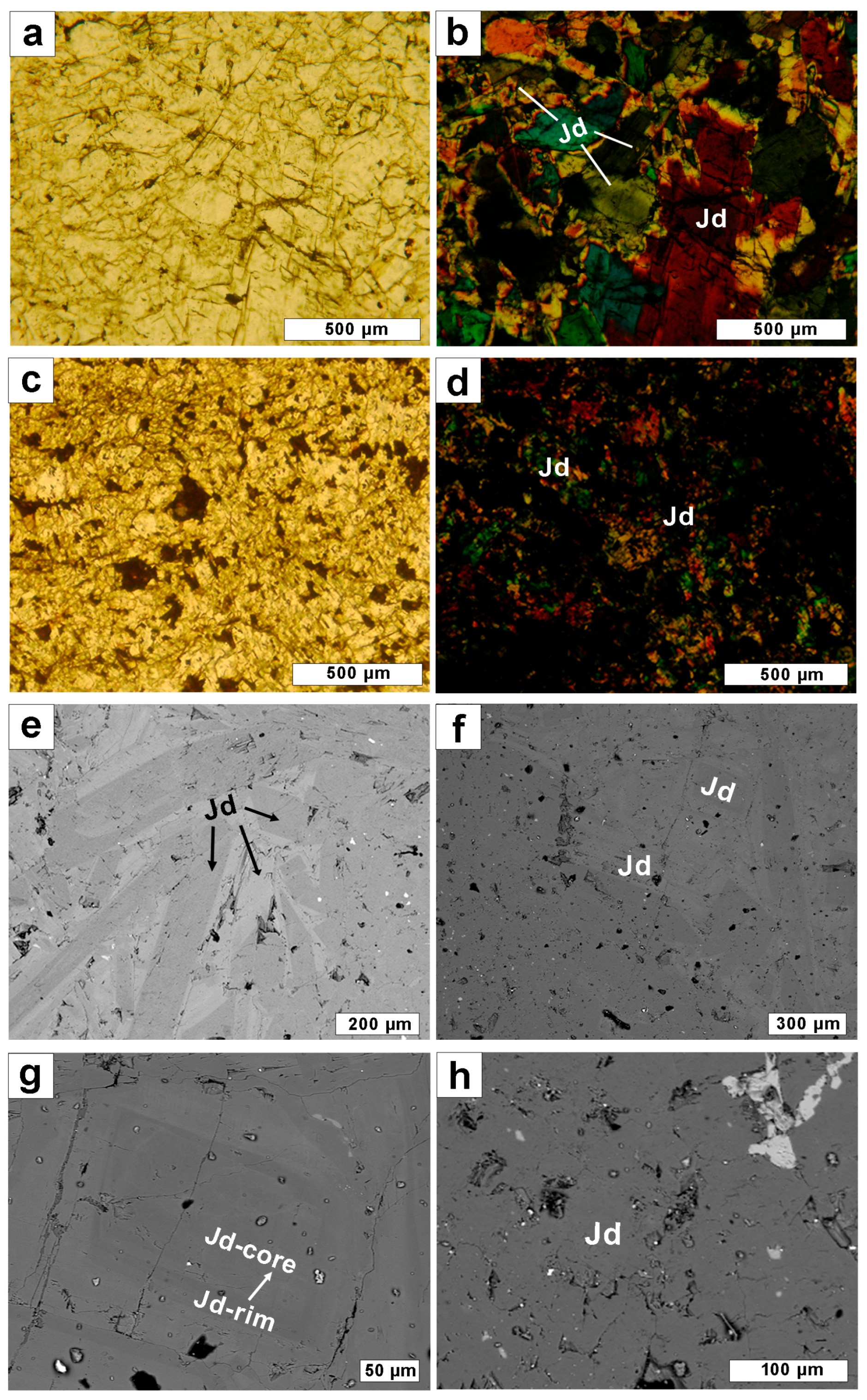
Jadeite crystals exceed 1 mm in length, exhibiting a texture ranging from porphyritic to inequigranular. Most occur as irregular masses with planar contacts and random orientations. Under cross-polarized light, variations in extinction angles within individual crystals indicate slight chemical zoning. The blue region consists of medium- to fine-grained jadeite with a massive fabric and curved mosaic granoblastic texture, defined by sinuous grain boundaries. This microstructure, along with the fine-grained recrystallization, indicates ductile deformation and dynamic metamorphism under relatively high-pressure, moderate-temperature conditions. Despite this overprinting, some primary structures are preserved (Figure 3a,b). The white region is composed of micro-grained jadeite with a subhedral microgranoblastic texture. The curved boundaries and fine grain size signify pronounced recrystallization, while a fractured appearance suggests intense dynamic metamorphism (Figure 3c,d). The blue area appears to have experienced less intense geological overprinting than the white area. In addition, it exhibits a significantly higher degree of recrystallinity than white area (Figure 3b,d).
Sample LSFC-1 is primarily composed of jadeite, with its contents ranging from approximately 92 to 100 mol% (Table 2). EPMA data were plotted on the ternary diagram used for clinopyroxene classification (WEF: wollastonite [Wo], enstatite [En], and ferrosilite [Fs])–jadeite (Jd)–aegirine (Ae). The white region consists of near-end-member jadeite, while the blue region shows greater chemical variability. Blue jadeite deviates more from the ideal composition (Figure 4).
The blue region shows a heterogeneous chemical composition with localized zoning (Figure 3e,f), while the white region is homogeneous (Figure 3h). Analytical points are divided into two categories: blue jadeite (points 1–6) and white jadeite (points 7–14). Blue jadeite has relatively higher Ti (0.05–0.27 wt.%), Fe (0.29–1.17 wt.%), Mg (0.55–1.75 wt.%), and Ca (0.61–2.02 wt.%), while white jadeite shows relatively lower levels of these elements (Ti, 0.00–0.17 wt.%, Fe, 0.02–0.28 wt.%; Mg, 0.02–0.37 wt.%; and Ca, 0.04–0.46 wt.%) (Table 2). The zoning pattern in the blue region, from core (point 5) to rim (point 6), shows increasing Ti, Fe, Ca, and Mg, with decreasing Na and Al (Figure 3g and Table 2).

2.5. UV–Vis Spectroscopy and Trace Element Composition (LA-ICP-MS)

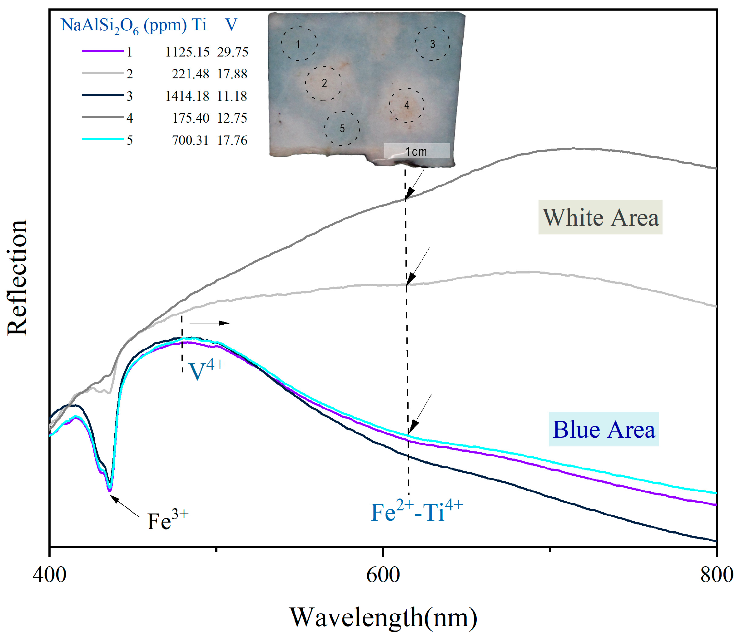
UV–Vis spectra were obtained of the blue (1, 3, 5) and white (2, 4) regions of sample LSFC-1. The blue regions show consistent absorption, with minor variation due to elemental differences. A strong 437 nm peak (Fe3+) is observed, along with gradually descending reflectance from 480 nm to a broad 610 nm trough. In contrast, the white regions exhibit increasing reflectance beyond 480 nm, with region 2 showing a weak trough from trace chromogenic elements. All spectra show a gradual decline beyond 700 nm (Figure 5).
LA-ICP-MS analysis reveals a distinct difference in trace element composition between the blue and white jadeite in the Myanmar samples (Table 3). The blue regions (1, 3, 5) have more Ti (700–1414 ppm), Cr, Mn, Ni, and Cu than the white regions (2, 4). Mg, Ca, Ti, and Fe are enriched in blue zones, while Mg, Ca, and Ti increase with color intensity. Ti shows a strong positive correlation with blue hue (Figure 6).
Notably, 4-1 exhibits high Fe content (similar to that of some blue points) and low Ti content in the white areas. However, it does not display any blue coloration. In contrast, relative to the blue region, 5-1 displays a rapid increase in Fe but relatively low Ti content, resulting in a distinctly lighter blue (Figure 6).

3. Discussion

3.1. Color and Chemical Composition of Blue Jadeite from Myanmar

The blue regions of the examined sample exhibit a grayish-blue coloration, in which the gray component arises from the relatively low lightness that diminishes chromatic vividness. In blue areas, a subtle greenish-blue tendency can be observed (Figure 1 and Figure 2). Given the compositional complexity of jadeite jade and the influence of its geological environment, objectively defining its chromatic range has long posed a significant challenge. The integration of colorimetric methods provides an objective and quantitative framework for characterizing jadeite color. In contrast to visual perception, which is susceptible to variations in illumination and subjective interpretation, standardized colorimetric systems (e.g., CIE L*a*b*) allow for precise and reproducible documentation of chromatic attributes, thereby offering a reliable reference for both scientific and gemological evaluation [16,36].
In jadeitite veins, jadeite is generally highly pure, with jadeite contents (XJd) typically exceeding 98 mol% (Figure 4) [3]. In terms of coloration, Myanmar blue jadeite jade is primarily composed of blue and white jadeite. The principal differences in trace element chemistry between the blue and white varieties are attributed to variations in Ca, Mg, Fe, and Ti contents (Table 2). Blue jadeite, characterized by enrichment in Ca, Mg, Fe, and Ti, exhibits a compositional trend toward the omphacite field, whereas white jadeite tends to approach the ideal end-member composition (Figure 4). Compared with the purple-violet blue or blue-bluish green jadeite series, Myanmar blue jadeite is characterized by high Fe and Ti contents with low Cr and Mn concentrations. In contrast, gem-quality Myanmar green jadeite contains Cr concentrations ranging from 1036 to 42,440 ppm (average 9056 ppm), with Ti concentrations between 327 and 3225 ppm (average 949 ppm) [37]. In blue-series jadeite from both Myanmar and Guatemala, Cr concentrations are generally low (0.01–0.03 wt.%). However, in the bluish-green series, Cr contents are slightly higher (approximately 0.09–0.10 wt.%) [23]. For Guatemalan bluish-green jadeite, coloration is predominantly controlled by Fe2+; the Cr absorption band is indistinct and the content is extremely low or negligible [28]. In Japanese blue jadeite, Cr concentrations fall below the detection limit, with no observable Cr absorption band, while the material displays an intense blue hue [6]. In sample LSFC-1, the Cr contents are only 29–111 ppm (average 53 ppm) and no Cr absorption band is observed, indicating that Cr does not contribute to the coloration of blue jadeite (Figure 5 and Table 3). The Cr content exerts a significant influence on jadeite coloration: when Cr contents are relatively high, the blue hue may be masked by green, whereas at low levels, or even below the detection limit, the blue tends to appear more vivid. In lavender jadeite from Myanmar, Mn concentrations range from 100 to 1000 ppm, accompanied by low Cr contents [38]. In natural purple jadeite, coloration is associated with Mn (35–360 ppm), while Fe (230–670 ppm) and Ti (60–230 ppm) contents remain relatively low. When Fe concentrations increase significantly (390–2500 ppm) together with Ti (180–1700 ppm), the jadeite exhibits a bluish-purple hue [21]. In sample LSFC-1, Mn concentrations in the blue regions only range from 25 to 72 ppm (average 45 ppm), showing no significant increase compared with the white regions. Combined with the absence of Mn3+ absorption bands, this indicates that Mn does not contribute to the coloration of blue jadeite. Unlike Myanmar blue jadeite jade, which is composed predominantly of jadeite, the blue jadeite jade from Guatemala (commonly referred to as “blue-water jadeite”) primarily consists of omphacite [23]. Nevertheless, the characteristics of the chromogenic elements remain consistent between the two.

3.2. Coloring Elements and Coloration Mechanism

The coloration of Myanmar blue jadeite is attributed to Fe2+-Ti4+ charge transfer. In the UV–Vis spectra, the Fe2+-Ti4+ charge-transfer absorption band occurs near 610 nm, while the absorption at 437 nm is assigned to Fe3+ (Figure 5). Moreover, the intensity of the blue coloration shows a linear correlation with Ti content (Figure 6). Several studies have also demonstrated that V4+ can induce increased absorption around 480 nm, resulting from the substitution of V4+ for Al3+ at the M1 sites [25,26,27]. However, since V4+ shows no clear correlation with the variation in blue coloration and its spectral features partially overlap with those of the Fe2+-Ti4+ charge-transfer band, it cannot be regarded as strong evidence for the origin of the blue color (Table 3).
In Myanmar green jadeite, Cr3+ is derived from chromite during the serpentinization-related metasomatic process. It diffuses into the jadeite lattice through the ore-forming fluids, substituting for Al3+ at the M1 sites, and produces a characteristic d-d transition absorption band in the visible spectrum (approximately 630–700 nm) [6,38,39]. This absorption of red light imparts the vivid emerald-green coloration of jadeite; the higher the Cr3+ concentration, the more saturated the color. Even a small amount of Cr3+ is sufficient to produce an emerald-green effect, whereas Fe (including Fe2+-Fe3+) can also generate green spectral features, but Cr3+ plays the more decisive role [39]. The chromophore in purple jadeite is Mn3+ occupying the M1 site of the lattice. In an octahedral crystal field, Mn3+ produces a characteristic d-d transition absorption band at approximately 560–580 nm, absorbing yellow-green light and thereby transmitting purple. The higher the Mn3+ content, the more vivid the purple coloration; additional impurities such as Fe, Ti, and Cr can further modulate the hue [6,18,21]. The primary source of coloration in blue jadeite is intervalence charge transfer between Fe2+ and Ti4+ ions occupying adjacent octahedral sites in the lattice. Within the crystal structure of jadeite, Fe2+ (3d6) and Ti4+ (3d0) can substitute for Al3+ at the octahedral M sites. When sufficient Fe2+-Ti4+ neighboring pairs are present, photo-induced electron transfer occurs under visible-light irradiation: an electron migrates from Fe2+ to Ti4+, producing Fe3+ and Ti3+ (Fe2+ + Ti4+ → Fe3+ + Ti3+) [6,12,40].
Neither high Fe with low Ti nor low Fe with high Ti is sufficient to produce a vivid blue color, as illustrated in samples 4-1 and 5-1 (Figure 6). The efficiency of Fe2+-Ti4+ charge transfer is constrained by the relative abundances of both elements. Only when the concentrations of Fe2+ and Ti4+ are simultaneously high can the number of adjacent ion pairs become adequate to generate an intense absorption band in the red–orange region (~600 nm), resulting in a deep blue coloration. Conversely, when either Fe2+ or Ti4+ concentrations are too low, the probability of ion-pair formation decreases, the absorption band weakens, and the color tends to appear pale blue or grayish [28].
Unlike the blue jadeite from Itoigawa, Japan, which exhibits a broad absorption band in the 500–750 nm range, attributed to Fe2+-Ti4+ charge transfer [6], Myanmar blue jadeite shows a spectral shift toward the purple region.
Fe2+, Ti4+, V4+, and Mg2+ substitute for Al3+ at the M1 site due to similar ionic radii and octahedral coordination (CN = 6), while Ca2+, with a radius close to Na+, substitutes at the larger M2 site (Table 4) [41,42].

3.3. Blue Jadeite Jade Formation Process

According to petrographic observations, the Myanmar blue jadeite sample contains three distinct grain size ranges: medium- to fine-grained jadeite in the blue regions and micro-grained jadeite in the white regions. Notably, the larger jadeite grains retain certain relic features of the primary texture (Figure 3a,b).
Jadeite evolves from NaAlSi2O6-rich cores to Ca-Mg-Fe-Ti-enriched rims, indicating a stable early system dominated by the jadeite end-member. With fluid evolution, increasing Ca, Mg, Fe, and Ti were introduced, causing rim compositions to deviate from the end-member. The zoning suggests episodic rather than continuous fluid input, leading to stepwise variations in crystal growth (Figure 3g). During the jadeite formation stage, large amounts of jadeitizing fluids (Na-Al-Si rich fluids) infiltrated the subduction zone [44,45]. These fluids could either directly crystallize to form P-type jadeite or react with the protolith through metasomatism to produce R-type jadeite [46].
In the first stage, jadeite crystals were relatively large and exhibited mosaic or granoblastic texture, reflecting crystallization under static metamorphic or hydrothermal conditions [47]. In the second stage, external dynamic metamorphism induced recrystallization. Some jadeite grains retained relics of the original texture, while others transformed into micro- to fine-grained aggregates. These finer jadeite grains were typically distributed around the larger crystals, giving the corresponding portions of the sample a superior texture and higher degree of translucency. In the final stage—which may have occurred concurrently with or subsequent to the second stage—intense dynamic metamorphism caused the jadeite to fracture and form a fragmented appearance. This led to the development of poorly textured white regions.

3.4. Gemmological Implications

Myanmar hosts abundant jadeite deposits in Pharkant-Tawmaw of the Kachin State, ranging from imperial green to vivid green in the green jadeite series, and from imperial violet to rosy violet in the purple jadeite series [35]. In terms of translucency and texture, commercial varieties include glassy, icy, and glutinous types. Myanmar has been widely recognized as the world’s leading source of jadeite in both quality and diversity; however, apart from a few reports mentioning jadeite with near-blue tones and some “blue-water” material (as noted in the Introduction), there had been no previous record of blue jadeite comparable in color to the experimental sample investigated here—let alone material reaching gem quality (Figure 7). Therefore, our report carries significant implications for both the economic potential and the aesthetic appreciation of blue jadeite.
The formation conditions of blue jadeite are relatively stringent. During its genesis, an adequate supply of Ti is essential, while the concentrations of Cr, Mn, and other chromogenic elements must remain low or even negligible. The degree of recrystallization is crucial for forming high-quality jadeite. If the recrystallization is too weak, primary structures may be partially preserved, resulting in a coarse-grained texture [47]. Conversely, if the degree of metamorphism is excessively intense, coupled with external stress, the jadeite may develop a fractured appearance, thereby diminishing its aesthetic value.
Although no jadeite deposits of blue coloration have been reported from Myanmar to date, and no grading system has yet been established for Myanmar blue jadeite, the appearance and structural characteristics of carved jade pieces (Figure 7) nonetheless indicate that they represent top-quality blue jadeite. This discovery provides valuable insights into the coloration and chromogenic mechanisms of blue jadeite, and contributes to a broader understanding of the genetic processes responsible for different jadeite color varieties.

4. Conclusions

This study confirms that this unique blue variety has been reported for the first time in Myanmar jadeite. The blue coloration in Myanmar jadeite is attributed to Fe2+-Ti4+ charge transfer, with the absorption band centered at 610 nm. The trace-element differences distinguishing blue jadeite from white jadeite are primarily Ca, Mg, Fe, and Ti. Among these, Ca and Mg serve mainly to maintain charge balance rather than act as chromogenic elements. Relatively low concentrations of Cr and Mn further contribute to enhancing the intensity of the blue color.
When Fe is high but Ti is low, the resulting blue color is relatively pale. Conversely, a high Ti content without sufficient Fe does not produce blue coloration. This pattern indicates a synergistic interaction between Fe2+ and Ti4+, consistent with the Fe2+-Ti4+ charge-transfer mechanism, with the intensity of the blue coloration being controlled by the number of Fe-Ti pairs. The formation of Myanmar blue jadeite likely involves two to three stages, with the white domains crystallizing later than the blue ones. The blue portions exhibit a superior texture and high translucency, whereas the white portions are characterized by a poorer texture.

Author Contributions

Conceptualization, S.D. and G.S.; methodology, S.D., Y.Z. and G.S.; software, S.D.; validation, S.D. and Y.Z.; formal analysis, S.D. and G.S.; investigation, S.D. and G.S.; resources, S.D., T.L. and G.S.; data curation, S.D.; writing—review and editing, S.D., Y.Z. and G.S.; visualization, S.D.; supervision, G.S. and Y.Z.; project administration, G.S.; and funding acquisition, G.S. All authors have read and agreed to the published version of the manuscript.

Funding

This investigation was supported by the National Natural Science Foundation of China, grant number 42273044.

Data Availability Statement

The data presented in this study are available within the article.

Acknowledgments

We thank the Gem Testing Laboratory within the School of Gemology, CUGB.

Conflicts of Interest

The authors declare no conflicts of interest.

References

  1. Yi, X.; Shi, G.H.; He, M.Y. Jadeitized omphacitite from Myanmar jadeite area. Acta Petrol. Sin. 2006, 22, 971–976. [Google Scholar]
  2. Shi, G.H.; Stöckhert, B.; Cui, W.Y. Kosmochlor and chromian jadeite aggregates from the Myanmar jadeitite area. Minerals 2007, 69, 1059–1075. [Google Scholar] [CrossRef]
  3. Shi, G.H.; Harlow, G.E.; Wang, J.; Wang, J.; Ng, E.; Wang, X.; Cao, S.M.; Cui, W.Y. Mineralogy of jadeitite and related rocks from Myanmar: A review with new data. Eur. J. Mineral. 2012, 24, 345–370. [Google Scholar] [CrossRef]
  4. Shi, G.H.; Cui, W.Y.; Tropper, P.; Wang, C.Q.; Shu, G.M.; Yu, H.X. The petrology of a complex sodic and sodic-calcic amphibole association and its implications for the metasomatic processes in the jadeitite area in northwestern Myanmar, formerly Burma. Contrib. Mineral. Petrol. 2003, 145, 355–376. [Google Scholar] [CrossRef]
  5. Harlow, G.E.; Sisson, V.B.; Sorensen, S.S. Jadeitite from Guatemala: New observations and distinctions among multiple occurrences. Geol. Acta 2011, 9, 363. [Google Scholar]
  6. Abduriyim, A.; Kazuko, S.; Yusuke, K. Japanese Jadeite: History, Characteristics, and Comparison with Other Sources. Gems Gemol. 2017, 53, 48–67. [Google Scholar] [CrossRef]
  7. Lesz, S.; Griner, S.; Nowosielski, R. Chemical microstructure of Franciscan jadeite from Pacheco Pass, California. Am. Mineral. 1998, 83, 273–279. [Google Scholar]
  8. Shi, G.H.; Jiang, N.; Liu, Y.; Wang, X.; Zhang, Z.Y.; Xu, Y.J. Zircon Hf isotope signature of the depleted mantle in the Myanmar jadeitite: Implications for Mesozoic intra-oceanic subduction between the Eastern Indian Plate and the Burmese Platelet. Lithos 2009, 112, 342–350. [Google Scholar] [CrossRef]
  9. Xing, B.Q.; Shi, G.H.; Zhang, J.H.; Long, C.; Zhang, Y.; He, L.Y.; Hu, R.J. Characteristics of the Guatemalan Feicui and Its Comparison to the Myanmar Feicui. Geoscience 2021, 35, 1769–1788. [Google Scholar]
  10. Shi, G.H.; Cui, W.Y. Petrology of jadeite-bearing serpentinized peridotite and its country rocks from Northwestern Myanmar (Burma). Acta Petrol. Sin. 2001, 17, 483–490. [Google Scholar]
  11. Wang, X.; Shi, G.H.; Qiu, D.F.; Wang, J.; Cui, W.Y. Grossular-bearing jadeite omphacite rock in the Myanmar jadeite area: A kind of jadeitized rodingite? Eur. J. Mineral. 2012, 24, 237–246. [Google Scholar] [CrossRef]
  12. Khomenko, V.M.; Platonov, A.N. Electronic absorption spectra of Cr3+ ions in natural clinopyroxenes. Physics. Chem. Miner. 1985, 11, 261–265. [Google Scholar] [CrossRef]
  13. Harder, H. Trace elements as coloring agents in jadeites. J. Gemmol. 1995, 24, 508–511. [Google Scholar] [CrossRef]
  14. Shi, G.H.; Cui, W.Y. Desalinization of Chromium within Myanmar Jadeite Jade and Implications. J. Gems Gemmol. 2005, 7, 7–12. [Google Scholar]
  15. Zhen, C.L.; Gao, Y.J.; Li, K. Identification Characteristic of Green Feicui from Myanmar and Guatemala. J. Gems Gemmol. 2023, 25, 17–28. [Google Scholar]
  16. Liu, Z.Y.; Guo, Y.; Shang, Y.R.; Yuan, B. Research on parameters optimization of digital imaging system in red–yellow jadeite color measurement. Sci. Rep. 2022, 12, 3619. [Google Scholar] [CrossRef] [PubMed]
  17. Pan, X.; Guo, Y.; Liu, Z.Y.; Zhang, Z.K.; Shi, Y.X. Impact of different standard lighting sources on red jadeite and color quality grading. Earth Sci. Res. J. 2019, 23, 371–378. [Google Scholar] [CrossRef]
  18. Rossman, G.R. Lavender jade. The optical spectrum of Fe3+ and Fe2+⤍Fe3+ intervalence charge transfer in jadeite from Burma. American. Mineral. 1974, 59, 868–870. [Google Scholar]
  19. Shang, Y.R.; Wang, W.Z.; Jin, T.L.; Huang, L.M.; Wu, Z.Y.; Guo, Y. Colouration in purple jadeite-jade from Myanmar A spectroscopy and chromaticity investigation. Acta Petrol. Et. Mineral. 2024, 43, 643–651. [Google Scholar]
  20. Li, X. The Color Causing Mechanism and Influencing Factors of Lavender Feicui in Myanmar. Master’s Thesis, China University of Geosciences (Beijing), Beijing, China, 2012. [Google Scholar]
  21. Han, W.; Liu, Y.; Zhang, J.; Lu, T.J. Color origins of two types of natural lavender jadeite jades. Acta Mineral. Sin. 2020, 40, 549–555. [Google Scholar]
  22. Wu, X.; Bao, Z.; Kang, Y.; Han, X.; Liu, X.; Qu, M. Color Origin of Burmese Lavender Jadeite. Laser Optoelectron. Prog. 2019, 56, 073001–073005. [Google Scholar] [CrossRef]
  23. Zhang, Y.; Shi, G.H. Origin of Blue-Water Jadeite Jades from Myanmar and Guatemala: Differentiation by Non-Destructive Spectroscopic Techniques. Crystals 2022, 12, 1448. [Google Scholar] [CrossRef]
  24. Yan, W.W.; Wang, J.H. Origin of the blue fog zone and black crust of Myanmar jadeite jade boulder. Acta Sci. Natralium Univ. Sunyatseni 2011, 50, 107–113. [Google Scholar]
  25. Yu, K.T.; Zhu, Z.B. A study on the chemical state of vanadium in turguoise pigment. Jourcal Ceram. 2000, 21, 186–189. [Google Scholar]
  26. Wang, D.; Mo, Z.R.; Li, X.M.; Wang, H.Q. Study on Colouring Mechanisms of Blue Jadeite. J. Gems Gemmol. 2013, 15, 14–17. [Google Scholar]
  27. Sujatha Devi, P.; Ganguli, D. Vanadyl ion stability in silica gels. J. Non-Cryst. Solids 1998, 240, 50–54. [Google Scholar] [CrossRef]
  28. Li, T.; Zhang, C.; Zhang, H.T.; Chen, Y.Q.; Li, Z.B.; Liu, Y. Color-Causing Mechanisms of Guatemala Jadeite Jade: Constraints from Spectroscopy and Chemical Compositions. Crystals 2023, 13, 1535. [Google Scholar] [CrossRef]
  29. Xie, Y.H.; Wang, C.Y. Trace Elements in Jadeites with Different Color and Their Infrared Spectra Characteristics. Rock Miner. Anal. 2003, 22, 183–187. [Google Scholar]
  30. Zhang, X.H. Jade Color Changes to the Color and Type and Content-Relations. Master’s Thesis, Chengdu University of Technology, Chengdu, China, 2008. [Google Scholar]
  31. Oberhänsli, R.; Bousquet, R.; Moinzadeh, H.; Moazzen, M.; Arvin, M. The Field of Stability of blue jadeite: A new occurrence of jadeite at Sorkhan, IRAN, As a case study. Mineralogist 2007, 45, 1501–1509. [Google Scholar] [CrossRef]
  32. Guo, Y.; Mo, T.; Cheng, S.H. Contribution of Green Jadeite-Jade’s Chroma Difference Based on CIE 1976 L*a*b* Uniform Color Space. Adv. Mater. Res. 2009, 177, 620–623. [Google Scholar] [CrossRef]
  33. Guo, Y.; Zhang, J.; Mo, T. Quality Evaluation of Green Jadeite-jade’s Lightness Based on CIE 1976 L*a*b* Uniform Color Space. Bull. Chin. Ceram. Soc. 2010, 29, 560–566. [Google Scholar]
  34. Morimoto, N. Nomenclature of Pyroxenes. Mineral. Petrol. 1988, 39, 55–76. [Google Scholar] [CrossRef]
  35. Zhang, Y.; Shi, G.H.; Wen, J.B. Chromite and Its Thin Kosmochlor and Cr-Omphacite Cortex in Amphibolite from the Myanmar Jadeite Deposits. Crystals 2025, 15, 79. [Google Scholar] [CrossRef]
  36. Guo, Y.; Zong, X.; Qi, M.; Zhang, Y.; Wang, H. Feasibility study on color evaluation of jadeite based on GemDialogue color chip images. EURASIP J. Image Video Process 2018, 2018, 95. [Google Scholar] [CrossRef]
  37. Liu, S.I.; Man, K.Y.; Seneewong-Na-Ayutthaya, M.; Jakkawanvibul, C.; Lee, A.T.-Y. Geographic Origin Deter-mination of High-quality Green Jadeite-Omphacite Jade (Fei Cui) from Myanmar, Guatemala and Italy Using Statistical Processing Coupled with Spectroscopic and Chemical Analyses. J. Gemmol. 2024, 39, 124–144. [Google Scholar] [CrossRef]
  38. Zhang, Y.; Shi, G.H.; Wen, J.B. Manganese-Rich Chromite in Myanmar Jadeite Jade: A Critical Source of Chromium and Manganese and Its Role in Coloration. Crystals 2025, 15, 704. [Google Scholar] [CrossRef]
  39. Coccato, A.; Karampelas, S.; Wörle, M.; Van Willigend, S.; Pétrequin, P. Gem quality and archeological green ‘jadeite jade’ versus ‘omphacite jade’(Article). J. Raman Spectrosc. 2014, 45, 1260–1265. [Google Scholar] [CrossRef]
  40. Fritsch, E.; Rossman, G.R. An Update on Color in Gems. Part 2: Colors Involving Multiple Atoms and Color Centers. Gems Gemol. 1988, 24, 3–15. [Google Scholar] [CrossRef]
  41. Pauling, L. The Principles Determining The Structure Of Complex Ionic Crystals. J. Am. Chem. Soc. 1929, 51, 1010–1026. [Google Scholar] [CrossRef]
  42. Goldschmidt, V.M. Geochemische Verteilungsgesetze und kosmische H?ufigkeit der Elemente. Naturwissenschaften 1930, 18, 999–1013. [Google Scholar] [CrossRef]
  43. Shannon, R.D. Revised effective ionic radii and systematic studies of interatomic distances in halides and chalcogenides. Acta Crystallogr. Sect. A 1976, 32, 751–767. [Google Scholar] [CrossRef]
  44. Harlow, G.H.; Sorensen, S. Jade (Nephrite and Jadeitite) and Serpentinite: Metasomatic Connections. Int. Geol. Rev. 2005, 47, 113–146. [Google Scholar] [CrossRef]
  45. Zhang, M.; Groat, L.A.; Salje, E.K.H.; Beran, A. The origin of jadeitite-forming subduction-zone fluids: CL-guided SIMS oxygen-isotope and trace-element evidence. Am. Mineral. 2006, 91, 979–996. [Google Scholar]
  46. Tsujimori, T.; Harlow, G.E. Petrogenetic relationships between jadeitite and associated high-pressure and low-temperature metamorphic rocks in worldwide jadeitite localities: A review. Eur. J. Mineral. 2012, 24, 371–390. [Google Scholar] [CrossRef]
  47. Shi, G.H.; Wang, X.; Chu, B.B.; Cui, W.Y. Jadeite jade from Myanmar: Its texture and gemmological implications. J. Gemmol. 2010, 31, 185–195. [Google Scholar] [CrossRef]
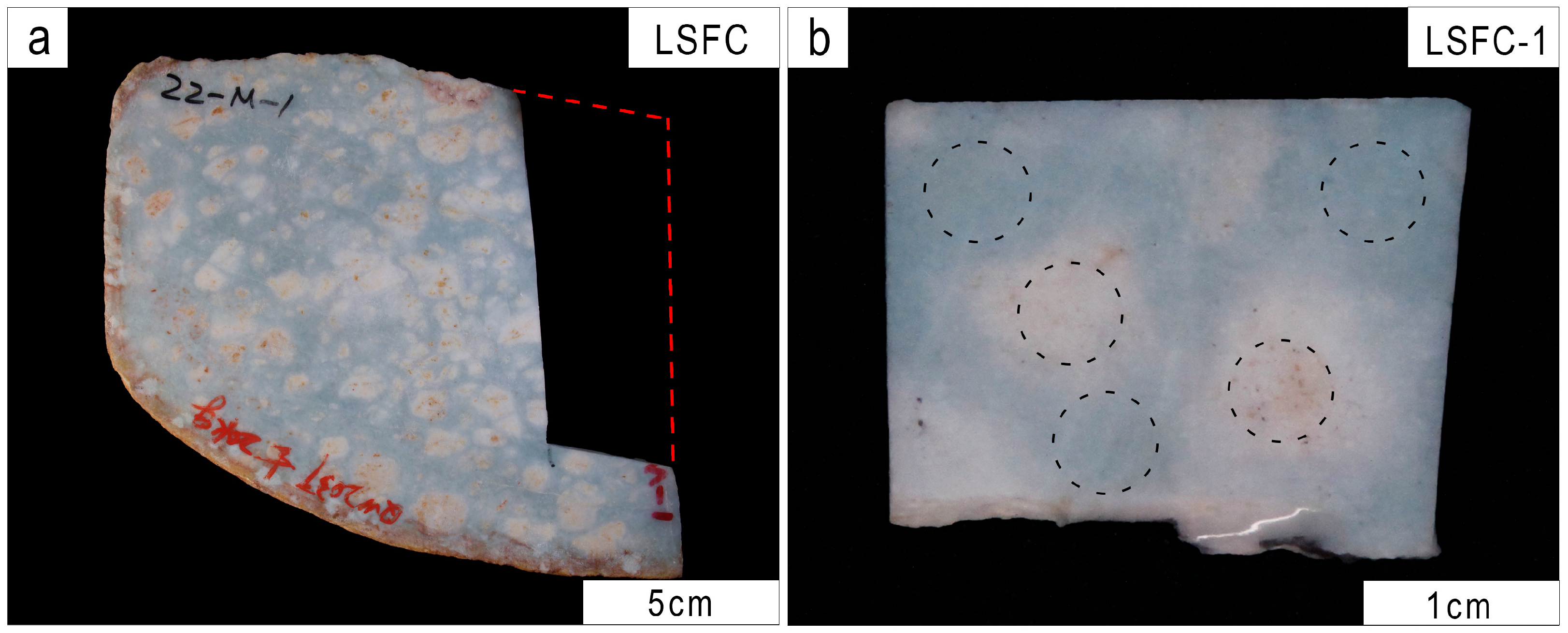
Figure 1. Photos of the Myanmar blue jadeite samples: (a) white areas within the blue region are distributed in a patchy pattern (LSFC); (b) blue jadeite with small white blocks (LSFC-1). Note: The dashed circles in (b) indicate the testing areas.
Crystals 15 00843 g001
Figure 2. Projection of different color regions of blue jadeite in the CIE 1976 L*a*b* color space (The chromaticity coordinates were analyzed in the CIELAB color space based on modified images captured by KONICA MINOLTA).
Crystals 15 00843 g002
Figure 3. Photomicrographs and BSE images of blue jadeite from Myanmar. (a,b) Blue jadeite area is primarily composed of medium- to fine-grained crystals (left: PPL and right: XPL). (c,d) White jadeite area consists of micro-grained white jadeite (left: PPL and right: XPL). (e,f) BSE images of the blue area show chemical heterogeneity. (g) Jadeite in the blue area exhibits well-developed compositional zoning. (h) BSE images of the white jadeite area reveal a homogeneous chemical composition.
Crystals 15 00843 g003
Figure 4. Ternary classification diagram of clinopyroxene compositions plotted in the WEF (wollastonite + enstatite + ferrosilite)–jadeite (Jd)–aegirine (Ae) system (Adapted from [34,35]).
Crystals 15 00843 g004
Figure 5. UV–Vis reflection spectrum of Myanmar blue jadeite. These features are assigned to absorptions caused by V4+ (480 nm), Fe3+ (437 nm), and Fe2+-Ti4+ charge transfer (610 nm) (regions 1, 3, 5—blue; regions 2, 4—white).
Crystals 15 00843 g005
Figure 6. Trace element composition chart of Myanmar blue jadeite (regions 1, 3, 5—blue; regions 2, 4—white).
Crystals 15 00843 g006
Figure 7. Photo of carved works from Myanmar blue jadeite jade (partial LSFC).
Crystals 15 00843 g007
Table 1. Chromaticity parameters of Myanmar blue jadeite in different color regions.
PointsBlueWhite
No.12341
a*−2.30−5.32−5.60−6.10−0.31
b*−3.29−2.10−2.89−2.04−0.63
L*57.9758.2856.4957.3166.44
Note: Partial representative dataset for sample LSFC-1, measured using an MDIS-f8 spectrometer (L*—lightness; a*—red–green axis; b*—yellow–blue axis).
Table 2. Chemical composition of the minerals in Myanmar jadeite (wt.%).
Points1234567891011121314
DescriptionsBlueWhite
CoreRim
SiO257.5057.9658.4657.5357.3257.2457.9357.2060.2659.4559.1957.4056.7059.25
TiO20.050.070.270.250.140.170.050.000.010.000.170.070.030.00
Al2O323.5624.7122.2621.3524.4922.6625.2624.4023.6523.6724.5024.3124.6624.18
FeO0.610.291.051.170.290.570.140.050.070.020.080.060.280.06
MnO0.030.010.000.100.040.000.010.020.050.010.070.000.040.02
MgO1.080.551.611.750.581.660.160.370.020.160.210.160.260.03
CaO1.480.771.962.020.611.910.260.370.080.230.280.210.460.04
Na2O14.1314.3713.7214.5414.4712.8014.5016.1915.5015.4914.6214.7615.8015.03
Cr2O30.020.040.030.010.000.000.000.000.040.000.020.000.000.00
NiO0.030.000.000.010.000.090.040.000.030.030.030.030.000.01
V2O50.050.000.020.000.000.010.060.000.000.020.000.020.050.00
CuO0.000.000.000.000.000.000.000.000.060.100.000.000.060.13
Total98.6698.9399.4998.7897.9897.1898.5598.3799.7899.1899.6197.1298.4198.76
Si1.981.982.001.991.982.001.981.972.042.022.011.991.962.02
Ti0.000.000.010.010.000.000.000.000.000.000.000.000.000.00
Al0.961.000.900.871.000.931.020.990.940.950.981.001.000.97
Fe0.020.010.030.030.010.020.000.000.000.000.000.000.010.00
Mn0.000.000.000.000.000.000.000.000.000.000.000.000.000.00
Mg0.060.030.080.090.030.090.010.020.000.010.010.010.010.00
Ca0.050.030.070.080.020.070.010.010.000.010.010.010.020.00
Na0.940.950.910.980.970.860.961.081.021.020.960.991.060.99
Cr0.000.000.000.000.000.000.000.000.000.000.000.000.000.00
Ni0.000.000.000.000.000.000.000.000.000.000.000.000.000.00
V0.000.000.000.000.000.000.000.000.000.000.000.000.000.00
Cu0.000.000.000.000.000.000.000.000.000.000.000.000.000.00
Quad0.050.020.070.070.020.070.010.010.010.010.010.010.010.01
Jd0.930.950.030.900.970.910.980.980.990.990.990.990.980.99
Ae0.020.030.900.030.010.020.010.010.000.000.000.000.010.00
Note: the chemical calculation for jadeite is based on 6 oxygen atoms. Mineral abbreviations: Jd—jadeite. The nomenclature for pyroxene is from [34]. Quad: quadrilateral pyroxene, i.e., WEF: wollastonite (Wo, Ca2Si2O6) + enstatite (En, Mg2Si2O6) + ferrosilite (Fs, Fe2Si2O6).
Table 3. Trace element concents (ppm) in blue and white areas of Myanmar blue jadeite.
Points1-11-21-33524-14-2
DescriptionsBlueWhite
Mg10,518.1712,220.1611,007.039782.207408.06806.51304.382471.81
Ca19,664.2919,212.0217,929.7316,066.0315,992.173313.972679.113174.62
Ti1232.931062.321080.191414.18700.31221.4864.11286.68
V42.4225.8520.9911.1817.7617.8814.1611.34
Cr111.2667.6556.3529.6646.5247.5837.8529.94
Mn71.8245.3547.7925.0833.1333.7526.6729.94
Fe3964.466445.966836.0412,960.8022,757.701783.826465.051330.49
Ni578.21363.49307.70182.34316.18280.02257.78177.36
Cu49.0328.3825.7813.4822.9422.5617.1413.44
La0.3000.1990.2890.1050.1940.1820.1149.380
Ce0.2900.1570.2580.1160.0940.1210.14227.160
Pr0.1680.0810.0630.1040.0760.0420.0630.063
Nd1.0500.3170.2720.2190.6300.3900.1600.310
Sm1.7900.9600.7900.4600.5200.4600.4300.225
Eu0.3600.1900.2220.1250.0940.2230.0800.072
Gd0.5500.6200.7200.2350.4600.7300.3800.330
Tb0.1500.1900.0820.0270.0730.0670.0660.053
Dy0.7200.5400.2550.1460.1560.2600.2400.450
Ho0.1600.1200.0800.0240.0330.0770.0610.052
Er0.7300.2700.3900.0650.1130.2590.1070.103
Tm0.1630.0560.0630.0130.0170.0480.0490.012
Yb0.9800.4200.3500.0780.0980.2880.2640.162
Lu0.3500.1930.0770.0380.0240.0640.0340.072
Table 4. Effective ionic radius (data from [43]).
Ion TypeCNIonic Radius (Å)
Ca2+61
Fe2+60.78
Ti4+60.605
V4+60.58
Mg2+60.72
Al3+60.535
Na+61.02
Disclaimer/Publisher’s Note: The statements, opinions and data contained in all publications are solely those of the individual author(s) and contributor(s) and not of MDPI and/or the editor(s). MDPI and/or the editor(s) disclaim responsibility for any injury to people or property resulting from any ideas, methods, instructions or products referred to in the content.