
Synthesis and Characterization of Iron–Sillenite for Application as an XRD/MRI Dual-Contrast Agent

 , ,
, ,  , and
, and 
Abstract
1. Introduction
2. Materials and Methods
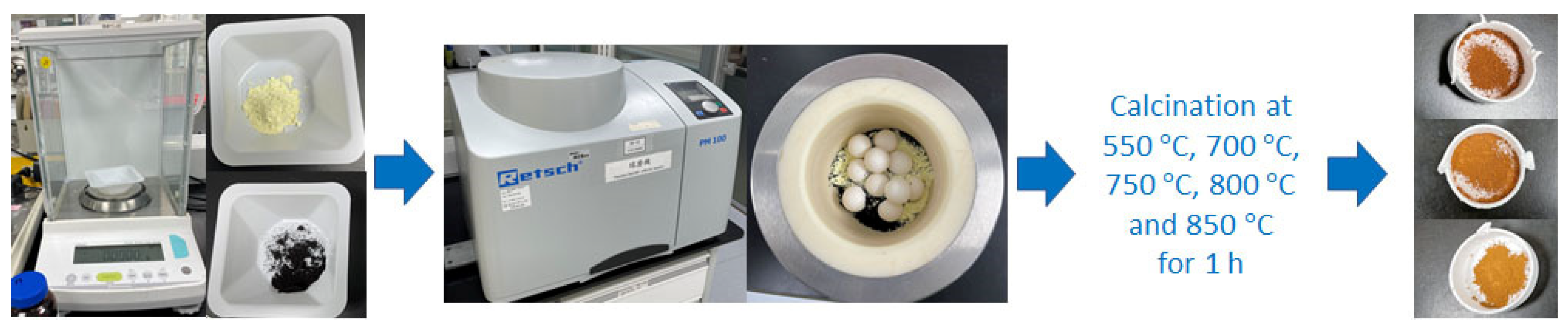
2.1. Synthesis
2.2. Characterization
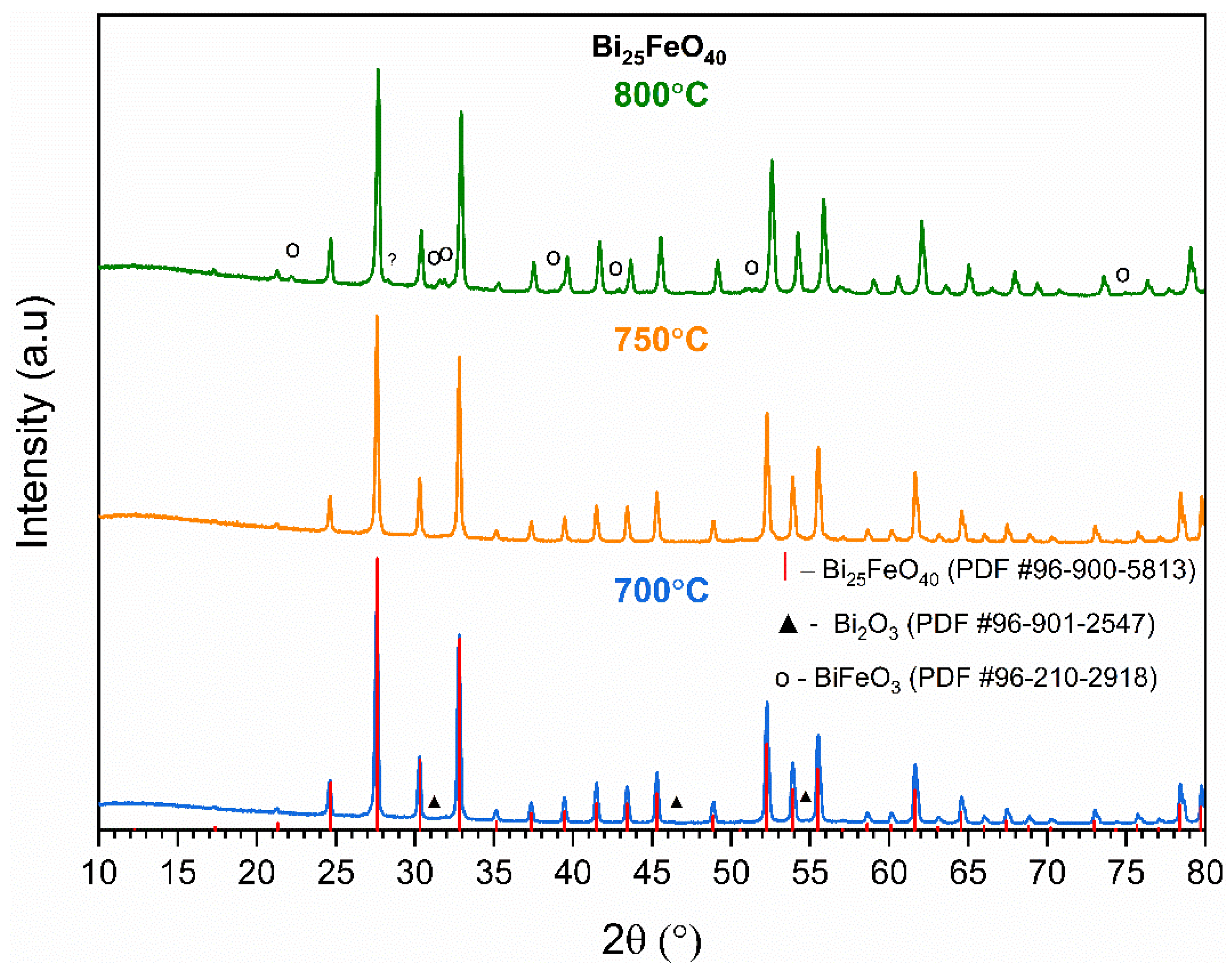
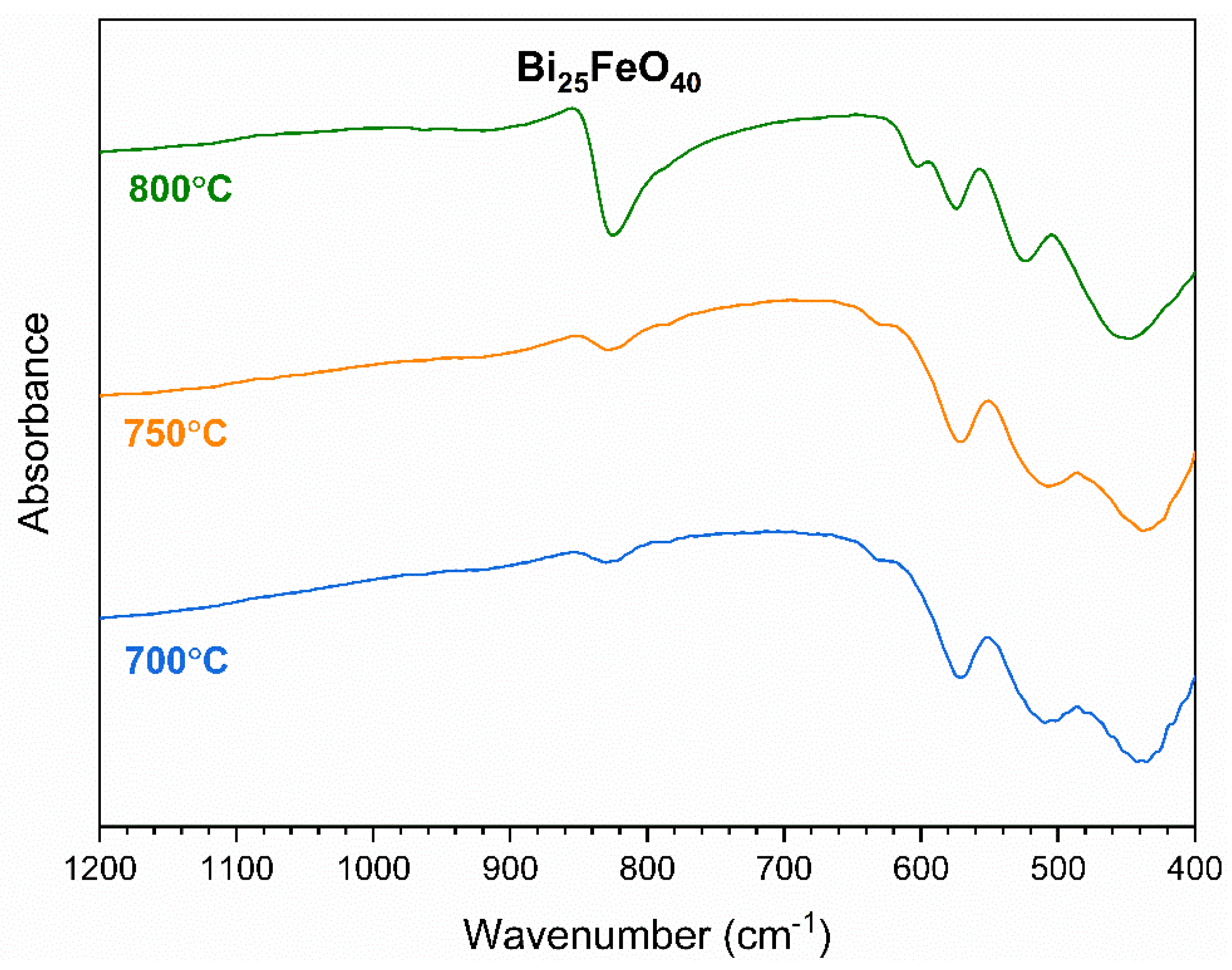
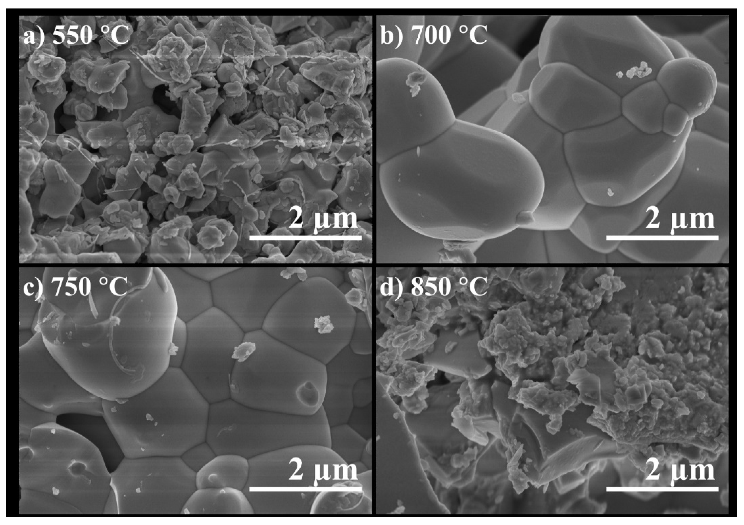
3. Results and Discussion
4. Conclusions
Supplementary Materials
Author Contributions
Funding
Data Availability Statement
Conflicts of Interest
References
- Hussain, S.; Mubeen, I.; Ullah, N.; Shahab, S.; Shah, U.D.; Khan, B.A.; Zahoor, M.; Ullah, R.; Khan, F.A.; Sultan, M.A. Modern Diagnostic Imaging Technique Applications and Risk Factors in the Medical Field: A Review. Biomed. Res. Int. 2022, 2022, 5164970. [Google Scholar] [CrossRef] [PubMed]
- Aimacana, C.M.C.; Perez, D.A.Q.; Pinto, S.R.; Debut, A.; Attia, M.F.; Santos-Oliveira, R.; Whitehead, D.C.; Terencio, T.; Alexis, F.; Dahoumane, S.A. Polytetrafluoroethylene-like Nanoparticles as a Promising Contrast Agent for Dual Modal Ultrasound and X-ray Bioimaging. ACS Biomater. Sci. Eng. 2021, 7, 1181–1191. [Google Scholar] [CrossRef] [PubMed]
- Ventura, M.; Sun, Y.; Rusu, V.; Laverman, P.; Borm, P.; Heerschap, A.; Oosterwijk, E.; Boerman, O.C.; Jansen, J.A.; Walboomers, X.F. Dual contrast agent for computed tomography and magnetic resonance hard tissue imaging. Tiss. Eng. C Methods 2013, 19, 405–416. [Google Scholar] [CrossRef] [PubMed]
- Zhu, T.; Ma, X.; Chen, R.; Ge, Z.; Xu, J.; Shen, X.; Jia, L.; Zhou, T.; Luo, Y.; Ma, T. Using fluorescently-labeled magnetic nanocomposites as a dual contrast agent for optical and magnetic resonance imaging. Biomater. Sci. 2017, 5, 1090–1100. [Google Scholar] [CrossRef] [PubMed]
- Farwell, M.D.; Pryma, D.A.; Mankoff, D.A. PET/CT imaging in cancer: Current applications and future directions. Cancer 2014, 120, 3433–3445. [Google Scholar] [CrossRef] [PubMed]
- Zhang, Y.; García-Gabilondo, M.; Grayston, A.; Feiner, I.V.J.; Anton-Sales, I.; Loiola, R.A.; Llop, J.; Ramos-Cabrer, P.; Barba, I.; Garcia-Dorado, D.; et al. PLGA protein nanocarriers with tailor-made fluorescence/MRI/PET imaging modalities. Nanoscale 2020, 12, 4988–5002. [Google Scholar] [CrossRef] [PubMed]
- Westbrook, C.; Talbot, J. MRI in Practice; Wiley: New York, NY, USA, 2018; p. 416. [Google Scholar]
- El-Hammadi, M.M.; Arias, J.L. Iron oxide-based multifunctional nanoparticulate systems for biomedical applications: A patent review (2008–present). Expert Opin. Ther. Pat. 2015, 25, 691–709. [Google Scholar] [CrossRef] [PubMed]
- Hsieh, S.-C.; Teng, N.-C.; Lin, C.-K.; Lee, P.-Y.; Ji, D.-Y.; Chen, C.-C.; Ke, E.-S.; Lee, S.-Y.; Yang, J.-C. A Novel Accelerator for Improving the Handling Properties of Dental Filling Materials. J. Endod. 2009, 35, 1292–1295. [Google Scholar] [CrossRef] [PubMed]
- Chen, C.-C.; Hsieh, S.-C.; Teng, N.-C.; Kao, C.-K.; Lee, S.-Y.; Lin, C.-K.; Yang, J.-C. Radiopacity and cytotoxicity of Portland cement containing zirconia doped bismuth oxide radiopacifiers. J. Endod. 2014, 40, 251–254. [Google Scholar] [CrossRef] [PubMed]
- Smart, L.E.; Moore, E.A. Solid State Chemistry: An Introduction, 4th ed.; Taylor & Francis: Boca Raton, FL, USA, 2012. [Google Scholar]
- Kumar, A.; Dutta, S.; Kim, S.; Kwon, T.; Patil, S.S.; Kumari, N.; Jeevanandham, S.; Lee, I.S. Solid-State Reaction Synthesis of Nanoscale Materials: Strategies and Applications. Chem. Rev. 2022, 122, 12748–12863. [Google Scholar] [CrossRef] [PubMed]
- Sharmin, F.; Basith, M.A. Simple Low Temperature Technique to Synthesize Sillenite Bismuth Ferrite with Promising Photocatalytic Performance. ACS Omega 2022, 7, 34901–34911. [Google Scholar] [CrossRef] [PubMed]
- Kumar, A.M.; Ragavendran, V.; Mayandi, J.; Ramachandran, K.; Jayakumar, K. Influence of PVP on Bi25FeO40 microcubes for Supercapacitors and Dye-Sensitized Solar Cells applications. J. Mater. Sci. Mater. Electr. 2022, 33, 9512–9524. [Google Scholar] [CrossRef]
- Jebari, H.; Tahiri, N.; Boujnah, M.; El Bounagui, O.; Boudad, L.; Taibi, M.; Ez-Zahraouy, H. Structural, optical, dielectric, and magnetic properties of iron-sillenite Bi25FeO40. Appl. Phys. 2022, 128, 842. [Google Scholar] [CrossRef]
- Nayak, A.K.; Gopalakrishnan, T. Phase- and Crystal Structure-Controlled Synthesis of Bi2O3, Fe2O3, and BiFeO3 Nanomaterials for Energy Storage Devices. ACS Appl. Nanomater. 2022, 5, 14663–14676. [Google Scholar] [CrossRef]
- Xiong, Z.W.; Cao, L.H. Tailoring morphology, enhancing magnetization and photocatalytic activity via Cr doping in Bi25FeO40. J. Alloys Compd. 2019, 773, 828–837. [Google Scholar] [CrossRef]
- Zhang, Y.; Cao, S.H.; Liang, C.; Shen, J.M.; Chen, Y.Q.; Feng, Y.C.; Chen, H.; Liu, R.; Jiang, F. Electrocatalytic performance of Sb-modified Bi25FeO40 for nitrogen fixation. J. Colloid Interf. Sci. 2021, 593, 335–344. [Google Scholar] [CrossRef] [PubMed]
- Juwita, E.; Sulistiani, F.A.; Darmawan, M.Y.; Istiqomah, N.I.; Suharyadi, E. Microstructural, optical, and magnetic properties and specific absorption rate of bismuth ferrite/SiO2 nanoparticles. Mater. Res. Express 2022, 9, 076101. [Google Scholar] [CrossRef]
- Wu, L.; Dong, C.H.; Chen, H.; Yao, J.L.; Jiang, C.J.; Xue, D.S. Hydrothermal Synthesis and Magnetic Properties of Bismuth Ferrites Nanocrystals with Various Morphology. J. Am. Ceram. Soc. 2012, 95, 3922–3927. [Google Scholar] [CrossRef]
- Yotburut, B.; Yamwong, T.; Thongbai, P.; Maensiri, S. Synthesis and characterization of coprecipitation-prepared La-doped BiFeO3 nanopowders and their bulk dielectric properties. Jpn. J. Appl. Phys. 2014, 53, 06JG13. [Google Scholar] [CrossRef]
- Koeferstein, R. Synthesis, phase evolution and properties of phase-pure nanocrystalline BiFeO3 prepared by a starch-based combustion method. J. Alloys Compd. 2014, 590, 324–330. [Google Scholar] [CrossRef]
- Goldman, A.R.; Fredricks, J.L.; Estroff, L.A. Exploring reaction pathways in the hydrothermal growth of phase-pure bismuth ferrites. J. Cryst. Growth 2017, 468, 104–109. [Google Scholar] [CrossRef]
- Sansom, G.; Rattanakam, R.; Jettanasen, J. Effects of Scaling Up on the Phase Evolution of Microcrystalline Bismuth Ferrite during Hydrothermal Process. E-J. Surf. Sci. Nanotechnol. 2022, 20, 85–89. [Google Scholar] [CrossRef]
- Yang, X.; Xu, G.; Ren, Z.H.; Weng, W.J.; Du, P.Y.; Shen, G.; Han, G.R. Effect of PVA Adding Amount on Phase-Controlled Synthesis and Morphology Evolution of the Bismuth Ferrite by Assisted Hydrothermal Reaction Route. Rare Met. Mater. Eng. 2012, 41, 247–249. [Google Scholar]
- Koeferstein, R.; Buttlar, T.; Ebbinghaus, S.G. Investigations on Bi25FeO40 powders synthesized by hydrothermal and combustion-like processes. J. Solid State Chem. 2014, 217, 50–56. [Google Scholar] [CrossRef]
- Ji, W.D.; Li, M.M.; Zhang, G.; Wang, P. Controlled synthesis of Bi25FeO40 with different morphologies: Growth mechanism and enhanced photo-Fenton catalytic properties. Dalton Trans. 2017, 46, 10586–10593. [Google Scholar] [CrossRef] [PubMed]
- Kumar, A.M.; Ragavendran, V.; Mayandi, J.; Ramachandran, K.; Jayakumar, K. Phase dependent electrochemical characteristics of bismuth ferrite: A bifunctional electrocatalyst for Supercapacitors and Dye-Sensitized Solar Cells. Colloids Surf. A Physicochem. Eng. Asp. 2023, 656, 130529. [Google Scholar] [CrossRef]
- Ren, L.; Lu, S.Y.; Fang, J.Z.; Wu, Y.; Chen, D.Z.; Huang, L.Y.; Chen, Y.F.; Cheng, C.; Liang, Y.; Fang, Z.Q. Enhanced degradation of organic pollutants using Bi25FeO40 microcrystals as an efficient reusable heterogeneous photo-Fenton like catalyst. Catal. Today 2017, 281, 656–661. [Google Scholar] [CrossRef]
- Zou, W.J.; Dong, J.T.; Ji, M.X.; Wang, B.; Li, Y.J.; Yin, S.; Li, H.M.; Xia, J.X. Synthesis of Bi25FeO40 Nanoparticles with Oxygen Vacancies via Ball Milling for Fenton Oxidation of Tetracycline Hydrochloride and Reduction of Cr(VI). ACS Appl. Nano Mater. 2023, 6, 4309–4318. [Google Scholar] [CrossRef]
- Sun, A.W.; Chen, H.; Song, C.Y.; Jiang, F.; Wang, X.; Fu, Y.S. Magnetic Bi25FeO40-graphene catalyst and its high visible-light photocatalytic performance. RSC Adv. 2013, 3, 4332–4340. [Google Scholar] [CrossRef]
- Li, F.H.; Zhou, J.K.; Gao, C.J.; Qiu, H.X.; Gong, Y.L.; Gao, J.H.; Liu, Y.; Gao, J.P. A green method to prepare magnetically recyclable Bi/Bi25FeO40-C nanocomposites for photocatalytic hydrogen generation. Appl. Surf. Sci. 2020, 521, 146342. [Google Scholar] [CrossRef]
- Jalil, M.A.; Chowdhury, S.S.; Sakib, M.A.; Yousuf, S.M.E.H.; Ashik, E.K.; Firoz, S.H.; Basith, M.A. Temperature-dependent phase transition and comparative investigation on enhanced magnetic and optical properties between sillenite and perovskite bismuth ferrite-rGO nanocomposites. J. Appl. Phys. 2017, 122, 084902. [Google Scholar] [CrossRef]
- Basith, M.A.; Ahsan, R.; Zarin, I.; Jalil, M.A. Enhanced photocatalytic dye degradation and hydrogen production ability of Bi25FeO40-rGO nanocomposite and mechanism insight. Sci. Rep. 2018, 8, 11090. [Google Scholar] [CrossRef] [PubMed]
- Huang, Y.; Zhang, X.Y.; Zhu, G.X.; Gao, Y.; Cheng, Q.F.; Cheng, X.W. Synthesis of silver phosphate/sillenite bismuth ferrite/graphene oxide nanocomposite and its enhanced visible light photocatalytic mechanism. Separ. Purif. Technol. 2019, 215, 490–499. [Google Scholar] [CrossRef]
- de Gois, M.M.; Araujo, W.P.; da Silva, R.B.; da Luz, G.E.; Soares, J.M. Bi25FeO40-Fe3O4-Fe2O3 composites: Synthesis, structural characterization, magnetic and UV-visible photocatalytic properties. J. Alloys Compd. 2019, 785, 598–602. [Google Scholar] [CrossRef]
- Wang, G.M.; Cheng, D.; He, T.C.; Hu, Y.Y.; Deng, Q.R.; Mao, Y.W.; Wang, S.G. Enhanced visible-light responsive photocatalytic activity of Bi25FeO40/Bi2Fe4O9 composites and mechanism investigation. J. Mater. Sci. Mater. Electron. 2019, 30, 10923–10933. [Google Scholar] [CrossRef]
- Wang, Y.F.; Xu, C.X.; Yan, L.; Li, J. Synthesis of BiFeO3/Bi25FeO40 heterojunction structure and precise adjustment of forbidden band width. Mater. Chem. Phys. 2023, 305, 127935. [Google Scholar] [CrossRef]
- Xu, C.X.; Wang, Y.F.; Wang, Q.; Li, J.; Yan, L. Phase transformation and heterojunction nanostructures of bismuth iron oxide. J. Mater. Sci. Mater. Electron. 2023, 34, 2236. [Google Scholar] [CrossRef]
- Lee, E.J.; Heo, W.C.; Park, J.W.; Chang, Y.; Bae, J.-E.; Chae, K.S.; Kim, T.J.; Park, J.A.; Lee, G.H. D-Glucuronic Acid Coated Gd(IO3)3·2H2O Nanomaterial as a Potential T1 MRI-CT Dual Contrast Agent. Eur. J. Inorg. Chem. 2013, 16, 2858–2866. [Google Scholar] [CrossRef]
- Sharma, V.K.; Alipour, A.; Soran-Erdem, Z.; Aykut, Z.G.; Demir, H.V. Highly monodisperse low-magnetization magnetite nanocubes as simultaneous T1-T2 MRI contrast agents. Nanoscale 2015, 7, 10519–10526. [Google Scholar] [CrossRef] [PubMed]
- Park, J.C.; Lee, G.T.; Kim, H.-K.; Sung, B.; Lee, Y.; Kim, M.; Chang, Y.; Seo, J.H. Surface Design of Eu-Doped Iron Oxide Nanoparticles for Tuning the Magnetic Relaxivity. ACS Appl. Mater. Interf. 2018, 10, 25080–25089. [Google Scholar] [CrossRef] [PubMed]
- Illert, P.; Waengler, B.; Waengler, C.; Zoellner, F.; Uhrig, T.; Litau, S.; Pretze, M.; Roeder, T. Functionalizable composite nanoparticles as a dual magnetic resonance imaging/computed tomography contrast agent for medical imaging. J. Appl. Polym. Sci. 2019, 136, 47571. [Google Scholar] [CrossRef]
- Eguia-Eguia, S.I.; Gildo-Ortiz, L.; Perez-Gonzalez, M.; Tomas, S.A.; Arenas-Alatorre, J.A.; Santoyo-Salazar, J. Magnetic domains orientation in (Fe3O4/γ-Fe2O3) nanoparticles coated by Gadolinium-diethylenetriaminepentaacetic acid (Gd3+-DTPA). Nano Express 2021, 2, 020019. [Google Scholar] [CrossRef]
- Kun Yang, K.; Peng, H.; Wen, Y.; Li, N. Re-examination of characteristic FTIR spectrum of secondary layer in bilayer oleic acid-coated Fe3O4 nanoparticles. Appl. Surf. Sci. 2010, 256, 3093–3097. [Google Scholar] [CrossRef]
- Husain, S.; Irfansyah, M.; Haryanti, N.H.; Suryajaya, S.; Arjo, S.; Maddu, A. Synthesis and characterization of Fe3O4 magnetic nanoparticles from iron ore. J. Phys. Conf. Ser. 2019, 1242, 012021. [Google Scholar] [CrossRef]
- Li, W. Facile synthesis of monodisperse Bi2O3 nanoparticles. Mater. Chem. Phys. 2006, 99, 174–180. [Google Scholar] [CrossRef]
- Labib, S. Preparation, characterization and photocatalytic properties of doped and undoped Bi2O3. J. Saudi Chem. Soc. 2017, 21, 664–672. [Google Scholar] [CrossRef]
- Wu, X.; Wang, X.; Chen, X.; Yang, X.; Ma, Q.; Xu, G.; Yu, L.; Ding, J. Injectable and thermosensitive hydrogels mediating a universal macromolecular contrast agent with radiopacity for noninvasive imaging of deep tissues. Bioact. Mater. 2021, 6, 4717–4728. [Google Scholar] [CrossRef] [PubMed]
- Dukic, W.; Delija, B.; Derossi, D.; Dadic, I. Radiopacity of composite dental materials using a digital X-ray system. Dent. Mater. J. 2012, 31, 47–53. [Google Scholar] [CrossRef] [PubMed]
- Hitij, T.; Fidler, A. Radiopacity of dental restorative materials. Clin. Oral Investig. 2013, 17, 1167–1177. [Google Scholar] [CrossRef] [PubMed]
- Fu, N.; Li, A.; Zhang, J.; Zhang, P.; Zhang, H.; Yang, S.; Zhang, J. Liposome-camouflaged iodinated mesoporous silica nanoparticles with high loading capacity, high hemodynamic stability, high biocompatibility and high radiopacity. Int. J. Pharmaceut. 2024, 650, 123700. [Google Scholar] [CrossRef] [PubMed]
- Emonde, C.V.; Eggers, M.E.; Wichmann, M.; Hurschler, C.; Ettinger, M.; Denkena, B. Radiopacity Enhancements in Polymeric Implant Biomaterials: A Comprehensive Literature Review. ACS Biomater. Sci. Eng. 2024, 10, 1323–1334. [Google Scholar] [CrossRef] [PubMed]

| Name of Control Sample | m(BaSO4), G | m(Laponite Solution), G |
|---|---|---|
| A1 | 0.1 | 0.9 |
| B1 | 0.2 | 0.8 |
| C1 | 0.3 | 0.7 |
| A2 | 0.2 | 1.8 |
| B2 | 0.4 | 1.6 |
| C2 | 0.6 | 1.4 |
| Name of Control Sample | Grayscale Value | mm Al |
|---|---|---|
| A1 | 10.773 | 0.355 |
| B1 | 84.596 | 3.387 |
| C1 | 142.910 | 2.922 |
| A2 | 53.893 | 0.681 |
| B2 | 83.008 | 4.257 |
| C2 | 159.511 | 5.092 |
| Name of Control Sample | Investigated Samples | Background | Iron–Sillenite | ||
|---|---|---|---|---|---|
| Grayscale Value | mm Al | Grayscale Value | mm Al | mm Al | |
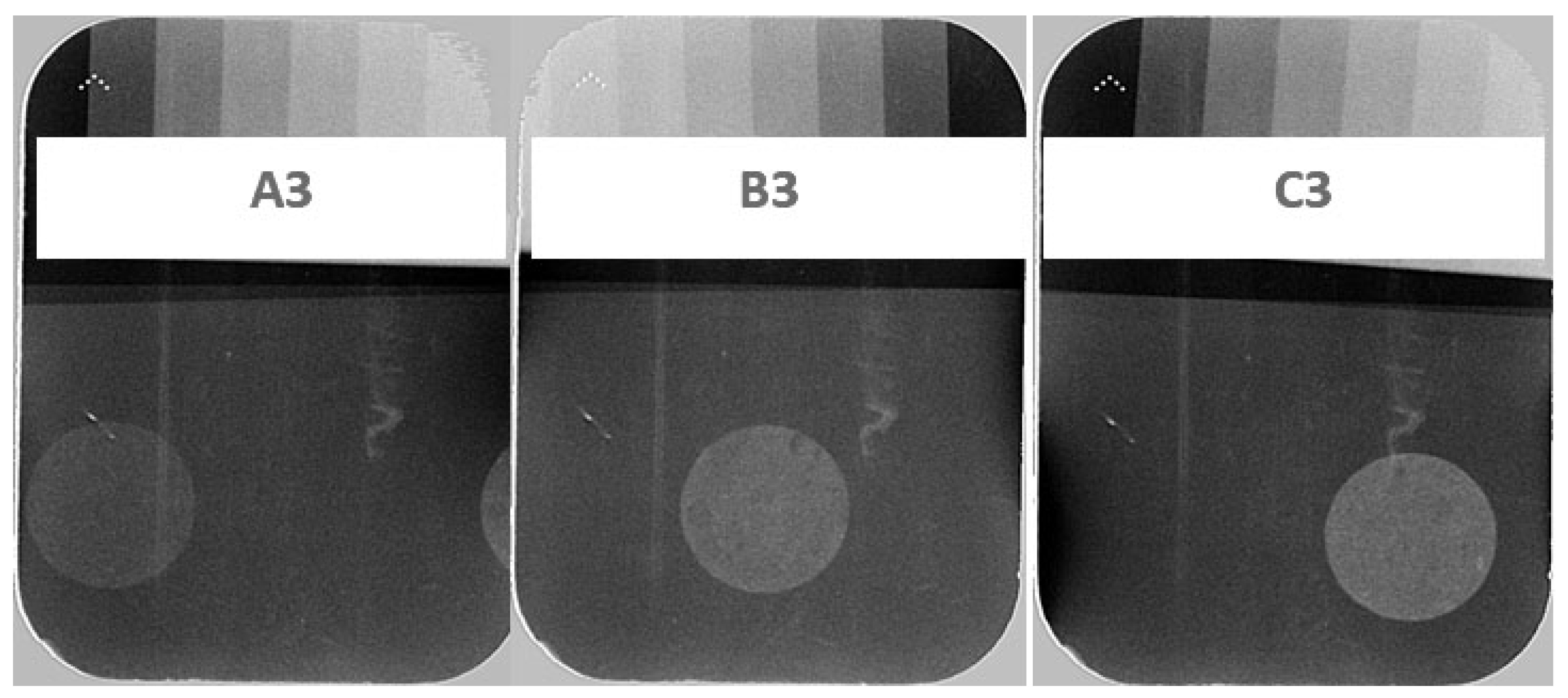
| A3 | 68.406 ± 6.534 | 2.279 | 60.143 ± 6.382 | 1.920 | 0.359 |
| B3 | 102.346 ± 7.559 | 3.379 | 75.137 ± 6.242 | 2.274 | 1.105 |
| C3 | 100.070 ± 7.654 | 3.790 | 60.731 ± 6.652 | 1.967 | 1.823 |
Disclaimer/Publisher’s Note: The statements, opinions and data contained in all publications are solely those of the individual author(s) and contributor(s) and not of MDPI and/or the editor(s). MDPI and/or the editor(s) disclaim responsibility for any injury to people or property resulting from any ideas, methods, instructions or products referred to in the content. |
© 2024 by the authors. Licensee MDPI, Basel, Switzerland. This article is an open access article distributed under the terms and conditions of the Creative Commons Attribution (CC BY) license (https://creativecommons.org/licenses/by/4.0/).
Share and Cite
Vistorskaja, D.; Yang, J.-C.; Wu, Y.-T.; Chang, L.-Y.; Lu, P.-W.; Zarkov, A.; Grigoraviciute, I.; Kareiva, A. Synthesis and Characterization of Iron–Sillenite for Application as an XRD/MRI Dual-Contrast Agent. Crystals 2024, 14, 706. https://doi.org/10.3390/cryst14080706
Vistorskaja D, Yang J-C, Wu Y-T, Chang L-Y, Lu P-W, Zarkov A, Grigoraviciute I, Kareiva A. Synthesis and Characterization of Iron–Sillenite for Application as an XRD/MRI Dual-Contrast Agent. Crystals. 2024; 14(8):706. https://doi.org/10.3390/cryst14080706
Chicago/Turabian StyleVistorskaja, Diana, Jen-Chang Yang, Yu-Tzu Wu, Liang-Yu Chang, Po-Wen Lu, Aleksej Zarkov, Inga Grigoraviciute, and Aivaras Kareiva. 2024. "Synthesis and Characterization of Iron–Sillenite for Application as an XRD/MRI Dual-Contrast Agent" Crystals 14, no. 8: 706. https://doi.org/10.3390/cryst14080706
APA StyleVistorskaja, D., Yang, J.-C., Wu, Y.-T., Chang, L.-Y., Lu, P.-W., Zarkov, A., Grigoraviciute, I., & Kareiva, A. (2024). Synthesis and Characterization of Iron–Sillenite for Application as an XRD/MRI Dual-Contrast Agent. Crystals, 14(8), 706. https://doi.org/10.3390/cryst14080706