Article

Potential of Y2Sn2O7:Eu3+, Dy3+ Inorganic Nanophosphors in Latent Fingermark Detection

1
Laboratory of Inorganic Chemistry, Faculty of Sciences of Sfax, Sfax University, Sfax 3018, Tunisia
2
Laboratory of Dielectric and Photonic Materials, Faculty of Sciences of Sfax, Sfax University, Sfax 3018, Tunisia
3
Dipartimento di Fisica, Università di Pisa, Largo B. Pontecorvo 3, I-56127 Pisa, Italy
4
Istituto Nanoscienze CNR, Piazza San Silvestro 12, I-56127 Pisa, Italy
5
Istituto Nazionale di Fisica Nucleare-Sezione di Pisa, Largo B. Pontecorvo 3, I-56127 Pisa, Italy
6
Electrical and Computer Engineering Department, Abu Dhabi University, Abu Dhabi 59911, United Arab Emirates
*
Author to whom correspondence should be addressed.
Crystals 2024, 14(4), 300; https://doi.org/10.3390/cryst14040300
Submission received: 9 February 2024 / Revised: 16 March 2024 / Accepted: 17 March 2024 / Published: 24 March 2024

Abstract

:
In this work, we investigated the potential of Eu3+/Dy3+-codoped Y2Sn2O7 fluorescent nanophosphors to visualize latent fingermarks. We prepared these nanophosphors with various doping concentrations by the conventional coprecipitation reaction. The crystal structure, morphology, luminescence properties, and energy transfer mechanisms were studied. The crystalline phase was characterized by X-ray diffraction and crystal structure refinement using the Rietveld method. XRD measurements showed that the samples crystallized in the pure single pyrochlore phase with few more peaks originated from secondary phases and impurities generated during phosphor production, and that Eu3+ ions occupied D3d symmetry sites. The average crystallite size after mechanical grinding was less than 100 nm for all compositions. The optical characterization showed that, when excited under 532 nm, the Eu3+/Dy3+-codoped Y2Sn2O7 samples’ main intense emission peaks were located at 580–707 nm, corresponding to the 5D07Fj (j = l, 2, 3, and 4) transitions of europium. In fact, the 5D07F2 hypersensitive transition is strongly dependent on the local environment and was quite weak in Eu3+:Y2Sn2O7 at low Eu3+ doping levels. We found that the presence of Dy3+ as a codopant permitted enhancing the emission from this transition. The calculated PL CIE coordinates for the synthesized nanophosphors were very close to those of the reddish-orange region and only slightly dependent on the doping level. Various surfaces, including difficult ones (wood and ceramic), were successfully tested for latent fingerprint development with the prepared Eu3+/Dy3+-codoped Y2Sn2O7 fluorescent nanophosphor powder. Thanks to the high contrast obtained, fingerprint ridge patterns at all three levels were highlighted: core (level 1) islands, bifurcation, and enclosure (level 2), and even sweat pores (level 3).

1. Introduction

Since each fingerprint is distinct from others and does not change with age, fingerprint identification technology is well-established in the field of forensic research. Sweat and sebum, which are continuously produced by perspiration, can leave residue on many surfaces when a finger touches them [1,2,3]. Fingerprints discovered at crime scenes are called latent fingerprints, which are unable to be seen with the naked eye; therefore, specific methods to develop them must be used. Different physical, chemical, and optical methods can be used to develop these latent fingermarks [1,4,5,6,7].
With state-of-the-art technology, detecting the first two levels of ridge details corresponding to cores (level 1), bifurcations, and terminations (level 2) is quite easy, but the third level (sweat pores) is rather complicated to see, mainly on difficult surfaces such as wood and ceramic. Level 1 details include broad morphological information such as the overall ridge pattern and the fingerprint ridge flow. The second level of details provides information regarding the pattern agreement of individual fingerprint ridges. Level 3 details include sweat pores, curvature, and spots, as well as fingerprint ridge dimensional elements. As a result, the rapid detection of latent fingerprints, which are originally generated by ridges and furrows in the sebum residue left by the finger skin, requires extra trial-and-error procedures.
Numerous research papers have been reported on organic nanophosphors, which may have significant advantages for latent fingerprint development [8] thanks to their interesting properties such as narrow emission lines, large Stokes shifts, long luminescence lifetime, and high-temperature synthesis. Moreover, such powders show their ability to detect latent fingerprints deposited on difficult surfaces such as wood and ceramic, but they usually develop weak fingerprints under ultraviolet (UV) or laser light.
So far, a variety of techniques have been developed for fingerprint visualization, ranging from quantum dots and single-metal deposition methods to superglue fuming and powder-dusting techniques. These traditional methods face several drawbacks, such as poor detection capability and high toxicity. In addition, very few methods are efficient enough to be able to visualize even the third level of fingerprint details, namely sweat pores. Among these techniques, powder dusting has attracted much attention because of its extreme simplicity and effectiveness, provided that the fluorescent powders have excellent absorption and luminescence characteristics.
However, the most common types of powders used at crime scenes have large and non-uniform sizes, so they can hinder the third-level details in the fingerprint pattern due to their airy nature during brush strokes. To overcome these limitations, trivalent rare earth ion-doped pyrochlore nanoparticles, with A2B2O7 chemical compositions, are among the most promising powders to improve the visualization of latent fingerprints thanks to their high fluorescence capability [9,10]. These nanometer-sized powders are particularly useful on rough surfaces such as wood and multi-colored non-porous objects where normal powders may clutter the surface. In addition, in order to visualize fingerprints well on surfaces of different colors, it is necessary to use fluorescent powders with different color shades to eliminate interference with the background color. Therefore, it is important to study and optimize new materials that have different color shades suitable for this purpose.
Among the various types of pyrochlore, nanopowders based on lanthanide ions (Ln3+) and tin tetracations (Sn4+) are excellent potential hosts for fluorescent ions due to their great chemical stability, high melting point, and small particle size (nm), which depend on the synthesis method and conditions.
In recent years, the red emission of europium ions has been extensively studied both in amorphous and crystalline materials [11,12]. The red emission intensity of Eu3+ ions strongly depends on its local environment [11,13]; therefore, a careful optimization of both the preparation procedure and composition of the compounds is usually crucial for achieving the best performance possible.
On the other hand, Dy3+ ions have been investigated for their photo- and radioluminescence properties [14,15,16]. In fact, they show a white-yellowish emission that appears to be rather stable against changes in the excitation wavelength. Moreover, Dy3+ ions can be used as codopants to tailor the emission properties of rare-earth ions [15].
Europium ions introduced in Y2Sn2O7 have been proven to generate red emission under UV excitation [17,18]. In fact, in our previous study [17], we observed that the hypersensitive 5D07F2 Eu3+ transition showed a low emission intensity at doping levels lower than 10% and became very bright at this level. Given its hypersensitive nature, this behavior can be ascribed to the local crystal environment. Therefore, to enhance the emission in the red region for better latent fingerprint visualization, we propose using dysprosium ions as codopants in this host material.
In fact, in this work, we successfully synthesized Y2Sn2O7:Eu3+, Dy3+ nanophosphors at various doping concentrations by a coprecipitation method and used them to detect even the third level of detail in fingerprints on wood and ceramic. Therefore, our work will be a step forward for detecting the third level of latent fingerprints in crime scenes.

2. Materials and Methods

2.1. Synthesis of Phosphor Samples

In this work, all the starting materials were purchased from Sigma-Aldrich (Merck KGaA, Darmstadt, DE, Germany). Stoichiometric amounts of SnCl2·2H2O, Eu2O3, Dy2O3, and Y2O3 were dissolved in 50 mL of 1 M HCl solution in a beaker, and the excess acid was evaporated out repeatedly. Then, ethylene glycol (40 mL) was added to this solution. The solution was slowly heated up to 100 °C followed by adding 2 g of urea, and the temperature was raised to 140 °C. In this step, ethylene glycol was used as the capping agent and urea was used for hydrolysis. At this temperature, the solution became turbid. The temperature was then increased to 150 °C and kept at this value for around 2 h. The precipitate was collected after the reaction by centrifugation and then washed two times with acetone followed by drying under ambient conditions (overnight). The samples thus prepared were finally calcined at 1300 °C in air at a heating rate of 10 °C per minute followed by 30 min of grinding. In a separate experiment, the undoped Y2Sn2O7 nanoparticles were also synthesized by a similar method taking only the yttrium and tin precursors. After this, the samples were ground for 60 min at 500 rpm, with a 5 min rest period in a zirconium jar containing zirconium balls. The relevant reaction formulas are shown below:
2 (SnCl2·2H2O) + (1 − x − y) Y2O3 + x Eu2O3 + y Dy2O3 → Y2−2x−2y Eu2xDy2y Sn2O7 + gaseous products

2.2. Characterization

The samples’ phase structures were examined utilizing a Panalytical Pro X’Pert MPD (Malvern Panalytical, Malvern, United Kingdom) (40 kV, 30 mA) with CuKa radiation (1.5404 A°) at 30 kV and 15 mA in the range of 10°–70° with a step size of 2θ = 0.02. The crystalline phases were identified by comparing the X-ray patterns of the JCPDS database structure. Refining of the crystallographic parameters was performed using the Rietveld fit program.
The hydrodynamic diameter of the powder was measured with Malvern Zetasizer Nano ZS90 (Malvern Panalytical Ltd., Malvern, UK). To separate nanoparticle aggregates, the powder was dispersed in deionized water and sonicated shortly before the test.
The measurement was performed in a glass cuvette with a round aperture at room temperature. The average particle size was calculated by Malvern zeta-sizer software Malvern Zetasizer Nano ZS90 using Dispersion Technology Software 5.1 starting from the autocorrelation function of the light scattered by the nanoparticles.
We observed the morphology of the samples with a high-resolution field emission scanning electron microscope performed with an FEI (Hillsboro, OR, USA) Quanta 450 FEG system operating in a low vacuum.
Photoluminescence (PL) spectra at room temperature were measured using an iHR320 (HORIBA, Ltd., Kyoto, Japan) spectrometer under a fixed excitation wavelength at 532 nm, utilizing a 50 mW laser as an excitation source for emission measurements. The emission monochromator was scanned in the wavelength region between 550 and 730 nm with a resolution of 0.06 nm.

3. Results and Discussion

3.1. Powder XRD Analysis
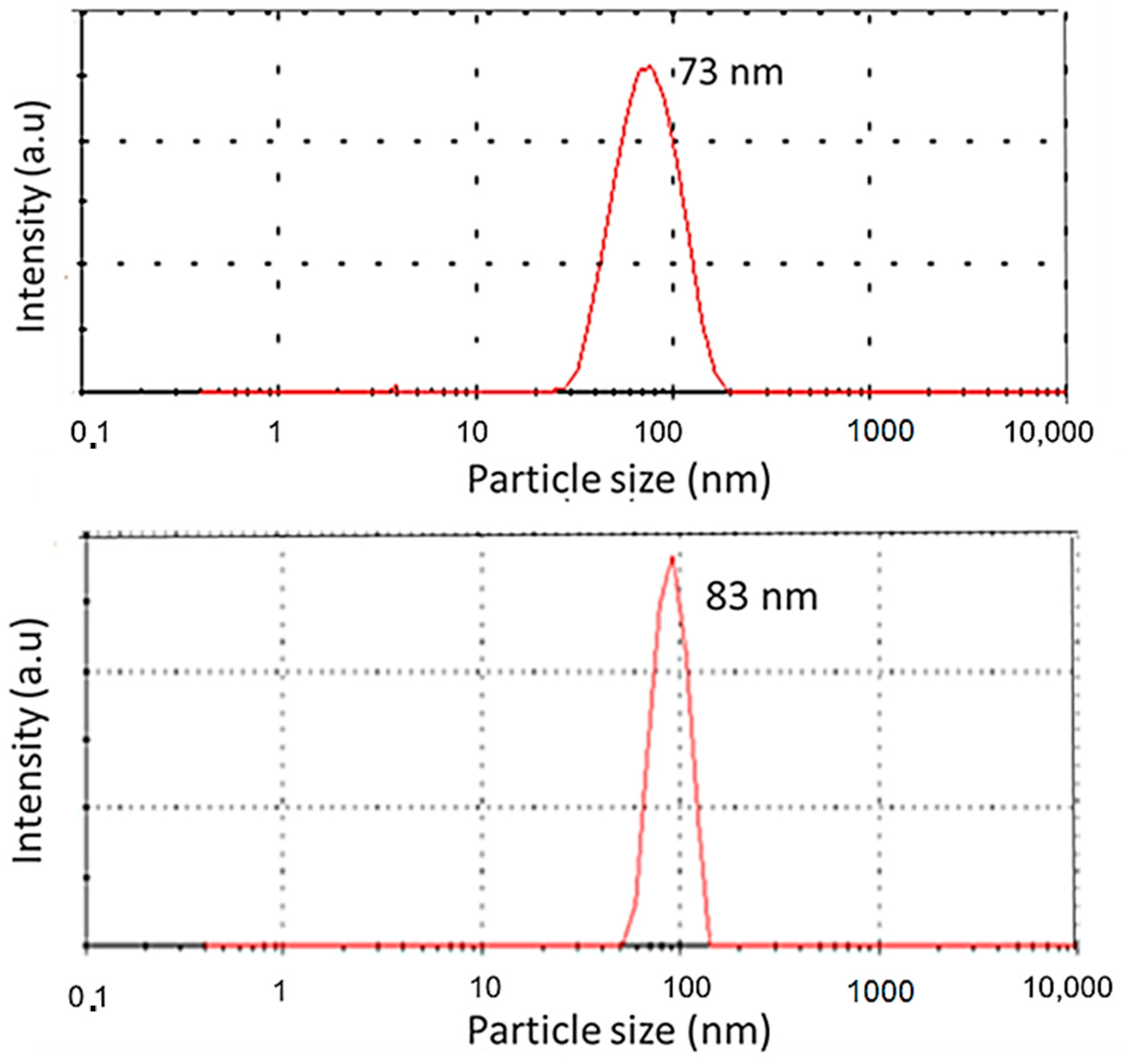
The room-temperature powder XRD patterns of the undoped and 2xEu3+/2yDy3+-codoped Y2Sn2O7 (2x/2y = 0.2/0.01, 0.05/0.05, 0.05/0.2, 0.2/0.05, and 0.1/0.1) samples are shown in Figure 1. The XRD pattern for the parent Y2Sn2O7 is provided for reference. For the (2x/2y = 0.05/0.05, 0.2/0.05, and 0.1/0.1) samples, the main crystalline peaks (222), (400), (440), and (622) correspond to the powder diffraction standards, as shown in Figure 1a. These results confirm that the samples annealed at 1300 °C crystallized in a single pyrochlore phase, and the crystal structure belonged to the F d 3 ¯ m cubic system space group. Since the ionic radius of the Eu3+ (r = 0.95 Å) ion [19] and of the Dy3+ (r = 0.91 Å) ion [20] are similar to that of the Y3+ ion (r = 0.92 Å), Eu3+ and Dy3+ ions can effectively bind to the Y2Sn2O7 host lattice; thus, replacing the Y3+ ion does not distort the crystal structure. However, for the (2x/2y = 0.2/0.01 and 0.05/0.2) samples with a high doping concentration, a few more peaks are visible in the experimental XRD pattern, possibly from secondary phases and impurities generated during phosphor production [21].
Using Rietveld refinements, the element parameters and atomic coordinates obtained by the least squares fitting procedure are shown in Table 1 [22,23]. Figure 1b shows an example of the result of the fitting procedure. There are two possible sites for oxygen ions, called O and O′. O′ ions are in undisturbed locations relative to the fluorite structure (3/8, 3/8, 3/8) and are coordinated by the Y/Eu/Dy cation tetrahedral. Instead, O (3/8, 1/8, 1/8) binds to Y/Eu/Dy and Sn at the next empty 8a site [24].
The Rietveld crystal structure refinement results are presented in Table 2. The cubic pyrochlore structure was used to find the lattice parameters, site mixing, and the number of phases. Structural parameters, such as Rp (profile fitting of R-value), Rwp (weighted profile of R-value), and χ2 (goodness-of-fit factor), obtained from the Rietveld refinement are also presented. The low values of χ2 and profile parameters (Rp, Rwp) indicate that the derived samples were of better quality and the refinements of the samples were effective.

3.2. DLS Analysis

The hydrodynamic diameters acquired from the measurements of the samples Y1.75Eu0.2Dy0.05Sn2O7 and Y1.8Eu0.1Dy0.1Sn2O7 were analyzed. We used deionized water as a solvent to ensure the suspensions had good stability, as reported in previous research works [17,25]. We compared the hydrodynamic radius of the samples before and after mechanical grinding. Before grinding, the lowest measured size value was around 291 nm, and the highest diameter was 456 nm. We used high-energy ball milling to crush and grind the materials to maintain the correct fluorescence and to obtain homogeneous and metastable nanocrystalline phases. The smallest-diameter powders were then separated by centrifugation. Usefully, we obtained good results up to 73 nm and 83 corresponding to the Y1.75Eu0.2Dy0.05Sn2O7 and Y1.8Eu0.1Dy0.1Sn2O7 nanopowders, respectively. Thus, the grinder performed an important role in reducing the size of the powders. Figure 2 shows the DLS measurements for the two samples after grinding.

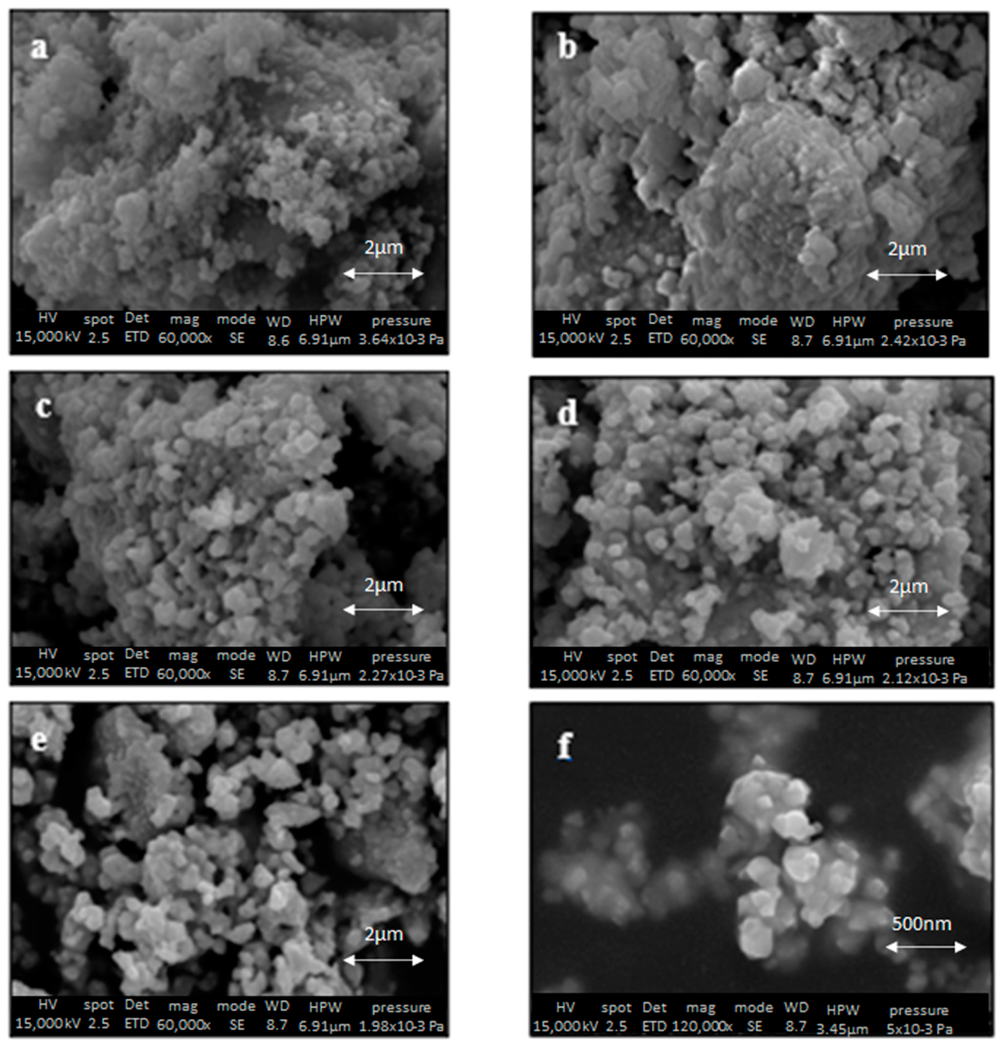
3.3. SEM Characterization

Figure 3 shows some representative SEM images of all the Y2Sn2O7:2xEu/2yDy samples. Figure 3f depicts a typical SEM image of the ground Y2Sn2O7:20%Eu3+/5%Dy3+ sample. The formation of very tiny nanometer-sized particles with diameters less than 100 nm can be plainly seen. Image analysis of more than 100 particles was performed to find the average diameter of each sample. Table 3 shows the different measured sizes of all samples after ball milling. In all cases, an average diameter of about 100 nm or less was observed.

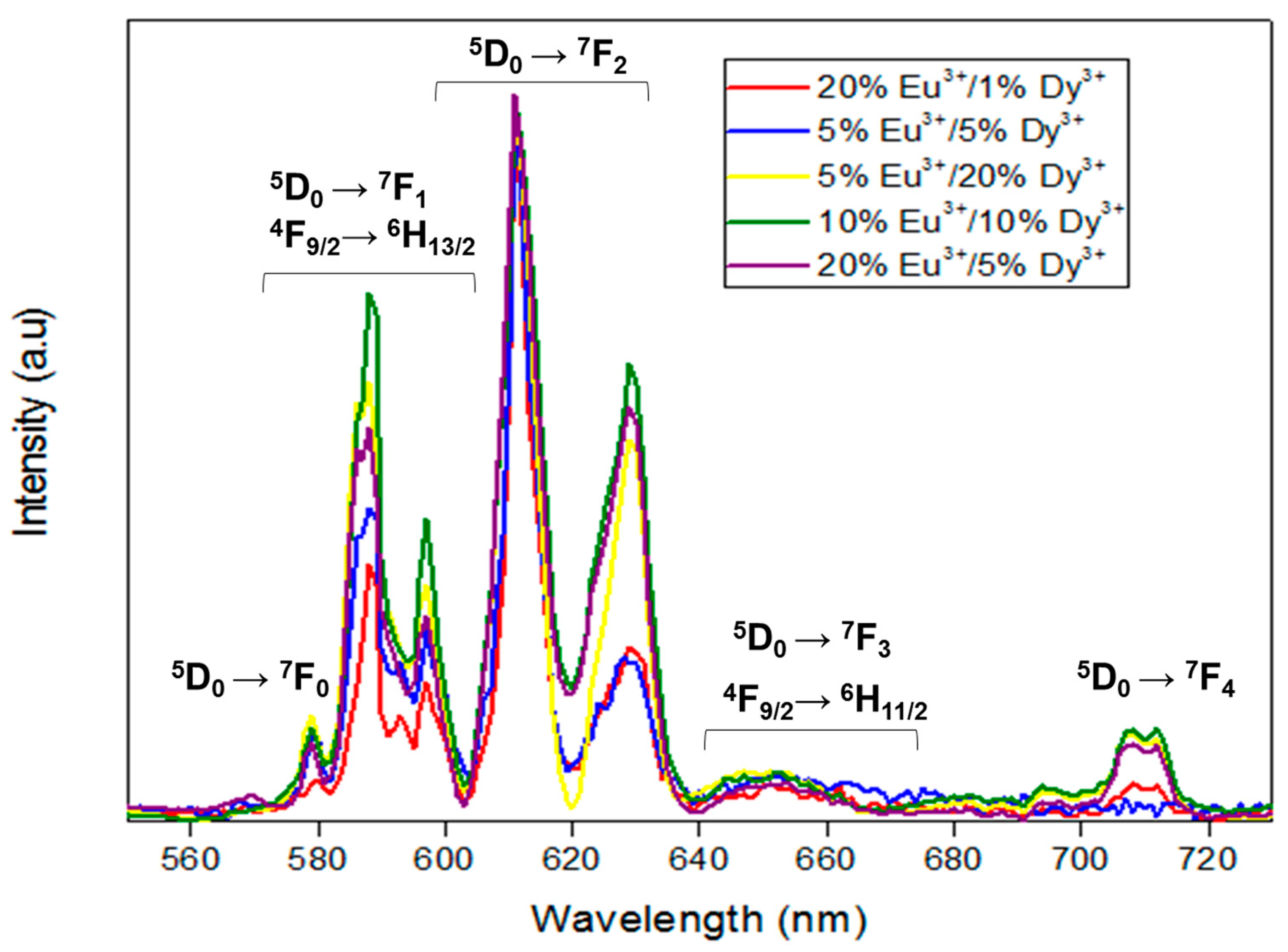
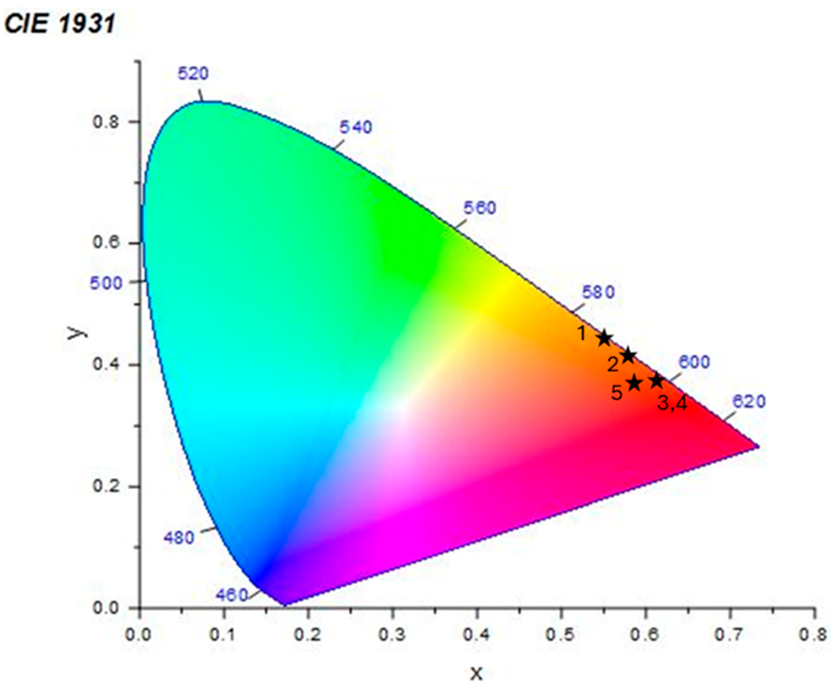
3.4. Photoluminescence

The emission spectra of the Y2Sn2O7:Dy3+, Eu3+ phosphors were recorded with λex = 532 nm. Figure 4 shows the PL spectra of the Y2−2x−2yEuxDySn2O7 (2x/2y = 0.2/0.05, 0.1/0.1, 0.05/0.2, 0.05/0.05, and 0.2/0.01) nanophosphors. All spectra were normalized to the highest peak at 611 nm.
These spectra are in good agreement with the literature [17,18,26,27,28], but, unlike the Y2Sn2O7:Eu3+ singly doped compound [17], this doubly doped composition shows a very intense emission in the red region from the hypersensitive Eu3+ 5D07F2 transition. Several sharp lines are located at 578, 588, 611, 628, and 707 nm [29]. These sharp emission peaks are mainly situated in the red spectral region and correspond to transitions from the excited state 5D0 to lower states 7FJ (J = 0, 1, 2, 3, 4), respectively [30,31]. Due to the screening effect of the outer 5s2 5p6 electrons, the crystal field hardly affects the position of the 4f energy levels of Eu3+; on the contrary, it strongly affects the transition probabilities and their selection rules. Eu3+ ions occupy a site with D3d symmetry in Y2−xEuxSn2O7 [18,32], and this causes electrical and magnetic dipole transitions to occur simultaneously. The 5D07F0 transition observed at 578 nm is forbidden by the Judd–Ofelt theory, but it is usually observed in these crystals due to J-mixing or to the mixing of low-lying charge transfer states into the wavefunctions of the 4f orbitals [17]. The 5D07F1 transition has peaks at 588 and 596 nm and it is magnetic dipolar in nature; therefore, it is relatively insensitive to the crystal field. The 5D07F2 transition is centered at 611 and 626 nm, and its intensity is hypersensitive to the crystal environment [13]. In a previous article, we observed highly different 5D07F2 transition intensities in Y2−xEuxSn2O7 at different doping levels, and we were not able to obtain an intense emission from this transition at a low Eu doping level. In this case, Dy codoping helps in enhancing this emission, probably due to a stabilization of the Eu3+ ions’ environment. Weaker emissions were also observed from the 5D07F3 (around 650 nm) and 5D07F4 (around 710 nm) transitions.
As for Dy:Y2Sn2O7, weak radioluminescence [14] and photoluminescence [15,16] were observed, with the main emission bands located at around 580 nm and 650 nm; therefore, we might expect to observe PL in the same regions. Unfortunately, these are superimposed to the Eu emission bands, so it is not easy to identify specific Dy peaks because their intensity is overwhelmed by the Eu emission bands. However, a careful investigation of the Dy PL features is beyond the scope of this work.
The energy level diagrams of Eu3+/Dy3+ with their main PL emission bands are sketched in Figure 5.
CIE 1931 color coordinates of the phosphors were calculated through commercial software (OriginPro 2019, OriginLab Corporation, Northampton, MA, USA). Figure 6 represents the CIE diagram of the Y2−2x−2yEu2xDy2ySn2O7 (2x/2y = 0.2/0.05, 0.1/0.1, 0.05/0.2, 0.05/0.05, and 0.2/0.01) phosphors. The CIE chromaticity coordinates (x, y) of the Y2Sn2O7:Eu3+/Dy3+phosphors are also reported in Table 4.
The color coordinates (x, y) of the Y1.75Eu0.2 Dy0.05Sn2O7 phosphors were established to be (0.61, 0.38), which are located in the near-red region, as shown in Figure 6. We noticed that with the increasing amount of Dy3+ and decreasing amount of the dopant Eu3+, we observed a small shift to the reddish-orange zone, such as in the case of Y1.75Eu0.05 Dy0.2Sn2O7 (0.56, 0.44) [33].

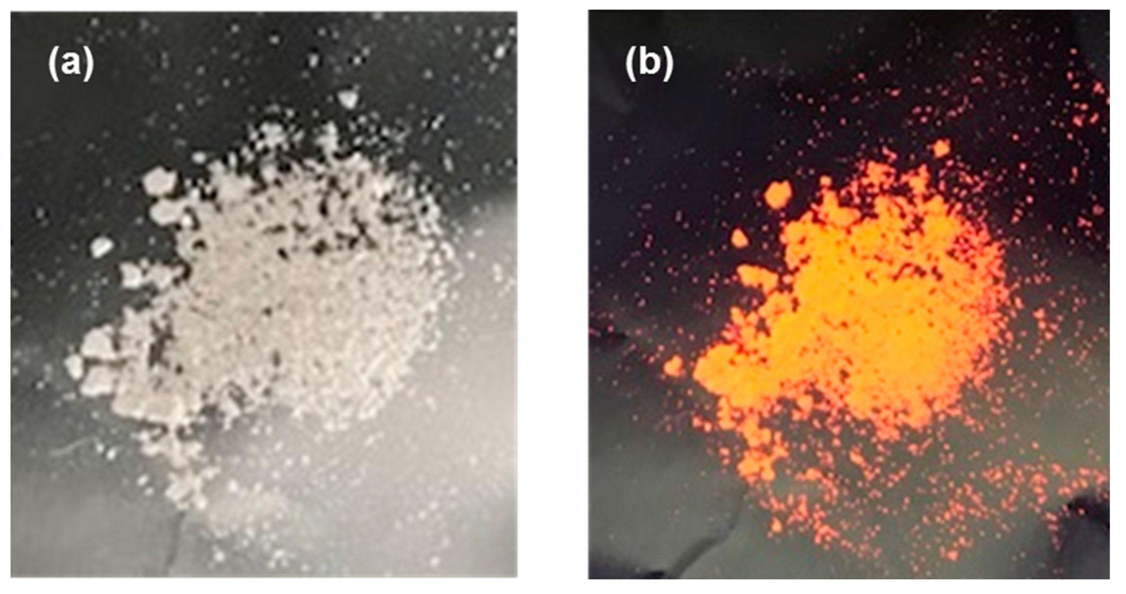
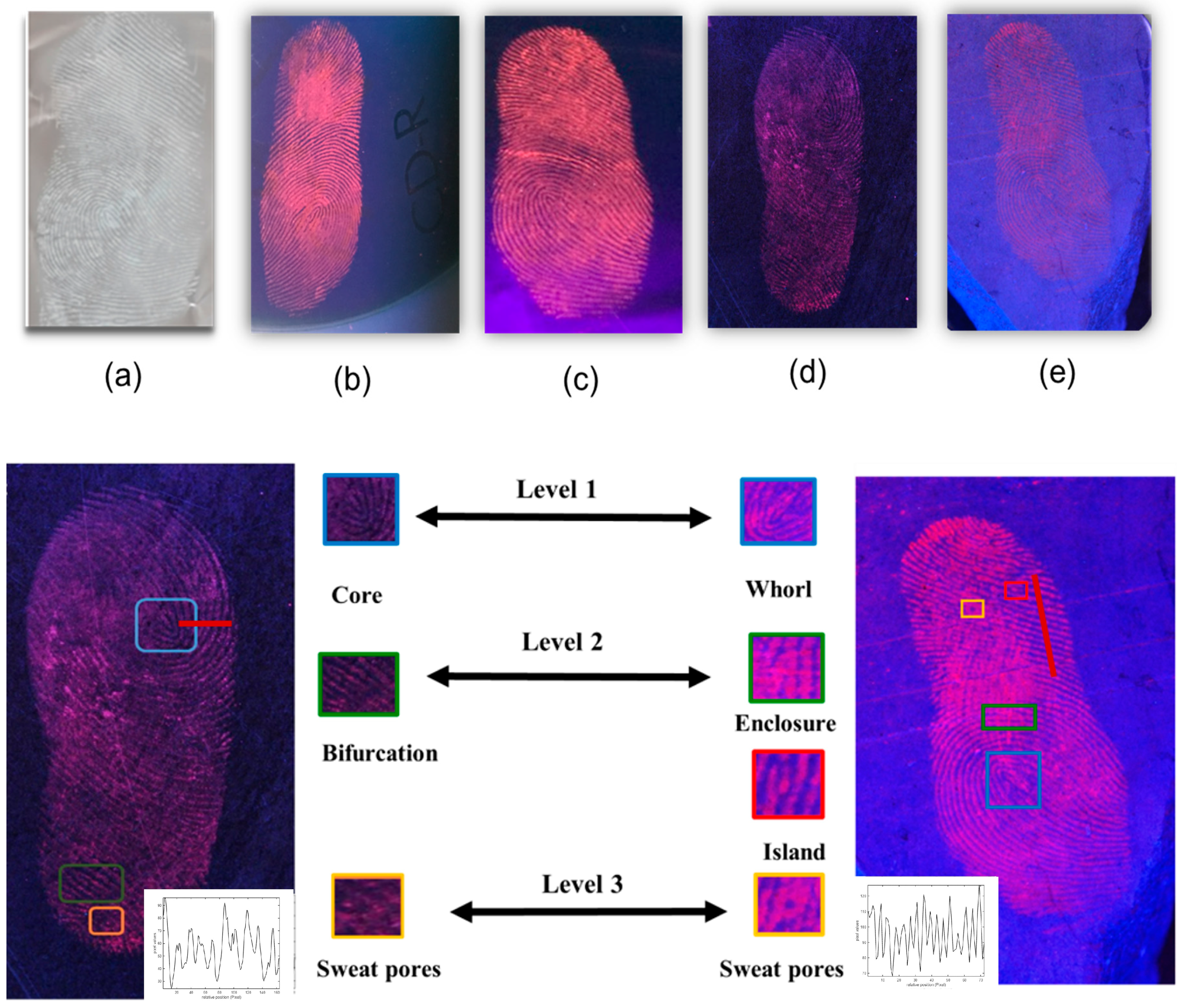
3.5. Detection of Latent Fingerprints

The luminescence properties of our powder Y1.8Eu0.1 Dy0.1Sn2O7 were demonstrated by using a UV lamp (Figure 7). This allowed us to work under an excitation of 254 nm and to observe with the naked eye the existence of luminescence. When the Y1.8Eu0.1 Dy0.1Sn2O7 nanopowder was analyzed under a UV lamp, it was evident that the powder, which was originally white in natural light, turned red-orange under UV light (λ = 254 nm), a characteristic of the existence of luminescence.
We tested the nanophosphors as latent fingerprint (LFP) developers; to this end, we chose different types of surfaces, both porous and non-porous, like CD, aluminum foil, wood, and ceramic. First, the donor washed and cleaned their hands; then, they pressed their finger on the various surfaces. Then, the Eu3+/Dy3+-codoped Y2Sn2O7 nanopowders were smoothly stained on the entire surface of the LFP with a feature brush and the excess was gently removed. Figure 8 shows fingerprint images developed under white light on aluminum and under 254 nm UV light on different surfaces. A good visualization of the LFP was obtained and, in fact, the ridges of the fingerprint were quite visible due to the small size of the powders. Furthermore, since these nanopowders emit a shimmering reddish-orange color, finer fingerprint details, such as pores (level 3), can be successfully developed, especially on wood surfaces and ceramic. Enlarged fingerprint images formed on wood and ceramic under UV light are also shown. The image clearly depicts all three layers of fingerprint ridge patterns: core and whorl (level 1), bifurcation, enclosure, and island (level 2), and sweat pores (Level 3). The pixel values along the red lines show a very good contrast, which clearly distinguishes the brightness of the ridges from the darkness of the furrows. This result confirmed that Y2Sn2O7:Eu3+/Dy3+ nanophosphors showed enhanced luminescence compared with the Y2Sn2O7:Eu3+ studied in our previous work [17]. In fact, thanks to Eu/Dy codoping, we succeeded in obtaining stable luminescence and detecting level 3 of the latent fingerprint on new surfaces such as ceramic, which was not detected in the case of simple doping with europium only [17].

4. Conclusions

In this report, 2xEu/2yDy:Y2Sn2O7 nanophosphors were successfully synthesized via the coprecipitation method followed by further calcining treatment, aiming for an efficient visualization of latent fingerprints in forensic science.
Based on the XRD and refinement results, all the synthesized samples crystallized in a pure single pyrochlore phase with few more peaks originating from secondary phases and impurities generated during phosphor production. In addition, using the DLS characterization confirmed by the SEM measurements and after mechanical grinding, the average size of all the samples was less than 100 nm. The room-temperature photoluminescence spectra of all the studied nanophosphors excited at 532 nm exhibited corresponding Eu3+ and Eu3+/Dy3+ bands, respectively, with the highest peak at 611 nm (5D07F2). The CIE coordinate values for Eu3+/Dy3+:Y2Sn2O7 nanophosphors were located within the reddish-orange region and were close to the ideal reddish-orange light. The presence of Dy3+ seemed to help in enhancing Eu luminescence in the red region. Using the powder-dusting method, we obtained latent fingerprint images on different surfaces, including difficult ones such as wood and ceramic, with the identification of all the three levels of details and good contrast. This demonstrates that the obtained nanophosphors are promising fluorescent agents for forensic applications.

Author Contributions

Conceptualization, L.B., R.M. and M.A. (Mohamed Abdelhedi); data curation, H.D., A.T. and M.A. (Mohamed Abdelhedi); funding acquisition, R.M. and A.T.; investigation, L.B., H.D. and A.T.; methodology, L.B., M.A. (Marwa Abid), A.T. and R.M.; project administration, H.D.; resources, A.T. and R.M.; software, M.A. (Mohamed Abdelhedi); supervision, A.T., R.M. and M.A. (Mohamed Abdelhedi); validation, L.B., H.D. and M.A. (Marwa Abid); writing—original draft, L.B. and H.D.; writing—review and editing, A.T. and M.Q. All authors have read and agreed to the published version of the manuscript.

Funding

This work was funded by the Tunisian Ministry of Higher Education and Scientific Research MHESR through the project “PBMLT” and the project PAQ Collabora T2-C3 “KIDAEM”, as well as by the University of Pisa through the project PRA_2022_2023_98 (“IMAGINATION”).

Data Availability Statement

Dataset available on request from the authors for privacy.

Acknowledgments

We thank CISUP for the access to the field emission scanning electron microscope laboratory facility.

Conflicts of Interest

The authors declare no conflicts of interest.

References

  1. Sodhi, G.S.; Kaur, J. Powder method for detecting latent fingerprints: A review. J. Forensic Sci. Int. 2001, 120, 172–176. [Google Scholar] [CrossRef]
  2. Wang, C.; Zhou, J.; Lulu, L.; Song, Q. Rapid Visualization of Latent Fingerprints with Color-Tunable Solid Fluorescent Carbon Dots. Part. Part. Syst. Charact. 2018, 35, 1700387. [Google Scholar] [CrossRef]
  3. Yang, Y.; Liu, X.; Lu, Y.; Tang, L.; Zhang, J.; Ge, L.; Li, F. Visualization of latent fingerprints using a simple “silver imaging ink”. Anal. Methods 2016, 8, 6293. [Google Scholar] [CrossRef]
  4. Saferstein, R. Criminalistics: An Introduction to Forensic Science; Prentice-Hall: Englewood Cliffs, NJ, USA, 2001. [Google Scholar]
  5. Meyers, R.A. Encyclopedia of Analytical Chemistry: Applications, Theory and Instrumentations, 1st ed.; Wiley: New York, NY, USA, 2001. [Google Scholar]
  6. Choi, M.J.; McDonagh, A.M.; Maynard, P.; Roux, C. Metal-containing nanoparticles and nano-structured particles in fingermark detection. Forensic Sci. Int. 2008, 179, 87–97. [Google Scholar] [CrossRef]
  7. Lee, H.C.; Gaensslen, R.E. Methods of Latent Fingerprint Development:Advances in Fingerprint Technology; CRC Press: Boca Raton, FL, USA, 2004; pp. 77–104. [Google Scholar]
  8. Sodhi, G.S.; Kaur, J. A novel cost-effective organic fingerprint powder based on fluorescent eosin yellow dye. Indian J. Criminol. 1999, 27, 73–74. [Google Scholar]
  9. Zhang, B.; Dewasurendra, S.; Zhang, F.X. Blue and red up-conversion light emission in TM-doped A2B2O7 oxides. Mater. Lett. 2016, 170, 53–57. [Google Scholar] [CrossRef]
  10. Belousov, A.S.; Suleimanov, E.V.; Fukina, D.G. Pyrochlore oxides as visible light-responsive photocatalysts. New J. Chem. 2021, 45, 22531–22558. [Google Scholar] [CrossRef]
  11. Anandkumar, M.; Bagul, P.M.; Deshpande, A.S. Structural and luminescent properties of Eu3+ doped multi-principal component Ce0.2Gd0.2Hf0.2La0.2Zr0.2O2 nanoparticles. J. Alloys Compd. 2020, 838, 155595. [Google Scholar] [CrossRef]
  12. Chen, Y.; Yang, Z.; Jin, J.; Qiao, J.; Wang, Y.; Molokeev, M.S.; Swart, H.C.; Xia, Z. Site Occupation Engineering toward Giant Red-Shifted Photoluminescence in (Ba,Sr)2LaGaO5:Eu2+. Phosphors Chem. Mater. 2023, 35, 8714–8721. [Google Scholar] [CrossRef]
  13. Binnemans, K. Interpretation of Europium (III) Spectra. Coord. Chem. Rev. 2015, 295, 1–45. [Google Scholar] [CrossRef]
  14. Ege, A.; Ayvacikli, M.; Dinçer, O.; Satılmış, S.U. Spectral emission of rare earth (Tb, Eu, Dy) doped Y2Sn2O7 phosphors. J. Lumin. 2013, 143, 653–656. [Google Scholar] [CrossRef]
  15. Chen, X.; Khamenok, A.V.; Khusainov, S.G.; Shestakov, M.V.; Moshchalkov, V.V. White Photoluminescence in Dy-Doped Oxyfluoride Glasses. Optics 2023, 4, 66–73. [Google Scholar] [CrossRef]
  16. Kumari, S.; Sharma, E.; Verma, J.; Dalal, J.; Kumar, A. Structural and photoluminescence properties of Dy-doped nanocrystalline ZrO2 for optoelectronics application. Ceram. Int. 2023, 49, 20185–20192. [Google Scholar] [CrossRef]
  17. Brini, L.; Bennour, I.; Toncelli, A.; Maalej, R.; Abdelhedi, M. Eu-Doped Pyrochlore Crystal Nano-Powders as Fluorescent Solid for Fingerprint Visualization and for Anti-Counterfeiting Applications. Materials 2022, 15, 2423. [Google Scholar] [CrossRef]
  18. Sharma, V.; Nigam, S.; Sudarsan, V.; Vatsa, R.K. High temperature stabilization of Y2Sn2O7: Eu luminescent nanoparticles—A facile synthesis. J. Lumin. 2016, 179, 248–253. [Google Scholar] [CrossRef]
  19. Vinothkumar, G.; Rengaraj, S.; Arunkumar, P.; Cha, S.W.; Suresh Babu, K. Ionic Radii and Concentration Dependency of RE3+ (Eu3+, Nd3+, Pr3+, and La3+)-Doped Cerium Oxide Nanoparticles for Enhanced Multienzyme-Mimetic and Hydroxyl Radical Scavenging Activity. J. Phys. Chem. C 2019, 123, 541–553. [Google Scholar] [CrossRef]
  20. Starecki, F.; Louvet, G.; Ari, J.; Braud, A.; Doualan, J.-L.; Chahal, R.; Hafienne, I.; Boussard-Pledel, C.; Nazabal, V.; Camy, P.; et al. Dy3+ doped GaGeSbSe fiber long-wave infrared emission Dy3+ doped GaGeSbSe fiber long-wave infrared emission. J. Lumin. 2020, 218, 116853. [Google Scholar] [CrossRef]
  21. Shrivastava, R.; Khaparde, S. Luminescence studies of diopside doped with various concentrations of Dysprosium (III). Res. Chem. Intermed. 2022, 48, 969–982. [Google Scholar] [CrossRef]
  22. Moran, R.F.; Mckay, D.; Tornstrom, P.C.; Aziz, A.; Fernandes, A.; Grau-Crespo, R.; Ashbrook, S.E. Ensemble-Based Modeling of the NMR Spectra of Solid Solutions: Cation Disorder in Y2(Sn,Ti)2O7. J. Am. Chem. Soc. 2019, 141, 17838–17846. [Google Scholar] [CrossRef]
  23. Sellami, M.; Caignaert, V.; Hanni, F.Z.K.; Bettahar, N.; Benhadria, N.; Sari-Mohammed, E.; Imine, S.; Bahmani, A. Synthesis and characterization of new pyrochlore solid solution Bi1.5Sb1.5–xNbxCuO7. Comptes Rendus Chim. 2014, 17, 899–904. [Google Scholar] [CrossRef]
  24. Pokhrel, M.; Dimakis, N.; Dannangoda, C.; Gupta, S.K.; Martirosyan, K.S.; Mao, Y. Structural Evolution and Magnetic Properties of Gd2Hf2O7 Nanocrystals: Computational and Experimental Investigations. Molecules 2020, 25, 4847. [Google Scholar] [CrossRef] [PubMed]
  25. Chaudhary, S.; Umar, A. Glycols functionalized fluorescent Eu2O3 nanoparticles: Functionalization effect on the structural and optical properties. J. Alloys Compd. 2016, 682, 160–169. [Google Scholar] [CrossRef]
  26. Lu, Z.; Wang, J.; Tang, Y.; Li, Y. Synthesis and Photoluminescence of Eu3+-Doped Y2Sn2O7 Nanocrystals. J. Solid State Chem. 2004, 177, 3075–3079. [Google Scholar] [CrossRef]
  27. Nigam, S.; Sudarsan, V.; Vatsa, R.K. Effect of Annealing Temperature on the Structural and Photoluminescence Properties of Y2Sn2O7: Eu Nanoparticles. Eur. J. Inorg. Chem. 2013, 2013, 357–363. [Google Scholar] [CrossRef]
  28. Fujihara, S.; Tokumo, K. Multiband Orange-Red Luminescence of Eu3+ Ions Based on the Pyrochlore-Structured Host Crystal. Chem. Mater. 2005, 17, 5587–5593. [Google Scholar] [CrossRef]
  29. Hansel, R.A.; Desai, S.K.; Allison, S.W.; Heyes, A.L.; Walker, D.G. Emission lifetimes of europium-doped pyrochlores for phosphor thermometry. J. Appl. Phys. 2010, 107, 016101. [Google Scholar] [CrossRef]
  30. Xia, J.; Lei, L.; Dai, X.; Ling, J.; Li, Y.; Xu, S. Excitation-dependent multi-color emissions in Yb/Er/Eu: Gd2Ti2O7 pyrochlore for anti-counterfeiting. Mater. Res. Bull. 2018, 107, 213–217. [Google Scholar] [CrossRef]
  31. Sebai, S.; Zambon, D.; Watras, A.; Dereń, P.J.; Megriche, A.; Mahiou, R. Synthesis and photoluminescence of Eu3+ activated alkali mixed (Li, Na)Y(PO3)4 under VUV-UV excitation. Opt. Mater. 2019, 92, 217–222. [Google Scholar] [CrossRef]
  32. Yang, M.; Sui, Y.; Wang, S.; Wang, X.; Wang, Y.; Lü, S.; Lü, T.; Liu, W. Correlation between the surface state and optical properties of S6 site and C2 site in nanocrystalline Eu3+:Y2O3. J. Alloys Compd. 2011, 509, 266–270. [Google Scholar] [CrossRef]
  33. Baig, N.; Dhoble, N.S.; Park, K.; Kokode, N.S.; Dhoble, S.J. Enhanced luminescence and white light emission from Eu3+-co-doped K3Ca2(SO4)3Cl:Dy3+ phosphor with near visible ultraviolet excitation for white LEDs. Luminescence 2015, 30, 479–484. [Google Scholar] [CrossRef]
Figure 1. (a) Powder XRD patterns of undoped and codoped Y2Sn2O7 samples, (b) Rietveld refinement of Y1.75Eu0.2Dy0.05Sn2O7, and (c) crystal structure of Y1.75Eu0.2Dy0.05Sn2O7.
Crystals 14 00300 g001
Figure 2. Dynamic light scattering (DLS) measurements of the size dispersion of Y1.75Eu0.2Dy0.05Sn2O7 and Y1.8Eu0.1Dy0.1Sn2O7 nanopowders.
Crystals 14 00300 g002
Figure 3. SEM image of (a) Y1.79Eu0.2Dy0.01Sn2O7, (b) Y1.9Eu0.05Dy0.05Sn2O7, (c) Y1.75Eu0.05Dy0.2Sn2O7, (d) Y1.75Eu0.2Dy0.05Sn2O7, and (e) Y1.8Eu0.1Dy0.1Sn2O7 nanoparticles after ball milling at a scale of 2 μm and (f) the ground Y1.75Eu0.2Dy0.05Sn2O7 sample at a scale of 500 nm.
Crystals 14 00300 g003
Figure 4. Emission spectra of synthesized Y2−2x−2yEuxDySn2O7 samples excited at 532 nm.
Crystals 14 00300 g004
Figure 5. Energy level diagram of Eu3+ and Dy3+ ions.
Crystals 14 00300 g005
Figure 6. CIE color chromaticity coordinates of Y2Sn2O7:Eu3+/Dy3+ phosphors. Data points for the various samples are represented as stars.
Crystals 14 00300 g006
Figure 7. Eu/Dy:Y2Sn2O7 powder: (a) in natural light; (b) under UV light (254 nm).
Crystals 14 00300 g007
Figure 8. Developed fingerprint images obtained with Eu/Dy: Y2Sn2O7 (a) under white light on aluminum and under UV light on different surfaces: (b) compact disc, (c) aluminum, (d) wood, and (e) ceramic. Below are enlargements of (d,e) with the three levels of fingerprint ridge patterns visualized and pixel values along the red line.
Crystals 14 00300 g008
Table 1. Atomic coordinates and site occupancy for Y2Sn2O7:20%Eu,5%Dy.
AtomSiteSymmetryxyz
Y/Eu/Dy16dD3d0.50.50.5
Sn16cD3d000
O48fC2v0.3370.1250.125
O′8bTd0.3750.3750.375
Table 2. Refined lattice constants, observed phases, and structural fitting parameters of the powder samples with nominal composition Y2−2x−2yEu2xDy2ySn2O7.
2x/2yLattice Constant (Å)Observed Phase RpRwpχ2
0.2/0.0110.3954Y1.79Eu0.2Dy0.01Sn2O723.124.65.93
0.05/0.0510.3857Y1.9Eu0.05Dy0.05Sn2O722.623.564.33
0.05/0.210.3861Y1.75Eu0.05Dy0.2Sn2O726.326.96.17
0.2/0.0510.3930Y1.75Eu0.2Dy0.05Sn2O717.317.82.29
0.1/0.110.3884Y1.8Eu0.1Dy0.1Sn2O720.521.54.52
Table 3. Measured size of all samples.
SampleSize (nm)
Y2Sn2O7:20%Eu/1%Dy102
Y2Sn2O7:5%Eu/5%Dy96
Y2Sn2O7:5%Eu/20%Dy119
Y2Sn2O7:20%Eu/5%Dy73
Y2Sn2O7:10%Eu/10%Dy86
Table 4. CIE chromaticity coordinates (x, y) of the Y2Sn2O7:Eu3+/Dy3+ phosphors.
Sample Number Eu3+/Dy3+ ConcentrationCIE
x

y
15/20 mol %0.5630.436
210/10 mol %0.5800.418
320/5 mol %0.6140.381
420/1 mol%0.6100.375
55/5 mol%0.5860.379
Disclaimer/Publisher’s Note: The statements, opinions and data contained in all publications are solely those of the individual author(s) and contributor(s) and not of MDPI and/or the editor(s). MDPI and/or the editor(s) disclaim responsibility for any injury to people or property resulting from any ideas, methods, instructions or products referred to in the content.

