Article

Interaction between the L-Ascorbic Acid and the HO2 Hydroperoxyl Radical: An Ab Initio Study

by
Iván Carrillo Díaz
1,2,
Ali Fransuani Jiménez González
2,
Juan Manuel Ramírez-de-Arellano
1,* and
Luis Fernando Magaña
2
1
Tecnologico de Monterrey, Escuela de Ingeniería y Ciencias, Calle del Puente 222, Mexico City 14380, Mexico
2
Instituto de Física, Universidad Nacional Autónoma de México, Mexico City 01000, Mexico
*
Author to whom correspondence should be addressed.
Crystals 2023, 13(7), 1135; https://doi.org/10.3390/cryst13071135
Submission received: 17 June 2023 / Revised: 12 July 2023 / Accepted: 17 July 2023 / Published: 20 July 2023

Abstract

:
We studied the interaction between the L-ascorbic acid C6H8O6 and the HO2 hydroperoxyl radical, using DFT ab initio methods. The purpose of this study is to explore whether the L-ascorbic acid would be able to interact with and possibly reduce the hydroperoxyl radical. We performed static calculations consisting of structural optimizations, using the pseudopotential formalism and the LDA, PBE, and BLYP density functional approximations, including van der Waals corrections. For all the cases considered, we found an interaction between C6H8O6 and HO2, reporting recovery times and absorption energies consistent with a physisorption process and confirming the ability of the L-ascorbic acid to act as a sensor of the HO2 radical.

1. Introduction

Free radicals—or simply radicals—are ions, atoms, or molecules with unpaired electrons, a feature that usually makes them highly reactive, as they try to capture an electron from other atoms to stabilize themselves [1]. When they achieve this, they start a chain reaction that generates more radicals. Though necessary for life, evidence shows that these radicals, particularly superoxide or hydrogen peroxide, can produce oxidative damage to living beings, which may be one cause for cellular aging [2,3].
L-ascorbic acid, or Vitamin C (C6H8O6), is an antioxidant substance that may deter the propagation and proliferation of free radicals, transforming them into inert radicals or oxidizing itself. L-ascorbic acid is soluble in water and occurs naturally in different foods—particularly fruits and vegetables—and beverages, as well as being available as a supplement [4]. An intake of 40–110 mg/day is recommended by different national agencies around the world [4,5]. Considering that the human body is unable to produce Vitamin C by itself [6], several studies have focused on the role that such intake might have in preventing or treating cancer, cardiovascular disease, age-related macular degeneration, and the common cold [7,8,9,10,11]. On the other hand, it is well-documented that the deficiency of L-ascorbic acid causes scurvy [12,13].
The structure, molecular force field, and different properties of L-ascorbic acid have been previously studied both experimentally and using DFT methods [14], both in the solid state and considering an aqueous solution. Other studies have also provided insights into the interaction between L-ascorbic acid and different radicals. For instance, it has been shown that ascorbic acid can be oxidized by losing one electron at a time and that disproportionation of the intermediate radical is thermodynamically favored at physiological pH [15]. Ascorbic acid may interact with the plasma membrane by donating electrons to the α-tocopheroxyl radical, and transplasma membrane oxidoreductase activity [16]. Other studies show that hydrogen oxoperoxonitrate oxidizes monohydrogen L-ascorbate by one electron [17]. And others have focused on the stability and surface properties of L-ascorbic acid alkyl esters and their differential interaction with lipid membranes [18]. Overall, the literature suggests that L-ascorbic acid can interact with free radicals and other species [19], but further studies are needed to fully understand the mechanisms and physiological significance of these interactions.
In an effort to contribute to the literature in that direction, in this work, we study the interaction between the L-ascorbic acid and the hydroperoxyl free radical (HO2), also known as hydrogen superoxide. This radical is formed by the transfer of an H atom onto molecular oxygen. Due to its high reactivity, it acts as an oxidant agent in different biological processes [20]. Understanding the possible interactions between these two substances is relevant to the development of health and safety solutions regarding the action of free radicals in biological systems.

2. Materials and Methods

We performed ab initio calculations using density functional theory (DFT) methods [21,22], within the Born–Oppenheimer approximation [23,24]. The calculations were performed using the code suite Quantum ESPRESSO, an open-source tool that considers plane waves for the basis set, and periodic boundary conditions [25,26,27]. We used Troullier–Martins norm-conserving pseudopotentials in the Kleinman–Bylander fully separable form [28,29]. For the exchange and correlation energies, we considered the following density functional approximations: Perdew–Zunger (LDA) [30], Perdew–Burke-Ernzerhof [31,32], and the hybrid BLYP functional [33,34]. For the PBE and BLYP functionals, we also considered the additional semiempirical Grimme’s DFT-D3 van der Waals correction [35].
The valence electronic states were taken as follows: for hydrogen, 1s; for carbon, 2s22p2; and for oxygen: 2s22p4. The cut-off radii for the pseudopotentials considered are as follows: for hydrogen: rs = 0.423 Å; for carbon: rs = 0.794 Å and rp = 0.815 Å; and for oxygen: rs = rp = 0.661 Å, with the s pseudo potential as the local component for all cases.
Non-relativistic, spin-polarized, and non-spin-polarized calculations were performed. The results were the same for both spin-polarized and non-polarized calculations, implying a nonmagnetic system. In this work, we report the non-polarized calculations for that reason. Several ab initio studies have been performed to study the HO2 radical structure and thermodynamic properties [36,37], and the spin effects on them [38], particularly to calculate in a precise way the formation enthalpies of said radical, when using the HLCC theory, based on the use of molecular orbitals. The influence of spin polarization is important when using molecular orbitals for solving the Kohn–Sham equations, as shown in [39], which focused on studying spin trapping and the influence in the single-point energies of all stationary points. However, the calculation of such properties lies outside of the scope of this work, as we are considering the use of cut-off radii for the plane-wave basis set used, rather than molecular orbitals. This could explain the equivalent results for both spin- and non-spin-polarized systems in our calculations.
The cut-off energy was set to 1200 eV or 88 Ry, and we considered a 5 × 5 × 5 k-points mesh, within the Monkhorst–Pack special k-point scheme [40]. The threshold energy convergence was 1.1 × 10⁻⁵ eV.
The structural relaxations performed are static calculations that correspond to the calculation option ‘relax’ in the Quantum ESPRESSO input file, and they consider a vacuum around the samples.
The absorption energies reported in this work were calculated using the following expression [41]:
E adsorption = E system   1 + system   2 E system   1 E system   2
In Equation (1), the first term on the right side corresponds to the total energy of the compound system, which, here, refers to the final configuration of the system involving both the L-ascorbic acid and the HO2 radical. The second and third terms on the right side of Equation (1) correspond to the total energies of the L-ascorbic acid on one hand, and the HO2 radical on the other, after being structurally relaxed separately.
We also calculated the corresponding recovery times for each case, a concept that is helpful for estimating the absorption time of the radical on the L-ascorbic acid. This is based on Eyring’s theory of state transitions, and can be calculated as [42,43]:
τ = h K B T e E ads / K B T
where Eads is the adsorption energy of HO2 on the C6H8O6 molecule, and KB and h are the Boltzmann and Planck’s constant, respectively. Recovery times are calculated for the static geometrical optimizations, and the temperature is set at T = 300 K in Equation (2) for all cases, although no molecular dynamics calculations are reported in this work.

3. Results

3.1. Structure Optimization

We first performed structural optimizations on the L-ascorbic acid and hydroperoxyl structures separately, to assess the validity of the pseudopotentials and XC-functionals used in this work. As we mentioned earlier, Quantum ESPRESSO considers periodic boundary conditions, and, for this reason, we placed each system at the center of a cubic unit cell with sides 20 Å long. In this way, we avoid spurious interactions between the system and its periodical repetitions.
The initial crystallographic structure for the L-ascorbic acid molecule, or C6H8O6, was obtained through the Crystallography Open Database [44,45,46,47,48,49,50,51,52], in particular, considering COD-ID Nr. 2300646 [53]. As we were interested in the interaction between one L-ascorbic acid molecule and the HO2 radical, we used the corresponding co-ordinates for one molecule among the several included in the COD file. Figure 1 shows the L-ascorbic acid molecule obtained in this way, after a structural relaxation is performed on it. In that figure, the labelling has been chosen to be consistent with the one used in previous DFT studies [14]. Table 1 shows the final bond distances obtained for this molecule after being optimized. Table 2 shows the corresponding angles. Similar geometrical relaxations were also performed on the hydroperoxyl radical HO2, shown in Figure 2. Table 3 summarizes the results of such optimizations, compared with the experimental values as reported in [54], which we used directly to build the initial hydroperoxyl structure. In both cases (C6H8O6 and HO2), calculations were made considering the following density functional approximations: LDA PZ, PBE, PBE + vdW, BLYP, and BLYP + vdW, where ‘vdW’ refers to the van der Waals correction previously mentioned. Overall, there is a good agreement with the experimental values (see [54,55]), with the PBE + vdW approximation being more accurate, as expected. The final structures obtained are qualitatively quite similar for all cases, but, for simplicity, in Figure 1 and Figure 2, only those obtained with the PBE-vdW approximation are shown.

3.2. Interaction between C6H8O6 and HO2: Structural Relaxations in a Vacuum

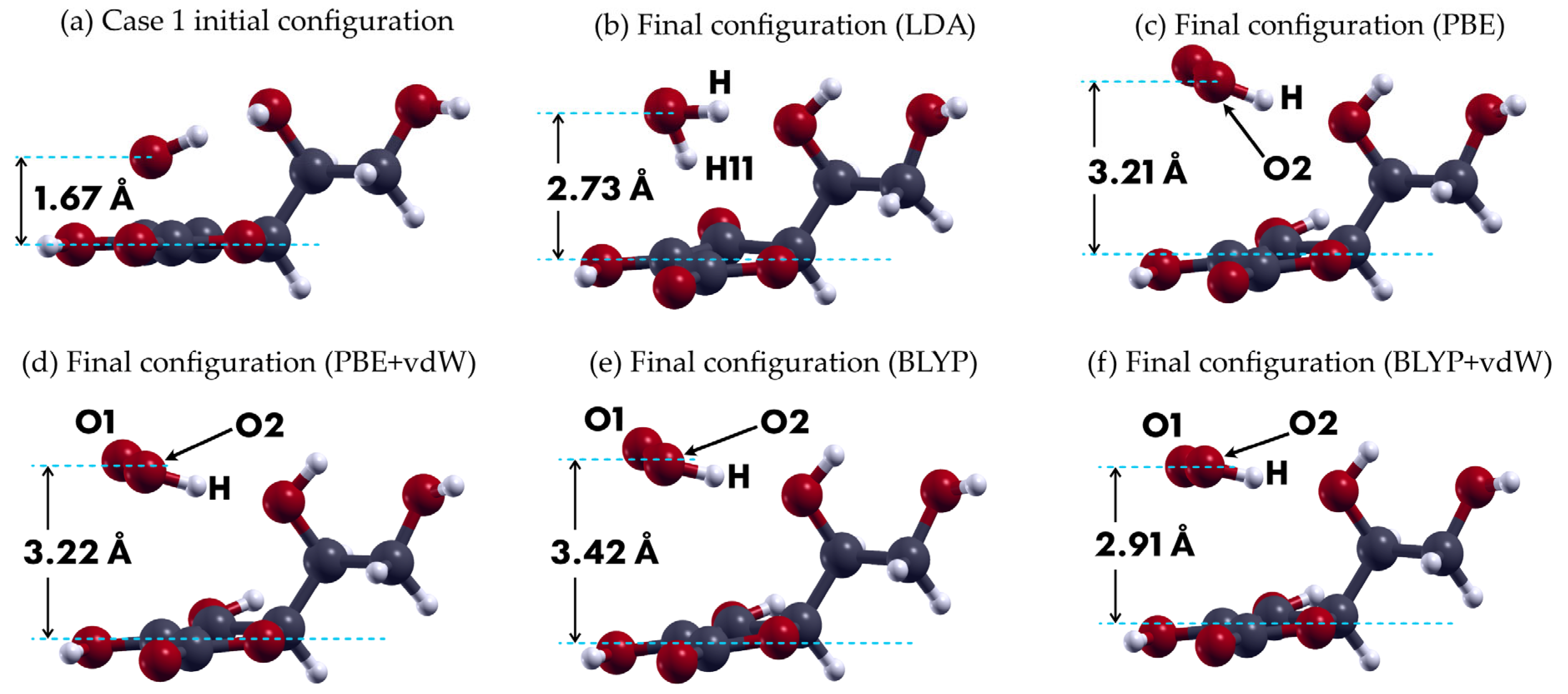
We first performed a structural relaxation in a vacuum, considering the HO2 radical at an initial vertical distance of 1.67 Å of the L-ascorbic acid C6H8O6, with respect to the plane that contains its pentagon. Two cases were considered, corresponding to the initial position of the H-atom in the hydroperoxyl radical, either pointing upwards (case 1) or downwards (case 2), with the C6H8O6 pentagon taken as reference. In both cases, the O-atoms axis of the radical is placed parallel to the C6H8O6 pentagon plane. Figure 1 shows the results for case 1.
In that case, the structural relaxation shows an increase in the distance between the HO2 radical and the C6H8O6, for all density functional approximations considered. In the LDA case, we observe a dissociation of one H-atom from the C6H8O6 onto the radical, something that is not present for the rest of the functionals considered, but, in all cases, the radical stabilizes around the distance shown in Figure 1, until convergence is achieved. Table 4 summarizes the absorption energies and recovery times obtained for the five structural relaxations performed. The results in this section are consistent among themselves, except for the case of the LDA functional, which overestimates the absorption energy, resulting in a rather large value for the recovery time.
Table 4. Case 1 adsorption energies Eads (in eV) and recovery times (in s) for the interaction between the HO2 radical and the L-ascorbic acid molecule, using different density functional approximations (see Figure 3). Calculations were structural relaxations in a vacuum.
Density Functional ApproximationEads τ
LDA−1.813.83 × 1017
PBE−0.432.31 × 10−6
PBE + vdW−0.361.82 × 10−7
BLYP−0.312.97 × 10−8
BLYP + vdW−0.444.59 × 10−6
Figure 3. Structural relaxations for the first case considered in this work, corresponding to the H-atom of the radical pointing upward. (a) Initial position: the O-atoms axis is placed parallel to the C6H8O6 pentagon plane. The rest of the images in this figure are shown from this same point of view and present the final configurations obtained with (b) the LDA, (c) PBE, (d) PBE + vdW, (e) BLYP, and (f) BLYP + vdW density functional approximations.
Crystals 13 01135 g003
The structural relaxation results corresponding to case 2—with the H-atom of the HO2 radical pointing downward—are shown in Figure 4, which qualitatively shows a similar behavior as case 1. Quantitatively, the final vertical distances in this case are smaller than the previous case. Table 5 shows the corresponding absorption energies and recovery times for calculations corresponding to case 2. We found reasonable values of those parameters when considering the PBE, PBE + vdW, and, to some extent, BLYP + vdW, in terms of sensor developing.

4. Discussion

The structural relaxations performed on both substances considered in this work resulted in bond distances and angles in good agreement with the experimental values. We used the LDA, PBE, PBE + vdW, BLYP, and BLYP + vdW density functional approximations in this work mainly to confirm the qualitative nature of the interaction studied, which was similar for all functionals considered. The use of different functionals improves the reliability of the results obtained and allows us to focus on the quantitative results corresponding to the functionals known to be more precise. In this case, PBE + vdW gave the most accurate results in terms of geometry and structure, when compared with the experimental parameters of the systems considered.
We found that the HO2 radical interacts in all cases with the L-ascorbic acid, which can be assessed as a physisorption in terms of the absorption energy values, according to the structural relaxations performed. Here, we are considering the typical binding energies for chemisorption to be above 0.5 eV per atom (see [41], page 195), and, under that criterion, we conclude that the interaction would rather be a physisorption, even at room temperature, according to the results obtained using the PBE + vW functional.
The absorption energies for case 2—in which the H atom of the radical points initially downward—are consistently larger than those for case 1, particularly when the van der Waals correction is included. This correction allows us to notice the action of the OH bonds from the acid molecule, in the interaction with the H-atom of the radical.
In all cases, the H-atom from the free radical interacts with an oxygen atom of the L-ascorbic acid molecule. The corresponding recovery times, calculated using Equation (2), suggest that L-ascorbic acid could properly detect the presence of the hydroperoxyl radical at room temperature, as suggested by the results using the PBE and PBE + vdW functionals in case 2. There, we obtained absorption energies of −0.78 eV and −0.73 eV, and recovery times of 2.09 and 0.329 s, respectively The double binding located in the pentagon of the L-ascorbic acid can be considered as a stable reaction site, although we did not find a reduction process of the HO2 radical.

Author Contributions

Conceptualization, I.C.D., J.M.R.-d.-A., and L.F.M.; data curation, I.C.D., A.F.J.G., and J.M.R.-d.-A.; formal analysis, A.F.J.G., J.M.R.-d.-A., and L.F.M.; funding acquisition, I.C.D., J.M.R.-d.-A., and L.F.M.; investigation, A.F.J.G., J.M.R.-d.-A., and L.F.M.; methodology, A.F.J.G. and J.M.R.-d.-A.; project administration, J.M.R.-d.-A. and L.F.M.; supervision, L.F.M.; validation, I.C.D., A.F.J.G., J.M.R.-d.-A., and L.F.M.; writing—original draft, I.C.D. and J.M.R.-d.-A.; writing—review and editing, A.F.J.G., J.M.R.-d.-A., and L.F.M. All authors have read and agreed to the published version of the manuscript.

Funding

Dirección General de Asuntos del Personal Académico de la Universidad Nacional Autónoma de México (DGAPA-UNAM), by grant number IN102323. The APC was funded by Tecnologico de Monterrey.

Data Availability Statement

Data unavailable due to privacy restrictions.

Acknowledgments

We thank (DGAPA-UNAM) for partial financial support by grant number IN102323. We also appreciate UNAM-Miztli-Super-Computing Center’s technical assistance by the project LANCADUNAM-DGTIC-030.

Conflicts of Interest

The authors declare no conflict of interest.

References

  1. Hayyan, M.; Hashim, M.A.; AlNashef, I.M. Superoxide Ion: Generation and Chemical Implications. Chem. Rev. 2016, 116, 3029–3085. [Google Scholar] [CrossRef] [PubMed] [Green Version]
  2. Halliwell, B. Free Radicals and Antioxidants: Updating a Personal View. Nutr. Rev. 2012, 70, 257–265. [Google Scholar] [CrossRef] [PubMed]
  3. Halliwell, B.; Gutteridge, J.M.C. Free Radicals in Biology and Medicine; Oxford University Press: Oxford, UK, 2015; ISBN 978-0-19-871747-8. [Google Scholar]
  4. Panel on Dietary Antioxidants and Related Compounds; Subcommittee on Upper Reference Levels of Nutrients; Subcommittee on Interpretation and Uses of Dietary Reference Intakes; Standing Committee on the Scientific Evaluation of Dietary Reference Intakes; Food and Nutrition Board; Institute of Medicine. Dietary Reference Intakes for Vitamin C, Vitamin E, Selenium, and Carotenoids; National Academies Press: Washington, DC, USA, 2000; p. 9810. ISBN 978-0-309-06935-9. [Google Scholar]
  5. World Health Organization. Vitamin and Mineral Requirements in Human Nutrition, 2nd ed.; World Health Organization: Geneva, Switzerland; FAO: Rome, Italy, 2004; ISBN 978-92-4-154612-6. [Google Scholar]
  6. Paasch, S.; Salzer, R. Solution to Spectroscopy Challenge 7. Anal. Bioanal. Chem. 2004, 380, 734–735. [Google Scholar] [CrossRef] [PubMed]
  7. Office of Dietary Supplements Vitamin, C. Fact Sheet for Health Professionals. Available online: https://ods.od.nih.gov/factsheets/VitaminC-HealthProfessional/ (accessed on 16 June 2023).
  8. Li, Y.; Schellhorn, H.E. New Developments and Novel Therapeutic Perspectives for Vitamin C. J. Nutr. 2007, 137, 2171–2184. [Google Scholar] [CrossRef] [Green Version]
  9. Carr, A.C.; Frei, B. Toward a New Recommended Dietary Allowance for Vitamin C Based on Antioxidant and Health Effects in Humans. Am. J. Clin. Nutr. 1999, 69, 1086–1107. [Google Scholar] [CrossRef] [Green Version]
  10. Honarbakhsh, S.; Schachter, M. Vitamins and Cardiovascular Disease. Br. J. Nutr. 2008, 101, 1113–1131. [Google Scholar] [CrossRef] [Green Version]
  11. Jacobs, C.; Hutton, B.; Ng, T.; Shorr, R.; Clemons, M. Is There a Role for Oral or Intravenous Ascorbate (Vitamin C) in Treating Patients With Cancer? A Systematic Review. Oncol. 2015, 20, 210–223. [Google Scholar] [CrossRef] [Green Version]
  12. Wang, A.H.; Still, C. Old World Meets Modern: A Case Report of Scurvy. Nutr. Clin. Pract. 2007, 22, 445–448. [Google Scholar] [CrossRef]
  13. Stephen, R.; Utecht, T. Scurvy Identified in the Emergency Department: A Case Report. J. Emerg. Med. 2001, 21, 235–237. [Google Scholar] [CrossRef]
  14. Bichara, L.C.; Lanús, H.E.; Nieto, C.G.; Brandán, S.A. Density Functional Theory Calculations of the Molecular Force Field of L -Ascorbic Acid, Vitamin C. J. Phys. Chem. A 2010, 114, 4997–5004. [Google Scholar] [CrossRef]
  15. Tu, Y.-J.; Njus, D.; Schlegel, H.B. A Theoretical Study of Ascorbic Acid Oxidation and HOO˙/O2˙ Radical Scavenging. Org. Biomol. Chem. 2017, 15, 4417–4431. [Google Scholar] [CrossRef]
  16. May, J.M. Is Ascorbic Acid an Antioxidant for the Plasma Membrane? FASEB J. 1999, 13, 995–1006. [Google Scholar] [CrossRef] [Green Version]
  17. Bartlett, D.; Church, D.F.; Bounds, P.L.; Koppenol, W.H. The Kinetics of the Oxidation of L-Ascorbic Acid by Peroxynitrite. Free Radic. Biol. Med. 1995, 18, 85–92. [Google Scholar] [CrossRef]
  18. Mottola, M.; Vico, R.V.; Villanueva, M.E.; Fanani, M.L. Alkyl Esters of L-Ascorbic Acid: Stability, Surface Behaviour and Interaction with Phospholipid Monolayers. J. Colloid Interface Sci. 2015, 457, 232–242. [Google Scholar] [CrossRef]
  19. Sharma, M.K.; Buettner, G.R. Interaction of Vitamin C and Vitamin E during Free Radical Stress in Plasma: An ESR Study. Free Radic. Biol. Med. 1993, 14, 649–653. [Google Scholar] [CrossRef]
  20. De Grey, A.D.N.J. HO2•: The Forgotten Radical. DNA Cell Biol. 2002, 21, 251–257. [Google Scholar] [CrossRef]
  21. Cottenier, S. Density Functional Theory and the Family of (L)APW-Methods: A Step-by-Step Introduction, 2nd ed.; Center for Molecular Modeling (CMM) & Department of Materials Science and Engineering (DMSE) Ghent University: Ghent, Belgium, 2013. [Google Scholar]
  22. Parr, R.G.; Yang, W. Density-Functional Theory of Atoms and Molecules; International Series of Monographs on Chemistry; Oxford University Press: Oxford, UK; Clarendon Press: New York, NY, USA, 1989; ISBN 978-0-19-504279-5. [Google Scholar]
  23. Marx, D.; Hutter, J. Ab Initio Molecular Dynamics: Basic Theory and Advanced Methods, 1st ed.; Cambridge University Press: Cambridge, UK, 2012; ISBN 978-1-107-66353-4. [Google Scholar]
  24. Born, M.; Oppenheimer, R. Zur Quantentheorie der Molekeln. Ann. Phys. 1927, 389, 457–484. [Google Scholar] [CrossRef]
  25. Giannozzi, P.; Andreussi, O.; Brumme, T.; Bunau, O.; Buongiorno Nardelli, M.; Calandra, M.; Car, R.; Cavazzoni, C.; Ceresoli, D.; Cococcioni, M.; et al. Advanced Capabilities for Materials Modelling with Quantum ESPRESSO. J. Phys. Condens. Matter 2017, 29, 465901. [Google Scholar] [CrossRef] [Green Version]
  26. Giannozzi, P.; Baroni, S.; Bonini, N.; Calandra, M.; Car, R.; Cavazzoni, C.; Ceresoli, D.; Chiarotti, G.L.; Cococcioni, M.; Dabo, I.; et al. QUANTUM ESPRESSO: A Modular and Open-Source Software Project for Quantum Simulations of Materials. J. Phys. Condens. Matter 2009, 21, 395502. [Google Scholar] [CrossRef]
  27. Giannozzi, P.; Baseggio, O.; Bonfà, P.; Brunato, D.; Car, R.; Carnimeo, I.; Cavazzoni, C.; de Gironcoli, S.; Delugas, P.; Ferrari Ruffino, F.; et al. QUANTUM ESPRESSO toward the Exascale. J. Chem. Phys. 2020, 152, 154105. [Google Scholar] [CrossRef] [Green Version]
  28. Troullier, N.; Martins, J. A Straightforward Method for Generating Soft Transferable Pseudopotentials. Solid State Commun. 1990, 74, 613–616. [Google Scholar] [CrossRef]
  29. Kleinman, L.; Bylander, D.M. Efficacious Form for Model Pseudopotentials. Phys. Rev. Lett. 1982, 48, 1425–1428. [Google Scholar] [CrossRef]
  30. Perdew, J.P.; Zunger, A. Self-Interaction Correction to Density-Functional Approximations for Many-Electron Systems. Phys. Rev. B 1981, 23, 5048–5079. [Google Scholar] [CrossRef] [Green Version]
  31. Perdew, J.P.; Burke, K.; Ernzerhof, M. Generalized Gradient Approximation Made Simple. Phys. Rev. Lett. 1996, 77, 3865–3868. [Google Scholar] [CrossRef] [PubMed] [Green Version]
  32. Perdew, J.P.; Burke, K.; Ernzerhof, M. Generalized Gradient Approximation Made Simple [Phys. Rev. Lett. 77, 3865 (1996)]. Phys. Rev. Lett. 1997, 78, 1396. [Google Scholar] [CrossRef] [Green Version]
  33. Becke, A.D. Density-Functional Exchange-Energy Approximation with Correct Asymptotic Behavior. Phys. Rev. A 1988, 38, 3098–3100. [Google Scholar] [CrossRef]
  34. Lee, C.; Yang, W.; Parr, R.G. Development of the Colle-Salvetti Correlation-Energy Formula into a Functional of the Electron Density. Phys. Rev. B 1988, 37, 785–789. [Google Scholar] [CrossRef] [Green Version]
  35. Grimme, S.; Antony, J.; Ehrlich, S.; Krieg, H. A Consistent and Accurate Ab Initio Parametrization of Density Functional Dispersion Correction (DFT-D) for the 94 Elements H-Pu. J. Chem. Phys. 2010, 132, 154104. [Google Scholar] [CrossRef] [Green Version]
  36. Liskow, D.H.; Schaefer, H.F.; Bender, C.F. Geometry and Electronic Structure of the Hydroperoxyl Radical. J. Am. Chem. Soc. 1971, 93, 6734–6737. [Google Scholar] [CrossRef]
  37. Flowers, B.A.; Szalay, P.G.; Stanton, J.F.; Kállay, M.; Gauss, J.; Császár, A.G. Benchmark Thermochemistry of the Hydroperoxyl Radical. J. Phys. Chem. A 2004, 108, 3195–3199. [Google Scholar] [CrossRef] [Green Version]
  38. Bil, A.; Latajka, Z. The Hydroperoxy Radical and Its Closed-Shell ‘analogues’: Ab Initio Investigations. Chem. Phys. Lett. 2004, 388, 158–163. [Google Scholar] [CrossRef]
  39. Durand, G.; Choteau, F.; Pucci, B.; Villamena, F.A. Reactivity of Superoxide Radical Anion and Hydroperoxyl Radical with α-Phenyl-N-Tert -Butylnitrone (PBN) Derivatives. J. Phys. Chem. A 2008, 112, 12498–12509. [Google Scholar] [CrossRef] [PubMed]
  40. Monkhorst, H.J.; Pack, J.D. Special Points for Brillouin-Zone Integrations. Phys. Rev. B 1976, 13, 5188–5192. [Google Scholar] [CrossRef]
  41. Oura, K. (Ed.) Surface Science: An Introduction; Advanced Texts in Physics; Springer: Berlin/Heidelberg, Germany; New York, NY, USA, 2003; ISBN 978-3-540-00545-2. [Google Scholar]
  42. Eyring, H. The Activated Complex in Chemical Reactions. J. Chem. Phys. 1935, 3, 107–115. [Google Scholar] [CrossRef]
  43. Popa, I.; Fernández, J.M.; Garcia-Manyes, S. Direct Quantification of the Attempt Frequency Determining the Mechanical Unfolding of Ubiquitin Protein. J. Biol. Chem. 2011, 286, 31072–31079. [Google Scholar] [CrossRef] [Green Version]
  44. Downs, R.T.; Hall-Wallace, M. The American Mineralogist Crystal Structure Database. Am. Mineral. 2003, 88, 247–250. [Google Scholar]
  45. Gražulis, S.; Chateigner, D.; Downs, R.T.; Yokochi, A.F.T.; Quirós, M.; Lutterotti, L.; Manakova, E.; Butkus, J.; Moeck, P.; Le Bail, A. Crystallography Open Database—An Open-Access Collection of Crystal Structures. J. Appl. Crystallogr. 2009, 42, 726–729. [Google Scholar] [CrossRef] [Green Version]
  46. Gražulis, S.; Daškevič, A.; Merkys, A.; Chateigner, D.; Lutterotti, L.; Quirós, M.; Serebryanaya, N.R.; Moeck, P.; Downs, R.T.; Le Bail, A. Crystallography Open Database (COD): An Open-Access Collection of Crystal Structures and Platform for World-Wide Collaboration. Nucleic Acids Res. 2012, 40, D420–D427. [Google Scholar] [CrossRef] [Green Version]
  47. Gražulis, S.; Merkys, A.; Vaitkus, A.; Okulič-Kazarinas, M. Computing Stoichiometric Molecular Composition from Crystal Structures. J. Appl. Crystallogr. 2015, 48, 85–91. [Google Scholar] [CrossRef] [Green Version]
  48. Le Bail, A. Inorganic Structure Prediction with It GRINSP. J. Appl. Crystallogr. 2005, 38, 389–395. [Google Scholar] [CrossRef] [Green Version]
  49. Merkys, A.; Vaitkus, A.; Butkus, J.; Okulič-Kazarinas, M.; Kairys, V.; Gražulis, S. COD::CIF::Parser: An Error-Correcting CIF Parser for the Perl Language. J. Appl. Crystallogr. 2016, 49, 292–301. [Google Scholar] [CrossRef] [Green Version]
  50. Merkys, A.; Vaitkus, A.; Grybauskas, A.; Konovalovas, A.; Quirós, M.; Gražulis, S. Graph Isomorphism-Based Algorithm for Cross-Checking Chemical and Crystallographic Descriptions. J. Cheminform. 2023, 15, 25. [Google Scholar] [CrossRef]
  51. Quirós, M.; Gražulis, S.; Girdzijauskaitė, S.; Merkys, A.; Vaitkus, A. Using SMILES Strings for the Description of Chemical Connectivity in the Crystallography Open Database. J. Cheminform. 2018, 10, 23. [Google Scholar] [CrossRef]
  52. Vaitkus, A.; Merkys, A.; Gražulis, S. Validation of the Crystallography Open Database Using the Crystallographic Information Framework. J. Appl. Crystallogr. 2021, 54, 661–672. [Google Scholar] [CrossRef]
  53. McMonagle, C.J.; Probert, M.R. Reducing the Background of Ultra-Low-Temperature X-Ray Diffraction Data through New Methods and Advanced Materials. J. Appl. Crystallogr. 2019, 52, 445–450. [Google Scholar] [CrossRef]
  54. Paukert, T.T.; Johnston, H.S. Spectra and Kinetics of the Hydroperoxyl Free Radical in the Gas Phase. J. Chem. Phys. 1972, 56, 2824–2838. [Google Scholar] [CrossRef] [Green Version]
  55. Hvoslef, J. The Crystal Structure of L -Ascorbic Acid, ‘vitamin C’. II. The Neutron Diffraction Analysis. Acta Crystallogr. B 1968, 24, 1431–1440. [Google Scholar] [CrossRef]
Figure 1. The structure of the L-ascorbic acid (C6H8O6) as considered in this work. It consists of 20 atoms, 6 carbon atoms, 6 oxygen atoms, and 8 hydrogen atoms. The atoms are labeled to identify bond lengths and angles, following the setup used in [14]. This configuration corresponds to the final structure obtained by using the PBE + vdW functional.
Crystals 13 01135 g001
Figure 2. Structure considered in this work for the hydroperoxyl radical HO2 or HOO. The experimental bond length between the H atom and the O atom (labeled O2–H) is 0.96 Å, while the bond length between O atoms is equal to 1.3 Å. The angle θ is experimentally equal to 108° (see [54]). The labels correspond to the length and angle identification of Table 3. The figure shows the optimized structure obtained by using the PBE + vdW functional, as in Figure 1.
Crystals 13 01135 g002
Figure 4. Structural relaxations for the second case considered in this work, corresponding to the H-atom of the radical pointing downward. (a) Initial position: the O-atoms axis is placed parallel to the C6H8O6 pentagon plane. The rest of the images in this figure are shown from this same point of view and present the final configurations obtained with (b) the LDA, (c) PBE, (d) PBE + vdW, (e) BLYP, and (f) BLYP + vdW density functional approximations. In both cases, the qualitative behavior is similar to those of case 1.
Crystals 13 01135 g004
Table 1. Values of bond lengths (Å) obtained after a structural relaxation (using different density functional approximations) for the L-ascorbic acid, and their corresponding experimental values in the last column (see [55]). The atom labelling corresponds to that of Figure 1, following the labelling structure used in [14].
ParameterLDAPBEPBE + vdWBLYPBLYP + vdWExp. [55]
C1–O71.2071.2151.2141.2161.2151.216
C5–O81.3321.3521.3521.3621.3621.361
C4–O101.3301.3501.3491.3601.3591.326
C1–O21.3701.3891.3891.4011.4011.355
C3–O21.4251.4461.4461.4611.4611.444
C12–O141.3931.4151.4151.4281.4291.427
C16–O191.4001.4241.4241.4391.4391.431
C4–C51.3431.3501.3501.3501.3501.338
C1–C51.4421.4561.4571.4621.4631.452
C3–C41.4891.5041.5041.5121.5101.493
C3–C121.5201.5441.5431.5531.5521.521
C12–C161.5091.5301.5301.5391.5381.521
Table 2. Values of bond angles (°) obtained after a structural relaxation (using different density functional approximations) for the L-ascorbic acid, and their corresponding experimental values (see [55]). As in Table 1, the labelling setup is based on [14].
ParameterLDAPBEPBE + vdWBLYPBLYP + vdWExp. [55]
C3–O2–C1108.4108.5108.5108.6108.5109.1
O2–C1–C5109.5109.1109.1108.7108.7109.5
C1–C5–C4108.1108.5108.5108.9108.9107.8
C5–C4–C3108.6108.9108.9109.3109.3109.5
C4–C3–O2105.4104.9105.0104.4104.6104.0
O2–C1–O7123.9123.1123.1123.0122.9121.4
O7–C1–C5126.6127.8127.8128.3128.4129.1
C1–C5–O8120.1121.5121.6121.8122.0124.6
O8–C5–C4131.8130.0129.9129.3129.2127.5
C5–C4–O10128.0126.9126.9126.4126.6133.5
C3–C4–O10123.4124.2124.1124.3124.0117.1
O2–C3–C12108.0108.7108.6109.1108.7114.8
C4–C3–C12114.2115.5115.1115.9115.1110.4
C3–C12–C16110.2110.6110.6110.9110.9111.7
C3–C12–O14110.8111.8111.6112.0111.5112.7
O14–C12–C16113.2113.3113.3113.2113.0106.9
C12–C16–O19108.3108.3108.1108.1107.8108.0
Table 3. Values of bond lengths (Å)—first two rows—and angle (°) after structural relaxations (using the LDA and GGA approximations) for the HO2 hydroperoxyl radical, and their corresponding experimental values (see [54]). The atom labelling corresponds to that of Figure 2.
ParameterLDAGGAPBE + vdWBLYPBLYP + vdWExp. [54]
O1–O2 1.3201.3431.3431.3331.3631.3
O2–H 0.9930.9890.9880.9710.9890.96
H–O2–O1 106.0105.1105.2104.3104.8108
Table 5. Case 2 adsorption energies Eads (in eV) and recovery times (in s) for the interaction between the HO2 radical and the L-ascorbic acid molecule, using different density functional approximations (see Figure 3). Calculations were structural relaxations in a vacuum.
Density Functional ApproximationEads τ
LDA−1.333.35 × 109
PBE−0.782.09
PBE + vdW−0.730.329
BLYP−0.171.18 × 10−10
BLYP + vdW−0.613.24 × 10−3
Disclaimer/Publisher’s Note: The statements, opinions and data contained in all publications are solely those of the individual author(s) and contributor(s) and not of MDPI and/or the editor(s). MDPI and/or the editor(s) disclaim responsibility for any injury to people or property resulting from any ideas, methods, instructions or products referred to in the content.