Abstract
In the present work, complexes of cobalt(II), copper(II), and zinc(II), 2-amino-4-methylpyrimidineand, and 2,3-diaminopyridine were successfully prepared and characterized using elemental analysis, UV-visible, and FTIR spectroscopy, as well as magnetic susceptibility measurements, molar conductance, TGA analysis, and X-ray diffraction. From elemental and spectral data, the formulae [M(L1)(L2)Cl2(H2O)] (where L1 = AMPY (2-amino-4-methylpyrimidine) and L2 = DAPY(2,3-diaminopyridine)) and M = Co(II) (2), Cu(II) (2), and Zn(II)) for the metal complexes have been proposed. The geometric structures of the mixed-ligand complexes were found to be octahedral around the metal ions, and the XRD patterns showed monoclinic crystal systems with space group P21. The mode of bonding was pentacoordinate for Cu and hexacoordinate for Zn and Co. Different features may result from the fact that not all molecules have the same electron distribution. For example, Zn and Co have larger electron densities in at least one of the chlorides in the HOMO compared with pentacoordinate Cu, which has a small electron distribution on the chloride. Thermal analysis indicated that all metal complexes are stable up to about 88 °C with thermodynamically favored overlapped chemical reactions. Excellent antibacterial and antifungal activity was shown by the three synthesized forms of the complexes. The Zn(II) complex had a high level of antioxidant activity with a DPPH scavenging of 91.5%, whereas the Cu(II) complex had a low level of antioxidant potential (16.5%). The docking tests also showed that all compounds had good binding energy levels (7.2–7.9 kcal mol−1). For this reason, all molecules can easily fit in the receptor protein’s catalytic sites. However, the Co(II) complex is shown to be more active.
1. Introduction
Transition element ions (TEI) have a good and important role in the self-assembly of complexes. Additionally, the wide field of chemistry using transition elements and organic ligands as building blocks is enriched with high design ability and a variety of structural architecture [1,2,3,4]. The cobalt(II), copper(II), and zinc(II) ions are of particular interest because they relate to labile metal centers with the coordination number of M2+ ranging from two to six and more. Co(II), Cu(II), and Zn(II) compounds are most commonly given with nitrogen-containing ligands with functional groups and different geometries. For example, pyridine, pyrimidine, and its derivatives react with metal ions (MIS) to give both polynuclear and discrete coordination compounds and participate in hydrogen-bonding and π-ring interactions, including weak interactions as well as coordinates to the transition element [5,6,7]. Moreover, it has an immense influence on various biological and chemical processes as well [8]. We have observed this through antimicrobial and antioxidant assays of the prepared compounds. 2-amino-4-methylpyrimidine (AMPY) and 2,3-di-aminopyridine (DAPY) serve as useful chelating ligands in a diversity of inorganic and organometallic applications and anti-bacterial, anti-fungal, antioxidant, anti-inflammatory, and pharmaceutical applications [9,10,11,12,13,14,15]. It can be assumed from several scientific works [9,10,11,12,13,14,15,16,17] that the qualities of antimicrobial and antioxidant actions depend on the type of ligands forming the bioactive complexes. For that, we have found that we can obtain different products with only a slight variation in the published procedure. That is why we have decided not only to optimize the discussed preparation method but also to determine the reaction products unambiguously and to collect information regarding the biological activity of the discussed complex better than that reported in the literature. On the basis of the stated facts, we have decided to prepare and characterize the biological activity of cobalt(II), copper(II), and zinc(II) complexes. Indeed, the chemistry of Co(II), Cu(II), and Zn(II) compounds with nitrogen donor ligands, especially aminopyridine and aminopyrimidine derivatives, has been extensively studied over the past few decades [16,17,18]. In this study, we prepared three complexes [Co(AMPY)(DAPY)Cl2(H2O)], [Cu(AMPY)(DAPY)Cl2(H2O)], and [Zn(AMPY)(DAPY)Cl2(H2O)] in aqueous media and characterized them by analytical and different spectroscopic methods.
2. Experimental Section
2.1. Chemicals
High purity 2-amino-4-methylpyrimidine (AMPY) (97%) and 2,3-diaminopyridine (DAPY) (95%) were supplied by Sigma–Aldrich. They were purchased and used without purification. Co(II), Cu(II), and Zn(II) compounds were synthesized in aqueous media using cobalt(II) chloride hexahydrates, copper(II) chloride dehydrated, zinc(II) chloride salts, and 2-amino-4-methylpyrimidine (AMPY) as primary ligands. 2,3-diaminopyridine (DAPY) was used as the auxiliary ligand for the three complexes, respectively.
2.1.1. Synthesis of [Co(AMPY)(DAPY)Cl2(H2O)]
An ethanolic solution (15 mL) of 2-amino-4-methylpyrimidine (AMPY) (0.5 g, 0.45 mmol) was slowly added to 20 mL water (CoCl2.6H2O, 1.09 g, 0.45 mmol), and to it an ethanolic solution (15 mL) of 2,3-diaminopyridine (DAPY) (0.5 g, 0.45 mmol) was added dropwise. The reaction mixture was heated at 60 °C for 60 min with constant stirring. The dark brown precipitate was washed with H2O and EtOH and then dried.
2.1.2. Synthesis of [Cu(AMPY)(DAPY)Cl2(H2O)]
It was prepared following the same procedure as in (1). The molar ratios were 1:1:1, CuCl2.2H2O (0.78 g, 0.45 mmol), AMPY (0.5 g, 0.45 mmol), and DAPY (0.5 g, 0.45 mmol). A dark green complex was isolated.
2.1.3. Synthesis of [Zn(AMPY)(DAPY)Cl2(H2O)]
A similar synthetic method as that for (1) was used in the preparation of the zinc(II) complex (15 mL), ZnCl2 (0.62 g, 0.45 mmol). A creamy white compound was obtained.
The structures of the ligands are presented in Figure 1.
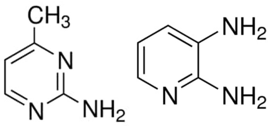
Figure 1.
The structures of the ligands: 2-amino-4-methylpyrimidine (AMPY) and 2,3-diaminopyridine (DAPY).
2.2. Characterization Methods
The infrared spectrum of the synthesized complexes was measured in the region of 4000–400 cm−1 with an Agilent spectrometer. Electronic UV-Vis spectra were recorded using a Shimadzu spectrophotometer (UV-1650PC, Japan). C, H, and N analyses were measured utilizing a Eurovector CHN (EA3000, Italy). X-ray diffraction patterns (XRD) were collected at room temperature on a Rigaku XRD diffractometer (Ultima IV, USA) with Cu-Kα radiation (λCu = 0.154059 Å). The used diffractometer is equipped with a monochromator. The structural and microstructural parameters were determined using the Rietveld method. The morphology of synthesized complexes was examined by scanning electron microscopy (SEM) in a DSM960A ZEISS microscope in secondary electron mode, operating at a voltage of 15 kV. The SEM was equipped with a Vega-Tescan energy dispersive X-ray spectrometry (EDS) analyzer. Thermal analyses were conducted using a Shimadzu simultaneous instrument in a nitrogen atmosphere in the range of 0–500 °C with a heating rate of 10 °C per minute.
2.3. Microbial Strains and Culture Media
In this study, different samples were tested against Gram (−ve) and Gram (+ve) bacterial strains, giving insight into their broad-spectrum effects. The pathogenic species used were two Gram-positive strains, Staphylococcus aureus and Micrococcus luteus, and two Gram-negative strains, Escherichia coli and Salmonella thyphimurium. The antifungal activity was evaluated against a pathogenic reference strain of the yeast Candida albicans. The species were grown in nutrient broth (Oxoid) at 37 °C for a day and cultivated on nutrient agar (Oxoid) for 24 h at 37 °C. The yeast strain was grown in Sabouraud chloramphenicol broth (Oxoid) at 25 °C for a day.
2.4. Antimicrobial Activity
The antimicrobial activity of the different extracts was tested with the reference agar disk diffusion method [19]. Before the test, 50 mg of each extract was dissolved in 1 mL of a solution of dimethylsulfoxyde “DMSO” (5%). The species were grown in Mueller–Hinton broth (Oxoid) at 37 °C for a day at 37 °C for 24 h and the suspensions were checked with 0.5 McFarland standard turbidity. Afterward, 100 μL of each precultured suspension was spread onto plates containing MH agar. Sterile filter paper discs (6 mm in diameter) were saturated with 20 μL of the different compounds and placed on agar. The treated plates were putted for 60 min at 4 °C and then incubated for a day at 37 °C. Then, during incubation, the diameter of the inhibition zone (clear halo) around the discs was measured.
2.5. Antioxidant Activity
(DPPH) Radical Scavenging Assay
The free radical scavenging effect of the compounds was assessed according to the method [20]. Briefly, 1 mL of sample (5 mg/mL) was mixed with 3 mL of a methanol solution of DPPH (2,2-diphenyl-1-picrylhydrazyl) (300 µM). The reaction mixture was vortexed and incubated for 30 min at room temperature. The absorbance of the solution was measured at 517 nanometers. Vitamin C was used as a standard. The inhibitory percentage of DPPH was measured by using the next Equation (1):
DPPH Scavenging effect (%) = [1 − (Abs compound/Abs control)] × 100
2.6. Computational Studies
The density functional theory (DFT) was performed using Gaussian09 program [21]. The optimization of the diverse complexes was performed by DFT through the functional B3LYP and Lanl2dz/6-31G(d) basis sets. LANL2DZ basis set was limited for the treatment of Zn(II), Cu(II), and Co(II) atoms and 6-31G(d) basis set for all other atoms. To confirm the stability of the structures, we calculated the vibrational frequencies at the same level of theory.
2.7. Molecular Docking Analysis
In silico molecular docking was used to study the interactions between S. aureus tyrosyl-tRNA synthetase and the three complexes Co(II), Cu(II), and Zn(II) to investigate the preferred occupation of the ligands in the binding active site. The crystal coordinates were obtained from the Protein Data Bank: S. aureus tyrosyl-tRNA synthetase (PDB: 1JIJ). All water species and the co-crystallized ligand have been deleted from the original structure. We designated the Gasteiger charge and polar hydrogens using AutoDockTools1.5.2 (ADT), and we prepared the PDBQT file format [22]. We used ADT to determine a docking grid. In 1JIJ, the grid box site was set at x: −10.908, y: 14.432 y, and z: 86.420 z Å. The size of the grid box was 25 Å for x, y, and z and 0.375 Å for the grid spacing. The structures of the compounds were those optimized in computational studies with B3LYP/LanL2dz/6-31G(d). We used AutoDock Vina software [23] with 32 as an exhaustiveness parameter to obtain the docking data. The docking conformation analysis was performed by ADT. Enzyme-ligand interactions are investigated by Discovery Studio Visualizer [24].
3. Results and Discussion
3.1. Synthesis and Spectroscopic Characterization
These components were found to react in the molar ratios of 1:1:1 metal:L1:L2. The compounds are air-stable. The molar conductivity values ΛM of the compounds in 10−3 M DMSO solutions vary from 23.2 to 38.8 to 62.1 S cm2 mol−1 for Zn(II), Cu(II), and Co(II) complexes, respectively, indicating that they are of a non-electrolytic nature.
Anal. Calc. for C10H16N6CoCl2O (complex 1): C, 32.79; H, 4.40; N, 22.95; Found: C, 32.04; H, 4.56; N, 21.98. m.p. 198 °C.
Anal. Calc. for C10H16N6CuCl2O (complex 2): C, 32.39; H, 4.34; N, 22.67. Found: C, 31.46; H, 4.28; N, 21.04. m.p. 192 °C.
Anal. Calc. for C10H16N6ZnCl2O (complex 3): C, 32.23; H, 4.32; N, 22.55. Found: C, 33.50; H, 4.11; N, 21.96. m.p. 186 °C.
Table 1 summarizes the infrared spectral data (cm−1) of the ligands and their complexes. The infrared spectral bands provide structural evidence for the coordination of the two ligands to the Co(II), Cu(II), and Zn(II) ions. The stretching vibration of the pyridine groups located at 1590 (ν) C=C and 1580 (ν) C=N cm−1 in the DAPY ligand exhibits a notable shift to a wave number of (1548–1567) and (1530–1540 cm−1) in all complexes [25,26,27]. The C-N stretching in the ring bands found in the complexes is in the range 1450–1495 cm−1 [28]. In the FTIR spectrum of the free AMPY, υ(C-NH2) occurs at 3312 cm−1 with a shift to a wave number (3296–3310 cm−1) in the spectra of the compounds [8]. A broad diffused band with medium intensity located in the range 3418–3498 cm−1 may be assigned to ν(OH) for lattice H2O in Co(II) and Cu(II) complexes [29]. For all complexes, the νOH stretching vibration of coordinated H2O appears in the 3306–3326 cm−1 region [30]. The IR spectra of the compounds appear as a band at 418–436 cm−1 assigned to (M-Cl) [31]. Metal-oxygen and M-nitrogen bonding are apparent from the manifestation of two bands at 554–570 and 468–475 cm−1, respectively [32].

Table 1.
The infrared spectral data (cm−1) of the ligands and their complexes.
3.2. Magnetic Moments
Magnetic susceptibility was measured at room temperature using a solid sample by the Gouy operation. The magnetic moment shows the Co(II) ion to be (4.78 BM) with configuration in an octahedral environment [33,34]. The value of the determined magnetic moment for the Cu(II) ion is (1.73 B.M.) based on the configuration in octahedral geometry [35,36]. Zn(II) is a nonmagnetic ion. It has been reported that when the value for Co is between 4.3 and 5.2, it has a high spin state.
3.3. Electronic Spectra
In the wavelength range 200–650 nm, the optical absorption coefficient was calculated for ligands (DMSO, 1×10⁻³ M) and their metal ions (Co(II), Cu (II), and Zn(II)), as shown in Figure 2 below. Absorption peaks were found at 31.746, 31.250, 32.476 cm−1 and 39.682, 39.062, 39.370 cm−1, which can be referred to as n-π* and π-π*, transitions of DAPY and AMPY [37,38]. The electronic spectra of the cobalt(II) complex have two absorption bands typical of high spin and low spin octahedral geometries, designated 4 T1 g(F)→4 A2g(F) and 2 Eg→2 T1g transitions, respectively [34]. The Cu(II) complex presents the absorption bands that need theoretical calculations in order to determine its coordination number. Zn+2 is a nonmagnetic ion. They have only bands for π-π*and n-π*, which are assigned to intra-ligand charge-transfer transitions. From the above data, the structures of the complexes can be postulated as follows (Figure 3 and Figure 4).
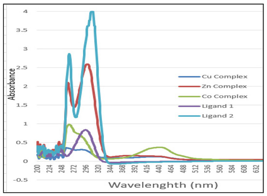
Figure 2.
Electronic spectra of L1, L2, Co(II), Cu(II), and Zn(II) mixed ligand complexes.
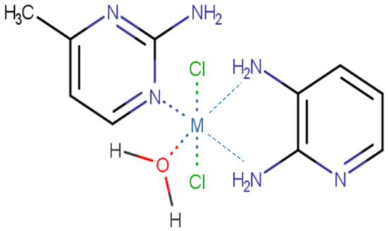
Figure 3.
Structure of [M(AMPY)(DAPY)Cl2(H2O)] M = Co(II), Cu(II), and Zn(II).
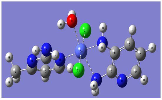
Figure 4.
A perspective view of the complete coordination around Co(II) complex.
3.4. Theoretical Study
Molecular modeling was performed for all compounds using the density functional theory DFT/B3LYP to study the structures and the frontier molecular orbitals (Figure 5). The mode of bonding is pentacoordinate for Cu and hexacoordinate for Zn and Co. Selected bond lengths (Å) calculated at B3LYP are presented in Table 2. Zn is coordinated to one nitrogen of L1 (Zn-N = 2.10Å) and to two nitrogens of L2 (2.28 and 2.38Å). Co is coordinated to one nitrogen of L1 (Co-N = 2.00 Å) and to two nitrogens of L2 (2.00 and 2.37 Å). Cu is bonded to one nitrogen of L1 (Cu-N = 2.10 Å) and one nitrogen of L2 (2.00 Å). Generally, the proposed model for metals is hexacoordinate, but density functional calculations show that the copper in our environment is pentacoordinate. In the literature, we find that copper can be pentacoordinated to some ligands [39].
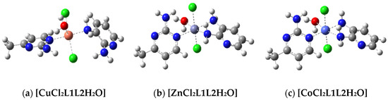
Figure 5.
Optimized geometry of the complexes. (a) Cu(II) (b) Zn(II), and (c) Co(II).
Table 2.
Selected bond lengths (Å) calculated at B3LYP.
The HOMO and LUMO frontier orbitals are exhibited in Figure 6. For Cu, the HOMO is localized on L2 and the LUMO is centralized on the second ligand, L1. For Zn, the HOMO is localized on one chloride, and the LUMO is localized on L1. For Co, both HOMO and LUMO are found with the metal and two chlorides. This shows that the electron distribution is not the same for all molecules, and this can lead to different properties [21]. For Cu, which is pentacoordinate, the electron distribution is small on the chloride, but for Zn and Co, the electron density is higher in at least one of the chlorides in the HOMO.
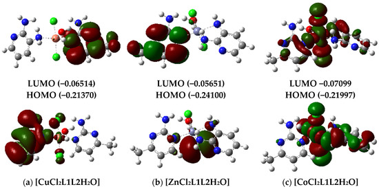
Figure 6.
Frontier molecular orbitals of HOMO and LUMO in complexes with (a) Cu, (b) Zn, and (c) Co.
3.5. Thermal Analysis
3.5.1. [Co(AMPY)(DAPY)Cl2(H2O)]
Figure 7a gives the TG and DTA curves of the [Co(AMPY)(DAPY)Cl2(H2O)] sample. As shown, the TG-analysis reveals that the thermal decomposition processes begin at a temperature of 35 °C and include four stages (Figure 7). The first stage corresponds to the dehydration process occurring in the temperature range of 65 to 95 °C. The mass loss (calc. at 4.91%, found at 4.75%) indicates the loss of H2O molecules. For this step (DTG minimum at 98 °C), an endothermic peak is observed in the DTA curve at 101 °C. The observed mass loss of the second step (154–230 °C) in the TG curve agrees with the loss of chlorine (calc. at 19.36%, found at 18.84%) and a DTG peak at 192 °C. This step is marked on the DTA curve by a broad exothermic peak at 194 °C. The third and fourth steps (232–500 °C) corresponded to the loss of decomposition of the rest of the organic ligands (calc. at 56.81%, found at 54.48%). For these steps, a DTG broad minimum at 298 and 310 °C and a DTA exotherm peak at 301 and 312 °C were observed. Cobalt oxides may occur as the thermal decomposition comes to an end at about 550 °C; given that Co(II) is less stable than Co(III) and that the first can spontaneously transform into the latter, the ultimate residue may be Co2O3 or perhaps a mixture of CoO and Co2O3. However, the calculation of the remaining mass at the end of TG is around 45% of the initial mass, and this can confirm the existence of Co2O3 instead of Co because its molar mass is 166 gmol−1, which corresponds to 45% of the initial mass of the complex (366 g.mol−1). Conversely, the decomposition processes usually resemble complex processes, and several decomposition reactions often overlap. Furthermore, the large exothermic peak shows that other processes besides chloride evaporation take place at this stage. However, even in the air, chloride prefers to evaporate as HCl rather than Cl2. It is quite likely that the intramolecular redox reaction and the exothermic impact that follow the formation of HCl result in some structural rearrangement.
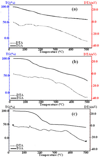
Figure 7.
TG- and DTA- thermograms of (a) Co(II), (b) Zn(II), and (c) Cu(II) complexes.
3.5.2. [Cu(AMPY)(DAPY)Cl2(H2O)]
As indicated by Figure 7c, the [Cu(AMPY)(DAPY)Cl2(H2O)] is stable up to 40 °C, when a first mass loss (calc. at 4.85%, found at 4.75%) occurs. Correspondingly, a DTG peak at 101 °C and an endothermic broad peak at 120 °C in the DTA curve were recorded. The observed mass loss of this first stage agrees well with the loss of one water molecule. The observed mass loss of the second step is associated with the loss of chlorine (calc. at 19.12%, found at 18.95%). This step corresponds to a DTG peak at 202 °C and an exothermic DTA peak at 204 °C. The third, fourth, and fifth steps of mass loss of AMPY and DAPY ligands (calc. at 56.14%, found at 53.16%) are manifested on the DTG curve as a peak at 251, 306, and 404 °C and an exothermic peak at 253, 308, and 406 °C in the DTA curve. The end product at 550 °C is consistent with CuO (calc. at 21.44%, found at 20.87%).
3.5.3. [Zn(AMPY)(DAPY)Cl2(H2O)]
As shown in the TG-curve of [Zn(AMPY)(DAPY)Cl2(H2O)], several step decompositions were observed at temperatures above 48.5 °C (Figure 7b); they occur in the temperature ranges of 82–112, 114–165, 167–268, 270–335, and 337–550 °C. The first mass loss (calc. at 4.83%, found at 4.49%) corresponds to the release of a water molecule with a DTG peak at 96 °C and an endothermic peak broad at 98 °C in the DTA curve. The observed weight loss of the second and third steps is correlated with the decomposition of AMPY and chlorine (calc. at 48.32%, found at 47.65%). The DTG peaks at 138 and 216 °C, and the DTA peaks at 140 and 218 °C, respectively. The fourth and fifth steps represent the decomposition of the remaining ligands (calculated at 29.29%, found at 27.10%). The final stable residue is ZnO (calc. at 21.84%, found at 20.76%).
3.6. X-ray Powder Diffraction (XRD)
Figure 8 shows a typical superposition of the XRD patterns of synthesized complexes. The crystallographic data refined by Rietveld are summarized in Table 3. As shown previously in Figure 4, the atoms were arranged in an octahedral state. In addition, as shown in Figure 8, the crystalline structure of the tree complexes is textured along the more intense prominent diffraction peak located at particular angles 2 thetas of 20, 24, and 18° for Cu(II), Zn(II), and Co(II) complexes (Figure 8a–c), respectively. As indicated by the known Sherrer’s formula, the crystallite size is inversely proportional to the full width at half maxima (FWHM). The most intense peaks are used in the crystallite size calculation, and sharp XRD peaks indicate that the particles were polycrystalline and belonged to the monoclinic crystal system. The calculated crystallite sizes for the Co(II), Cu(II), and Zn(II)- complexes are around 119, 88, and 175 nm, respectively.
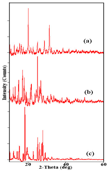
Figure 8.
X-ray powder diffraction (XRD) patterns of (a) Cu(II), (b) Zn(II), and (c) Co(II) complexes.
Table 3.
XRD data of the Co(II), Cu(II), and Zn(II) complexes.
3.7. Scanning Electron Microscopy (SEM)
Figure 9 presents SEM images of the Co(II), Cu(II), and Zn(II) complexes. As shown, one can see that the morphology of the Co(II) complex particles is consistent with spherical particles (Figure 9a). Whereas for Cu(II) and Zn(II) compounds, herbal assembly and cluster slice morphology are observed (Figure 9b,c), respectively.
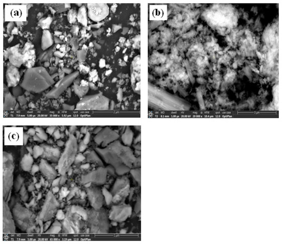
Figure 9.
SEM images of (a) Co(II), (b) Cu(II), and (c) Zn(II) complexes.
3.8. Antimicrobial and Antioxidant Assays
The antimicrobial results are summarized in (Table 4). The Cu(II) complex and the Co(II) complex have the most important effect. They showed a qualitative antibacterial effect with an inhibitory zone ranging between 1 cm and 2.5 cm for Gram-positive strains, such as Staphylococcus aureus and Micrococcus luteus, and between 1.2 cm and 1.4 cm for Gram-negative strains. The tested compounds have an antifungal effect against Candida albicans with a maximum inhibitory zone of 2.5 cm. As summarized in Table 5, the results demonstrated a high antioxidant potential for the Zn(II) complex with a DPPH scavenging of 91.5%; however, the Cu(II) complex was low (16.5%).
Table 4.
Antimicrobial and antioxidant effects. (Inhibitory zone expressed in cm ±SD).
Table 5.
Docking binding energy (kcal mol−1) of the complexes into the active site of the TyrRS receptor (PDB: 1JIJ).
3.9. In Silico Docking Study
In order to explore potential novel antibacterial compounds, we can study one of the enzymes that contribute to this potential property, which is the tyrosyl-tRNA synthetase. It is among the aminoacyl-tRNA synthetases (aaRSs) and oversees the catalysis of the covalent bond of amino acids to their corresponding tRNAs to make charged tRNA. Therefore, aaRSs inhibition affects the growth of the cell owing to its role in the process of protein biosynthesis. We must notice that the complex can be degraded in the medium, and the antimicrobial effect can also be due to the interaction of the individual components with the enzyme.
To explore the possible antibacterial activity of the compounds against pathogenic species, docking of the compounds was achieved with the catalytic site of TyrRS from S. aureus built from the structure of the TyrRS receptor (pdb: 1JIJ). The three compounds showed different values of binding energies with the TyrRS model (Table 5; Figure 10). The results showed that all compounds have good values of binding energy (7.2–7.9 kcal mol−1). The docking data presented showed that all compounds fit very well in the catalytic pockets of the proteins of the receptor. The most potent compound, Co(II), represented appropriate interactions with different residues of tyrosyl-tRNA synthetase, and the results are revealed in Figure 9. The obtained results describe that the compound Co(II) having the potent inhibitory activity occupies perfectly the catalytic site of Tyrosyl-tRNA synthetase with a value for the binding energy of −7.2 kcal mol−1. The Co(II) complex shows four hydrogen bonds: two between the hydrogens of the water molecule attached to the Co metal with Asp195, and two between the hydrogens of the two NH2 molecules attached to the metal with Asp40. The rings of the ligand show π-cation interaction with His50 and π-π, T-shaped interaction with Gly38. It shows Van der Waals interactions with the amino acids of the active site: Asp 80, His47, Lys84, Thr42, Gln196, Ala39, Gln174, Leu70, and Tyr170. These interactions contribute to the stabilization of the complex and suggest that it is involved in its inhibitory effect. These interactions with His47, Ala39, Gln174, Leu70, and Tyr170 are common to the catalytic sites found in compounds having antimicrobial activity [40]. This suggests that they contribute to this property.
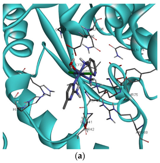
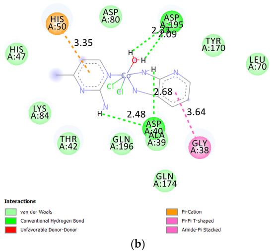
Figure 10.
(a) Three-dimensional (3D) and (b) two-dimensional (2D) interaction of Co(II) complex with the active site of tyrosyl tRNA synthetized (PDB: 1JIJ).
4. Conclusions
Three new Co(II), Cu(II), and Zn(II) metal complexes were prepared with success. Elemental analysis, UV-visible, FTIR, molar conductance, TGA analysis, X-ray diffraction, magnetic susceptibility studies, and X-ray diffraction were used to characterize the complexes. The possible geometries of the three synthesized complexes are octahedral structures, and these are six coordinated metal-ligand complexes. The XRD patterns indicate that the particles were polycrystalline and belonged to the monoclinic crystal system with space group P121. A spherical morphology of the particle powder of the Co(II) complex was observed, while for the Cu(II) and Zn(II) compounds, the particles have herbal assembly and cluster slice morphologies, respectively. Thermal analysis showed that the thermal stability of various compounds ranged from Co-complex at 35 °C to Cu-complex at 40 °C to Zn-complex at 48.5 °C. Above their stability temperature, one can observe several overlapping chemical reactions that are thermodynamically favorable. Conversely, the Cu(II) complex exhibited a modest antioxidant potential (16.5%), but the Zn(II) complex showed significant antioxidant activity with a DPPH scavenging of 91.5%. Additionally, the docking assays revealed that all substances had high amounts of binding energy (7.2–7.9 kcal mol−1). For this reason, all molecules can readily fit in the catalytic regions of the receptor protein. The Co(II) complex, however, appears to be more active. Advanced in vitro investigations will be carried out to confirm these chemicals’ potential impacts, and further attributes of these molecules will be investigated in subsequent research.
Author Contributions
All authors contributed equally to this work. All authors have read and agreed to the published version of the manuscript.
Funding
This research was funded by the Deputyship for Research & Innovation, Ministry of Education: Saudi Arabia, project number (QU-IF-2-5-4-26289).
Data Availability Statement
By e-mail: m.alfakeh@qu.edu.sa.
Acknowledgments
The authors extend their appreciation to the Deputyship for Research & Innovation, Ministry of Education, Saudi Arabia, for funding this research work through project number (QU-IF-2-5-4-26289). The authors also thank Qassim University for technical support.
Conflicts of Interest
The authors declare no conflict of interest.
References
- Kokunov, Y.V.; Gorbunova, Y.E.; Kovalev, V.V.; Kozyukhin, S. Silver complexes with 2-amino-4-methylpyrimidine: Synthesis, crystal structure, and luminescent properties. Russ. J. Coord. Chem. 2013, 39, 565–570.–570. [Google Scholar] [CrossRef]
- Navarro, J.A.; Barea, E.; Galindo, M.A.; Salas, J.M.; Romero, M.A.; Quirós, M.; Masciocchi, N.; Galli, S.; Sironi, A.; Lippert, B. Soft functional polynuclear coordination compounds containing pyrimidine bridges. J. Solid State Chem. 2005, 178, 2436–2451. [Google Scholar] [CrossRef]
- Cook, T.R.; Zheng, Y.-R.; Stang, P.J. Metal−organic frameworks and self-assembled supramolecular coordination complexes: Comparing and contrasting the design, synthesis, and functionality of metal−organic materials. Chem. Rev. 2013, 113, 734–777. [Google Scholar] [CrossRef] [PubMed]
- Sharma, C.V.K.; Griffin, S.T.; Rogers, R.D. Simple routes to supramolecular squares with ligand corners: 1: 1 Ag I: Pyrimidine cationic tetranuclear assemblies. Chem. Commun. 1998, 2, 215–216. [Google Scholar] [CrossRef]
- Pettinari, C.; Tabăcaru, A.; Galli, S. Coordination polymers and metal−organic frameworks based on poly (pyrazole)-containing ligands. Coord. Chem. Rev. 2016, 307, 1–31. [Google Scholar] [CrossRef]
- Ye, Y.; Xiong, S.; Wu, X.; Zhang, L.; Li, Z.; Wang, L.; Ma, X.; Chen, Q.-H.; Zhang, Z.; Xiang, S. Microporous Metal−Organic Framework Stabilized by Balanced Multiple Host−Couteranion Hydrogen-Bonding Interactions for High-Density CO2 Capture at Ambient Conditions. Inorg. Chem. 2016, 55, 292–299. [Google Scholar] [CrossRef]
- Shahbazi, M.; Mehrzad, F.; Mirzaei, M.; Eshtiagh-Hosseini, H.; Mague, J.T.; Ardalani, M.; Shamsipur, M. Synthesis, single crystal X-ray characterization, and solution studies of Zn (II)-, Cu (II)-, Ag (I)- and Ni (II)-pyridine-2, 6-dipicolinate N-oxide complexes with different topologies and coordination modes. Inorg. Chim. Acta 2017, 458, 84–96. [Google Scholar] [CrossRef]
- Mandal, T.; Dey, A.; Seth, S.K.; Ortega-Castro, J.; Rheingold, A.L.; Ray, P.P.; Frontera, A.; Mukhopadhyay, S. Influence of 2-Amino-4-methylpyridine and 2-Aminopyrimidine Ligands on the Malonic Acid-Cu(II) System: Insights through Supramolecular Interactions and Photoresponse Properties. ACS Omega 2020, 5, 460–470. [Google Scholar] [CrossRef]
- Yenikaya, C.; Sarı, M.; Bülbül, M.; İlkimen, H.; Çelik, H.; Büyükgüngör, O. Synthesis, characterization and antiglaucoma activity of a novel proton transfer compound and a mixed-ligand Zn(II) complex. Bioorg. Med. Chem. 2010, 18, 930–938. [Google Scholar] [CrossRef]
- Fuhrmann, H.; Brenner, S.; Arndt, P.; Kempe, R. Octahedral group 4 metal complexes that contain amine, amido, and aminopyridinato ligands: synthesis, structure, and application in α-olefin oligo- and polymerization. Inorg. Chem. 1996, 35, 6742–6745. [Google Scholar] [CrossRef]
- London, B.K.; Claville, M.O.F.; Babu, S.; Fronczek, F.R.; Uppu, R.M. A co-crystal of nona-hydrated disodium(II) with mixed anions from m-chlorobenzoic acid and furosemide. Acta Crystallogr. 2015, 71, 1266–1269. [Google Scholar]
- Yenikaya, C.; Büyükkıdan, N.; Sarı, M.; Keşli, R.; İlkimen, H.; Bülbül, M.; Büyükgüngör, O. Synthesis, characterization, and biological evaluation of Cu(II) complexes with the proton transfer salt of 2,6-pyridinedicarboxylic acid and 2-amino-4-methylpyridine. J. Coord. Chem. 2011, 64, 3353–3365. [Google Scholar] [CrossRef]
- Mistri, S.; Zangrando, E.; Manna, S.C. Cu(II) complexes of pyridine-2,6-dicarboxylate and N-donor neutral ligands: Synthesis, crystal structure, thermal behavior, DFT calculation and effect of aromatic compounds on their fluorescence. Inorg. Chim. Acta. 2013, 405, 331–338. [Google Scholar] [CrossRef]
- Yenikaya, C.; Poyraz, M.; Sarı, M.; Demirci, F.; İlkimen, H.; Büyükgüngör, O. Synthesis, characterization and biological evaluation of a novel Cu(II) complex with the mixed ligands 2,6-pyridinedicarboxylic acid and 2-aminopyridine. Polyhedron 2009, 28, 3526–3532. [Google Scholar] [CrossRef]
- Ilkimen, H. Synthesis and characterization of mixed ligand Cu (II) complexes of 2-methoxy-5-sulfamoylbenzoic acid and 2-aminopyridine derivatives. Maced. J. Chem. Chem. Eng. 2019, 38, 13–17. [Google Scholar] [CrossRef]
- Poddar, R.K.; Agarwala, U. Reactions of Ru(PPh3)2Cl2 and [Ru(AsPh3)2Cl2]2 with various donor molecules. J. Inorg. Nucl. Chem. 1973, 35, 3769–3779. [Google Scholar] [CrossRef]
- Raso, A.G.; Fiol, J.J.; Zafra, A.L.; Cabrero, A.; Mata, I.; Molins, E. Crystal structures of the N-salicylidene–Lserinatoaquacopper(II) monohydrate and its ternary derivative with 2-aminopyridine. Polyhedron 1999, 18, 871–878. [Google Scholar] [CrossRef]
- Al-Fakeh, M.S.; Alsaedi, R.O.; Amiri, N.; Allazzam, G.A. Synthesis, Characterization, and Antimicrobial of MnO and CdO Nanoparticles by Using a Calcination Method. Coatings 2022, 12, 215. [Google Scholar] [CrossRef]
- Mahdhi, A.; Leban, N.; Chakroun, I.; Chaouch, M.A.; Hafsa, J.; Fdhila, K.; Mahdouani, K.; Majdoub, H. Extracellular polysaccharide derived from potential probiotic strain with antioxidant and antibacterial activities as a prebiotic agent to control pathogenic bacterial biofilm formation. Microb. Pathog. 2014, 109, 220. [Google Scholar] [CrossRef]
- Dbeibia, A.; Ben Taheur, F.; Altammar, K.A.; Haddaji, N.; Mahdhi, A.; Amri, Z.; Mzoughi, R.; Jabeur, C. Control of Staphylococcus aureus methicillin resistant isolated from auricular infections using aqueous and methanolic extracts of Ephedra alata. Saudi J. Biol. Sci. 2022, 29, 1021–1028. [Google Scholar] [CrossRef]
- Frisch, M.J.; Trucks, G.W.; Schlegel, H.B.; Scuseria, G.E.; Robb, M.A.; Cheeseman, J.R.; Montgomery, J.A.; Vreven, T.; Kudin, K.N.; Burant, J.C.; et al. Gaussian 09, Revision D.01; Gaussian, Inc.: Wallingford, CT, USA, 2013. [Google Scholar]
- Morris, G.M.; Huey, R.; Olson, A.J. Using AutoDock for ligand-receptor docking. Curr. Protoc. Bioinform. 2008, 24, 8–14. [Google Scholar] [CrossRef] [PubMed]
- Trott, O.; Olson, A.J. AutoDock Vina: Improving the speed and accuracy of docking with a new scoring function, efficient optimization, and multithreading. J. Comput. Chem. 2010, 31, 455–461. [Google Scholar] [CrossRef] [PubMed]
- BIOVIA. Dassault Systèmes, v16. 1.0. 15350; Discovery studio visualizer; Dassault Systèmes: San Diego, CA, USA, 2015. Available online: https://www.3ds.com/products-services/biovia/products/molecular-modeling-simulation/biovia-discovery-studio/ (accessed on 7 December 2022).
- Jose, S.P.; Mohan, S. Vibrational Spectra and Normal Co-Ordinate Analysis of 2-Aminopyridine and 2-Amino Picoline. Spectrochim. Acta Part A Mol. Biomol. Spectrosc. 2006, 64, 240–245. [Google Scholar] [CrossRef] [PubMed]
- Yuoh, A.C.B.; Agwara, M.O.; Yufanyi, D.M.; Conde, M.A.; Jagan, R.; Eyong, K.O. Synthesis, Crystal Structure, and Antimicrobial Properties of a Novel 1-D Cobalt Coordination Polymer with Dicyanamide and 2-Aminopyridine. Int. J. Inorg. Chem. 2015, 2015, 9–12. [Google Scholar] [CrossRef]
- Al-Fakeh, M.S.; Allazzam, G.A.; Yarkandi, N.H. Ni (II), Cu (II), Mn (II), and Fe (II) Metal Complexes Containing 1,3-Bis (diphenylphosphino) propane and Pyridine Derivative: Synthesis, Characterization, and Antimicrobial Activity. Int. J. Biomater. 2021, 2021, 1–12. [Google Scholar] [CrossRef] [PubMed]
- Raducka, A.; Czylkowska, A.; Gobis, K.; Czarnecka, K.; Szymanski, P.; Swiatkowski, M. Characterization of Metal-Bound Benzimidazole Derivatives, Effects on Tumor Cells of Lung Cancer. Materials 2021, 14, 2958. [Google Scholar] [CrossRef]
- Al-Fakeh, M.S. Synthesis, thermal stability and kinetic studies of copper (II) and cobalt (II) complexes derived from 4-aminobenzohydrazide and 2-mercaptobenzothiazole. Eur. Chem. Bull. 2020, 9, 403–409. [Google Scholar] [CrossRef]
- Aly, A.A.M.; Ghandour, M.; Alfakeh, M.S. Synthesis and characterization of transition metal coordination polymers derived from 1,4-Benzenedicarboxylic acid and Azoles. Turk. J. Chem. 2012, 36, 69–79. [Google Scholar]
- Kevin, W.; Wellington, B.; Perry, T.; Kaye, A.; Watkin, G.M. Designer ligands. Part 14. Novel Mn(lI), Ni(II) and Zn(II) complexes of benzamide- and biphenyl-derived ligands. ARKIVOC 2008, 17, 248–264. [Google Scholar]
- Ghandour, M.A.; Aly, A.; Al-Fakeh, M.S. Synthesis and characterization of Cu(II), Cd(II) and Pb(II) coordination polymers derived from 1,4-benzene- and 1,1’-ferrocenedicarboxylate and 2-aminobenzothiazole. J. Indian Chem. Soc. 2011, 88, 1633–1638. [Google Scholar]
- Al-Fakeh, M.S. Synthesis and characterization of coordination polymers of 1,3-di (4-pyridyl)-propane and 2-aminobenzothiazole with Mn(II), Co(II), Cu(II) and Ni(II) ions. J. Chem. Pharm. Res. 2018, 10, 77–83. [Google Scholar]
- Al-Fakeh, M.; Alsaedi, R. Synthesis, Characterization and Antimicrobial Activity of CoO Nanoparticles from a Co (II) Complex Derived from Polyvinyl Alcohol and Aminobenzoic Acid Derivative. Sci. World J. 2021, 2021, 1–11. [Google Scholar] [CrossRef] [PubMed]
- Aly, A.; Ghandour, M.A.; Abu-Zied, B.M.; Al-Fakeh, M.S. Synthesis, properties and environmentally important nanostructured and antimicrobial supramolecular coordination polymers containing 5-(3-pyridyl)-1,3,4-oxadiazole-2-thiol and benzimidazole. J. Environ. Anal. Toxicol. 2012, 2, 2–7. [Google Scholar] [CrossRef]
- Al-Fakeh, M.; Alminderej, F. New method for the preparation and biological activity of CuO nanoparticles from a mixed PVA and 2-Aminobenzothiazole complex. Int. J. ChemTech Res. 2018, 11, 442–449. [Google Scholar]
- El-ajaily, M.M.; Ben-Gweirif, S.F.; El-zweay, R.S.; Maihub, A.A. Synthesis of Some Mixed Ligand Complexes Derived from Catechol and 2-Aminopyridine and Their Biological Activity. J. Pure Appl. Sci. 2007, 6, 5. [Google Scholar]
- Uçar, I.; Karabulut, B.; Bulut, A.; Büyükgüngör, O. Synthesis, structure, spectroscopic and electrochemical properties of (2-amino-4-methylpyrimidine)-(pyridine-2, 6-dicarboxylato) copper (II) monohydrate. J. Mol. Struct. 2007, 834, 336–344. [Google Scholar] [CrossRef]
- Bukharov, M.S.; Shtyrlin, V.G.; Mamin, G.V.; Stapf, S.; Mattea, C.; Mukhtarov, A.S.; Serov, N.Y.; Gilyazetdinov, E.M. Structure and Dynamics of Solvation Shells of Copper(II) Complexes with N,O-Containing Ligands. Inorg. Chem. 2015, 54, 9777–9784. [Google Scholar] [CrossRef]
- Alminderej, F.; Bakari, S.; Almundarij, T.I.; Snoussi, M.; Aouadi, K.; Kadri, A. Antimicrobial and Wound Healing Potential of a New Chemotype from Piper cubeba L. Essential Oil and In Silico Study on S. aureus tyrosyl-tRNA Synthetase Protein. Plants 2021, 10, 205. [Google Scholar] [CrossRef]


Disclaimer/Publisher’s Note: The statements, opinions and data contained in all publications are solely those of the individual author(s) and contributor(s) and not of MDPI and/or the editor(s). MDPI and/or the editor(s) disclaim responsibility for any injury to people or property resulting from any ideas, methods, instructions or products referred to in the content. |
© 2023 by the authors. Licensee MDPI, Basel, Switzerland. This article is an open access article distributed under the terms and conditions of the Creative Commons Attribution (CC BY) license (https://creativecommons.org/licenses/by/4.0/).