Review

Recent Trends in Elpasolite Single Crystal Scintillators for Radiation Detection

1
MIIT Key Laboratory of Critical Materials Technology for New Energy Conversion and Storage, School of Chemistry and Chemical Engineering, Harbin Institute of Technology, Harbin 150080, China
2
State Key Laboratory of Urban Water Resource and Environment, Harbin Institute of Technology, Harbin 150090, China
*
Author to whom correspondence should be addressed.
Crystals 2022, 12(7), 887; https://doi.org/10.3390/cryst12070887
Submission received: 17 May 2022 / Revised: 14 June 2022 / Accepted: 17 June 2022 / Published: 22 June 2022
(This article belongs to the Special Issue Advances in Optoelectric Functional Crystalline Materials)

Abstract

:
Scintillation detection has attracted great interest in nuclear medicine, nuclear radiation detection, high-energy physics, and non-destructive inspection. The elpasolite crystals with Ce3+ dopants are promising for these endeavors due to their modest light yield and extremely good proportionality when excited by the gamma ray. Moreover, the 6Li and 35Cl isotopes in elpasolite crystals endow them with excellent neutron detection capability. These features allow not only a high energy resolution but also a high detection sensitivity. The elpasolite scintillators also enable the precisely dual detection of gamma/neutron signals through pulse height discrimination (PHD) or pulse shape discrimination (PSD). In this work, we review recent investigations on using the typical elpasolite scintillators, including Ce3+-doped Cs2LiYCl6 (CLYC), Cs2LiLaCl6 (CLLC), and Cs2LiLaBr6 (CLLB), for the monitoring of gamma rays and neutrons. The scintillation properties, detection mechanism, and elpasolite crystal structure are also discussed with the aim of improving high-energy ray detection ability.

1. Introduction

Scintillation detection plays an important role in a wide range of applications, such as nuclear medicine, nuclear radiation detection, high-energy physics, and border control, due to its ability to precisely monitor multiple rays [1,2]. A scintillation detector is composed of two parts: scintillation crystal, and photoelectric converter for converting the (ultraviolet) UV–visible lights into electric signals. In these detectors, the scintillation crystals act as conversion media, which can convert high-energy radiations into detectable low-energy photons. The dielectric or semiconductor wide bandgap materials are able to accomplish this purpose by absorbing high-energy radiation energy and converting into UV or visible photons through three stages: conversion, transport, and luminescence emission, shown in Figure 1. In the conversion stage, hot electrons and holes are produced through multistep interaction between scintillator lattice and ionizing radiation. Then, the produced electrons and holes are gradually thermalized and transferred to the bottom of the conduction band and the top of the valence band, respectively. For the transport process, electrons and holes migrates from the conduction valence bands of host materials to the emission centers. Finally, the recombination of electrons and holes emits detectable photons in the luminescence emission stage. In addition, the core-to-valence luminescence (CVL) could also be detected in some elpasolite scintillators. The electrons in the core band are excited to the conduction band by ionizing radiation. The holes generated in the core band immediately recombine with electrons in the valence and emit short-wavelength (220 nm), fast emission (~1 ns) fluorescence, favoring their use in gamma/neutron discrimination.
The solid scintillators, including organic and inorganic scintillation materials, have successfully been manufactured into scintillation detectors [3]. Among them, the inorganic scintillators have become the most widely used materials nowadays due to the outstanding scintillation characteristics [4]. The first inorganic scintillator Ba[Pt(CN)4] powder, emitting visible photons under X-ray irradiation, was discovered by Edison [5]. In the late 1940s, the alkali halides single crystals, such as NaI:Tl [6] and CsI:Tl [7], were developed as the better scintillators. Indeed, the NaI:Tl is still one of the most widely used inorganic scintillators due to its modest scintillation properties, relatively low cost, and availability of large-size single crystals [8]. The NaI:Tl crystal is often employed as the benchmark to estimate the performance of newly explored scintillation materials. Subsequently, the Lus2SiO5 crystal was also exploited as standard material for positron emission tomography (PET) equipment (one of the most typical applications of scintillators) [9]. The LaBr3 crystal was also demonstrated to be the one of the best inorganic materials for the use of scintillation detectors [10]. However, these scintillators described above are severely limited by the inferior energy resolution for NaI:Tl and internal radioactive contaminants for LaBr3 [11].
The elpasolites materials as a new generation of halide scintillators have achieved high light output and excellent proportionality, and more outstanding scintillators are exploited from the elpasolite crystal family. Benefiting from the high proportionality, the excellent energy resolution of 3.6% at 662 keV was achieved in the Cs2LiYCl6 crystal [12]. Cs2LiLaBr6 has been found to be the brightest elpasolite scintillator, and it achieves 60,000 photons/MeV light yield and 2.9% energy resolution at 662 KeV [13]. A great number of experiments and theoretical studies on existing elpasolite crystals have been implemented with the aim of developing unprecedented scintillators for high-efficiency detection of gamma rays [14,15,16,17]. The neutron detection capabilities of elpasolite crystal scintillators were also further exploited [18,19,20,21]. The Li-containing elpasolites can provide extraordinary capability to monitor thermal neutrons by the large cross section of 6Li to thermal neutrons [22,23]. Elpasolite scintillators with special compositions, such as Cs2LiYCl6 and Cs2LiLaBr6 crystals, exhibit ideally simultaneous detection ability for gamma rays and neutrons through pulse shape discrimination [24]. These above investigations highlight the significant value of halide elpasolite scintillators for high-energy ray discrimination.
In this review, we summarize recent works on the exploration of various elpasolite crystals for sensing gamma rays and neutrons. This mainly includes the typical Cs2LiYCl6 (CLYC), Cs2LiLaCl6 (CLLC), and Cs2LiLaBr6 (CLLB) crystals and the other elpasolite crystals. We also briefly summarize the scintillation properties, detection mechanism, and structure of these elpasolite materials.

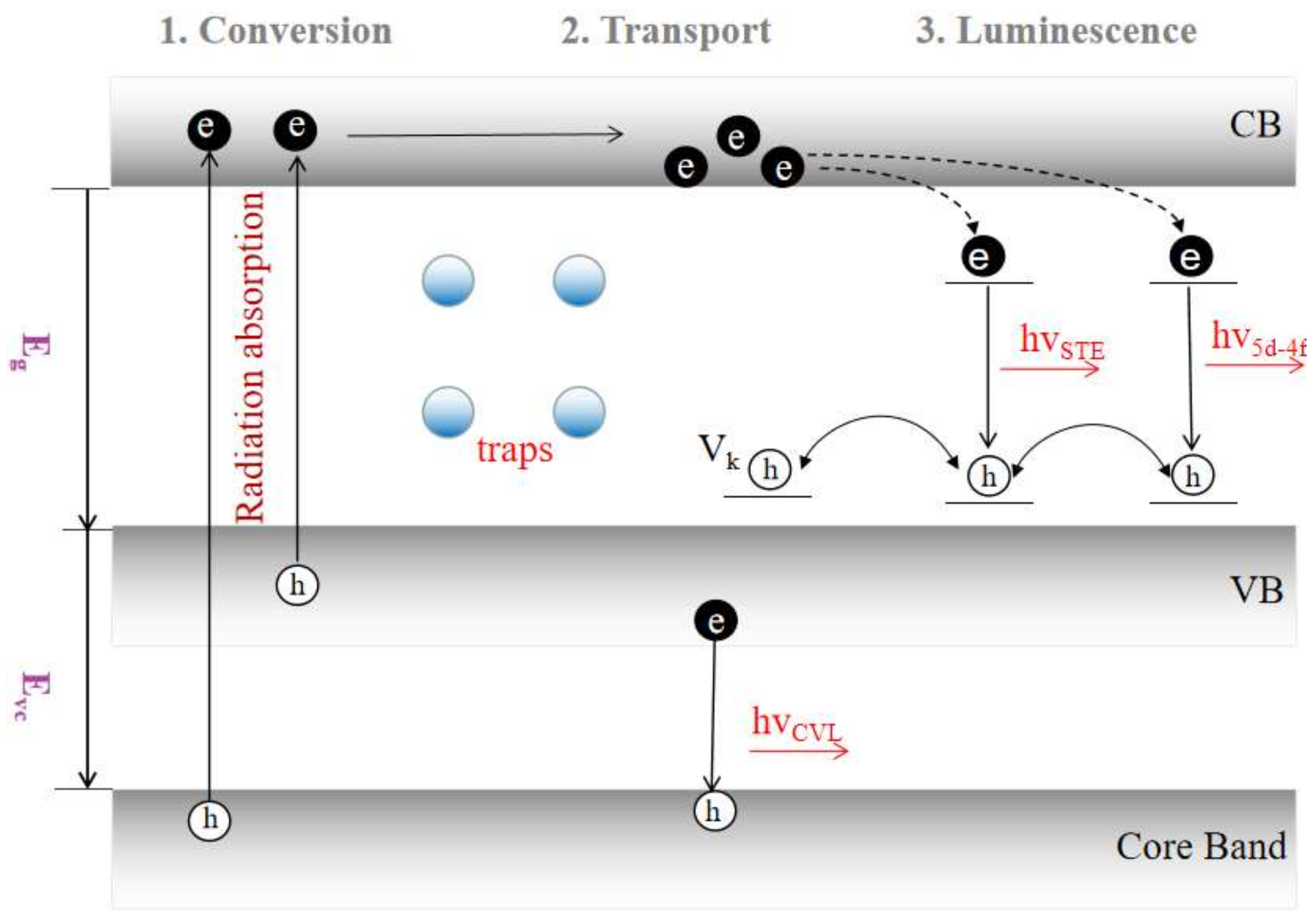
2. Scintillator Properties

The light yield (LY) and energy resolution (ER) are considered as the most important scintillation characteristics of scintillation materials. LY refers to the total number of emitting photons after the scintillator, absorbing radiation energy within a certain detection time. In a certain band gap range, relatively small band gap enables the higher light output. The total light yield also depends on the energy of excitation source and this dependence relation is called nonproportionality. The luminescence quenching, a nonlinear relationship with excitation density, is considered to be the root cause of nonproportionality. For practical detection applications, it is generally required that the emission light of the scintillator is located at the visible region, favoring the photomultiplier tube or silicon photodiode on the detector to monitor these photons. The energy resolution of scintillators is related to the LY and nonproportionality to a great extent [25,26,27,28]. Indeed, the scintillators with sufficiently high energy resolution have this ability to exactly distinguish the radiation species from the close interfere signals. The physical and scintillation properties of various scintillation crystals are also given in Table 1.

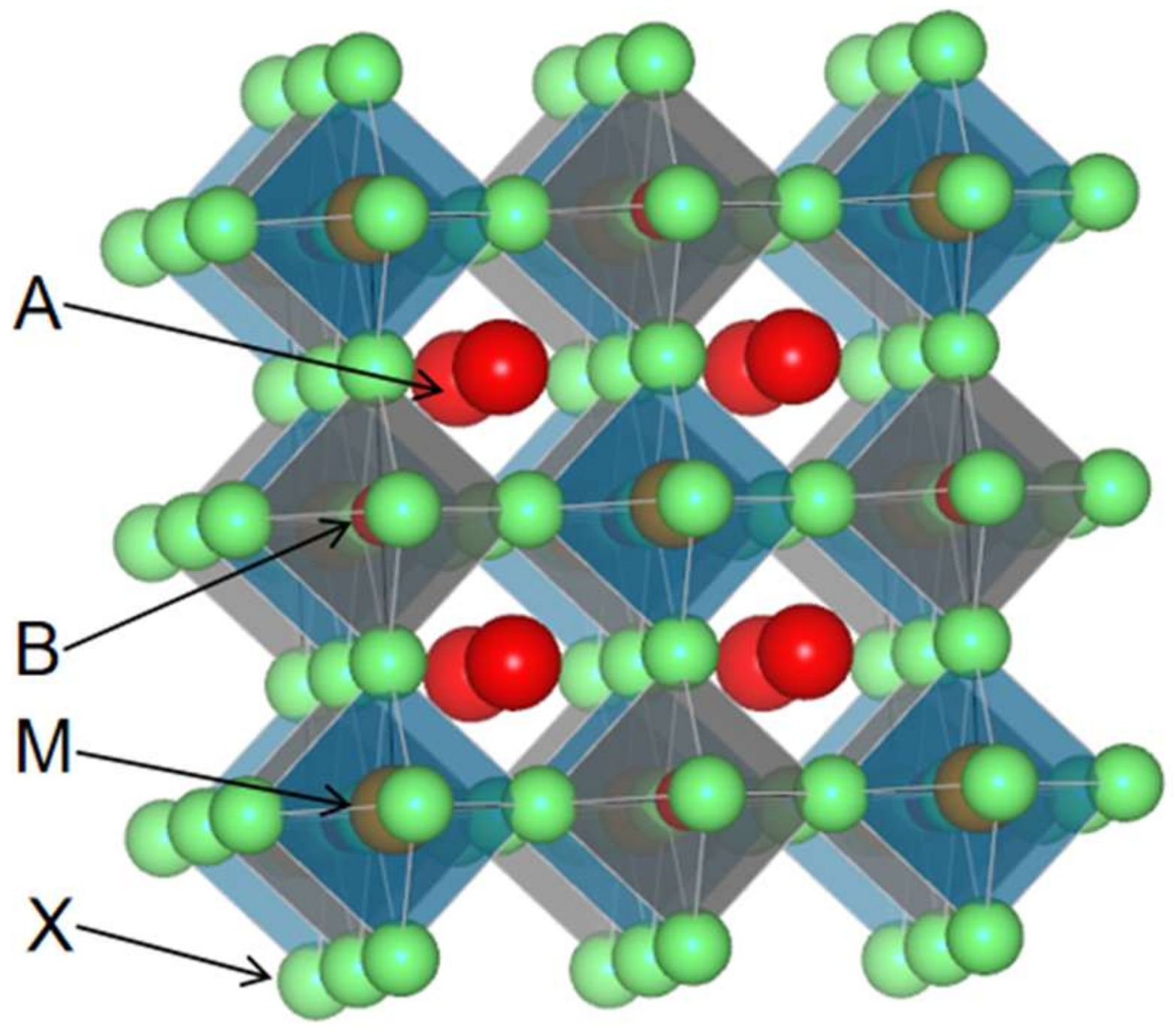
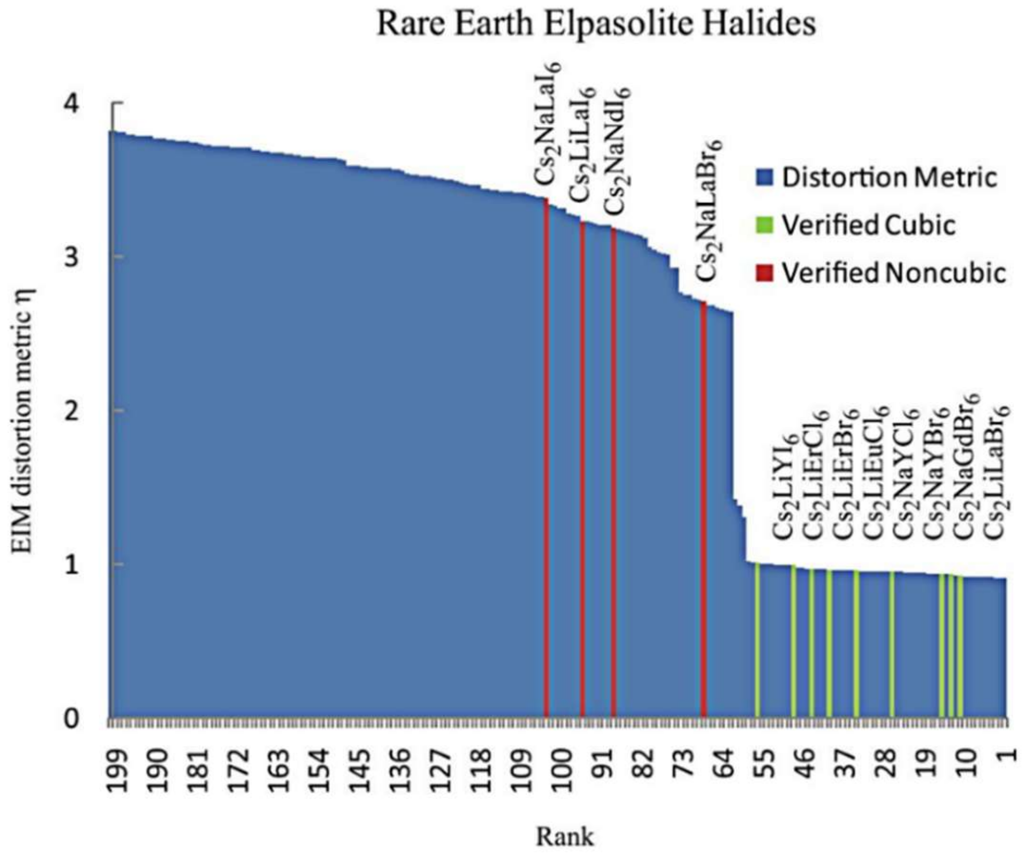
3. Structure of Elpasolite Scintillators
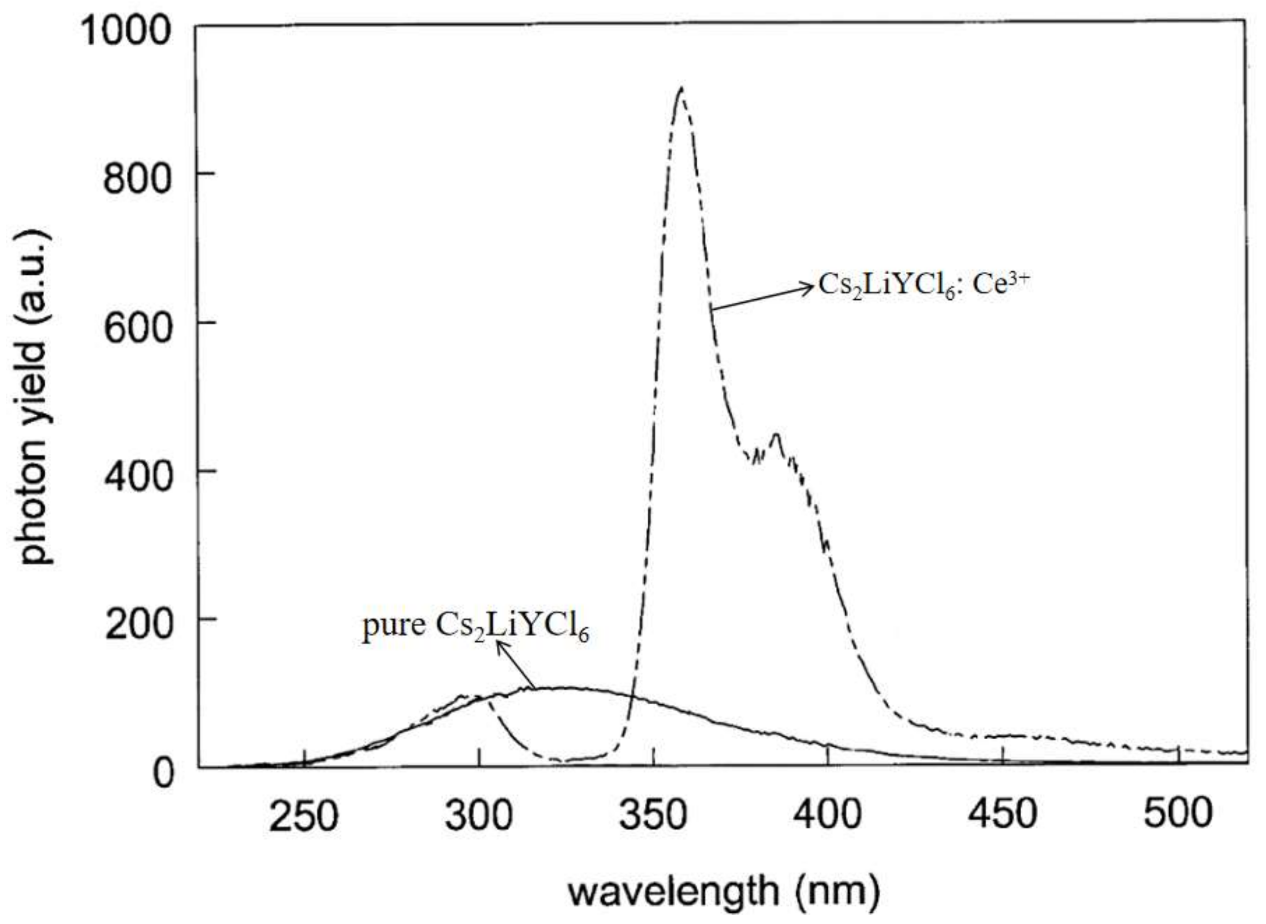
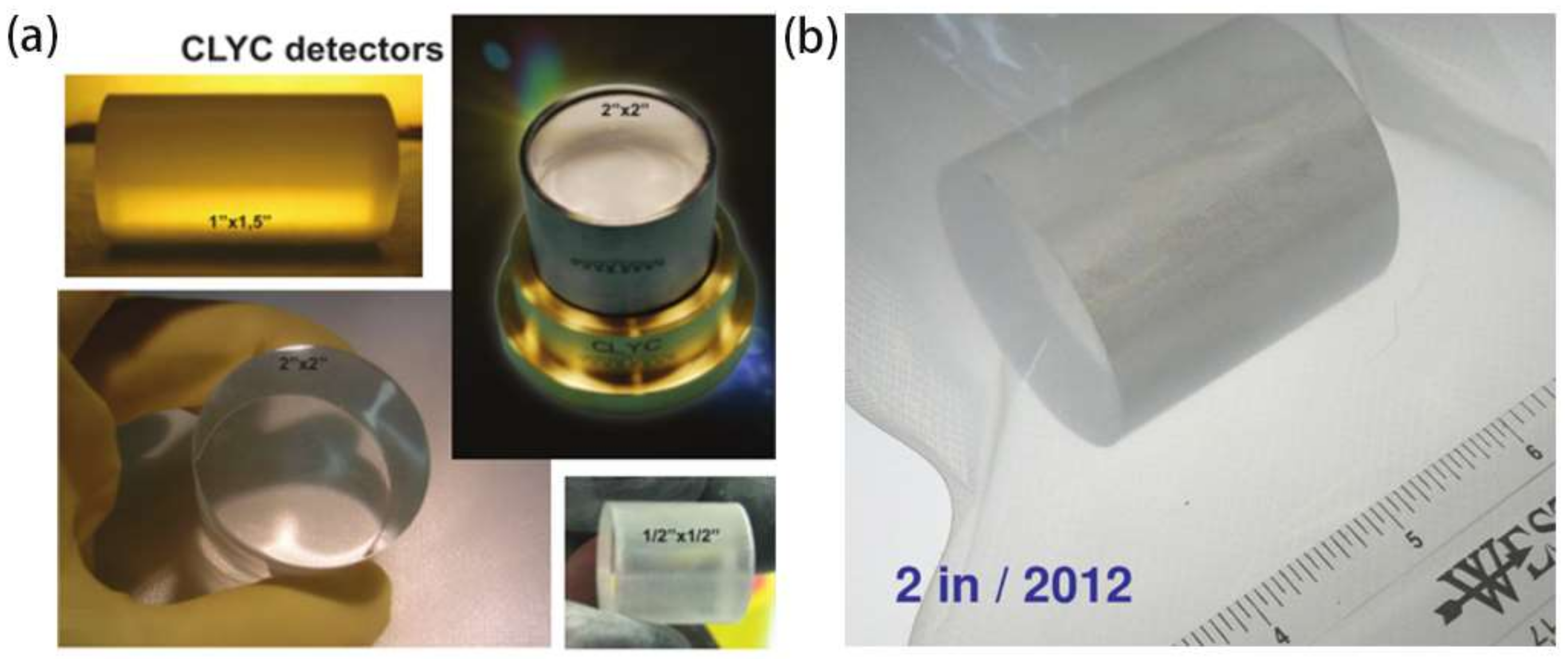
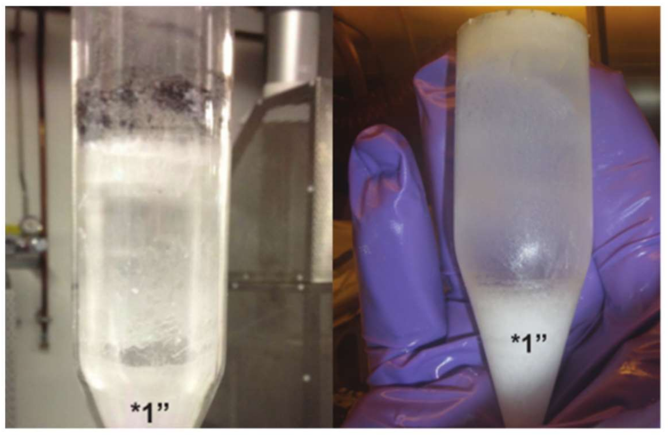
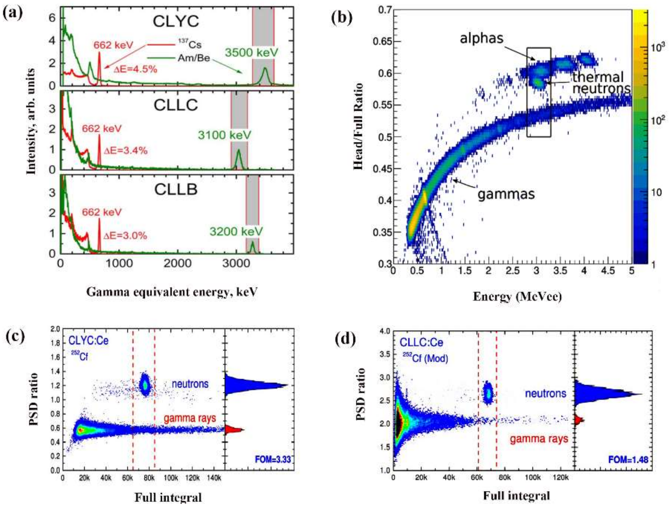
The absolute multitude of investigations on elpasolite materials have been focused on crystals with formula A2BMX6. Figure 2 shows the typical double-perovskite structure diagram of an elpasolite crystal. Three cations (A, B, and M) and one anion (X) constitute the A2BMX6 elpasolite crystal, which can be regarded as the linking of octahedra BX6 and MX6. Most of them possess cubic or near-cubic structure. The most prominent feature of perovskite-related architecture is the structure distortions accompanied by phase transitions [29]. Flerov, et al. demonstrated the lattice distortion in the phase transformation process of elpasolite crystal [30]. The Goldschmidt’s tolerance factor (t) has been used to determine stability range of elpasolite structures at room temperature. The cubic phase is stable in the range of 0.8 < t < 1 (for varied cations and anions, t value is slightly different). The smaller or larger t value leads to a distorted variant of the cubic structure. When the t value is close to the boundary of stability phase, a phase transition from P4/nbm tetragonal to cubic is also observed in the Rb2NaTmCl6 crystal. The grown crystals with large size may crack during cooling after growth, which has been the main obstacle for growing the large LaBr3 crystals with low cost. Understanding phase transformation from crystallization temperature to room temperature is useful for avoiding crystal cracking during post-growth cooling and fabricating devices.
The distortion metric predicting the stable structure of crystals at room temperature is defined by analyzing embedded-atom method (EIM) database and molecular statics study of crystal stability in Ref. [31]. A number of A2BMX6 crystals have been studied and their corresponding distortion metric versus rank number sorted by this metric, shown in Figure 3, which successfully screens out a number of cubic elpasolite compositions. This favors guiding the growth of large crystals with scintillation performance. The anions also influence crystal structure. The elpasolite chlorides with smaller anion radius have stable cubic structure at room temperature. In contrast, the elpasolite iodides with larger anion exhibit non-cubic structure, which hampers the production of high-quality scintillators.

4. Cs2LiYCl6 (CLYC)

The CLYC:Ce3+ is one of the most typical scintillators in the elpasolite crystal family, and it was developed as an effective thermal neutron detector in 1999 by Combes, et al. [32]. Subsequently, William M. Higgins, et al. further studied the CLYC:Ce3+ crystals and finally fabricated the commercial dual-detection scintillators [33]. The initially reported energy resolution of CLYC:Ce3+ crystals is 7.3% at 662 keV of 137Cs radiation, as shown in Figure 4. Benefiting from the improvement in vertical Bridgman technology and purification method, the energy resolution of one inch CLYC:Ce3+ crystal has been elevated to 3.6% at 662 keV in 2012 [12]. It is worth noting that the energy resolution value of CLYC:Ce3+ outperforms many commercial NaI:Tl products, which stems from exceptionally good energy proportionality. In addition, the shaping time, as another important parameter controlled by photomultipliers (PMT), can also influence the measured energy resolution of CLYC crystals. For instance, the energy resolution measured with 4 us shaping time was significantly superior to that obtained with 0.5 us shaping time. The virtually absent direct capture of electrons and holes by Ce3+ activators results in the slow decay time of CLYC scintillation crystals, which may explain the shaping time dependence of energy resolution.
The emission spectra of CLYC scintillation crystals were also extensively studied. A broad emission band ranging from 240 to 460 nm was observed in the X-ray-excited emission spectrum of pure CLYC, which is attributed to the self-trapped exciton (STE) emission shown in Figure 5 [32]. The spontaneous formation of STE, the luminescent defect itself, plays a significant role in the luminescent process of elpasolite crystals. As a result, the decay time of elpasolite crystals is relatively long compared to other commercially available scintillators. The very fast decay component was also found in the emission spectrum, which can be ascribed to the intrinsic core–valence luminescence (CVL) [24]. CVL appears at 310 nm with a fast decay time of several ns. It has been demonstrated that the smaller energy gap between the core band and the top of valence band than band gap favors the intense CVL. The slow decay process of the CLYC scintillation crystal is found to be a significant drawback, leading to the limited applications [35]. To address this problem, the Ce3+ activators were introduced into the CLYC lattice. For a Ce3+-doped CLYC system, the emission spectrum obtained from X-ray excitation shows the typical doublets emission peak, which is caused by transition from the lowest 5d level to the 2F5/2 and 2F7/2 levels of Ce3+ activators, shown in Figure 5. The Bessiere group reported that the regulation of the Ce3+ concentration can improve the scintillation performance, and the optimal doping concentration was determined to be less than 1% [36]. As shown in the literature, 0.1% Ce3+-doped CLYC exhibits a 700 photons/MeV LY with 8% energy resolution at 662 keV of 137Cs radiation. The best energy resolution value of 7% was achieved in 0.02% Ce3+-doped CLYC. The most optimum doping concentration varies slightly with different crystal growth processes, and 0.5% Ce3+ concentration is considered as the standard doping level nowadays [37]. The 0.5% Ce3+-doped CLYC outputs the typical Ce emissions with 40 ns decay time. The slow decay component still accounts for most of the light yield due to the localized band structure of CLYC:Ce3+. Developing new elpasolite compositions with delocalized band structure may be a valid way to reduce the decay time.
Growing of CLYC:Ce3+ scintillation crystals has been attempted in the last ten years. In 2010, a 3 inch inclusion-free single crystal was successfully grown by using the vertical Bridgman technique at the Radiation Monitoring Devices (RMD) company for the first time [33]. Figure 6a presents the photo of 2 inch inclusion-free CLYC:Ce3+ crystals grown by the Saint-Gobain company. Figure 6b illustrates the photo of a 2 inch inclusion-free single crystal grown by RMD with energy resolution of 4.12%. However, the key growth technology was not provided by both of them. As we know, the complex components of a CLYC:Ce3+ crystal, as well as the incongruent melting characteristic, is not conducive for the growth of the single crystals. This leads to the generation of defects such as bubbles, inclusions, and cracks in single crystals. The CLYC:Ce3+ phase diagram based on Cs2YCl5 and LiCl was studied to decrease the defect and acquire the optimized melting characteristic for a single crystal [38]. The 1 × 1 × 0.5 cm CLYC crystal was fabricated into a portable scintillation detector with a solid-state photomultiplier (1 cm × 1 cm) in ref. [39]. The neutron counts of fabricated CLYC:Ce3+ detector were compared with the 3-He tubes under 241Am/Be neutron source (4 MeV of average neutron energy). The result indicates that when the distance between neutron source and detector is greater than 6 mm, the CLYC:Ce3+ detector could outperform traditional 3-He tubes in count rates and sensitivity. In addition, the fabricated detector also shows the excellent detection ability for gamma signals. However, since the elpasolite scintillators are hygroscopic, the packaging technique was employed to protect the crystal and avoid the degradation of light output. The performance of CLYC:Ce3+ detectors can be also influenced by packing processes. In the future, the improved packaging techniques are expected to assist the CLYC:Ce3+ detector for radiation detection.

5. Cs2LiLaCl6 (CLLC)

CLLC:Ce3+ single crystals were also exploited as scintillation material for monitoring gamma rays. The luminescent emitting properties of the CLLC:Ce3+ crystals were measured by vacuum ultraviolet (VUV) synchrotron radiation [41]. The luminescence spectra contain two emission bands: core–valence luminescence (CVL) emitting band peaking at 4.4 eV with decay time of 1.4 ns and Ce3+ emitting band peaking at 3.3 eV with decay time of 36 ns, shown in Figure 7. The first CLLC:Ce3+ crystal based on Bridgman growth technique was reported by Glodo, et al. and its size reached 1 inch in diameter [42]. However, the as-grown crystal was opaque because it had a great number of inclusions. Compared with CLYC:Ce3+, the main advantage of CLLC:Ce3+ possesses a higher light yield. Their light yields reach 35,000 photons/MeV and 110,000 photons/neutron under irradiation from 137Cs (662 keV) and 241Am/Be (thermalized neutrons) sources, respectively. A 3.4% energy resolution at 662 KeV was achieved by gamma ray excitation, which is superior to that of CLYC:Ce3+. The 36 ns decay time of typical Ce3+ luminescence (372 nm and 400 nm) in 1% Ce3+-doped CLLC was obtained, which also outperforms the CLYC:Ce3+ crystals. Despite the excellent scintillation performance, there is no report on the successful growth of a CLLC single crystal. This is attributed to the Cs3LaCl6 s phase existing in the melt during crystal growth arising from the incongruently melting characteristic of the CLLC crystal. The inclusions were thus formed in the grown crystal, resulting in opacity of the grown crystal. In order to solve this problem, Hebing Zhu designed and employed the rich LiCl melt with high axial temperature gradient to produce the CLLC:Ce3+ single crystal [43]. The rich LiCl melts favor the suppression of Cs3LaCl6 s phase. In addition, the high axial temperature gradient enables the sufficient impurity transport, benefiting the production of transparent CLLC crystals. The 12 mm crystal wafer cut from the transparent section of the as-grown crystal exhibits better optical transmittance than that grown by Glodo, et al. However, the low energy resolution of 7.1% implies a poor crystal quality.

6. Cs2LiLaBr6 (CLLB)

CLLB:Ce3+, as another elpasolite crystal, is exploited for the purpose of dual detection of gamma ray and neutron. The Ouspenski group demonstrated that the light yield of CLLB:Ce3+ crystal reaches 60,000 photons/MeV, which is brighter than CLYC:Ce3+ and CLLC:Ce3+ [44]. As a result, an excellent energy resolution of 2.9% at 662 keV is achieved. Moreover, they found that the light yield increases and the decay time decreases with increasing temperature, which can be ascribed to the more effective energy transfer from the CLLB to the Ce3+ activators. Shirwadkar, et al. studied the CLLB doped with varied Ce3+ concentration under 137Cs excitation [45]. The low light yield was observed when the Ce3+ concentration was less than 0.2%. However, the light yield indicated a significant increase as the Ce3+ doping concentration exceeded 0.5%. This phenomenon is distinctly different from that of the CLYC crystal. The energy resolution of the CLYC crystal reduced significantly at high-concentration Ce3+-doping. X-ray induced emission spectra of CLLB are presented in Figure 8. The pure CLLB has a broad response, whereas Ce3+-doped CLLB exhibits typically dual bands peaking at 390 and 420 nm. With increasing Ce3+ concentration, the intensity ratio of 420 nm/390 nm emissions shows an increased trend. This may be attributed to the absorption of Ce3+ activator to 390 nm light and re-emission of 420 nm photons. Under the excitation of gamma ray, the Ce3+-doped CLLB showed a relatively long decay time of 55 ns. This can be attributed to the improved efficiency of direct capture of energy by Ce3+ activators. With regard to La-containing scintillators, such as LaBr3:Ce3+ and CLLB:Ce3+, the alpha contamination degrading the energy resolution is a thorny problem. The relationship between the internal alpha contamination and the energy resolution was studied and presented in ref. [46].
Similar to the growth of the CLLC crystal, the growth of the CLLB crystal mainly faces two major obstacles: (I) the raw materials are extremely sensitive to air and moisture, requiring vacuum seal in a quartz crucible during growth; (II) the incongruent melting property always lead to bubbles and cracks in as-grown crystals, degrading the scintillation performance. Lin, et al. demonstrated the CLLB:Ce3+ crystal can be grown by using the Bridgeman growth technique [47]. The transparent 1 inch crystal with energy resolution of 4.09% at 662 keV has been successfully grown by Saint-Gobain Crystals, shown in Figure 9. The CLLB:Ce3+ scintillator can be fabricated into portable gamma/neutron dual detectors using silicon photomultipliers by Saint-Gobain Crystals [48]. The parameters, including Ce3+ concentration, shape, size, silicon photomultiplier (SiPM) model, and SiPM placement on detector, are systematically researched. The energy resolution of detecting gamma ray reaches 6.0% at 662 keV and achieves a PSD figure of merit (PSD FOM) of 1.9 under241Am/Be source (4 MeV of average neutron energy).

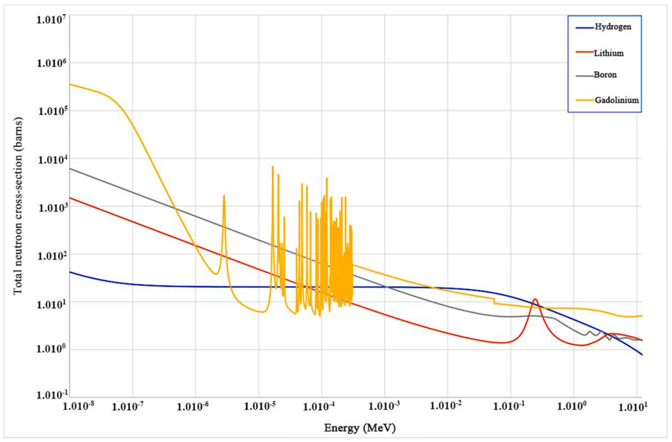
7. The Elpasolite Scintillators for Neutron Detection

Highly efficient neutron detection is conducive to prevent proliferation of nuclear material. In recent years, dwindling supplies of 3He and growing emphasis on global nuclear threat have triggered great demands for developing appropriate neutron detection scintillators to accomplish the mission [49]. Scintillators for neutron detection can be divided into solid, liquid, and gas according to the physical state of the material, and solid scintillators mainly include plastic, glass, and crystal scintillators. In this paper, we focus on the elpasolite crystal scintillators. A great number of isotopes were exploited for monitoring neutron [50]. Figure 10 presents the neutron capture cross sections of lithium, boron, gadolinium, and hydrogen as a function of neutron energy. Among them, gadolinium isotope (yellow line) shows the highest cross section area at low energy region. However, the high-energy gamma rays were produced when the Gd isotope of high effective atomic number (Zeff) interacted with neutrons, escaping from the material and resulting in low detection efficiency. Boron isotope also exhibited high cross section value for low-energy neutron detection. The low light yield, however, severely limits its use for neutron detection. The hydrogen isotope has a high cross section in the high-energy region, favoring the detection of fast neutrons.
It is worth noting that the lithium isotope with small effective atomic number (Zeff) shows large neutron capture cross section in the high-energy region, which benefits the improvement in resolution and produces considerable light yield with respect to thermal neutron. A great number of 6Li-based neutron detection scintillators (6Li-glass, 6LiI:Eu, 6LiF/ZnS:Ag etc.) have been exploited [51,52]. However, the 6Li-glass shows a low light yield of 6000 ph/neutron due to severe scintillation quenching. Despite that the 6LiI:Eu exhibits high light yield, it cannot discriminate gamma and neutron signal reliably. The 6LiF/ZnS:Ag has been developed as the most widely used neutron detection scintillator with the advantages of high light yield (160,000 ph/neutron), low sensitivity to gamma rays, and good gamma/neutron discrimination ability. However, the preparation of 6LiF/ZnS:Ag scintillator achieved by mixing ZnS:Ag and 6LiF leads to the opacity due to their sharply different refractive indices, which severely limits the thickness of scintillator layer (≤0.4 mm) and results in low neutron detection efficiency. To address this problem, efforts have been devoted to the elpasolite scintillators containing Li for differentiating neutron from gamma signals. For instance, CLYC:Ce3+, CLLC:Ce3+, and CLLB:Ce3+ crystals containing 6Li achieved, respectively, 2.9, 2.6 and 1.8% energy resolution [34]. Among them, the best neutron detector was demonstrated to be the CLYC:Ce3+ crystal, which provided light yield as high as 70,000 photons/neutron and better gamma ray discrimination ability with maximum FOM value of 3.33 under 241Am/Be source (4 MeV of average neutron energy). The light yield value of 70,000 ph/neutron is higher than that of 6Li-glass (6000 ph/neutron), 6LiI:Eu (50,000 ph/neutron), and LiBaF3:Ce (3600 ph/neutron). It is only smaller than that of 6LiF/ZnS:Ag (160,000 ph/neutron). Notably, the relatively high price and strong hygroscopicity are the main obstacles for their widespread application.
It is of note that almost all neutron facilities have gamma ray background. Therefore, the reliable discrimination of neutron from gamma signals requires the gamma rejection ratio (GRR) larger than 10−6 [53]. As shown in Figure 11a, the full energy peaks of gamma equivalent energy of neutron in CLYC:Ce3+, CLLC:Ce3+, and CLLB:Ce3+ appear at 3.5 MeV, 3.1 MeV, and 3.2 MeV, respectively. Under 137Cs gamma source and 241Am/Be source irradiation, such high gamma equivalent energy can identify neutron peak from gamma ray peaks at 662 KeV by pulse height discrimination (PHD). In addition, pulse shape discrimination (PSD) has been also applied as a reliable method for neutron detection [54]. The various characteristics of pulse shapes between gamma rays and neutron signals are of importance for the PSD technique. As an important parameter, the PSD ratio is defined by the following formula:
PSD   Ratio = A w 2 A w
where A[w2] is the integral of the signals over head region of the trace; A[w] is the integral of the signals over the full region of the trace. Another parameter for effectively separating two peaks can be acquired from the following formula:
FOM = D n D g F n + F g
where Fn is the full-width half-maximum (FWHM) of neutron peaks, Fg is the FWHM of gamma ray peaks, and Dn and Dg are the Gaussian mean of neutron and gamma peaks, respectively. FOM is figure of merit. When the FOM value is larger than one, the gamma rays and neutron signals can be clearly distinguished. The head region in Formula (1) ranging from 0 to 450 ns often contributes to great FOM value, and head region should be optimized for different materials due to various signal pulse characteristics [54]. Figure 11b presents the PSD scatter plot of CLLB:Ce3+, in which the obvious difference of pulse shape is observed in the tail region. The FOM value reaches 1.9 under241Am/Be source (4 MeV of average neutron energy); therefore, the gamma rays and neutron signals can be well distinguished. Figure 11c,d give the PSD scatter plots of CLYC:Ce3+ and CLLC:Ce3+. As can be seen, their gamma rays and neutron signals can be well separated and detected, which can be attributed to the relatively large FOM values of 3.33 and 1.48 under241Am/Be source (4 MeV of average neutron energy). It is notable that the larger FOM value from the CLYC:Ce3+ leads to the better ability for dual detection of gamma rays and neutron. Concerning the CLYC:Ce3+, the very fast core–valence luminescence (CVL) seems to provide marked variations in the scintillation temporal response. However, for crystals with large volume or high Ce3+ concentration, signal pulse spectrum does not exhibit CVL component due to the self-absorption effect. Thus, the CVL can only be exploited in small-volume CLYC with low Ce3+ concentration for the PSD method [33].

8. Other Elpasolites

Great efforts have been also devoted to the exploration of other elpasolite crystals with various scintillation characteristics, listed in Table 2. The Ce3+-doping concentration and the most important characteristics, including g-ray light yield (LY), energy resolution, decay time, and FOM, are provided. For comparing the ray detection performance of these elpasolite crystals, the commonly-used NaI(Tl) is also included in Table 2. A number of Li-containing elpasolites were developed for the specific purpose of thermal neutron detection by exploiting the 6Li isotope with large cross section area. Among the elpasolite chloride crystals, Cs2LiYCl6 with 0.5% Ce3+ shows a larger FOM value of 3.33 under241Am/Be source (4 MeV of average neutron energy), favoring the accurate discrimination of neutron and gamma radiations. Cs2LiLaBr6 with a light yield of 60,000 photons/MeV enables the most efficient gamma ray detections. The elpasolite crystals with 35Cl can implement the monitoring of fast neutrons. Due to the detection ability of 6Li for neutrons, the elpasolite chloride crystals are endowed with the simultaneous-sensing performance of thermal and fast neutrons. The high-atomic-number Tl-based elpasolite scintillators are developed for the purpose of increasing photo-fraction of detected gamma events, such as Tl2LiYCl6 and Tl2LiGdCl6, exhibiting higher light output than Cs2LiYCl6. It is assumed that the elpasolite scintillators with less electronegative anions show higher light output and relatively slower decay time. This is because their less electronegative anions contribute to smaller band gap and more delocalized valence band edge states. The decay time decreases with the sequence of Cl → Br → I and the light yield of elpasolite bromides is generally higher than that of chlorides counterpart. Moreover, the narrow band gap also favors the elevation of light yield and enhancement of the stabilization of STEs by inhibiting STEs quenching at room temperature [56].

9. Conclusions

This review summarizes recent progress on the scintillation properties, detection mechanism, crystal structure, and high-energy detection applications of elpasolite scintillators. Novel detection strategies are always based on the development of materials science with new and novel properties. The typical elpasolite scintillators, including Cs2LiYCl6 (CLYC), Cs2LiLaCl6 (CLLC), and Cs2LiLaBr6 (CLLB) crystals, and the other elpasolite crystals indeed provide great opportunities for gamma ray and neutron detection. The optimal Ce3+ concentration is determined to be 0.5% in Cs2LiYCl6 and 2% in Cs2LiLaBr6, which benefits the elevation of energy resolution and light yield. Obviously, challenges still remain; for instance, the development of new elpasolite materials with less localized band structure are required for overcoming the slow decay time of existing elpasolite scintillators. Despite that the CLLC crystals exhibits better energy resolution and pulse shape discrimination properties, the poor crystal qualities must be improved greatly. In addition, the highly efficient elpasolite crystal growth technology for eliminating inclusions and elevating optical quality needs to be explored.

Author Contributions

Writing—original draft preparation, T.J.; writing—review and editing, T.J., S.H. and C.Y.; formal analysis, Y.S., Z.L.; supervision S.H. and C.Y. All authors have read and agreed to the published version of the manuscript.

Funding

This work is supported by the National Natural Science Foundation of China (No. 52072086, 52172002 and 52102003). Supported by Open Project of State Key Laboratory of Urban Water Resource and Environment, Harbin Institute of Technology (No. QA202126). Supported by the fellowship of the China Postdoctoral Science Foundation (2021M690816).

Institutional Review Board Statement

Not applicable.

Informed Consent Statement

Not applicable.

Data Availability Statement

Not applicable.

Conflicts of Interest

There are no conflicts to declare.

References

  1. Xie, J.J.; Yang, P.Z.; Liao, J.Y. Progress on LnX(3) (Ce) scintillator crystals. J. Inorg. Mater. 2005, 20, 522–528. [Google Scholar]
  2. Nikl, M.; Yoshikawa, A.; Kamada, K.; Nejezchleb, K.; Stanek, C.R.; Mares, J.A.; Blazek, K. Development of LuAG-based scintillator crystals-A review. Prog. Cryst. Growth Charact. Mater. 2013, 59, 47–72. [Google Scholar] [CrossRef]
  3. Maddalena, F.; Tjahjana, L.; Xie, A.; Zeng, S.; Wang, H.; Coquet, P.; Drozdowski, W.; Dujardin, C.; Dang, C.; Birowosuto, M.D. Inorganic, Organic, and Perovskite Halides with Nanotechnology for High-Light Yield X- and gamma-ray Scintillators. Crystals 2019, 9, 88. [Google Scholar] [CrossRef] [Green Version]
  4. McGregor, D.S. Materials for Gamma-Ray Spectrometers: Inorganic Scintillators. Annu. Rev. Mater. Sci. 2018, 48, 245–277. [Google Scholar] [CrossRef]
  5. Rontgen, W.C. On a New Kind of Rays. Science 1896, 3, 227–231. [Google Scholar] [CrossRef]
  6. Sakai, E. Recent Measurements on Scintillator-photodetector Systems. IEEE Trans. Nucl. Sci. 1987, 34, 418–422. [Google Scholar] [CrossRef]
  7. Chowdhury, M.A.H.; Imrie, D.C. Thermal annealing and optical darkening effects in CsI (Tl) crystals. Nucl. Instrum. Methods Phys. Res. Sect. Accel. Spectrom. Detect. Assoc. Equip. 1999, 432, 138–146. [Google Scholar] [CrossRef]
  8. Boukeffoussa, K.; Bouakaz, K. Analytical method for calculating the efficiency and solid angle of an NaI (Tl) detector. Appl. Radiat. Isot. 2021, 173, 138–146. [Google Scholar] [CrossRef]
  9. Mao, R.H.; Wu, C.; Dai, L.E.; Lu, S. Crystal growth and scintillation properties of LSO and LYSO crystals. J. Cryst. Growth 2013, 368, 97–100. [Google Scholar] [CrossRef]
  10. Yamamoto, S.; Imaizumi, M.; Shimosegawa, E.; Hatazawa, J. Development of a compact and high spatial resolution gamma camera system using LaBr3 (Ce). Nucl. Instrum. Methods Phys. Res. Sect. Accel. Spectrom. Detect. Assoc. Equip. 2010, 622, 261–269. [Google Scholar] [CrossRef]
  11. Yanagida, T. Study of rare-earth-doped scintillators. Opt. Mater. 2013, 35, 1987–1992. [Google Scholar] [CrossRef]
  12. Hawrami, R.; Hines, C.; Abselem, I.; Biteman, V.; Vaghini, J.; Glodo, J.; O’Dougherty, P.; Shah, K.S.; Cherepy, N.; Payne, S.; et al. Latest Advances in Large Diameter SrI:Eu & CLYC: Ce Scintillators for Isotope Identification. SPIE Conf. Hard X-Ray Gamma-Ray Neutron Detect. Phys. XIV 2012, 8507, 155–162. [Google Scholar]
  13. Shirwadkar, U.; Glodo, J.; van Loef, E.V.; Hawrami, R.; Mukhopadhyay, S.; Churilov, A.; Higgins, W.M.; Shah, K.S. Scintillation properties of Cs2LiLaBr6 (CLLB) crystals with varying Ce3+ concentration. Nucl. Instrum. Methods Phys. Res. Sect. Accel. Spectrom. Detect. Assoc. Equip. 2011, 652, 268–270. [Google Scholar] [CrossRef]
  14. Tremsin, A.S.; Perrodin, D.; Losko, A.S.; Vogel, S.C.; Bourke, M.A.; Bizarri, G.A.; Bourret, E.D. Real-time Crystal Growth Visualization and Quantification by Energy-Resolved Neutron Imaging. Sci. Rep. 2017, 7, 1–10. [Google Scholar] [CrossRef] [PubMed] [Green Version]
  15. Shi, H.L.; Du, M.H. Discrete Electronic Bands in Semiconductors and Insulators: Potential High-Light-Yield Scintillators. Phys. Rev. Appl. 2015, 3, 054005. [Google Scholar] [CrossRef]
  16. Gundiah, G.; Brennan, K.; Yan, Z.; Samulon, E.C.; Wu, G.; Bizarri, G.A.; Derenzo, S.E.; Bourret-Courchesne, E.D. Structure and scintillation properties of Ce3+-activated Cs2NaLaCl6, Cs3LaCl6, Cs2NaLaBr6, Cs3LaBr6, Cs2NaLaI6 and Cs3LaI6. J. Lumin. 2014, 149, 374–384. [Google Scholar] [CrossRef]
  17. Dong, L.W.; Liu, Y.P.; Wen, K.C.; Chen, D.J.; Rao, D.W.; Liu, J.P.; Yuan, B.T.; Dong, Y.F.; Wu, Z.; Liang, Y.F.; et al. High-Polarity Fluoroalkyl Ether Electrolyte Enables Solvation-Free Li+ Transfer for High-Rate Lithium Metal Batteries. Adv. Sci. 2022, 9, 2104699. [Google Scholar] [CrossRef]
  18. Van Eijk, C.W.E. Neutron detection and neutron dosimetry. Radiat. Prot. Dosim. 2004, 110, 5–13. [Google Scholar] [CrossRef]
  19. Thomas, D.J. Neutron spectrometry for radiation protection. Radiat. Prot. Dosim. 2004, 110, 141–149. [Google Scholar] [CrossRef]
  20. Runkle, R.C.; Bernstein, A.; Vanier, P.E. Securing special nuclear material: Recent advances in neutron detection and their role in nonproliferation. J. Appl. Phys. 2010, 108, 111101. [Google Scholar] [CrossRef]
  21. Dong, L.W.; Liu, Y.P.; Chen, D.J.; Han, Y.P.; Ji, Y.P.; Liu, J.P.; Yuan, B.T.; Dong, Y.F.; Li, Q.; Zhou, S.Y.; et al. Stabilization of high-voltage lithium metal batteries using a sulfone-based electrolyte with bi-electrode affinity and LiSO2F-rich interphases. Energy Storage Mater. 2022, 44, 527–536. [Google Scholar] [CrossRef]
  22. Shirwadkar, U.; Hawrami, R.; Glodo, J.; van Loef, E.V.D.; Shah, K.S. Novel Scintillation Material CS(2)LiLaBr(6-x)Clx: Ce for Gamma-ray and Neutron Spectroscopy. In Proceedings of the IEEE Nuclear Science Symposium/Medical Imaging Conference Record (NSS/MIC)/19th Room-Temperature Semiconductor X-ray and Gamma-ray Detector Workshop, Anaheim, CA, USA, 29 October–3 November 2012; pp. 1963–1967. [Google Scholar]
  23. Dong, L.W.; Liu, J.P.; Chen, D.J.; Han, Y.P.; Liang, Y.F.; Yang, M.Q.; Yang, C.H.; He, W.D. Suppression of Polysulfide Dissolution and Shuttling with Glutamate Electrolyte for Lithium Sulfur Batteries. Acs Nano. 2019, 13, 14172–14181. [Google Scholar] [CrossRef] [PubMed]
  24. Glodo, J.; Higgins, W.M.; van Loef, E.V.D.; Shah, K.S. Cs2LiYCl6: Ce Scintillator for Nuclear Monitoring Applications. IEEE Trans. Nucl. Sci. 2009, 56, 1257–1261. [Google Scholar] [CrossRef]
  25. Lecoq, P. Development of new scintillators for medical applications. Nucl. Instrum. Methods Phys. Res. Sect. Accel. Spectrom. Detect. Assoc. Equip. 2016, 809, 130–139. [Google Scholar] [CrossRef]
  26. Dujardin, C.; Auffray, E.; Bourret-Courchesne, E.; Dorenbos, P.; Lecoq, P.; Nikl, M.; Vasil’ev, A.N.; Yoshikawa, A.; Zhu, R.Y. Needs, Trends, and Advances in Inorganic Scintillators. IEEE Trans. Nucl. Sci. 2018, 65, 1977–1997. [Google Scholar] [CrossRef] [Green Version]
  27. Bizarri, G.; Cherepy, N.J.; Choong, W.S.; Hull, G.; Moses, W.W.; Payne, S.A.; Singh, J.; Valentine, J.D.; Vasilev, A.N.; Williams, R.T. Progress in Studying Scintillator Proportionality: Phenomenological Model. IEEE Trans. Nucl. Sci. 2009, 56, 2313–2320. [Google Scholar] [CrossRef] [Green Version]
  28. Khodyuk, I.V.; Dorenbos, P. Trends and Patterns of Scintillator Nonproportionality. IEEE Trans. Nucl. Sci. 2012, 59, 3320–3331. [Google Scholar] [CrossRef]
  29. Onken, D.R.; Perrodin, D.; Bourret, E.D.; Vogel, S.C. The crystal structure and temperature dependence of the elpasolite Tl2LiYCl6. J. Appl. Crystallogr. 2021, 54, 604–611. [Google Scholar] [CrossRef]
  30. Flerov, I.N.; Gorev, M.V.; Aleksandrov, K.S.; Tressaud, A.; Grannec, J.; Couzi, M. Phase transitions in elpasolites (ordered perovskites). Mater. Sci. Eng. R Rep. 1998, 24, 81–151. [Google Scholar] [CrossRef]
  31. Doty, F.P.; Zhou, X.W.; Yang, P.; Rodriguez, M.A. Elpasolite scintillators. In Office of Scientific & Technical Information Technical Reports; Sandia National Laboratories: Albuquerque, NM, USA; Livermore, CA, USA, 2012. [Google Scholar]
  32. Combes, C.M.; Dorenbos, P.; van Eijk, C.W.E.; Kramer, K.W.; Gudel, H.U. Optical and scintillation properties of pure and Ce3+-doped Cs2LiYCl6 and Li3YCl6: Ce3+ crystals. J. Lumin. 1999, 82, 299–305. [Google Scholar] [CrossRef]
  33. Higgins, W.M.; Glodo, J.; Shirwadkar, U.; Churilov, A.; van Loef, E.; Hawrami, R.; Ciampi, G.; Hines, C.; Shah, K.S. Bridgman growth of Cs2LiYCl6: Ce and Li-6-enriched (Cs2LiYCl6)-Li-6: Ce crystals for high resolution gamma ray and neutron spectrometers. J. Cryst. Growth 2010, 312, 1216–1220. [Google Scholar] [CrossRef]
  34. Glodo, J.; Hawrami, R.; Shah, K.S. Development of Cs2LiYCl6 scintillator. J. Cryst. Growth 2013, 379, 73–78. [Google Scholar] [CrossRef]
  35. Li, K.N.; Zhang, X.P.; Gui, Q.; Jin, P.; Tian, G. Characterization of the new scintillator Cs2LiYCl6: Ce3+. Nucl. Sci. Technol. 2018, 29, 11. [Google Scholar] [CrossRef]
  36. Bessiere, A.; Dorenbos, P.; van Eijk, C.W.E.; Kramer, K.W.; Gudel, H.U. Luminescence and scintillation properties Of Cs2LiYCl6: Ce3+ for gamma and neutron detection. Nucl. Instrum. Methods Phys. Res. Sect. Accel. Spectrom. Detect. Assoc. Equip. 2005, 537, 242–246. [Google Scholar] [CrossRef]
  37. Lee, D.W.; Stonehill, L.C.; Klimenko, A.; Terry, J.R.; Tornga, S.R. Pulse-shape analysis of Cs2LiYCl6: Ce scintillator for neutron and gamma-ray discrimination. Nucl. Instrum. Methods Phys. Res. Sect. Accel. Spectrom. Detect. Assoc. Equip. 2012, 664, 1–5. [Google Scholar] [CrossRef]
  38. Ruta, F.L.; Swider, S.; Lam, S.; Feigelson, R.S. Understanding phase equilibria and segregation in Bridgman growth of Cs2LiYCl6 scintillator. J. Mater. Res. 2017, 32, 2373–2380. [Google Scholar] [CrossRef]
  39. Whitney, C.; Stapels, C.; Johnson, E.; Chapman, E.; Alberghini, G.; Glodo, J.; Shah, K.; Christian, J. 6-Li Enriched Cs(2)LiYCl(6): Ce Based Thermal Neutron Detector Coupled with CMOS Solid-State Photomultipliers for a Portable Detector Unit. In Proceedings of the Conference on Medical Imaging 2011-Physics of Medical Imaging, Valencia, Spain, 23–29 October 2011; Volume 7961. [Google Scholar]
  40. Lejay, J.; Blahuta, S.; Ouspenski, V.; Menge, P.; Richaud, D. Large CLYC: Ce and CLLB: Ce Crystals for Gamma-Neutron Detection Systems; Saint-Gobain Crystals: Hiram, OH, USA, 2013; p. 1. [Google Scholar]
  41. Rodnyi, P.A.; Mikhailik, V.B.; Stryganyuk, G.B.; Voloshinovskii, A.S.; van Eijk, C.W.E.; Zimmerer, G.F. Luminescence properties of Ce-doped Cs2LiLaCl6 crystals. J. Lumin. 2000, 86, 161–166. [Google Scholar] [CrossRef]
  42. Glodo, J.; Hawrami, R.; Loef, E.V.; Higgins, W.; Shah, K.S. Dual gamma neutron detection with Cs2LiLaCl6. In Proceedings of the SPIE-The International Society for Optical Engineering, San Diego, CA, USA, 1 January 2009. [Google Scholar]
  43. Zhu, H.B.; Zhang, P.; Pan, S.K.; Li, H.Y.; Jiang, Y.; Zhang, J.Y.; Zhang, Z.; Ren, G.H.; Pan, J.G.; Chen, H.B. Growth and characterization of Cs2LiLaCl6: Ce single crystals. J. Cryst. Growth 2019, 507, 332–337. [Google Scholar] [CrossRef]
  44. Yang, K.; Menge, P.R.; Lejay, J.L.; Ouspenski, V. Scintillation Properties and Temperature Responses of Cs2LiLaBr6: Ce3+. In Proceedings of the 60th IEEE Nuclear Science Symposium (NSS)/Medical Imaging Conference (MIC)/20th International Workshop on Room-Temperature Semiconductor X-ray and Gamma-ray Detectors, Seoul, South Korea, 27 October–2 November 2013. [Google Scholar]
  45. Glodo, J.; van Loef, E.; Hawrami, R.; Higgins, W.M.; Churilov, A.; Shirwadkar, U.; Shah, K.S. Selected Properties of Cs2LiYCl6, Cs2LiLaCl6, and Cs2LiLaYBr6 Scintillators. IEEE Trans. Nucl. Sci. 2011, 58, 333–338. [Google Scholar] [CrossRef]
  46. Woolf, R.S.; Wulf, E.A.; Phlips, B.F.; Chowdhury, P.; Jackson, E.G. Identification of internal radioactive contaminants in elpasolites (CLYC, CLLB, CLLBC) and other inorganic scintillators. Nucl. Instrum. Methods Phys. Res. Sect. Accel. Spectrom. Detect. Assoc. Equip. 2020, 954, 161228. [Google Scholar] [CrossRef]
  47. Lin, J.; Wei, Q.H.; Zhang, D.; Tang, G.; Qin, L.S.; Shi, H.S. Crystal Growth and Scintillation Properties of Non-Stoichiometric Cs2LiLaBr6: Ce. Cryst. Res. Technol. 2019, 54, 1900047. [Google Scholar] [CrossRef]
  48. Menge, P.R.; Lejay, J.; Ouspenski, V. Design and Performance of a Compact Cs2LiLaBr6 (Ce) Neutron/Gamma Detector Using Silicon Photomultipliers. In Proceedings of the IEEE Nuclear Science Symposium and Medical Imaging Conference (NSS/MIC), San Diego, CA, USA, 31 October–7 November 2015. [Google Scholar]
  49. Kouzes, R.T.; Ely, J.H.; Erikson, L.E.; Kernan, W.J.; Lintereur, A.T.; Siciliano, E.R.; Stephens, D.L.; Stromswold, D.C.; van Ginhoven, R.M.; Woodring, M.L. Neutron detection alternatives to He-3 for national security applications. Nucl. Instrum. Methods Phys. Res. Sect. Accel. Spectrom. Detect. Assoc. Equip. 2010, 623, 1035–1045. [Google Scholar] [CrossRef]
  50. Cieslak, M.J.; Gamage, K.A.A.; Glover, R. Critical Review of Scintillating Crystals for Neutron Detection. Crystals 2019, 9, 480. [Google Scholar] [CrossRef] [Green Version]
  51. Hirai, M.; Niimura, N.; Ishida, A. Epithermal and thermal-neutron beamline monitor using LI-6 glass scintillator. Nucl. Instrum. Methods Phys. Res. Sect. Accel. Spectrom. Detect. Assoc. Equip. 1987, 259, 497–500. [Google Scholar] [CrossRef]
  52. Cowles, C.; Behling, S.; Baldez, P.; Folsom, M.; Kouzes, R.; Kukharev, V.; Lintereur, A.; Robinson, S.; Siciliano, E.; Stave, S.; et al. Development of a lithium fluoride zinc sulfide based neutron multiplicity counter. Nucl. Instrum. Methods Phys. Res. Sect. Accel. Spectrom. Detect. Assoc. Equip. 2018, 887, 59–63. [Google Scholar] [CrossRef]
  53. Bessiere, A.; Dorenbos, P.; van Eijk, C.W.E.; Kramer, K.W.; Gudel, H.U. New thermal neutron scintillators: Cs2LiYCl6: Ce3+ and Cs2LiYBr6: Ce3+. IEEE Trans. Nucl. Sci. 2004, 51, 2970–2972. [Google Scholar] [CrossRef] [Green Version]
  54. Glodo, J.; Hawrami, R.; van Loef, E.; Shirwadkar, U.; Shah, K.S. Pulse Shape Discrimination with Selected Elpasolite Crystals. IEEE Trans. Nucl. Sci. 2012, 59, 2328–2333. [Google Scholar] [CrossRef]
  55. Mesick, K.E.; Coupland, D.D.S.; Stonehill, L.C. Pulse-shape discrimination and energy quenching of alpha particles in Cs2LiLaBr6: Ce3+. Nucl. Instrum. Methods Phys. Res. Sect. Accel. Spectrom. Detect. Assoc. Equip. 2017, 841, 139–143. [Google Scholar] [CrossRef] [Green Version]
  56. Rooh, G.; Kim, H.J.; Park, H.; Kim, S. Investigation of scintillation and luminescence properties of cerium doped Rb2LiGdCl6 single crystals. J. Cryst. Growth 2013, 377, 28–31. [Google Scholar] [CrossRef]
  57. Ferrulli, F.; Labalme, M.; Silari, M. Investigation of CLYC-6 for thermal neutron detection and CLYC-7 for fast neutron spectrometry. Nucl. Instrum. Methods Phys. Res. Sect. Accel. Spectrom. Detect. Assoc. Equip. 2022, 1029, 166460. [Google Scholar] [CrossRef]
  58. Mishin, A.N.; Rodnyi, P.A.; Sidorenko, A.V.; Voloshinovskii, A.S.; Dorenbos, P. Search for new scintillators for x- and gamma-ray detectors. In Proceedings of the 4th International Workshop on Nondestructive Testing and Computer Simulations in Science and Engineering, St. Petersburg, Russia, 12–17 June 2000; Volume 4348, pp. 47–51. [Google Scholar]
  59. Rooh, G.; Kang, H.; Kim, H.J.; Park, H.; Kim, S. The growth and scintillation properties of Cs2NaCeCl6 single crystal. J. Cryst. Growth 2009, 311, 2470–2473. [Google Scholar] [CrossRef]
  60. Cho, J.Y.; Kim, H.J.; Khan, A.; Park, J.M. Crystal growth and characterization of K2LiCeCl6, a novel elpasolite scintillator. Radiat. Meas. 2021, 141, 106524. [Google Scholar] [CrossRef]
  61. Kim, H.J.; Rooh, G.; Park, H.; Kim, S. Luminescence and scintillation properties of the new Ce-doped Tl2LiGdCl6 single crystals. J. Lumin. 2015, 164, 86–89. [Google Scholar] [CrossRef]
  62. Zhang, X.G.; Cai, Z.C.; Kang, Z.; Zhai, H.W.; Yin, Z.; Jie, W.Q.; Wang, T. Crystal Growth and Optimization of Cs2LiLaBr6 Scintillator via the Cs2LaBr5-LiBr Phase Diagram Construction. IEEE Trans. Nucl. Sci. 2022, 69, 56–60. [Google Scholar] [CrossRef]
  63. Shirwadkar, U.; Glodo, J.; van Loef, E.; Hawrami, R.; Mukhopadhyay, S.; Shah, K.S. Investigating Scintillation Properties of Ce Doped Cs2LiYBr6. In Proceedings of the IEEE Nuclear Science Symposium (NSS)/Medical Imaging Conference (MIC)/17th International Workshop on Room-Temperature Semiconductor X-ray and Gamma-ray Detectors, Knoxville, TN, USA, 30 October–6 November 2010; pp. 1585–1588. [Google Scholar]
  64. Jang, J.; Kim, H.J.; Rooh, G.; Kim, S. Development and measurement of luminescence properties of Ce-doped Cs2LiGdBr6 crystals irradiated with X-ray, gamma-ray and proton beam. J. Korean Phys. Soc. 2017, 71, 796–801. [Google Scholar] [CrossRef]
  65. Kim, S.; Rooh, G.; Kim, H.J.; Kim, D.S.; Kang, S.J. Crystal growth and scintillation properties of Cs2LiLuBr6:Ce3+. J. Cryst. Growth 2011, 317, 84–86. [Google Scholar] [CrossRef]
  66. Kim, S.; Rooh, G.; Kim, H.J.; Kim, W.; Hong, U. Crystal Growth and Scintillation Properties of Cs2NaCeBr6. IEEE Trans. Nucl. Sci. 2010, 57, 1251–1254. [Google Scholar] [CrossRef]
  67. Rooh, G.; Kim, H.; Park, H.; Kim, S. Luminescence and Scintillation Characteristics of Rb2LiCeBr6 Single Crystal. IEEE Trans. Nucl. Sci. 2010, 57, 3836–3840. [Google Scholar]
  68. Glodo, J.; van Loef, E.V.D.; Higgins, W.M.; Shah, K.S. Scintillation Properties of Cs2NaLaI6: Ce. In Proceedings of the 15th International Workshop on Room-Temperature Semiconductor X- and Gamma-Ray Detectors/2006 IEEE Nuclear Science Symposium, San Diego, CA, USA, 29 October–4 November 2006; pp. 1208–1211. [Google Scholar]
Figure 1. Schematic representation of scintillation mechanism. CB: Conduction band, VB: Valence band, Eg: Energy band gap, Evc: Energy difference between the top of the valence band and the outermost-core state, Vk: Self-trapped hole, vSTE: Photon frequency of self-trapped excitonic luminescence photons, v5d-4f: Photon frequency of Ce 5d–4f transition, vCVL: Photon frequency of core to valence luminescence, h: Planck’s constant.
Crystals 12 00887 g001
Figure 2. Structure of elpasolites (A2BMX6, where X = F, Cl, Br, I; M = Al, Sc, V, Cr, Fe, Ga, Y, In, Ln, Bi; A and B = Li, Na, K, Rb, Cs, Tl, NH4, Ag).
Crystals 12 00887 g002
Figure 3. Distortion metric versus rank number by analyzing embedded-atom method (EIM) database and molecular statics study of crystal stability. All nine predicted cubic elpasolites were experimentally found to be cubic, and all four predicted non-cubic elpasolites were determined to have non-cubic structure. Reproduced with permission [31]. Copyright 2012, Pleiades Publishing Inc.
Crystals 12 00887 g003
Figure 4. 137Cs energy spectra measured for various CLYC crystals over time, showing the progress in the energy resolution and size (BA: bi-alkali, SBA: super bi-alkali PMTs). Reproduced with permission [34]. Copyright 2013, Elsevier B.V.
Crystals 12 00887 g004
Figure 5. X-ray-excited emission spectra of pure Cs2LiYCl6 (solid line) and Cs2LiYCl6:Ce3+ (dashed line) recorded at room temperature. The emission intensity of crystals was normalized at 300 nm. Reproduced with permission [32]. Copyright 1999, Elsevier Science B.V.
Crystals 12 00887 g005
Figure 6. (a) CLYC crystals grown by RMD. Reproduced with permission [40]. Copyright 2013, Elsevier B.V. (b) CLYC crystals grown by the Saint-Gobain company. Reproduced with permission [34]. Copyright 2013, Saint-Gobain Crystals.
Crystals 12 00887 g006
Figure 7. Emission spectra of Cs2LiLaCl6:Ce3+ (1.0 mol%) under vacuum ultraviolet (VUV) excitation (19 eV) measured at 300 K. (1) Integral spectrum, (2) fast-emission component, and (3) slow emission component [41]. Copyright 2000, Elsevier.
Crystals 12 00887 g007
Figure 8. X-ray-induced emission spectra measured for CLLB samples with varying Ce3+ concentration ranging from 0 to 20%. Philips X-ray generator and C31034 photomultiplier tube (PMT) were used [13]. Copyright 2010, Elsevier B.V.
Crystals 12 00887 g008
Figure 9. The photo of as-grown CLLC crystals by Saint-Gobain company. Reproduced with permission [40]. Copyright 2013, Saint-Gobain Crystals.
Crystals 12 00887 g009
Figure 10. Neutron capture cross sections of hydrogen, lithium, boron, and gadolinium isotopes. Hydrogen isotope: blue line; lithium isotope: red line; boron isotope: grey line; gadolinium isotope: yellow line. Reproduced with permission [50]. Copyright 2019, MDPI.
Crystals 12 00887 g010
Figure 11. (a) A comparison of Cs and 241Am/Be energy spectra measured for CLYC, CLLC, and CLLB samples. Reproduced with permission [45]. Copyright 2013, Elsevier B.V. (b) A PSD scatter plot of CLLB. Reproduced with permission [55]. Copyright 2017, Elsevier B.V. (c,d) A PSD scatter plot of CLYC and CLLC. Reproduced with permission [54]. Copyright 2012, IEEE-INST Electrical Electronics Engineers Inc.
Crystals 12 00887 g011
Table 1. The physical and scintillation characteristics of various scintillator crystals. M.P.: melting point; E.R.: energy resolution; HY: hygroscopicity.
Chemical
Component
Density
(g/cm3)
M. P.
(K)
Gamma-ray LY
(ph/MeV)
E. R.
At 662
keV (%)
Decay
Times
(ns)
Neutron
Detection
HYPrice
NaI (Tl)3.6792440,0006.5230YesYesLow
CsI (Tl)4.5189418,0004.31250NoSlightLow
SrI2 (Eu)4.5567580,00031.2103NoYesMed
LaCl3 (Ce)3.9114850,0003.124NoYesHigh
LaBr3 (Ce)5.63105670,0002.416NoYesHigh
LSO (Ce)7.41232030,0009.040NoNoHigh
YSO (Ce)4.45220018,5009.342NoNoHigh
BGO7.131323360020300NoNoLow
Cs2LiYCl63.3191320,0003.63, 50YesYesHigh
NaI (Tl)3.6792440,0006.5230YesYesLow
Table 2. Scintillation characteristics of various elpasolite crystals. FOM value was measured under241Am/Be source (4 MeV of average neutron energy). Ce Con: Ce concentration; E. R.: energy resolution. For comparison, the NaI:Tl crystal is also included [8,29,57,58,59,60,61,62,63,64,65,66,67,68].
Chemical
Component
Ce
Con.
(mol %)
Gamma
Ray
LY
(ph/MeV)
E. R.
At 662
keV
(%)
Decay
Times (ns)
Thermal
Neutron
Detection
Fast
Neutron
Detection
FOMReferences
Cs2LiYCl60.520,0003.63, 50,
1.0103
3.33[57]
Cs2LiLaCl6135,0003.485, 450, 1.31031.48[58]
Cs2NaCeCl6020,0008.391,
3.2103
[59]
K2LiCeCl6021,0001670, 360, 2.01030.81[60]
Tl2LiYCl61.527,0004.037, 341, 947 [29]
Tl2LiGdCl61058,0004.634, 191, 1.2103 [61]
Cs2LiLaBr6260,0002.9122, 661 1.9[62]
Cs2LiYBr6124,0004.150, 270 1.23[63]
Cs2LiGdBr61537,8006.262, 224, 1.0103 [64]
Cs2LiLuBr6125,0001937, 337, >103 [65]
Cs2NaCeBr6025,0006.7140, 880 [66]
Rb2LiCeBr6033,0006.355, 284 [67]
Cs2NaLaI6546,0005.045, 170 [68]
NaI4 (Tl)40,0006.5230 [8]
Publisher’s Note: MDPI stays neutral with regard to jurisdictional claims in published maps and institutional affiliations.

