Article

Enhancement of Protein Crystallization Using Nano-Sized Metal–Organic Framework

1
Guangdong Provincial Key Laboratory of Bioengineering Medicine, Department of Cell Biology, College of Life Science and Technology, Jinan University, Guangzhou 510632, China
2
CAS Key Laboratory of Quantitative Engineering Biology, Shenzhen Institute of Synthetic Biology, Shenzhen Institute of Advanced Technology, Chinese Academy of Sciences, Shenzhen 518055, China
3
Department of Chemistry, City University of Hong Kong, 83 Tat Chee Avenue, Kowloon, Hong Kong, China
*
Authors to whom correspondence should be addressed.
Crystals 2022, 12(5), 578; https://doi.org/10.3390/cryst12050578
Submission received: 30 March 2022 / Revised: 13 April 2022 / Accepted: 14 April 2022 / Published: 21 April 2022
(This article belongs to the Section Macromolecular Crystals)

Abstract

:
Protein crystallization plays a fundamental role in structural biology and chemistry, drug discovery, and crystallography itself. Determining how to improve the crystal growth is necessary and vital during the whole process. According to the recently published data, crystallizing proteins on nanoporous surfaces (i.e., metal–organic framework, abbreviated as MOF) is faster and demands less protein. However, dispersing micro-sized MOF materials uniformly is still a challenge and limiting process in protein crystallization. Here, we investigate the uniformity of micro-sized MOF under the treatment of the high-pressure homogenizer. At various pressures, the MOF is split into particles of different sizes, including the uniform and stable nano-sized MOF. Crystallization experiments demonstrated its enhancement in protein crystallization, and the number of crystals is significantly increased in the presence of nano-sized MOF. This work explores the use of nano-sized MOF solids to crystallize proteins of limited availability (i.e., insufficient for conventional methods) or of a hard-to-crystallize nature.

1. Introduction

X-ray crystallography is the most powerful methodology for atomic-resolution structural determination of proteins, as highlighted by the fact that about 90% of all entries are crystal structures in the Protein Data Bank (PDB) [1,2,3]. This usually requires single crystals of high diffraction quality. Since there is no way to rationally predict crystallization conditions for successful crystal growth, one is obliged to empirically screen a wide range of crystallization space, such as precipitant type and concentration, salt concentration, pH, and temperature at all usable levels [4,5,6]. In general, the crystallization process consists of two steps, nucleation and crystal growth. Nucleation is the crucial step that determines the entire crystallization process, such as optimizing the diffraction quality of crystals, as well as discovering new crystallization conditions [7,8,9,10]. Therefore, the ability to control nucleation has a profound effect on the success of protein crystallization.
Crystalline porous materials have been investigated as promising nucleating agents in protein crystallization [11,12,13,14,15]. To date, various porous materials have been explored and proven to be effective for protein crystallization, including bioglass [16,17], coated carbon nanotubes [18,19], nanoporous gold [20], and metal–organic frameworks (MOFs) [21], and many high-quality crystals of both model and target proteins have been produced as a result. The use of porous materials as the nucleating agents for promoting protein crystallization has, therefore, attracted much attention [22,23,24]. Particularly, metal–organic frameworks (MOFs) as crystalline co-ordination polymers [25] exhibit tailorable pores and cavities in dimensions/sizes similar to the protein molecules in solutions [11]. The hypothesis was that the pores would entrap protein molecules, thereby encouraging them to aggregate in crystalline order. These properties endow MOFs with great potential for applications in protein crystallization. Some researchers report the use of MOFs as nucleating agents to promote nucleation of protein, and the best promotion of lysozyme crystallization with MOFs compared favorably with the control in the absence of MOFs [21]. However, improvement in protein crystallization experiments is always desired but remains challenging.
Herein, we selected an MOF, HKUST-1 (Hong Kong University of Science and Technology), as a showcase. This material, initially synthesized by Stephen et al. [26] exhibits a crystalline porous structure with a unit cell dimension at 26.3 Å (Figure 1A,B). It looks promising for designing nucleating agents in protein crystallization. To validate this assumption, we first compared the crystallization of lysozymes in the absence/presence of HKUST-1. The results suggested that the number of lysozyme crystals is increased in the HKUST-1 groups compared with the control. Like most MOF materials, its size is usually in a micrometer scale (Figure 1C,D), and its surface is hydrophobic. These properties make it very difficult to disperse in an aqueous solution. Therefore, it is not easy to accurately control the number and quality of nucleation. To solve this problem, a high-pressure homogenizer has been used for the mechanical treatment of HKUST-1, thus breaking the micro-sized MOF crystals into uniform particles. Notably, the enhancement of nucleation of protein crystals is observed with increased homogenizer pressure.

2. Materials and Methods

2.1. Materials

Lysozyme from chicken egg white (lyophilized powder, protein ≥ 90%, ≥40,000 units/mg protein, Lot: SIGMA-L6876) and sodium acetate (anhydrous, for molecular biology, ≥99%, Lot: SIGMA-S2889) were purchased from Sigma-Aldrich Inc. Sodium chloride (for molecular biology, ≥99.5% (AT)) was purchased from Aladdin Industrial Inc. (Shanghai, China).

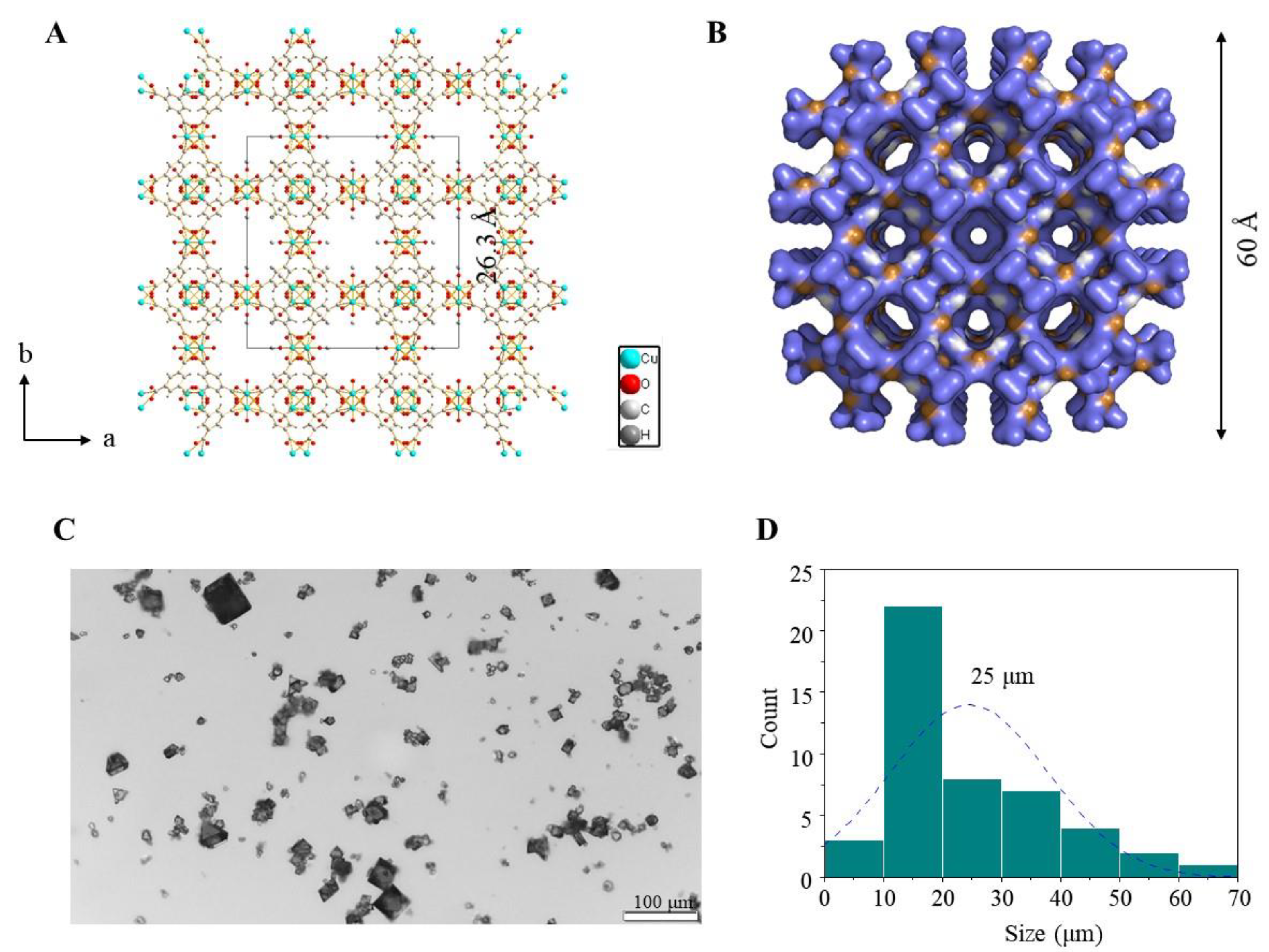
2.2. Synthesis, Characterization of HKUST-1

HKUST-1 was synthesized according to the literature procedure [26]. The blue powder was filtered and washed with ethanol before being dried at 100 °C in a furnace with flowing nitrogen for at least 4 h, resulting in a blue powder. Powder X-ray diffraction (XRD) patterns were obtained on a Rigaku SmartLab XE diffractometer (Cu Kα) over the range of 5−50° 2θ with a step size of 0.01° 2θ. The infrared spectra were determined on a Nicolet Magna series FT-IR 550 spectrometer using KBr pellets. Particle size was obtained using ImageJ 1.8.0 software, where at least 50 particles were counted for each sample. SEM (ZEISS, Jena, Germany) imaging was employed to characterize the morphology. DLS size measurements of hydrodynamic diameter were made on a Malvern Zetasizer Nano-ZS. Results were averaged over three measurements.

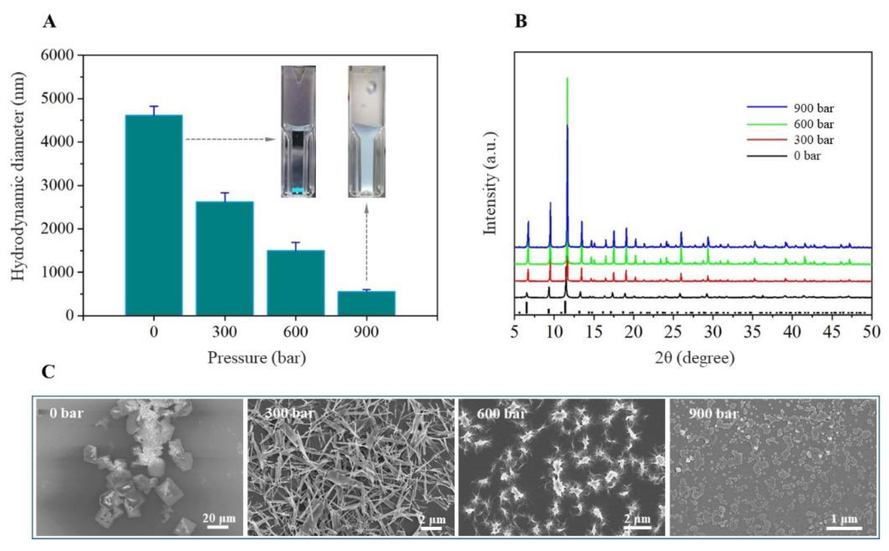
2.3. Preparation of Nano-Sized HKUST-1

A total of 30 mg as-synthesized HKUST-1 was washed once with absolute ethanol and then twice with deionized water. Then, 30 mL HKUST-1 suspensions were homogenized at 0, 300, 600, and 900 bar, respectively, using a UH-06 homogenizer (Union-Biotech Co., Shanghai, China). At each level of homogenization pressure, the aqueous suspensions were treated for 5 min. After each homogenization pass, the aqueous suspensions were cooled down to 10 °C due to the increasing temperature of samples with increasing homogenization pressure. All aqueous suspensions were stored at 4 °C in the fridge.

2.4. Sample Preparation

Lysozyme was directly dissolved in 0.05 M NaAc (pH 4.5) buffer solution, with a final concentration of 20 mg·mL−1. The protein solution was centrifuged at 277 K and 12,000 rpm for about 10 min before use. Stock solutions were prepared by dissolving sodium acetate and sodium chloride in Milli-Q water at the concentration of 1 M and 5 M, respectively. All the solutions used for crystallization were filtered through filters of pore size 0.22 μm.

2.5. Crystallization Experiment

HKUST-1 was used for protein crystallization in two forms: (a) 0.1 mg·mL1 HKUST-1 treated at 300 bar, 600 bar, and 900 bar pressure was added to 20 mg·mL−1 lysozyme, and untreated HKUST-1 was used as a control. (b) Untreated HKUST-1 was added with 20 mg·mL−1 lysozyme at concentrations of 0 mg·mL−1, 0.05 mg·mL−1, 0.1 mg·mL−1, and 0.15 mg·mL−1, and lysozyme without HKUST-1 was used as a control group. Each group was repeated 12 times in parallel. Protein crystallization was performed with 96-well plates by the conventional hanging-drop vapor-diffusion method. In all crystallizations, 1.25 M solution of NaCl in 0.1 M sodium acetate (pH 4.5) was used as a reservoir solution. A total of 0.8 μL of protein solution was mixed with 0.8 μL of reservoir solution, and the volume of the reservoir solution in the well was 60 μL. After the crystallization set-up, the crystallization plate was stored at 289 K.

3. Results

3.1. Preparation of Nano-Sized HKUST-1

HKUST-1 particles with different particle sizes were prepared using a mechanical treatment to break the micro-sized MOF crystals to form uniform particles. The regulator pressure of the high-pressure homogenizer was set at 0, 300, 600, and 900 bars, respectively, to obtain different homogenized MOF particles in size scale, even nanoparticles. From the results of the statistical analysis, it was observed that pressure treatment conditions had a greater effect on particle size. The mean sizes of HKUST-1 micro- and nanoparticles were measured by the dynamic light scattering method (DLS). Figure 2A shows that the particles were divided into four levels in size, including a homogenized nanoparticle with 557 ± 42 nm in size. Thus, a high-pressure homogenizer appears to be an effective instrument to produce uniform micro- and nanoparticles with narrow size distribution and excellent uniformity by controlling the pressure on the regulator. The X-ray diffraction patterns of HKUST-1 under different treatment conditions are presented in Figure 2B. The main peaks at about 11.6°, 13.4°, 17.4°, and 19° indicate the structure of HKUST-1. For all different pressure conditions, the main peaks observed were similar, indicating that the structure remains and crystallinity does not decrease. The morphological analysis of HKUST-1 micro- and nanoparticles was performed by SEM. SEM allowed the visualization of particles with homogeneous size distributions, and these observations were in good agreement with the results of the DLS analysis experiment. The particles produced possessed various shape characteristics with a rough surface. Therefore, the pressure of the homogenizer significantly influenced the surface morphology, resulting in the production of particles with gross surface defects. Furthermore, morphological analysis of the sample at 900 bar revealed nanoparticles with laminate shape and rough surface. A certain degree of adhesion between the particles was observed. This may attribute to stacking of laminate particles during sample preparation for SEM. These results demonstrate that the high-pressure homogenization method could be used with pressure adjustment to successfully prepare uniform MOFs.

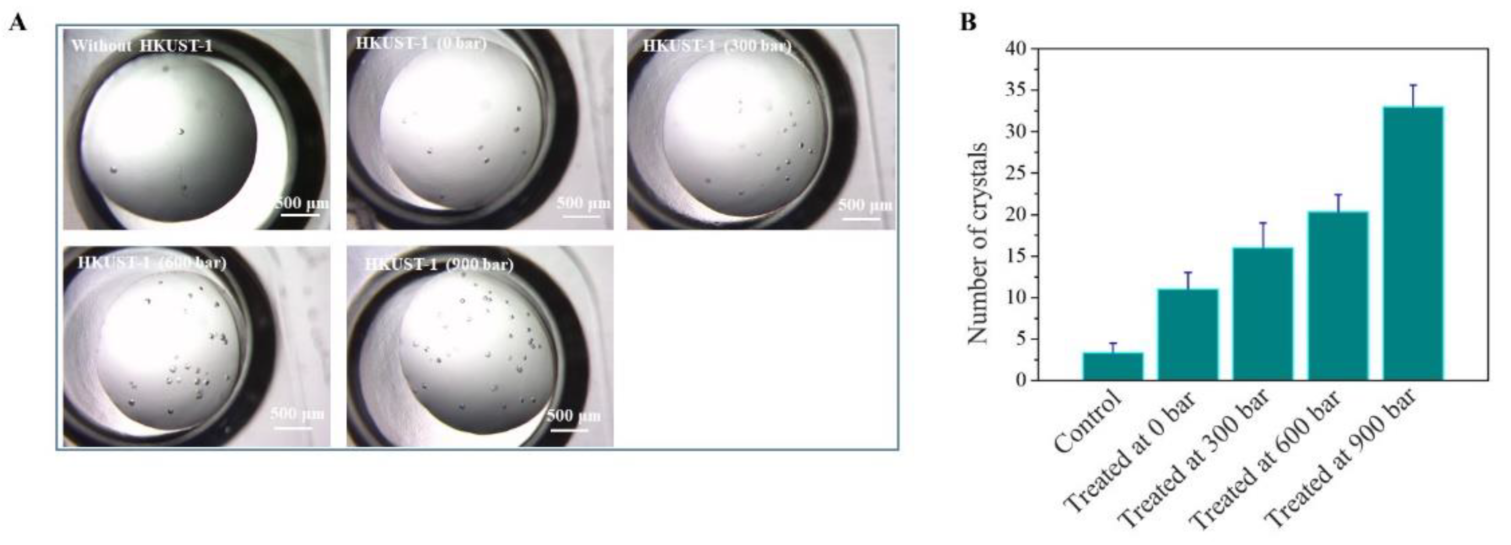
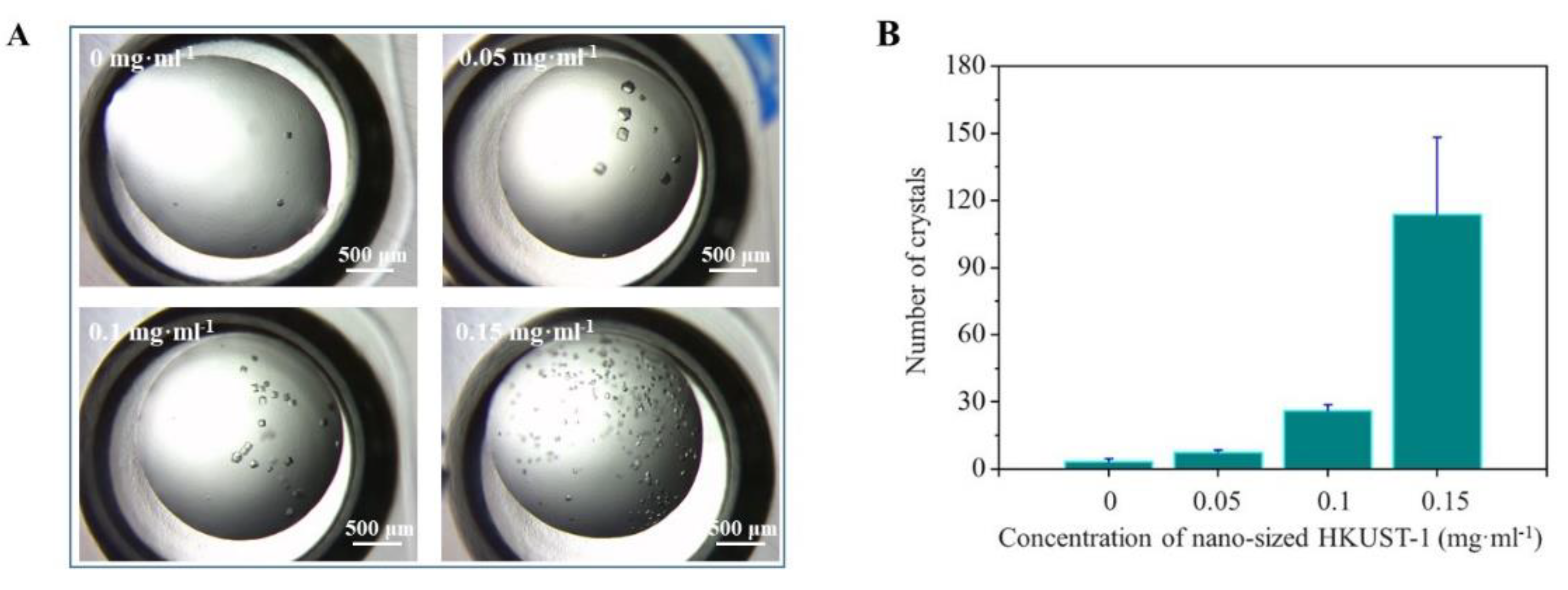
3.2. Crystallization Enhanced by Nano-Sized HKUST-1

Initial lysozyme crystallization experiments were carried out, for optimization purposes, using various concentrations of NaCl (0.75, 1.0, 1.25, 1.5, 1.75, 2.0, 2.5, and 3.0 M). As a result, 1.25 M is chosen as the concentration of NaCl in the following crystallization experiments because its supersaturation is on the edge of crystal nucleation. The nucleation ability of protein crystals on HKUST-1 was tested using the optimized buffer condition. In addition, the effect of HKUST-1 with different treatments on nucleation is also tested. As illustrated in Figure 3A, the results indicated that the number of lysozyme crystals is increased in the HKUST-1 groups compared with the control. Further analysis of the number of crystals associated with differences in homogenizer pressure showed that higher pressure-treated material will achieve stronger nucleation properties in the experiments (Figure 3B). Notably, the enhancement of nucleation of protein crystals is observed with increased homogenizer pressure. This may not only attribute to the homogeneous dispersion of HKUST-1, but also to more nucleation sites that are exposed as the specific surface area increases. Similar phenomena are observed in carbon nanomaterials, such as graphene quantum dots [27] and multi-walled carbon nanotubes [28]. In previous research, the influence of surface curvature of nucleating agents is also considered as an important factor in the nucleation process [29].
Besides, we investigated the effects of the concentration of HKUST-1 in the protein crystallization process by counting the number of crystals, using an optical microscope. The number and size of crystals are related to the concentration of nucleating agents, which is important for obtaining sufficient diffraction intensity for structure analysis. The details of the crystallization conditions of lysozyme and the measurement methods used are provided in Materials and Methods. Formations of more crystals are observed in the lysozyme solution mixed with HKUST-1 (0.05 mg·mL−1) compared to that without HKUST-1, as illustrated in Figure 4. The increased crystal numbers in lysozyme solution with 0.05 mg·mL−1 HKUST-1 were consistent as per observation in lysozyme crystallization with other HKUST-1 concentrations (0.1, and 0.15 mg·mL−1), as shown in Figure 4A. The number of crystals formed in the presence of 0.15 mg·mL−1 HKUST-1 was ∼10 times larger than that when HKUST-1 was not included (Figure 4B). The HKUST-1 not only increased the total number of crystals formed, but also increased the rate of crystallization, as shown in Figure 4A. In the concentration of 0.15 mg·mL−1 HKUST-1, the number of crystals increased abruptly in the lysozyme solution, while the number of crystals in the solution increased negligibly in absence of HKUST-1.
In order to investigate the generality of nano-sized MOF in protein crystallization, a tricky protein sample, ergothioneine synthase (EgtB), was selected for our study. Five commercial crystallization kits were used for micro-batch crystallization at 295 K. One condition yielded microcrystals: condition 51 from Molecular Dimensions Morpheus HT-96 screen. After the optimization of precipitant composition and protein concentrations, the crystals reached a final size of approximately 0.05 mm × 0.05 mm × 0.1 mm in one week. Sixteen crystals were sent to the synchrotron at beamline BL19U1 of the Shanghai Synchrotron Radiation Facility for data collection. After data processing and scaling, the maximum resolution of the data is 3.6 Å, which is not sufficient for further catalytic mechanism analysis. To overcome that barrier, nano-sized HKUST-1 is used as an additive in the crystallization screen. Encouragingly, a new condition (NO. 63 from Molecular Dimensions Morpheus HT-96 screen) is discovered for growing EgtB crystals (Figure S1), and a 2.3 Å dataset is collected for determining its structure (Figure S2). The structure analysis and catalytic mechanism investigation are in progress. These results demonstrated the potential use of nano-sized MOF as a promising nucleating agent in protein crystallization.

4. Conclusions

In this work, we have developed a method for preparing uniform-sized MOF particles that are functionalized as a nucleating agent in protein crystallization. Controlling the pressure of the high-pressure homogenizer, it readily yields uniform MOFs in the desired sizes, including uniformly nano-size MOF particles. Here, notable improvements were observed in the generation of protein crystals in the presence of uniform MOFs, whilst additionally reducing the number of conditions remaining clear during the crystallization trial. Uniformly MOF particles have been shown to improve the reproducibility of crystallization, and aid crystal generation from lower protein concentrations. In practice, the nucleating agent is used as a tool during the crystallization screening process where nucleus formation or conditions are lacking. In addition, our methodology provides a robust approach for protein crystallization screens. Findings from this work would allow the development of new methodologies for studying protein crystallization and their relevant applications.

Supplementary Materials

The following supporting information can be downloaded at: https://www.mdpi.com/article/10.3390/cryst12050578/s1, Figure S1: Optical microscope image of EgtB crystals in presence of nano-size MOF particles; Figure S2: X-ray diffraction of EgtB crystals prepared by using nano-size MOF particles.

Author Contributions

L.L., X.X. and J.Z. designed the project and wrote the manuscript. X.Z. and L.L. performed all experiments. Z.X. contributed invaluable suggestions for improving the manuscript. All authors have read and agreed to the published version of the manuscript.

Funding

This research was funded by Shenzhen-Hong Kong-Macau Technology Research Programme (Type C) (SGDX2020110309300301, to L.L.).

Institutional Review Board Statement

Not applicable.

Informed Consent Statement

Not applicable.

Data Availability Statement

Date is contained within the article.

Conflicts of Interest

The authors declare no conflict of interest.

References

  1. Zundert, G.; Moriarty, N.W.; Sobolev, O.V.; Adams, P.D.; Borrelli, K.W. Macromolecular refinement of X-ray and cryoelectron microscopy structures with Phenix/OPLS3e for improved structure and ligand quality. Structure 2021, 29, 913–921. [Google Scholar] [CrossRef] [PubMed]
  2. Gauto, D.F.; Estrozi, L.F.; Schwieters, C.D.; Effantin, G.; Macek, P.; Sounier, R.; Sivertsen, A.C.; Schmidt, E.; Kerfah, R.; Mas, G.; et al. Integrated NMR and cryo-EM atomic-resolution structure determination of a half-megadalton enzyme complex. Nat. Commun. 2019, 10, 2697. [Google Scholar] [CrossRef] [PubMed] [Green Version]
  3. Yu, C.; Huang, L. Cross-Linking Mass Spectrometry: An Emerging Technology for Interactomics and Structural Biology. Anal. Chim. 2018, 90, 144–165. [Google Scholar] [CrossRef] [PubMed]
  4. Ancarik, J.; Kim, S.H. Sparse matrix sampling: A screening method for crystallization of proteins. J. Appl. Crystallogr. 1991, 24, 409–411. [Google Scholar] [CrossRef]
  5. Gorrec, F. The MORPHEUS protein crystallization screen. J. Appl. Crystallogr. 2009, 42, 1035–1042. [Google Scholar] [CrossRef] [PubMed]
  6. Tarver, C.L.; Yuan, Q.; Pusey, M.L. Ionic Liquids as Protein Crystallization Additives. Crystals 2021, 11, 1166. [Google Scholar] [CrossRef]
  7. Santesson, S.; Cedergren-Zeppezauer, E.S.; Johansson, T.; Laurell, T.; Nilsson, J.; Nilsson, S. Screening of nucleation conditions using levitated drops for protein crystallization. Anal. Chim. 2003, 75, 1733–1740. [Google Scholar] [CrossRef]
  8. Vekilov, P.G. Nucleation of protein crystals. Prog. Cryst. Growth Charact. 2016, 62, 136–154. [Google Scholar] [CrossRef]
  9. Georgieva, D.G.; Kuil, M.E.; Oosterkamp, T.H.; Zandbergen, H.W.; Abrahams, J.P. Heterogeneous nucleation of three-dimensional protein nanocrystals. Acta Crystallogr. D Biol. Crystallogr. 2007, 63, 564–570. [Google Scholar] [CrossRef]
  10. Sleutel, M.; Van Driessche, A. Nucleation of protein crystals—A nanoscopic perspective. Nanoscale 2018, 10, 12256–12267. [Google Scholar] [CrossRef]
  11. Chayen, N.E.; Saridakis, E.; El-Bahar, R.; Nemirovsky, Y. Porous silicon: An effective nucleation-inducing material for protein crystallization. J. Mol. Biol. 2001, 312, 591–595. [Google Scholar] [CrossRef] [PubMed]
  12. Khurshid, S.; Saridakis, E.; Govada, L.; Chayen, N.E. Porous nucleating agents for protein crystallization. Nat. Protoc. 2014, 9, 1621–1633. [Google Scholar] [CrossRef] [PubMed]
  13. Chayen, N.E.; Saridakis, E.; Sear, R.P. Experiment and theory for heterogeneous nucleation of protein crystals in a porous medium. Proc. Natl. Acad. Sci. USA 2006, 103, 597–601. [Google Scholar] [CrossRef] [PubMed] [Green Version]
  14. Nanev, C.; Govada, L.; Chayen, N.E. Theoretical and experimental investigation of protein crystal nucleation in pores and crevices. IUCrJ 2021, 8, 270–280. [Google Scholar] [CrossRef]
  15. Chen, W.; Park, S.J.; Kong, F.; Li, X.; Heng, J.; Yang, H. High protein-loading silica template for heterogeneous protein crystallization. Cryst. Growth Des. 2019, 20, 866–873. [Google Scholar] [CrossRef]
  16. Stewart, P.S.; Kolek, S.A.; Briggs, R.A.; Chayen, N.E.; Baldock, P. Random microseeding: A theoretical and practical exploration of seed stability and seeding techniques for successful protein crystallization. Cryst. Growth Des. 2011, 11, 3432–3441. [Google Scholar] [CrossRef]
  17. Nanev, C.N.; Saridakis, E.; Chayen, N.E. Protein crystal nucleation in pores. Sci. Rep. 2017, 7, 35821. [Google Scholar] [CrossRef]
  18. Marchesan, S.; Prato, M. Under the lens: Carbon nanotube and protein interaction at the nanoscale. Chem. Commun. 2015, 51, 4347–4359. [Google Scholar] [CrossRef]
  19. Asanithi, P.; Saridakis, E.; Govada, L.; Jurewicz, I.; Brunner, E.W.; Ponnusamy, R.; Cleaver, J.A.; Dalton, A.B.; Chayen, N.E.; Sear, R.P. Carbon-nanotube-based materials for protein crystallization. ACS Appl. Mater. Interfaces 2009, 1, 1203–1210. [Google Scholar] [CrossRef] [Green Version]
  20. Kertis, F.; Khurshid, S.; Okman, O.; Kysar, J.W.; Govada, L.; Chayen, N.; Erlebacher, J. Heterogeneous nucleation of protein crystals using nanoporous gold nucleants. J. Mater. Chem. 2012, 22, 21928–21934. [Google Scholar] [CrossRef]
  21. Leite, J.; Rodrigues, D.; Ferreira, S.; Figueira, F.; Paz, F.; Gales, L. Mesoporous metal–organic frameworks as effective nucleating agents in protein crystallography. Cryst. Growth Des. 2019, 19, 1610–1615. [Google Scholar] [CrossRef]
  22. Stolyarova, S.; Saridakis, E.; Chayen, N.E.; Nemirovsky, Y. A model for enhanced nucleation of protein crystals on a fractal porous substrate. Biophys. J. 2006, 91, 3857–3863. [Google Scholar] [CrossRef] [PubMed] [Green Version]
  23. Luo, Z.; Morey, J.R.; McDevitt, C.A.; Kobe, B. Heterogeneous nucleation is required for crystallization of the ZnuA domain of pneumococcal AdcA. Acta Crystallogr. F Struct. Biol. Commun. 2015, 71, 1459–1464. [Google Scholar] [CrossRef] [PubMed] [Green Version]
  24. Sugahara, M.; Asada, Y.; Morikawa, Y.; Kageyama, Y.; Kunishima, N. Nucleant-mediated protein crystallization with the application of microporous synthetic zeolites. Acta Crystallogr. D Biol. Crystallogr. 2008, 64, 686–695. [Google Scholar] [CrossRef] [PubMed]
  25. Ferey, G. Hybrid porous solids: Past, present, future. Chem. Soc. Rev. 2008, 37, 191–214. [Google Scholar] [CrossRef]
  26. Chui, S.S.; Lo, S.M.; Charmant, J.P.; Orpen, A.G.; Williams, I.D. A chemically functionalizable nanoporous material. Science 1999, 283, 1148–1150. [Google Scholar] [CrossRef]
  27. Kang, M.; Lee, G.; Jang, K.; Jeong, D.W.; Lee, J.; Kim, H.; Kim, Y.J. Graphene Quantum Dots as Nucleants for Protein Crystallization. Cryst. Growth Des. 2022, 22, 269–276. [Google Scholar] [CrossRef]
  28. Leese, H.S.; Govada, L.; Saridakis, E.; Khurshid, S.; Menzel, R.; Morishita, T.; Clancy, A.J.; White, E.R.; Chayen, N.E.; Shaffer, M. Reductively PEGylated carbon nanomaterials and their use to nucleate 3D protein crystals: A comparison of dimensionality. Chem. Sci. 2016, 7, 2916–2923. [Google Scholar] [CrossRef] [Green Version]
  29. Gomez, L.R.; Garcia, N.A.; Vitelli, V.; Lorenzana, J.; Vega, D.A. Phase nucleation in curved space. Nat. Commun. 2015, 6, 6856. [Google Scholar] [CrossRef] [Green Version]
Figure 1. Micro-sized porous HKUST-1. (A) The crystal structure of the HKUST-1, ball-and-stick model (cubic, a = 26.3 Å). The formula of HKUST-1 is Cu3(C9H3O6)2(H2O)3·ꭓH2O, abbreviated Cu3(BTC)2, BTC = 1,3,5-benzene tricarboxylate. (B) The porous surface of HKUST-1 was calculated by the software PyMol. (C) The optical microscope photographs of as-prepared HKUST-1. (D) Size distribution (histogram and Gaussian fit) determined from (C) with ImageJ software. The average grain size was determined using the equivalent circle diameter method.
Crystals 12 00578 g001
Figure 2. High-pressure homogenization treatment of as-prepared HKUST-1. (A) Pressure-dependent hydrodynamic diameter evolution of HKUST-1 particles. The inserted picture shows the solution state after mechanical treatment at the corresponding pressure. X-ray diffraction patterns (B) and scan electron microscopy images (C) of the HKUST-1 treated under different pressure.
Crystals 12 00578 g002
Figure 3. (A) Protein crystallization trials of lysozyme at a final concentration of 10 mg·mL−1 in presence of different treated HKUST-1, lysozyme control (top left), 0 bar (top middle), 300 bar (top right), 600 bar (bottom left), and 900 bar (bottom right) of nano-sized HKUST-1. (B) Nucleation density (number of crystals/drop) of lysozyme upon HKUST-1 under different treatments. The concentration of HKUST-1 is 0.1 mg·mL−1.
Crystals 12 00578 g003
Figure 4. (A) Protein crystallization trials of lysozyme at a final concentration of 10 mg·mL−1 in presence of nano-sized HKUST-1, lysozyme control (top left), 0.05 mg·mL−1 (top right), 0.1 mg·mL−1 (bottom left), 0.15 mg·mL−1 (bottom right) of nano-sized HKUST-1. (B) Nucleation density (number of crystals/drop) of lysozyme upon the increase in the concentration of nano-sized HKUST-1.
Crystals 12 00578 g004
Publisher’s Note: MDPI stays neutral with regard to jurisdictional claims in published maps and institutional affiliations.

