Article

Structural and Spectroscopic Features of the Bixbyite-Type Yttrium Scandate Doped by Rare-Earth Ions

Prokhorov General Physics Institute of the Russian Academy of Sciences, 119991 Moscow, Russia
*
Author to whom correspondence should be addressed.
Crystals 2022, 12(12), 1745; https://doi.org/10.3390/cryst12121745
Submission received: 17 October 2022 / Revised: 24 November 2022 / Accepted: 28 November 2022 / Published: 2 December 2022
(This article belongs to the Special Issue Structural and Spectroscopic Studies of Rare Earth Doped Crystals)

Abstract

:
Yttrium scandate crystal fiber has been obtained through laser-heated pedestal growth. The crystal belongs to a bixbyite crystal structure and crystallizes in Ia 3 ¯ space group. X-ray diffraction method shows a lattice parameter of a = 10.228(1) Å. Factor-group analysis of YScO3 Raman spectra points to high degree of disorder in crystal structure of the new compound. Spectral-kinetic investigation of the crystal fibers doped by rare-earth ions points to the presence of two independent active optical centers of rare-earth ions. Moreover, the character of rare-earth impurities’ distribution is independent on a rare-earth ionic radius size.

1. Introduction

In recent years, there has been a global tendency to replace radio communication lines within satellite constellations, as well as between satellites and ground objects [1]. There is a fundamental problem of searching for an active laser medium for use in atmospheric and space optical communication systems (FSO). To organize space or long-range atmospheric optical communication, laser systems with high energy characteristics and, therefore, laser media that can withstand extreme optical, radiational and thermal loads are needed.
High melting rare-earth-doped sesquioxides can be promising materials for FSO applications. Due to the disordered crystal structure, rare-earth-doped mixed sesquioxides (such as yttrium scandate YScO3) have inhomogeneously broadened spectral lines [2,3] which provides an advantage for its highly efficient pumping with modern diodes with low requirements for their thermal stabilization. Instead of the Czochralski method [4,5], we propose the use of the laser-heated pedestal method, which is more suitable for the growth of the high-melting crystals of good optical quality and small size required for use in equipment. In this approach, crystal synthesis is accompanied by high-temperature heating with laser radiation and subsequent rapid cooling.
Scandia (Sc2O3) and yttria (Y2O3) crystallize in a bixbyite structural type and have high thermal conductivity (two times more than yttrium aluminum garnet) [6] and a wide transparency window of 0.25 to 9.6 μm [7]. These properties are critically important for solid-state laser application. [3,8,9] Therefore, yttria and scandia may become alternative materials and replace YAG crystals in some applications. Ceramics based on rare-earth oxides [10] and mixed sesquioxides [11,12,13,14,15,16] are declared as excellent laser materials. Moreover, sesquioxides are advanced materials for application as white light emitting phosphors, [17,18] high effective luminescent materials, [19,20] rare-earth magnets, [21,22] optical recording and storage [23].
Yttrium scandate has a polymorphic nature. Its high-temperature modification [24,25], similar to yttria [26] and scandia [27], belongs to a bixbyite structural type with the space group Ia 3 ¯ . The crystal structure includes one site of oxygen ion with local symmetry C1 (Wykoff position 48e) and two symmetrically independent sites of cations with local symmetry C3i (Wykoff position 8b) and C2 (Wykoff position 24d). The ionic radii of yttrium (0.9 Å), scandium (0.83 Å) and rare-earths are close, and the substitution of basic cations by rare-earth impurities does not require charge compensation and it is crystallochemically unforbidden.
Here we have synthesized and characterized the YScO3 crystal fibers, pure and doped by 0.1 at. % Nd3+ and Tm3+ ions. Nd3+ and Tm3+ ion situate in different ends of lanthanoid group and have a maximal different ionic radius. Nd3+ belongs to large rare-earth ions while Tm3+ is a small ion. Moreover, Nd3+ is a Kramers ion with an odd number of electrons, and Tm3+ belongs to non-Kramers ions, with an even number of electrons. Therefore, Nd3+ and Tm3+ are maximally different ions and would presumably have different spectral behavior, which is related to the local structure of optically active centers. We have used selective laser spectroscopy methods, in which the luminescence spectra are recorded at selective excitation and, conversely, the excitation spectra are measured at selective detection. This approach allows us to trace local changes at included ion-activators. In this work, we have focused on studying the influence of rare-earth ion radius size on spectroscopic properties and the character of rare-earth impurities’ distribution.

2. Results and Discussion

To grow the crystal fiber, high pure powders of scandium and yttrium oxides have been mixed in an agate mortar up to a homogeneous state. The mixture of Y2O3, Sc2O3 and RE2O3 (Tm2O3 or Nd2O3) was used as precursor to grow RE-doped crystal fibers. Then, the mixture has been transferred to a platinum crucible and annealed at a temperature of 1100 °C for 48 h. The powder rod of Y2O3-Sc2O3 composition has been obtained at the pressure of 20 MPa. The powder rod had length of 50 mm and diameter of 1 mm.
The rod was fixed vertically, and its tip was heated and melted at CO2 laser irradiation. An yttria-stabilized zirconia (YSZ) crystal was used as a seed. It was dipped into the melt and raised slowly, pulling a crystal fiber with an approximately round cross-section from the melt. At a growth rate of about 80 mm/h, a YScO3 crystal fiber with a diameter of 0.85 mm and length of 50 mm has been obtained.
The image in back-scattered electrons, the brightness of which depends on the average atomic number and the density of the substance, demonstrates the homogeneity of the crystal composition (Figure 1a). According to X-ray spectral analysis (SEM-EDS) the amounts of Sc and Y are almost equal and unchanging across the fiber (Figure 1b). Therefore, the crystal composition may be presented as Y0.95Sc1.05O3, while taking into account three oxygen atoms per formula unit.
Structural characteristics of the crystal fiber, as well as powders of Y2O3 and Sc2O3 precursors, have been obtained with the X-ray phase analysis. The X-ray pattern of YScO3 includes good narrow peaks that point to a high degree of crystallinity (Figure 1c). According to X-ray data, the YScO3 crystal fiber is homogeneous and belongs to the bixbyite structural type (space group Ia 3 ¯ ), similar to yttrium and scandium sesquioxides (Figure 1c, insert). The lattice parameters of YScO3, Y2O3 and Sc2O3 were obtained as a (YScO3) = 10.228(1) Å, a (Y2O3) = 10.604(1) Å and a (Sc2O3) = 9.841(4) Å, respectively. Due to the low concentration of Nd3+ and Tm3+ ions in the YScO3 host (about 0.1 at. %), RE impurity does not influence bixbyite-type crystal structure, phonon structure and crystal quality. XRD (Supplementary Materials Figure S1, Table S1) and Raman spectra (Figure S2) of the pure and rare-earth doped YScO3 crystal fibers have been added as Supporting Information. Table 1 shows indexation of the most intensive peaks.
The micro-Raman spectra have been obtained for Y2O3 and Sc2O3 powders and the YScO3 crystal fiber (Figure 1d).
The unit cell of the bixbyite-type yttrium scandate includes 40 atoms (8 formula units). Therefore, factor-group analysis leads to the following distribution of the 120 phonon modes in the Brillouin zone point Γ = 0:
Γ = ( 3 A g + 3 A u + 3 E g + 3 E u + 9 F g + 9 F u ) C 1   local   symmetry + A g + A u + E g + E u + 5 F g + 5 F u   C 2   local   symmetry + A u + E u + 3 F u   C 3 i   S 6     local   symmetry
After subtracting 1 F u of the acoustic modes there remain 16 F u of the infra-red active, ( 4 A g + 4 E g + 14 F g ) of the Raman-active and 5 A u + 5 E u   of the optically inactive phonon modes. In the Sc2O3 → YScO3 → Y2O3 series, we observed “mass effect” when vibrational modes are shifted to the range of lower frequencies with increasing cation mass. The spectral lines of yttrium scandate are broadened due to the presentence of both Y3+ and Sc3+ cations in statistically equal structural positions. Due to partial overlap, a number of experimental lines are lower than predicted with factor-group analysis. Table 2 shows identification of the Raman-active modes [28].
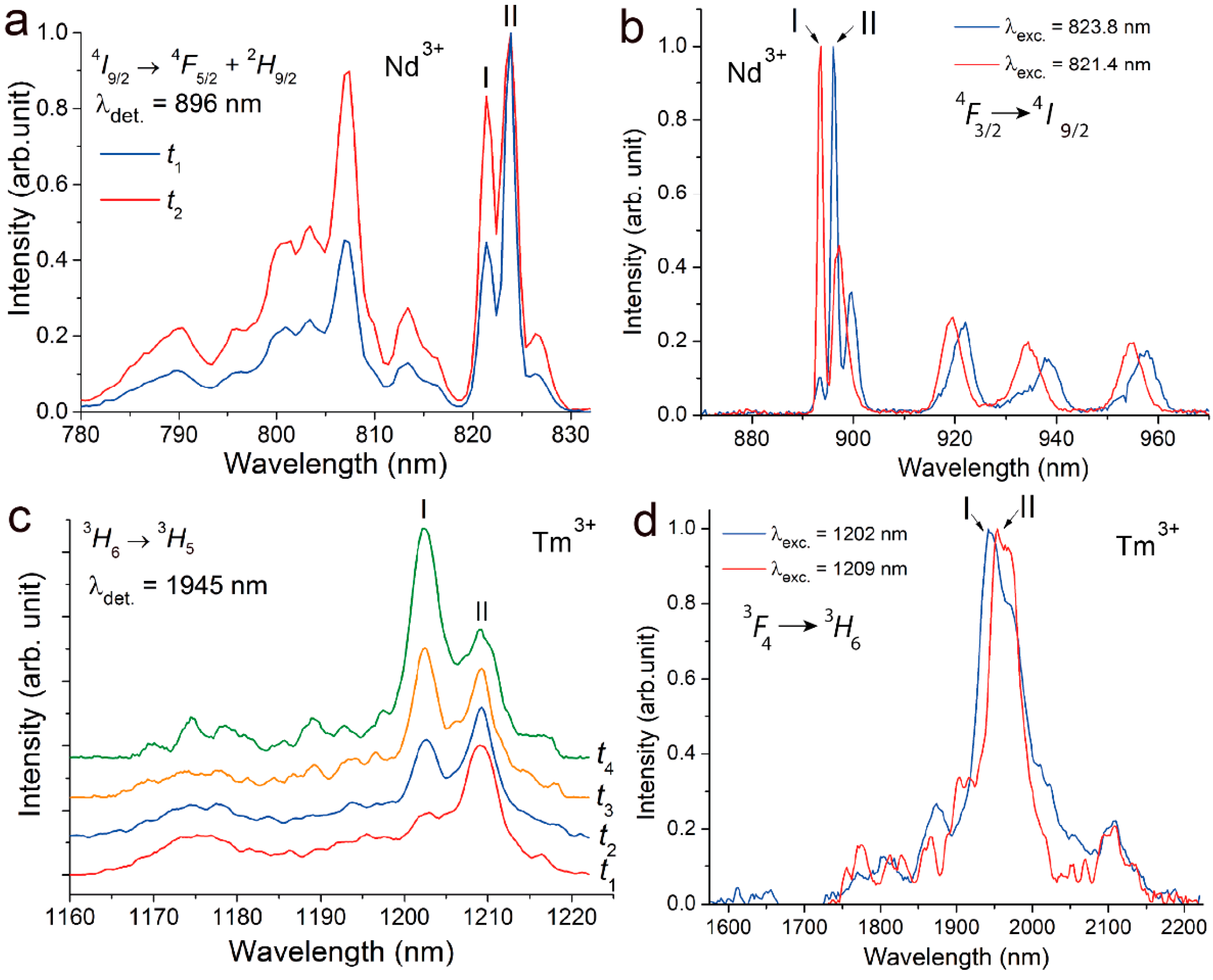
Time-resolved luminescence excitation spectra of Nd3+:YScO3 have been obtained for 4I9/2 → 4F5/2 + 2H9/2 electro-dipole transitions in the Nd3+ ion at selective detection of 896 nm (4F3/2 → 4F9/2 transition) at a temperature of 77 K (Figure 2a). Two intensive peaks are observed at 821.4 and 823.8 nm. The ratio of line intensities depends on delay in registration.
Figure 2b shows the luminescence emission spectra of Nd3+ ions being measured in the 4F3/2 → 4I9/2 electron transitions at 77 K. Nd3+ ions have been excited at 823.6 and 821.4 nm, which corresponds to a maximum of peaks being observed in luminescence excitation spectra.
Analysis of the spectra allows us to distinguish two independent optical centers of Nd3+ ions. The luminescence kinetics have been recorded on the 3F3/2 → 4I9/2 electron transition at two different excitation wavelengths (823.6 and 821.4 nm). In both cases, decay curves are described by exponential law with lifetimes of τ1(77 K) = 290 μs (at 823.6 nm excitation) and τ2(77 K) = 250 μs (at 821.4 nm excitation). The close lifetimes and relatively equal intensities of the spectral lines obtained from two different centers indicate the same C2 symmetry in both cases. As is known [29], electro-dipole transitions are forbidden for rare-earth ion occupied centro-symmetric positions, in particular, the C3i position in the bixbyite structure. The luminescence for these centers must be non-active. We assume that both of the centers have local symmetry of C2. Probably, one of them forms as a result of the local substitution of the Y3+ cation by an Nd3+ ion, and another one is a result of the local substitution of the Sc3+ cation by an Nd3+ ion in the YScO3 host.
The Tm3+ ion has the same behavior in Tm3+:YScO3. Luminescence excitation spectra of Tm3+:YScO3 have been obtained for of 3H6 → 3H5 magneto-dipole transitions in the Tm3+ ion at selective detection of 1945 nm (3F4 → 3H6 transition) at a temperature of 77 K (Figure 2c). Two intensive peaks are observed at 1202.4 and 1209.4 nm. Ratio of line intensities depends on delay in registration in time-resolved spectra. This indicates the presence of two optical centers of Tm3+ ion with two different lifetimes.
Figure 2d shows luminescence spectra of Tm3+ ions being measured in the 3F4 → 3H6 electron transition at selective excitation of 1202.4 and 1209.4 nm. The luminescence kinetics have been recorded on the 3F4 → 4H6 electron transition at two different excitations of 1202.4 and 1209.4 nm. In both cases, decay curves are described by exponential law with lifetimes of τ1(77 K) = 4.25 ms (at 1202.4 nm excitation) and τ2(77 K) = 3.7 ms (at 1209.4 nm excitation). The close lifetimes and relatively equal intensities of the spectral lines obtained from two different centers indicate the same C2 symmetry in both cases. One of Tm3+ optical center forms as a result of the local substitution of the Y3+ cation by a Tm3+ ion, and another one is a result of the local substitution of the Sc3+ cation by a Tm3+ ion in the YScO3 host.

3. Materials and Methods

To grow the YScO3 crystal fiber, the laser heated pedestal growth (LHPG) method has been used [25,30,31]. This approach allows us to synthesize crystal in the form of a fiber and to obtain high-melting materials and compounding with disordered crystal structure. To prepare RE2O3−Y2O3−Sc2O3 powder, preform scandium, yttrium and rare-earth (Nd3+ or Tm3+) oxides (purity of more than 99.999%, Sigma Aldrich, Burlington, MA, USA) were well mixed in a stoichiometric proportion (of mole ratio RE2O3/Y2O3/Sc2O3 = 0.1:99.9:100) in an agate mortar to form a homogeneous mixture. The mole ratio of 1:1 was used to prepare undoped Y2O3−Sc2O3 powder preform. Then, the mixture was transferred to a platinum crucible and annealed at 1100 °C for 48 h in an air atmosphere to exclude the presence of H2O. The final powder mixture was pressed at 20 MPa to form a rod with about 1 mm diameter and 50 mm length.
Visualization and chemical composition analysis of the YScO3 crystal fiber were carried out on the EVO 10 scanning electron microscope (Zeiss Microscopy GmbH, Jena, Germany). The samples were examined at an accelerating voltage of 20 kV with a working distance of 9.0 mm in low vacuum operation (EP = 50 Pa) using a LaB6 cathode.
To obtain SEM imaging of the sample, part of the crystal fiber was placed onto a carbon tape. The observations of the sample were carried out at a beam current of 85 pA. The images were captured in backscattered electron mode (BSE) to assess sample homogeneity (Figure 1a).
The semi-quantitative estimation of the main cation (Y3+ and Sc3+) ratio was based on the data of the Smart METEK EDX spectrometer. Scanning was carried out in line-scan mode at a step of 2 μm and a beam current of 149 pA.
A part of the crystal fiber has been ground in an agate mortar and characterized with X-ray phase analysis. The X-ray pattern has been recorded on the Bruker D8 Discover A25 DaVinci Design diffractometer in the range of 2θ = 15–80° with a step of 0.02° and an exposition time of 1.0 s. The Siemens KFL X-ray tube with Cu (U = 40 kV, I = 40 mA, λ = 0.154 nm) irradiation has been used as the X-ray source. The spectra have been resolved with the EVA 2.1 software and identified with the PDF-2 2011 database. The TOPAS 4.2 software has been used to index spectral peaks and to determine lattice cell parameters.
Micro-Raman spectra of the YScO3 crystal fiber and precursor (Y2O3 and Sc2O3) powders have been recorded on the EnSpectr R532 express analyzer equipped with the Olympus microscope (40× objective) at a laser excitation of 532 nm. The laser beam has been focused on a core of the fiber. Each spectrum has been obtained as the difference between the spectrum taken with the laser running and the dark spectrum. The subtraction of the first spectrum from the second one was carried out automatically using the EnSpectr Pro software. The final Raman spectra were obtained by averaging 20 runs acquired during the total time of 90 s (4.5 s for a run).
Spectral-kinetic characteristics of the Nd3+- and Tm3+-doped YScO3 crystal fibers have been obtained with the selective spectroscopy method. Luminescence spectra have been recorded on the MDR–23 monochromator. The Hamamatsu R5108 и FEU-83 photomultipliers have been used as detectors. The LQ529B–LP 604 (Solar LS, Belarus) laser complex has been used as the excitation source.

4. Conclusions

A YScO3 crystal fiber has been obtained through laser-heated pedestal growth (LHPG). According to X-ray spectral analysis (SEM-EDS), the amounts of Sc and Y are almost equal and unchanging across the fiber. The YScO3 crystal fiber is homogeneous and belongs to the bixbyite structural type (space group Ia 3 ¯ ). The factor group analysis predicted 22 Raman-active phonon modes. However, due to partial overlap, a number of experimental lines are lower than predicted with factor-group analysis. Luminescence excitation and luminescence spectra of Tm3+:YScO3 and Nd3+:YScO3 show the presence of two active centers of the rare-earth ions in both cases. We suppose that one of rare-earth optical centers forms as a result of the local substitution of the Y3+ cation by an RE3+ ion, and another one is a result of the local substitution of the Sc3+ cation by an RE3+ ion in the YScO3 host. Therefore, the nature of the inclusion of a rare-earth impurity into the YScO3 bixbyite-type crystal structure does not depend on the ionic radius size of rare-earths.

Supplementary Materials

The following supporting information can be downloaded at: https://www.mdpi.com/article/10.3390/cryst12121745/s1, Figure S1: The X-ray pattern of YScO3 and Nd3+:YScO3 crystal fibers (black and red ones, respectively); Table S1: Miller indices (hkl), interplanar distances d, and Bragg angles 2θ of the pure and Nd3+-doped YScO3 crystal fibers with a bixbyite-type structure; Figure S2: Micro-Raman spectra of YScO3, Nd3+:YScO3, and Tm3+:YScO3 crystal fibers (black, red and blue ones, respectively).

Author Contributions

Conceptualization, V.T.; methodology, O.A. and S.R.; validation, E.D., O.A. and S.R.; formal analysis, E.D., O.A., V.V. (Valery Voronov) and L.B.; investigation, E.D., O.A., D.G., V.V. (Valery Voronov), S.R., L.B. and I.N.; resources, S.R., V.K., S.K. and V.V. (Viktor Vlasov); writing—original draft preparation, E.D., D.G., Valery Voronov., S.R. and L.B.; writing—review and editing, E.D., O.A. and V.T.; visualization, E.D. and O.A.; supervision, V.T.; project administration, E.D.; funding acquisition, E.D. and O.A. All authors contributed equally to this manuscript. All authors have read and agreed to the published version of the manuscript.

Funding

This research was funded by the Russian Science Foundation, grant number 22-22-00968.

Data Availability Statement

Not applicable.

Conflicts of Interest

The authors declare no conflict of interest. The funders had no role in the design of the study; in the collection, analyses, or interpretation of data; in the writing of the manuscript; or in the decision to publish the results.

References

  1. Garlinska, M.; Pregowska, A.; Masztalerz, K.; Osial, M. From Mirrors to Free-Space Optical Communication—Historical Aspects in Data Transmission. Future Internet 2020, 12, 179. [Google Scholar] [CrossRef]
  2. Liu, W.; Lu, D.; Pan, S.; Xu, M.; Hang, Y.; Yu, H.; Zhang, H.; Wang, J. Ligand Engineering for Broadening Infrared Luminescence of Kramers Ytterbium Ions in Disordered Sesquioxides. Cryst. Growth Des. 2019, 19, 3704–3713. [Google Scholar] [CrossRef]
  3. Liu, W.; Lu, D.; Guo, R.; Wu, K.; Pan, S.; Hang, Y.; Sun, D.; Yu, H.; Zhang, H.; Wang, J. Broadening of the Fluorescence Spectra of Sesquioxide Crystals for Ultrafast Lasers. Cryst. Growth Des. 2020, 20, 4678–4685. [Google Scholar] [CrossRef]
  4. Suzuki, A.; Kalusniak, S.; Tanaka, H.; Brützam, M.; Ganschow, S.; Tokurakawa, M.; Kränkel, C. Spectroscopy and 2.1 μm Laser Operation of Czochralski-Grown Tm3+:YScO3 Crystals. Opt. Express 2022, 30, 42762–42771. [Google Scholar] [CrossRef]
  5. Kränkel, C.; Uvarova, A.; Haurat, É.; Hülshoff, L.; Brützam, M.; Guguschev, C.; Kalusniak, S.; Klimm, D. Czochralski Growth of Mixed Cubic Sesquioxide Crystals in the Ternary System Lu2O3-Sc2O3-Y2O3. Acta Crystallogr. B Struct. Sci. Cryst. Eng. Mater. 2021, 77, 550–558. [Google Scholar] [CrossRef]
  6. Klein, P.H.; Croft, W.J. Thermal Conductivity, Diffusivity, and Expansion of Y2O3, Y3Al5O12, and LaF3 in the Range 77–300° K. J Appl. Phys. 1967, 38, 1603. [Google Scholar] [CrossRef]
  7. Nigara, Y. Measurement of the Optical Constants of Yttrium Oxide. Jpn. J. Appl. Phys. 1968, 7, 404. [Google Scholar] [CrossRef]
  8. Heuer, A.M.; von Brunn, P.; Huber, G.; Kränkel, C. Dy3+:Lu2O3 as a Novel Crystalline Oxide for Mid-Infrared Laser Applications. Opt. Mater. Express 2018, 8, 3447–3455. [Google Scholar] [CrossRef]
  9. Tokurakawa, M.; Shirakawa, A.; Ueda, K.; Peters, R.; Fredrich-Thornton, S.T.; Petermann, K.; Huber, G. Ultrashort Pulse Generation from Diode Pumped Mode-Locked Yb3+:Sesquioxide Single Crystal Lasers. Opt. Express 2011, 19, 2904–2909. [Google Scholar] [CrossRef]
  10. Loiko, P.; Koopmann, P.; Mateos, X.; Serres, J.M.; Jambunathan, V.; Lucianetti, A.; Mocek, T.; Aguilo, M.; Diaz, F.; Griebner, U.; et al. Highly Efficient, Compact Tm3+: RE2O3 (RE = Y, Lu, Sc) Sesquioxide Lasers Based on Thermal Guiding. IEEE J. Sel. Top. Quantum Electron. 2018, 24, 1600713. [Google Scholar] [CrossRef]
  11. Li, D.; Kong, L.; Xu, X.; Liu, P.; Xie, G.; Zhang, J.; Xu, J. Spectroscopy and Mode-Locking Laser Operation of Tm:LuYO3 Mixed Sesquioxide Ceramic. Opt. Express 2019, 27, 24416–24425. [Google Scholar] [CrossRef] [PubMed]
  12. Hao, Z.; Zhang, L.; Wang, Y.; Wu, H.; Pan, G.-H.; Wu, H.; Zhang, X.; Zhao, D.; Zhang, J. 11 W Continuous-Wave Laser Operation at 209 μm in Tm:Lu1.6Sc0.4O3 Mixed Sesquioxide Ceramics Pumped by a 796 nm Laser Diode. Opt. Mater. Express 2018, 8, 3615–3621. [Google Scholar] [CrossRef]
  13. Pirri, A.; Patrizi, B.; Maksimov, R.N.; Shitov, V.A.; Osipov, V.V.; Vannini, M.; Toci, G. Spectroscopic Investigation and Laser Behaviour of Yb-Doped Laser Ceramics Based on Mixed Crystalline Structure (ScxY1−x)2O3. Ceram. Int. 2021, 47, 29483–29489. [Google Scholar] [CrossRef]
  14. Toci, G.; Pirri, A.; Patrizi, B.; Maksimov, R.N.; Osipov, V.V.; Shitov, V.A.; Yurovskikh, A.S.; Vannini, M. High Efficiency Emission of a Laser Based on Yb-Doped (Lu,Y)2O3 Ceramics. Opt. Mater. 2018, 83, 182–186. [Google Scholar] [CrossRef]
  15. Pirri, A.; Maksimov, R.N.; Shitov, V.A.; Osipov, V.V.; Sani, E.; Patrizi, B.; Vannini, M.; Toci, G. Continuously Tuned (Tm0.050Sc0.252Y0.698)2O3 Ceramic Laser with Emission Peak at 2076 nm. J. Alloys Compd. 2022, 889, 161585. [Google Scholar] [CrossRef]
  16. Pirri, A.; Maksimov, R.N.; Li, J.; Vannini, M.; Toci, G. Achievements and Future Perspectives of the Trivalent Thulium-Ion-Doped Mixed-Sesquioxide Ceramics for Laser Applications. Materials 2022, 15, 2084. [Google Scholar] [CrossRef]
  17. Reenabati Devi, K.; Dorendrajit Singh, S.; David Singh, T. Photoluminescence Properties of White Light Emitting La2O3:Dy3+ Nanocrystals. Indian J. Phys. 2018, 92, 725–730. [Google Scholar] [CrossRef]
  18. Antoinette, M.M.; Israel, S.; Berchmans, J.L.; Manoj, G.J. Enhanced Photoluminescence and Charge Density Studies of Novel (Sm1−xGdx)2O3 Nanophosphors for WLED Applications. J. Mater. Sci. Mater. Electron. 2018, 29, 19368–19381. [Google Scholar] [CrossRef]
  19. Barrera, E.W.; Cascales, C.; Pujol, M.C.; Park, K.H.; Choi, S.B.; Rotermund, F.; Carvajal, J.J.; Mateos, X.; Aguiló, M.; Díaz, F. Synthesis of Tm:Lu2O3 Nanocrystals for Phosphor Blue Applications. Phys. Procedia 2010, 8, 142–150. [Google Scholar] [CrossRef] [Green Version]
  20. Kumar, A.; Tiwari, S.P.; Kumar, K.; Rai, V.K. Structural and Optical Properties of Thermal Decomposition Assisted Gd2O3:Ho3+/Yb3+ Upconversion Phosphor Annealed at Different Temperatures. Spectrochim. Acta A Mol. Biomol. Spectrosc. 2016, 167, 134–141. [Google Scholar] [CrossRef]
  21. El-Kelany, K.E.; Ravoux, C.; Desmarais, J.K.; Cortona, P.; Pan, Y.; Tse, J.S.; Erba, A. Spin Localization, Magnetic Ordering, and Electronic Properties of Strongly Correlated Ln2O3 Sesquioxides (Ln=La, Ce, Pr, Nd). Phys. Rev. B 2018, 97, 245118. [Google Scholar] [CrossRef]
  22. Tseng, K.P.; Yang, Q.; McCormack, S.J.; Kriven, W.M. High-Entropy, Phase-Constrained, Lanthanide Sesquioxide. J. Am. Ceram. Soc. 2020, 103, 569–576. [Google Scholar] [CrossRef] [Green Version]
  23. Balabanov, S.; Demidova, K.; Filofeev, S.; Ivanov, M.; Kuznetsov, D.; Li, J.; Permin, D.; Rostokina, E. Influence of Lanthanum Concentration on Microstructure of (Ho1–xLax)2O3 Magneto-Optical Ceramics. Phys. Status Solidi B Basic Res. 2020, 257, 1900500. [Google Scholar] [CrossRef]
  24. Clark, J.B.; Richter, P.W.; Du Toit, L. High-Pressure Synthesis of YScO3, HoScO3, ErScO3, and TmScO3, and a Reevaluation of the Lattice Constants of the Rare Earth Scandates. J. Solid State Chem. 1978, 23, 129–134. [Google Scholar] [CrossRef]
  25. Alimov, O.; Dobretsova, E.; Guryev, D.; Kashin, V.; Kiriukhina, G.; Kutovoi, S.; Rusanov, S.; Simonov, S.; Tsvetkov, V.; Vlasov, V.; et al. Growth and Characterization of Neodymium-Doped Yttrium Scandate Crystal Fiber with a Bixbyite-Type Crystal Structure. Cryst. Growth Des. 2020, 20, 4593–4599. [Google Scholar] [CrossRef]
  26. Hanic, F.; Hartmanová, M.; Knab, G.G.; Urusovskaya, A.A.; Bagdasarov, K.S. Real Structure of Undoped Y2O3 Single Crystals. Acta Crystallogr. Sect. B 1984, 40, 76–82. [Google Scholar] [CrossRef] [Green Version]
  27. Geller, S.; Romo, P.; Remeika, J.P. Refinement of the Structure of Scandium Sesquioxide. Zeitschrift Kristallographie New Cryst. Struct. 1967, 124, 136–142. [Google Scholar] [CrossRef]
  28. Abrashev, M.V.; Todorov, N.D.; Geshev, J. Raman Spectra of R2O3 (R—Rare Earth) Sesquioxides with C-Type Bixbyite Crystal Structure: A Comparative Study. J. Appl. Phys. 2014, 116, 103508. [Google Scholar] [CrossRef] [Green Version]
  29. Kaminskii, A.A. Crystalline Lasers: Physical Processes and Operating Schemes; CRC Press: Boca Raton, FL, USA, 2020; ISBN 9781003067962. [Google Scholar]
  30. Feigelson, R.S. Pulling Optical Fibers. J. Cryst. Growth 1986, 79, 669–680. [Google Scholar] [CrossRef]
  31. Bufetova, G.A.; Kashin, V.V.; Nikolaev, D.A.; Rusanov, S.Y.; Seregin, V.F.; Tsvetkov, V.B.; Shcherbakov, I.A.; Yakovlev, A.A. Neodymium-Doped Graded-Index Single-Crystal Fibre Lasers. Quantum Electron. 2006, 36, 616. [Google Scholar] [CrossRef]
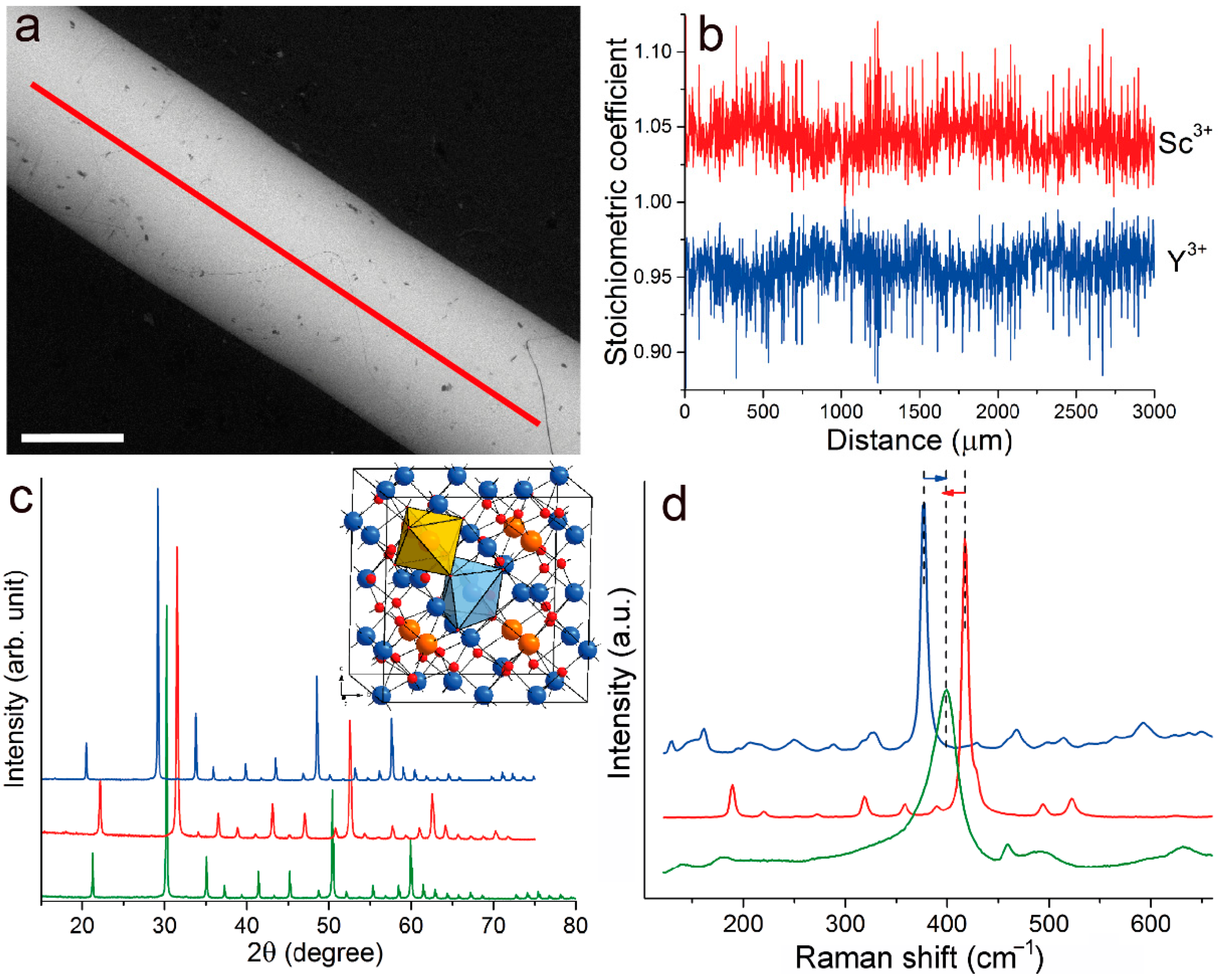
Figure 1. (a) An image of the YScO3 crystal fiber captured in backscattered electron mode (BSE). A scalebar is 0.5 mm; (b) Stoichiometric coefficients of Y3+ (blue one) and Sc3+ (red one) ions calculated based on three oxygen atoms per formula unit; (c) The X-ray pattern of the YScO3 crystal fiber (green one) and Y2O3 and Sc2O3 precursors (blue and red ones, respectively). Insert: bixbyite-type crystal structure of YScO3. Symmetrically diverse MO6 polyhedra both centered by statistically distributed Y3+ and Sc3+ ions are shown as orange (C3i site) and blue (C2 site); red balls are O atoms; (d) Raman spectra of Y2O3 and Sc2O3 precursors and YScO3 crystal fiber (blue, red and green lines, respectively).
Crystals 12 01745 g001
Figure 2. (a) Luminescence excitation spectra of Nd3+:YScO3 being obtained at a time delay of: t1 = 20 μs; t2 = 254 μs. (b) Luminescence spectra of Nd3+:YScO3 being obtained at excitation wavelength of 823.8 and 821.4 nm. (c) Luminescence excitation spectra of Tm3+:YScO3 being obtained at a time delay of: t1 = 0.6 ms; t2 = 6 ms; t3 = 10 ms; t4 = 12 ms. (d) Luminescence spectra of Tm3+:YScO3 being obtained at excitation wavelength of 1202 and 1209 nm. All measurements were accomplished at 77 K.
Crystals 12 01745 g002
Table 1. Miller indices (hkl), interplanar distances d, Bragg angles 2θ and Bragg peak intensities I of the Y2O3, Sc2O3 and YScO3 with a bixbyite-type structure.
Y2O3; a = 10.604(1) ÅSc2O3; a = 9.841(4) ÅYScO3; a = 10.228(1) Å
Indexhklm2θdI2θdI2θdI
12112420.4944.330213.9022.1394.012119.30 21.2614.175613.54
2222829.1623.0598100.00 31.5052.8374100.00 30.2462.9526100.00
3004633.7982.650021.90 36.532.45788.30 35.0652.557018.45
44112435.9182.49824.40 38.8342.31713.50 37.2682.41084.62
53322439.8622.25974.70 43.1212.096211.90 41.3722.18068.36
63412443.512.07835.80 47.0891.92848.60 45.1662.00599.11
72512446.9251.93471.70 50.8141.79543.30 48.7241.86743.27
80441248.5561.873527.80 52.6071.738339.10 50.4321.808147.56
94332450.1611.81721.3054.3441.68681.6052.0981.75412.14
106112453.2451.71902.70 57.7371.59554.10 55.3241.65923.47
115412456.2111.63512.10 61.0021.51773.50 58.4291.57824.54
126222457.6571.597513.60 62.5951.482814.20 59.9421.541927.23
136312459.0821.56232.70 64.1661.45034.20 61.4331.50816.56
14444860.4781.52962.20 65.7141.41981.20 62.9031.47633.66
157212464.5751.44211.60 70.2551.33872.90 67.2051.39193.08
16008671.1231.32451.50 74.0981.27854.70
Table 2. Raman-active modes in spectra of Y2O3, Sc2O3 and YScO3; exp.—experimental, calc.—calculated.
Assignments [28]Sc2O3, exp. [28]Sc2O3, calc. [28]Sc2O3YScO3Y2O3, exp. [28]Y2O3, calc. [28]Y2O3
Ag221220220180162162161
391351358--306318
495498494-431460467
623593-590-553542
Eg273273273-194198194
359332--325290288
430416429400-366358
626590---555567
Fg189192189140129132129
202199-180138138152
252250253-182181207
319306--235234217
329311319--244250
359341358-325288288
-368---299-
391386390--338327
419412418400377368377
-472-460-416429
-491494--442467
523536522495469495499
587605624590-545514
669646670630592581592
Publisher’s Note: MDPI stays neutral with regard to jurisdictional claims in published maps and institutional affiliations.