Article

Three-Dimensional CA-LBM Numerical Model and Experimental Verification of Cs2AgBiBr6 Perovskite Single Crystals Grown by Solution Method

School of Materials Science and Engineering, Hebei University of Technology, Tianjin 300401, China
*
Authors to whom correspondence should be addressed.
Crystals 2021, 11(9), 1101; https://doi.org/10.3390/cryst11091101
Submission received: 17 August 2021 / Revised: 1 September 2021 / Accepted: 7 September 2021 / Published: 10 September 2021

Abstract

:
A three-dimensional cellular automata-lattice Boltzmann (CA-LBM) coupling model is established to simulate the facet growth process and the controlled cooling growth process of Cs2AgBiBr6 perovskite single crystals. In this model, the LBM method is used to calculate the real-time solute field, the CA method is used to simulate the crystal growth process driven by supersaturation of solute, and the geometric parameter g related to the adjacent grid is introduced to reduce the influence of grid anisotropy. The verification of the model is achieved by comparing the simulation results with the experimental results. The comparison results show that a smaller cooling rate is helpful for the growth of large-size single crystals, which verifies the rationality and correctness of the model.

1. Introduction

Recently, perovskite materials have received widespread attention due to their excellent photoelectric properties [1]. A perovskite single crystal has extremely low defect density and minimal interface defects compared to polycrystalline materials [2,3,4,5]. However, the application of solution methods to perovskite single-crystal materials has strict solvent requirements [3], so it is difficult to obtain single crystals with large sizes. Large-sized perovskite single crystals are of great significance for improving their optical and electrical properties [5].
Organic–inorganic hybrid perovskite materials (CH3NH3PbX3, X = I, Br, Cl) have attracted wide attention in the fields of solar cells [6,7,8], photodetectors [9,10,11,12,13], light-emitting diodes [14], etc., because of their excellent optical and electrical properties [15,16]. However, organic-inorganic hybrid perovskite materials have two important disadvantages: (1) their properties are easily affected by temperature, humidity, and light; and (2) the Pb2+ contained in the component leads to their toxicity and decreased environmental friendliness [16,17], and the application of these perovskite is limited to some extent.
Lead-free double perovskite Cs2AgBiBr6 is a stable and nontoxic photoelectric material [16,17,18,19] with suboptimal photon to charge carrier conversion efficiency. Compared with other lead-free double perovskites, Cs2AgBiBr6 perovskite has better stability in response to moisture, air, heat, and light [20], which is very suitable for high-energy photon detection applications [21].
So far, scholars have conducted much research on the growth of perovskite crystals by the solution method. The most common method is the solution temperature-lowering (STL) route. Su et al. [15] synthesized high-purity polycrystalline Cs2AgBiBr6 powder by the solution method by using hydrobromic acid as a solvent; millimeter-sized Cs2AgBiBr6 crystals were obtained. Yin et al. [22] obtained Cs2AgBiBr6 single crystals with smooth surfaces and relatively high resistivity with good reproducibility by controlling cooling. Zhang et al. [23] grew Cs2AgBiBr6 crystals with maximum dimensions of 10 mm and a flat plane by the solution cooling method with the addition of toluene. Zhu et al. [24] proposed an additive CH3COONa-controlled nucleation route toward the generalized growth of all inorganic double perovskite single crystals. A Cs2AgBiBr6 single crystal with a maximum size of 13 mm was successfully grown by this method. Dang et al. [25] successfully grew centimeter-sized Cs2AgBiBr6 single crystals by the TSSG method, using MABr as the flux in the mother solution.
The above methods are all experimental studies that do not fully reveal the growth mechanism of Cs2AgBiBr6 perovskite crystals. The numerical simulation method can reproduce the evolution process of crystal growth from the perspective of the full space and time domain and provide process guidance for crystal preparation. For example, Zhang et al. [26,27,28] studied the directional growth of polycrystalline silicon by numerical simulation based on the cellular automaton (CA) method and studied the growth mechanism of facet silicon crystals in detail. Chen et al. [29], based on the phase field (PF) method, studied the growth of arbitrary symmetry facet dendrites. Although the driving force for the growth of silicon crystals is temperature and that of Cs2AgBiBr6 perovskite crystals is solution supersaturation, both of them are facet crystals, so the same numerical method can be used to reveal the growth mechanism of Cs2AgBiBr6 perovskite.
In this paper, the solution temperature-lowering growth process of Cs2AgBiBr6 single crystals is simulated by the CA-LBM method and compared with the experiment to verify the correctness of the model. Then, in order to guide the preparation of larger-size single crystals, the CA-LBM method is used to simulate the growth behavior of single crystals at different cooling rates. In this paper, the experimental and simulation results are respectively compared qualitatively and quantitatively to further verify the model.

2. Preparation of Cs2AgBiBr6 Single Crystals by the Solution Method

Materials: CsBr (99.9%) and BiBr3 (99%) were purchased from Xi’an Polymer Light Technology Corp. (PLT) and Alfa Aesar, respectively. AgBr (99.9%) and hydrobromic acid (HBr, 48% wt % in H2O) were purchased from Aladdin. All these commercially available chemicals were used without any further purification.
Facet Growth Procedure: The concentration was calculated from the solution volume and the mass of the solute evaporated from the sample. First, the precursor of a target concentration (0.1 M, M is the concentration unit: mol/L) was prepared by CsBr, AgBr, BiBr3, and 3 mL HBr acid (CsBr:AgBr:BiBr3 = 2:1:1) in a 5 mL glass bottle. Every point was averaged from three samples for a more accurate result. The bottle was sealed by a silicone plug and silicone grease and heated at 120 °C in an oil bath. Second, after the solute fully dissolved, the temperature was lowered at a rate of 0.5 °C·h−1 to 60 °C to avoid excessive nucleation and crystal growth. Then, the crystals were obtained by pouring out the residual solution.
Controlled Cooling Rate Growth Procedure: The Cs2AgBiBr6 crystals were grown by the method of controlling the cooling rate. First, 3 mL of the respective precursor solution with the same concentration (0.1 M) was prepared in four 5 mL glass bottles. The preparation method of the precursor solution followed the Facet Growth Procedure. Second, the sealed glass bottles were heated at 120 °C in an oil bath to fully dissolve the solute and then cooled to 60 °C at the cooling rates of 2 °C·h−1, 1.5 °C·h−1, 1 °C·h−1, and 0.5 °C·h−1, respectively. Then, the crystals were obtained by pouring out the residual solution.

3. Numerical Model of Cs2AgBiBr6 Single-Crystal Growth Based on the CA Method

A three-dimensional CA-LBM coupling model with interface energy anisotropy was established to simulate Cs2AgBiBr6 single-crystal growth. The solute field was calculated by the LBM method, the solid-liquid interface advancing process was calculated by the CA method, and the CA method and LBM method were coupled according to the solute content decreased by the solid-state rate growth of Cs2AgBiBr6 crystals. The experimental results were compared with the simulation results to verify the rationality and correctness of the model.

3.1. LBM Model

The D3Q15 model [28,30], with the single relaxation time lattice Bhatnagar–Gross–Krook (LBGK) method, was used to calculate the solute field.
gi(r + eit, t + ∆t) − gi(r, t) = (gieq(r, t) − gi(r, t))/τc + Gi
G i = 1000 ρ s ω i f s M   ·   ( C m a x C m i n )
where r, t, Δt, and τc represent location, time, time step, and the relaxation time of the solute field, respectively; gi is the distribution function of particle solute content; and gieq is the equilibrium distribution function of particle solute content. Gi is the solute sink term, which represents solute content consumed due to crystal growth. ρs and M represent the solid density and molar mass, respectively, of Cs2AgBiBr6 perovskite. (CmaxCmin) represents the difference between the initial content and the end content, which is used for the dimensionless solid density.

3.2. CA Model

In this paper, the three-dimensional CA model was used to simulate the growth of perovskite single crystals. Due to the slow growth rate of the Cs2AgBiBr6 single crystals and the limitations of computation capacity, it was difficult to simulate the growth of the perovskite crystals in the macro scale. In order to reduce the calculation time as much as possible under the condition of ensuring calculation accuracy, 61 × 61 × 61 cube cells were selected in the calculation area, and the cell size was 1 µm.
The following is a three-dimensional model to simulate the nucleation, growth, and capture of Cs2AgBiBr6 single crystals.

3.2.1. Model for the Nucleation

The continuous nucleation model based on Gaussian distribution is used in the nucleation model [31].
n(S) = ∫ (dn/dS)dS
dn/dS = nmax · (2πSσ)1/2 · exp(−0.5((SSmea)/Sσ)2)
Here, S is supersaturation, and dn/dS is the nucleation distribution function. nmax, Sσ, and Smea represent the maximum nucleation density, standard deviation supersaturation, and average nucleation supersaturation, respectively. These three parameters are determined by the experimental conditions.
For the nucleation on the wall of a three-dimensional container, the nucleation probability in a time step can be expressed as
P = δn · VC
where δn is the increasing nucleation density in one time step, and VC is the volume of a cell.
When the supersaturation of a cell is higher than the critical supersaturation of nucleation, and the probability of nucleation is greater than a random number of 0–1, the cell nucleates. The nucleation position is randomly selected in the cell space that satisfies the basic conditions of nucleation.

3.2.2. Crystal Growth Model

In the process of crystal growth, the transition from interface cell to solid cell is determined by the growth of the solid fraction. For solution growth, without considering the evaporation of the solution, the solid fraction increment can be expressed by
fs = g · S · M/1000ρs
where S is the supersaturation of the solution, and M and ρs represent the molar mass and solid density of Cs2AgBiBr6, respectively. In order to maintain the solid-liquid interface of the single layer as well as the solidification and growth, the geometric parameter g related to the state of the adjacent grid is introduced [28].
g = min((∑ζmχ1 + 21/2ζmχ2)/2, 1)
ζχ1, ζχ2 = 0, (fs < 1) ζχ1, ζχ2 = 1, (fs = 1)
Here, ζχ1 is the state parameter of the nearest six grid cells, and ζχ2 is the state parameter of the next nearest 12 grid cells.
In this model, the driving force of growth is the saturation (S) of the solution, which is the difference between the solution concentration (C) and solubility (Csat) at a certain temperature.
S = CCsat
The nucleation and growth concentrations are two key factors to control crystal growth. The temperature-concentration distribution curve of Cs2AgBiBr6 in HBr can be divided into three zones by the solubility curve and supersolubility curve [22], as shown in Figure 1.
In Figure 1, nucleation can occur when the concentration is in the nucleation zone. When the concentration is in the growth zone, the crystal can grow stably without nucleation. Therefore, in order to grow larger single crystals, the concentration should be controlled in the growth range as much as possible. By fitting the above two curves, the solubility and supersolubility of Cs2AgBiBr6 can be expressed as polynomials of temperature as Equations (10) and (11), respectively.
Csat = 3.34174 × 10⁻⁷Tt3 − 6.57362 × 10⁻⁵Tt2 + 5.11715 × 10⁻³Tt − 0.08669
Cssat = 4.77122 × 10⁻⁷Tt3 − 8.58122 × 10⁻⁵Tt2 + 5.46878 × 10³Tt 0.05521
Here, Csat is solubility, and Cssat is supersolubility. Tt is the equivalent temperature after considering the influence of interface energy anisotropy and the curvature of the interface, which can be calculated using Equation (12).
Tt = T + Г · wmc
Here, T is the liquidus temperature, Γ is the Gibbs–Thomson coefficient, and wmc is the weighted mean curvature. For cubic crystals with a fourfold anisotropy, wmc can be calculated from Equation (13) [32]:
wmc = (3ε − 1) · (xnx + yny + znz) 48ε · (nx2 · xnx + ny2 · yny + nz2 · znz)
   + 12ε · Q · (xnx + ∂yny + ∂znz) + 12ε · (nx · ∂xQ + ny · ∂yQ + nz · ∂zQ)
where ε is the degree of anisotropy of the surface energy, and nx = xfs/|∇fs|, ny = yfs/|∇fs|, nz = zfs/|∇fs|, where |∇fs| = ( x f s ) 2 + ( y f s ) 2 + ( z f s ) 2 . Q ≡ nx4 + ny4 + nz4.

3.2.3. Model for the Capture

During the CA simulation, the transition of the cell state from liquid to interface is governed by the capture rule. Since the change of cellular state has a great influence on the subsequent growth process, it is particularly important to choose an appropriate capture rule.
For the three-dimensional CA model, the number of adjacent cells is large, so the 3D capture rule is more complex than the 2D capture rule. The traditional capture rules are Von Neumann’s and Moore’s rules, but no matter which capture rule, it will inevitably lead to artificial anisotropy. Therefore, based on Von Neumann’s rule, this paper introduces the geometric parameter g [28] to reduce the artificial anisotropy, and a three-dimensional low anisotropy capture model is established.

4. Results and Discussion

4.1. The Physical Parameters of Cs2AgBiBr6 Precursor Solution

The physical parameters used in the present computations are listed in Table 1.

4.2. Simulation and Verification of Facet Growth

In order to reveal the growth rule of Cs2AgBiBr6, a Cs2AgBiBr6 facet growth morphology is simulated firstly.
Since both the simulation and experiment adopt the all-area synchronous cooling method, 61 × 61 × 61 cubic cells with the same cooling conditions are selected in the calculation area, and the cell size is 1 µm. The initial simulation conditions are set as central point nucleation, and the all-area temperature is cooled by 0.5 °C every 1800 time steps. The physical parameters used in the calculation are listed in Table 1, which contains both the initial temperature and the initial concentration. Crystal growth is calculated by the CA model driven by solute supersaturation, and the temperature–solubility curve used is shown in Figure 1.
The calculation results when the time steps are 100,000, 140,000 and 180,000 are shown in Figure 2a–c, respectively. The real time after conversion is 55.5 h, 77.7 h, and 100 h, respectively. The upper part of each drawing shows the 3D simulation result, and the lower part shows the solute distribution and the solid-liquid interface of the Y–Z section corresponding to the 3D drawing.
The facet morphology cannot be seen clearly in Figure 2a because it is in the early stage of growth after nucleation. The corresponding solute cross-section figure shows that the solute distribution is quite uniform, and the anisotropy is not relatively distinct. Figure 2b shows that the crystal is in an octahedral shape, and the morphology of facet growth appeared after a period of growth. The corresponding solute cross-section in Figure 2e shows that under the influence of anisotropy and geometric parameters g, the growth of the <111> direction (45° direction) is restrained, and crystal growth occurs along the <100> direction (axial direction). The solute in the <100> direction is poorer than that in the <111> direction, resulting in faster growth in the <111> direction, thus maintaining the facet growth morphology. It can be seen from Figure 2c that the crystal is characterized by four-fold anisotropic symmetry and continues to grow in an octahedral shape.
The analysis of the above simulation results reveals the growth mechanism of Cs2AgBiBr6; the <100> and <111> directions restrict each other during the growth process due to the influence of solute content, forming alternating growth behaviors of the <100> and <111> directions, and finally forming a typical octahedral morphology.
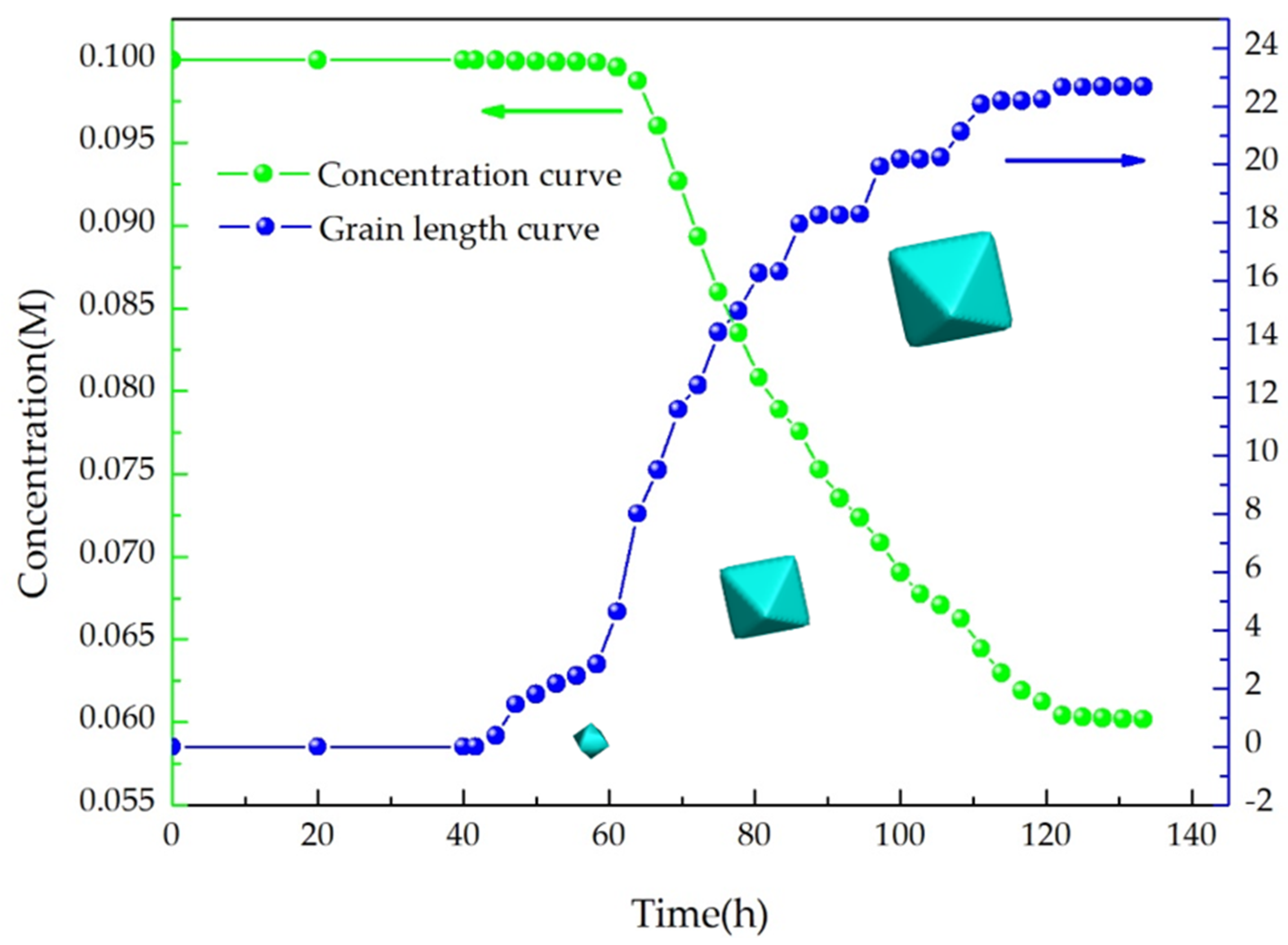
The concentration of solute has a great influence on the process of grain growth. As shown in Figure 3, the grain growth is divided into two stages, namely nucleation and growth. The concentration curve in Figure 3 does not change, and the grain length is 0 before 42 h (about 95 °C). After 42 h, nucleation occurs and enters the growth stage, and the solute concentration decreases with the increase in grain length. It is worth noting that the solute consumption rate has an obvious acceleration process and then gradually slows down. This is due to the increase in solute capture rate at the solid–liquid interface with the cooling growth process, and with the gradual consumption of solute, the supersaturation of solution decreases, which is not enough to maintain rapid growth. When the cooling is stopped, the supersaturation tends to 0, and the solution concentration tends to a certain value.
It can be seen from the above analysis that the concentration curve in Figure 3 shows that the trend of constant-gradient decline tends to be stable. This trend can be explained by comparison with Figure 1. At the beginning stage of cooling growth, the solute concentration is lower than the solubility curve and supersolubility curve, indicating that the concentration is in the dissolution zone. During this period, neither nucleation nor growth can occur. As the temperature gradually lowers, the solute concentration enters the nucleation zone, where nucleation occurs and consumes part of the solute and then enters the growth zone. In the subsequent cooling process, it alternately enters the nucleation zone and growth zone, so the concentration curve in Figure 3 shows a gradient declining trend. In the final stage of growth, the solute concentration is close to the solubility, and the growth stops and reaches a stable state, as seen in Figure 3. The above analysis shows that the concentration curve in Figure 3 is consistent with the change trend of the temperature–solubility curve in Figure 1, which verifies the correctness of the CA model driven by supersaturation in this paper.
The grain length curve in Figure 3 does not increase linearly but has some steps. This is because the supersaturation of the solution is consumed after a certain period of growth at a certain temperature, resulting in temporary slow growth. When the solution is cooled, the supersaturation increases again, resulting in the acceleration of growth again. Therefore, a stepped curve is formed.
Analysis of the above simulation results shows that the grain will continue to grow with an octahedral morphology. Therefore, in this paper, the simulation results can be approximately compared with the experimental results to verify the rationality and correctness of the model. Figure 4 shows the comparison between simulation results (Figure 4a,b) and experimental results (Figure 4c,d) of Cs2AgBiBr6 single-crystal growth.
Figure 4c is the single-crystal morphology prepared by the author of this paper by the solution method, while Figure 4d is the single crystal prepared in Reference [16]. The comparison between Figure 4a and c show that the (111) characteristic surface of the simulated single crystal is in good agreement with the experimental results. The comparison between Figure 4b and d show that the simulation results are also in good agreement with the experimental results in Reference [16]. The two sets of results are cross-validated, which demonstrate that the model established in this paper can simulate the facet growth morphology of Cs2AgBiBr6 correctly. The surface of the single crystal in Figure 4c is comparatively rough, which is caused by the simultaneous growth of many nucleation sites, while in Figure 4a, only one nucleation site is set, so the single-crystal surface is smooth.

4.3. Simulation and Verification of Controlled Cooling Growth

In order to reveal the effect of solution cooling rate on the size and number of the Cs2AgBiBr6 single crystal, the grain growth process at different cooling rates is simulated.
The calculation area also selects 61 × 61 × 61 cubic cells with the same cooling conditions, and the cell size is 1 µm. In order to facilitate the observation of the grain size distribution, the initial conditions are set as random point nucleation on the Z = 30 plane (central horizontal plane), and the all-area temperature is cooled by 2 °C, 1.5 °C, 1 °C, and 0.5 °C every 1800 time steps, respectively. The physical parameters used are listed in Table 1. The crystal growth process is also calculated by the CA model driven by the supersaturation.
Figure 5 shows the simulation and experimental results when the cooling rate is 2 °C·h−1, 1.5 °C·h−1, 1 °C·h−1, and 0.5 °C·h−1. The upper part of each drawing shows the 3D simulation results, and the lower part shows the experimental results corresponding to the 3D drawing.
From the above analysis, it can be predicted that the number of final grains will gradually decrease as the cooling rate decreases, and the maximum grain size will gradually increase. The comparison results of Figure 5d,h verify this prediction. When the cooling rate is reduced to 0.5 °C·h−1, the number of final grains is reduced to 4, and the maximum grain size increases considerably.
There are some very small grains in Figure 5d,h, which is because the number of initial nucleation is small, resulting in the solute mass required for grain growth being insufficient to deplete the supersaturated solute mass increased by cooling and then precipitate in the form of a few grains. However, the solute content at this time can no longer maintain its rapid growth, so the subsequent precipitated grain size is much smaller than the initial grain.
The qualitative comparison between the above simulation results and the experimental results verifies the rationality of the model established in this paper. In order to verify the correctness of the model, Figure 6 shows the quantitative comparison between the simulation results and experimental results. Due to the limitation of the amount of calculation, the volume of the calculation area is smaller than the volume of the actual growth solution, and the quantitative comparison results cannot be clearly presented by using the maximum grain size. Therefore, the dimensionless method is adopted for treatment—the grain size corresponding to different cooling rates is divided by the maximum grain size corresponding to 0.5 °C·h−1 to obtain the dimensionless grain size corresponding to different cooling rates (grain size ratio).
It can be seen intuitively from Figure 6a,b that as the cooling rate increases, the number of grains gradually increases, while the maximum grain size shows a decreasing trend. The two curves in Figure 6a are in good agreement, and there is a slight deviation when the cooling rate is larger. This is because the random nucleation model is adopted in this paper, the number of nucleations will be within a certain range, and the nucleation position is random, so it is more in line with the real grain growth situation.
In Figure 6b, when the cooling rate is larger, the simulation results are lower than the experimental results. This is because the simulation results have more grains than the experimental results when the cooling rate is larger, resulting in a smaller size of the simulated grains than the experimental grains. The quantitative comparison between Figure 6a,b shows that the simulation results are in good agreement with the experimental results, which verifies the correctness of the model established in this paper.

5. Conclusions

In this paper, a three-dimensional CA-LBM coupling model is established to simulate the facet growth process and controlled cooling growth process of Cs2AgBiBr6 perovskite single crystals. The CA model takes the supersaturation of solute as the driving force and considers the influence of interface energy anisotropy on the morphology of the liquid–solid interface. The geometric parameter g is introduced to reduce the influence of grid anisotropy. The qualitative and quantitative comparison between the simulation results and the experimental results verified the rationality and correctness of the model, indicating that the model established in this paper can reproduce the solution growth process of the Cs2AgBiBr6 perovskite single crystal well and can guide the preparation of larger-size Cs2AgBiBr6 single crystals.

Author Contributions

Conceptualization, R.L. and H.C. (Hongjian Chen); data curation, H.C. (Hui Chen); formal analysis, H.C. (Hui Chen); methodology, C.W., R.L. and H.C. Hongjian Chen; software, H.C. (Hui Chen); validation, H.C. (Hui Chen) and C.W.; investigation, H.C. (Hui Chen); resources, C.W., R.L. and H.C. (Hongjian Chen); writing—original draft preparation, H.C. (Hui Chen); writing—review and editing, H.C. (Hui Chen), R.L. and H.C. (Hongjian Chen); supervision, R.L. and H.C. (Hongjian Chen); project administration, R.L. and H.C. (Hongjian Chen); funding acquisition, H.C. All authors have read and agreed to the published version of the manuscript.

Funding

This research was funded by grants from the National Natural Science Foundation of China (grant No. 51475138 and No. 51975182) and the 2019 Tongliao Science and Technology Innovation Guidance Incentive Fund.

Data Availability Statement

Not applicable.

Acknowledgments

Hui Chen would like to thank Cuncun Wu, Ri Li and Hongjian Chen (Hebei University of Technology) for providing materials used for experiments and academic guidance.

Conflicts of Interest

The authors declare no conflict of interest.

References

  1. Canil, L.; Cramer, T.; Fraboni, B.; Ricciarelli, D.; Meggiolaro, D.; Singh, A.; Liu, M.; Rusu, M.; Wolff, C.M.; Phung, N.; et al. Tuning halide perovskite energy levels. Energy Environ. Sci. 2021, 14, 1429–1438. [Google Scholar] [CrossRef]
  2. Rong, S.S.; Faheem, M.B.; Li, Y.B. Perovskite single crystals: Synthesis, properties, and applications. J. Electron. Sci. Technol. 2021, 19, 100081. [Google Scholar] [CrossRef]
  3. Ma, L.; Yan, Z.; Zhou, X.; Pi, Y.; Du, Y.; Huang, J.; Wang, K.; Wu, K.; Zhuang, C.; Han, X. A polymer controlled nucleation route towards the generalized growth of organic-inorganic perovskite single crystals. Nat. Commun. 2021, 12, 1–10. [Google Scholar] [CrossRef] [PubMed]
  4. Lian, Z.; Yan, Q.; Gao, T.; Ding, J.; Lv, Q.; Ning, C.; Li, Q.; Sun, J.L. Perovskite CH3NH3PbI3(Cl) Single Crystals: Rapid Solution Growth, Unparalleled Crystalline Quality, and Low Trap Density toward 108 cm−3. J. Am. Chem. Soc. 2016, 138, 9409–9412. [Google Scholar] [CrossRef] [PubMed]
  5. Di, J.; Chang, J.; Liu, S. Recent progress of two-dimensional lead halide perovskite single crystals: Crystal growth, physical properties, and device applications. Eco. Mat. 2020, 2, 12036. [Google Scholar] [CrossRef]
  6. Kojima, A.; Teshima, K.; Shirai, Y.; Miyasaka, T. Organometal Halide Perovskites as Visible-Light Sensitizers for Photovoltaic Cells. J. Am. Chem. Soc. 2009, 131, 6050–6051. [Google Scholar] [CrossRef]
  7. Yang, W.S.; Park, B.-W.; Jung, E.H.; Jeon, N.J.; Kim, Y.C.; Lee, D.U.; Shin, S.S.; Seo, J.; Kim, E.K.; Noh, J.H.; et al. Iodide management in formamidinium-lead-halide—Based perovskite layers for efficient solar cells. Science 2017, 356, 1376–1379. [Google Scholar] [CrossRef] [Green Version]
  8. NREL. Transforming Energy. Available online: https://www.nrel.gov/pv/cell-efficiency.html (accessed on 4 August 2021).
  9. Lin, Q.; Armin, A.; Burn, P.L.; Meredith, P. Near infrared photodetectors based on sub-gap absorption in organohalide perovskite single crystals. Laser Photonics Rev. 2016, 10, 1047–1053. [Google Scholar] [CrossRef]
  10. Wei, W.; Zhang, Y.; Xu, Q.; Wei, H.; Fang, Y.; Wang, Q.; Deng, Y.; Li, T.; Gruverman, A.; Cao, L.; et al. Monolithic integration of hybrid perovskite single crystals with heterogenous substrate for highly sensitive X-ray imaging. Nat. Photonics 2017, 11, 315–321. [Google Scholar] [CrossRef]
  11. Yakunin, S.; Dirin, D.N.; Shynkarenko, Y.; Morad, V.; Cherniukh, I.; Nazarenko, O.; Kreil, D.; Nauser, T.; Kovalenko, M.V. Detection of gamma photons using solution-grown single crystals of hybrid lead halide perovskites. Nat. Photonics 2016, 10, 585–589. [Google Scholar] [CrossRef]
  12. Birowosuto, M.D.; Cortecchia, D.; Drozdowski, W.; Brylew, K.; Lachmanski, W.; Bruno, A.; Soci, C. X-ray Scintillation in Lead Halide Perovskite Crystals. Sci. Rep. 2016, 6, 37254. [Google Scholar] [CrossRef] [PubMed] [Green Version]
  13. Wangyang, P.; Sun, H.; Zhu, X.; Yang, D.; Gao, X.; Liu, W.; Chen, Y.; Tian, H.; Huanglong, S. Solution-Processable Methyl Ammonium Lead Iodide Single Crystal Photodetectors for Visible Light and X-ray. Phys. Status Solidi A 2017, 214, 1700538. [Google Scholar] [CrossRef]
  14. Cao, Y.; Wang, N.; Tian, H.; Guo, J.; Wei, Y.; Chen, H.; Miao, Y.; Zou, W.; Pan, K.; He, Y.; et al. Perovskite light-emitting di-odes based on spontaneously formed submicrometre-scale structures. Nature 2018, 562, 249–253. [Google Scholar] [CrossRef] [PubMed]
  15. Su, J.; Huang, Y.-Q.; Chen, H.; Huang, J. Solution Growth and Performance Study of Cs2AgBiBr6 Single Crystal. Cryst. Res. Technol. 2020, 55, 1900222. [Google Scholar] [CrossRef]
  16. Giustino, F.; Snaith, H.J. Toward Lead-Free Perovskite Solar Cells. ACS Energy Lett. 2016, 1, 1233–1240. [Google Scholar] [CrossRef] [Green Version]
  17. Wu, H.; Erbing, A.; Johansson, M.B.; Wang, J.; Kamal, C.; Odelius, M.; Johansson, E.M.J. Mixed-Halide Double Perovskite Cs2 AgBiX6 (X = Br, I) with Tunable Optical Properties via Anion Exchange. Chem. Sus. Chem. 2021. [Google Scholar] [CrossRef]
  18. Greul, E.; Petrus, M.L.; Binek, A.; Docampo, P.; Bein, T. Highly stable, phase pure Cs2AgBiBr6 double perovskite thin films for optoelectronic applications. J. Mater. Chem. A 2017, 5, 19972–19981. [Google Scholar] [CrossRef] [Green Version]
  19. Wu, C.; Zhang, Q.; Liu, Y.; Luo, W.; Guo, X.; Huang, Z.; Ting, H.; Sun, W.; Zhong, X.; Wei, S.; et al. The Dawn of Lead-Free Perovskite Solar Cell: Highly Stable Double Perovskite Cs2AgBiBr6 Film. Adv. Sci. 2018, 5, 1700759. [Google Scholar] [CrossRef] [PubMed] [Green Version]
  20. Kumar, S.; Hassan, I.; Regue, M.; Gonzalez-Carrero, S.; Rattner, E.; Isaacs, M.A.; Eslava, S. Mechanochemically synthesized Pb-free halide perovskite-based Cs2AgBiBr6–Cu–RGO nanocomposite for photocatalytic CO2 reduction. J. Mater. Chem. A 2021, 9, 12179–12187. [Google Scholar] [CrossRef]
  21. Keshavarz, M.; Debroye, E.; Ottesen, M.; Martin, C.; Zhang, H.; Fron, E.; Küchler, R.; Steele, J.A.; Bremholm, M.; Van De Vondel, J.; et al. Tuning the Structural and Optoelectronic Properties of Cs2AgBiBr6 Double-Perovskite Single Crystals through Alkali-Metal Substitution. Adv. Mater. 2020, 32, 2001878. [Google Scholar] [CrossRef]
  22. Yin, L.; Wu, H.; Pan, W.; Yang, B.; Li, P.; Luo, J.; Niu, G.; Tang, J. Controlled cooling for synthesis of Cs2AgBiBr6 single crystals and its application for X-ray detection. Adv. Opt. Mater. 2019, 7, 1900491. [Google Scholar] [CrossRef]
  23. Zhang, W.; Gong, Z.; Pan, S.; Zhang, Y.; Chen, D.; Pan, J. Growth and photodetection properties of Cs2AgBiBr6 crystals with large flat (1 1 1) plane grown from the solution by adding toluene. J. Cryst. Growth 2020, 552, 125922. [Google Scholar] [CrossRef]
  24. Zhang, W.; Zhu, H.; Pan, S.; Li, H.; Zhang, J.; Gong, Z.; Zhang, Y.; Pan, J. Growth and properties of centimeter-sized lead free all inorganic perovskite Cs2AgBiBr6 crystal by additive CH3COONa. J. Cryst. Growth 2020, 532, 125440. [Google Scholar] [CrossRef]
  25. Dang, Y.; Tong, G.; Song, W.; Liu, Z.; Qiu, L.; Ono, L.K.; Qi, Y. Interface engineering strategies towards Cs2AgBiBr6 single-crystalline photodetectors with good Ohmic contact behaviours. J. Mater. Chem. C 2020, 8, 276–284. [Google Scholar] [CrossRef]
  26. Lian, Q.; Liu, W.; Li, R.; Yan, W.; Liu, C.; Zhang, Y.; Wang, L.; Chen, H. Numerical Simulation of Multi-Crystalline Silicon Crystal Growth Using a Macro–Micro Coupled Method during the Directional Solidification Process. Appl. Sci. 2016, 7, 21. [Google Scholar] [CrossRef] [Green Version]
  27. Wang, J.; Li, R.; Li, N.; Yan, W.; Ma, W.; Chen, H. Cellular Automaton Modeling of Silicon Facet Formation during Directional Solidification. Crystals 2018, 8, 399. [Google Scholar] [CrossRef] [Green Version]
  28. Ma, W.; Li, R.; Chen, H. Three-Dimensional CA-LBM Model of Silicon Facet Formation during Directional Solidification. Crystals 2020, 10, 669. [Google Scholar] [CrossRef]
  29. Chen, Z.; Chen, P.; Gong, H.; Duan, P.; Hao, L.; Jin, K. Phase field method simulation of faceted dendrite growth with arbitrary symmetries. Trans. Nonferrous Met. Soc. China 2018, 28, 290–297. [Google Scholar] [CrossRef]
  30. Chen, R.; Xu, Q.; Liu, B. Cellular automaton simulation of three-dimensional dendrite growth in Al–7Si–Mg ternary aluminum alloys. Comput. Mater. Sci. 2015, 105, 90–100. [Google Scholar] [CrossRef] [Green Version]
  31. Rappaz, M.; Gandin, C.-A. Probabilistic modelling of microstructure formation in solidification processes. Acta Met. Mater. 1993, 41, 345–360. [Google Scholar] [CrossRef]
  32. Pan, S.; Zhu, M. A three-dimensional sharp interface model for the quantitative simulation of solutal dendritic growth. Acta Mater. 2010, 58, 340–352. [Google Scholar] [CrossRef]
Figure 1. The temperature–concentration distribution curve of Cs2AgBiBr6 in HBr.
Crystals 11 01101 g001
Figure 2. Numerical simulation results of different time steps: (a) t = 100,000 ∆t (55.5 h); (b) t = 140,000 ∆t (77.7 h); (c) t = 180,000 ∆t (100 h); (df) the corresponding Y–Z section of solute distribution.
Crystals 11 01101 g002
Figure 3. Evolution diagram of concentration at the origin of the coordinate (0, 0, 0) and grain length.
Crystals 11 01101 g003
Figure 4. Comparison between simulation results and experimental results of Cs2AgBiBr6 single-crystal growth. (a) Simulation results after coordinated axis rotation; (b) simulation results without rotating axis; (c) experimental results in this paper; (d) experimental results in Ref. [16].
Crystals 11 01101 g004
Figure 5. Grain size distribution at different cooling rates. (ad) The simulation results of 2 °C·h−1, 1.5 °C·h−1, 1 °C·h−1, and 0.5 °C·h−1, respectively; (eh) the experimental results corresponding to the respective upper simulation results.
Crystals 11 01101 g005
Figure 6. Quantitative comparison between simulation results and experimental results. (a) Cooling rate-number of grains curve (the inset is the detailed view), and (b) cooling rate-maximum grain size ratio curve (the inset is the detailed view).
Crystals 11 01101 g006
Table 1. Physical properties and calculation parameters used in the present model.
PropertyValue
Initial solution concentration, C (M)0.1
Initial temperature, T (K)393.0
Solid density, ρ (g·cm−3)4.915
Liquid solute diffusion coefficient, D (m2·s−1)3.0 × 10⁻⁹
Gibbs–Thomson coefficient, Γ (mK)2.6 × 10⁻⁷
Anisotropic coefficient of interface energy, ε1.5 × 10⁻²
Maximum nucleation density, nmax (m−3)8.4 × 10¹⁰
Standard deviation supersaturation, Sσ (M)1.0 × 10⁻³
Maximum nucleation supersaturation, Smax (M)1.0 × 10⁻²
Cell size, a (m)1.0 × 10⁻⁶
Time step, ∆t (s)5.0 × 10⁻⁵
Publisher’s Note: MDPI stays neutral with regard to jurisdictional claims in published maps and institutional affiliations.