Article

Facile Synthesis of Stacked Ni(OH)2 Hexagonal Nanoplates in a Large Scale

School of Mechanical Engineering, North University of China, Taiyuan 030051, China
*
Authors to whom correspondence should be addressed.
Crystals 2021, 11(11), 1407; https://doi.org/10.3390/cryst11111407
Submission received: 26 October 2021 / Revised: 10 November 2021 / Accepted: 16 November 2021 / Published: 18 November 2021

Abstract

:
Using NiCl2 and NaOH as raw materials, stacked Ni(OH)2 hexagonal nanoplates with different edge lengths were prepared in a large scale by a simple hydrothermal route. The stacked Ni(OH)2 structure was composed of a certain amount of parallel Ni(OH)2 hexagonal nanoplates along the (001) direction. Each parallel Ni(OH)2 nanoplate had a single-crystal structure, and the exposed planes were (001), (00−1), and (100). The formation mechanism of the stacked Ni(OH)2 structures was discussed on the basis of the Ni(OH)2 crystal structure.

1. Introduction

As an important inorganic functional material, Ni(OH)2 has a wide application in many areas, especially for the electrode material of various alkaline rechargeable batteries (Ni/Cd, Ni/H2, and Ni/Zn) and supercapacitors [1,2,3,4,5]. Nanotechnology has gained more and more attention since it endows materials with unique physical and chemical properties—notably, an improved electrochemical performance. The size, morphology, and crystallography of nanomaterials have significant effects on the performance of nanomaterials that are closely related to the synthesis process [6,7,8]. So far, different morphologies of Ni(OH)2, including nanosheets, nanofilms, nanotubes, nanoflakes, and 3D porous structures, have been prepared [9,10,11]. Ni(OH)2 hexagonal plates have frequently been reported, while the stacked Ni(OH)2 hexagonal structure can seldomly be found in previous studies. Here, we introduce a simple method for the large-scale preparation of regular stacked Ni(OH)2 hexagonal nanoplates. The formation mechanism of the stacked structures was established in light of the Ni(OH)2 crystal structure, which is expected to promote the practical application of the stacked Ni(OH)2 hexagonal nanoplates in alkaline rechargeable batteries.

2. Experimental Details

2.1. Synthesis of Ni(OH)2 Hexagonal Nanoplates

NiCl2∙6H2O (5.94 g) and NaOH (2.00 g) were separately dissolved in 25 mL of distilled water with magnetic stirring. The prepared solutions were mixed together to form the insoluble floc Ni(OH)2 under magnetic stirring. Repeated centrifugations were carried out to remove the excess Na+ and Cl to prepare the uniform Ni(OH)2 nanoplates. Then, the centrifugal precipitation was dissolved in distilled water (~40 mL) and divided into four parts. Afterwards, three of the four parts were transferred to 60 mL Teflon-lined stainless steel autoclaves and mixed with 40 mL of distilled water. The autoclaves were subsequently sealed and kept at 80, 140, and 180 °C for 10 h, respectively and then cooled to room temperature. The green product was collected and washed with distilled water and absolute ethanol several times. Finally, the product was dried under vacuum at 60 °C for several hours. The real yields of the product at RT, 80, 140, and 180 °C were evaluated as 94.8%, 84.5%, 89.7% and 87.9%, respectively. Although product loss during washing and centrifugation was inevitable, the real yield of the product was still above 84.5%, at least.

2.2. Material Characterization

The crystal phase was characterized by X-ray powder diffraction (XRD) using a Smartlab X-ray diffractometer (Rigaku Corporation, Akishima-shi, Tokyo, Japan) with Cu-Kα radiation (Kα = 1.5418 Å). The morphology and the electron diffraction pattern of the prepared Ni(OH)2 were examined on scanning electron microscope (MIRA3 LMH, TESCAN Corporation, Brno, Czech) and transmission electron microscope (JEOL 2100F, JEOL ltd, Akishima, Tokyo, Japan), respectively.

3. Results and Discussion

To reveal the phase constituent of green products at room temperature, 80, 140, and 180 °C, X-ray diffraction was carried out, and the results are shown in Figure 1. All the diffraction peaks of each prepared sample can be indexed as hexagonal β-Ni(OH)2 (JCPDS No. 14-0117). The diffraction peaks at about 19.3°, 33.1°, 38.5°, 52.1°, 59.1°, 62.7°, 70.5°, and 72.7° were indexed as (001), (100), (101), (102), (110), (111), (103), and (201), respectively. The edge lengths of the Ni(OH)2 crystallites were inversely proportional to the FWHM of the diffraction peaks. With the increases in the reaction temperature, the diffraction peaks become sharper and the FWHM became narrower, meaning that the edge length of the Ni(OH)2 crystallite increased.
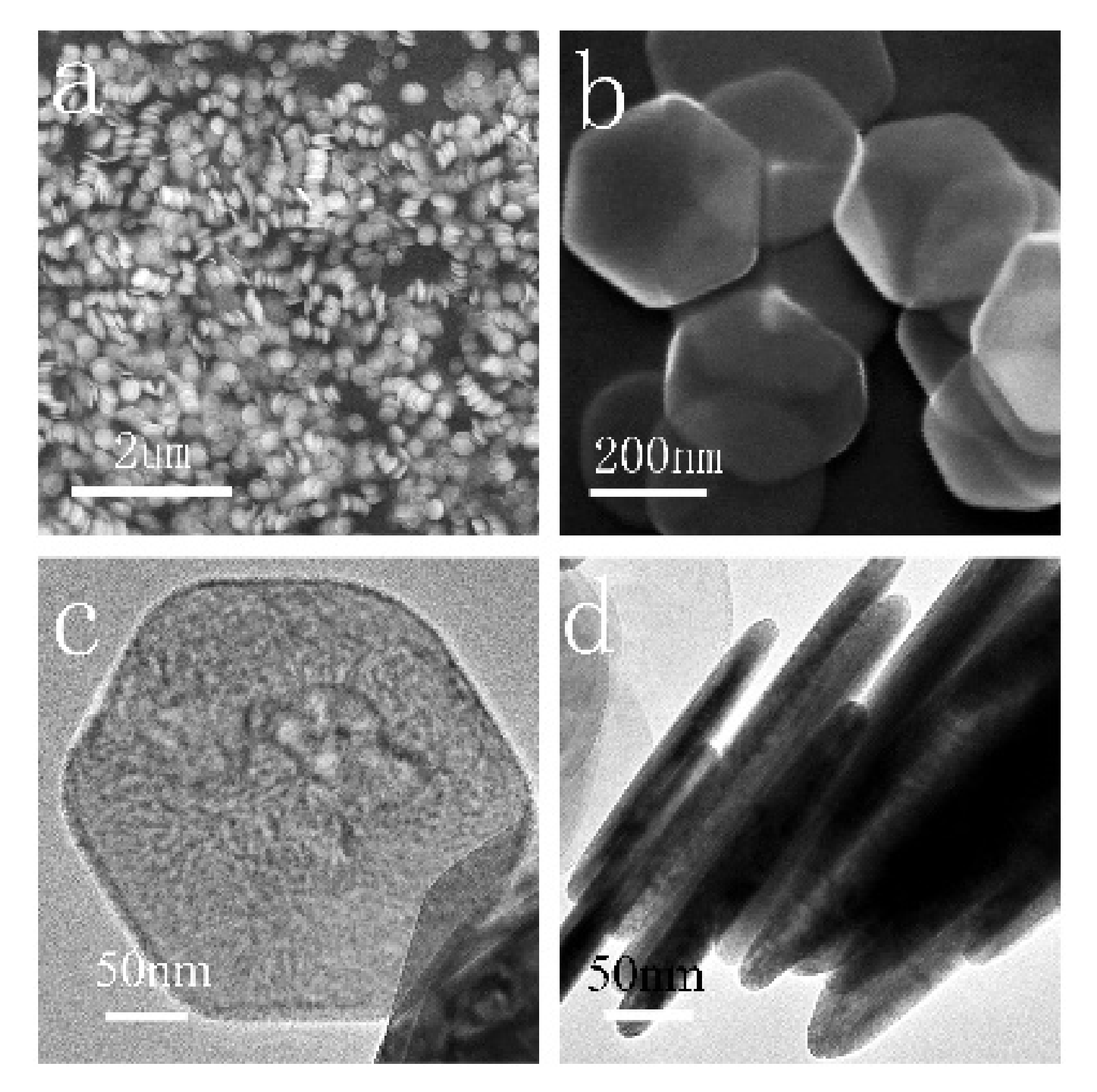
To check the morphology of the Ni(OH)2 crystallites, the sample prepared at 180 °C was examined due to its high crystallinity. Figure 2a shows the morphology of Ni(OH)2 under low magnification. The nanoplates were uniform, and the average side length was evaluated as 259 nm. Many nanoplates were stacked with a dozen to several dozens of single Ni(OH)2 plates. Figure 2b shows an SEM image of Ni(OH)2 nanoplates under high magnification. The Ni(OH)2 nanoplates exhibited regular hexagonal morphology. The frontal and lateral TEM images of a Ni(OH)2 nanoplate with a thickness of 25 nm are shown in Figure 2c,d, respectively. No evident pores were found on the surface of the nanoplate. Figure 2d indicates that the hexagonal Ni(OH)2 nanoplates were stacked from one single Ni(OH)2 plate to another neighboring plate in sequence without overlapping.
Figure 3a shows a simulated crystal structure model of the Ni(OH)2 hexagonal nanoplate. A TEM image of one individual hexagonal nanoplate is shown in Figure 3b,c, with the diffraction spots of the squared area shown in Figure 3b, suggesting the single-crystal nature. The sixfold symmetry of a β-Ni(OH)2 nanoplate was viewed along the (001) direction, further confirming its hexagonal structure. The interplanar spacing of 0.27 nm in Figure 3d was indexed as the (100) plane. According to a recent study [12], the continuous lateral planes of the Ni(OH)2 hexagonal nanoplate should be (100), (010), and (−100). The results shown in Figure 3b,c are consistent with the simulated model of a β-Ni(OH)2 nanoplate (Figure 3a). Figure 3,f shows the lateral TEM image and corresponding SAED of the stacked Ni(OH)2 nanoplates, respectively. The one-way diffraction spots were positioned along the (hk0) direction.
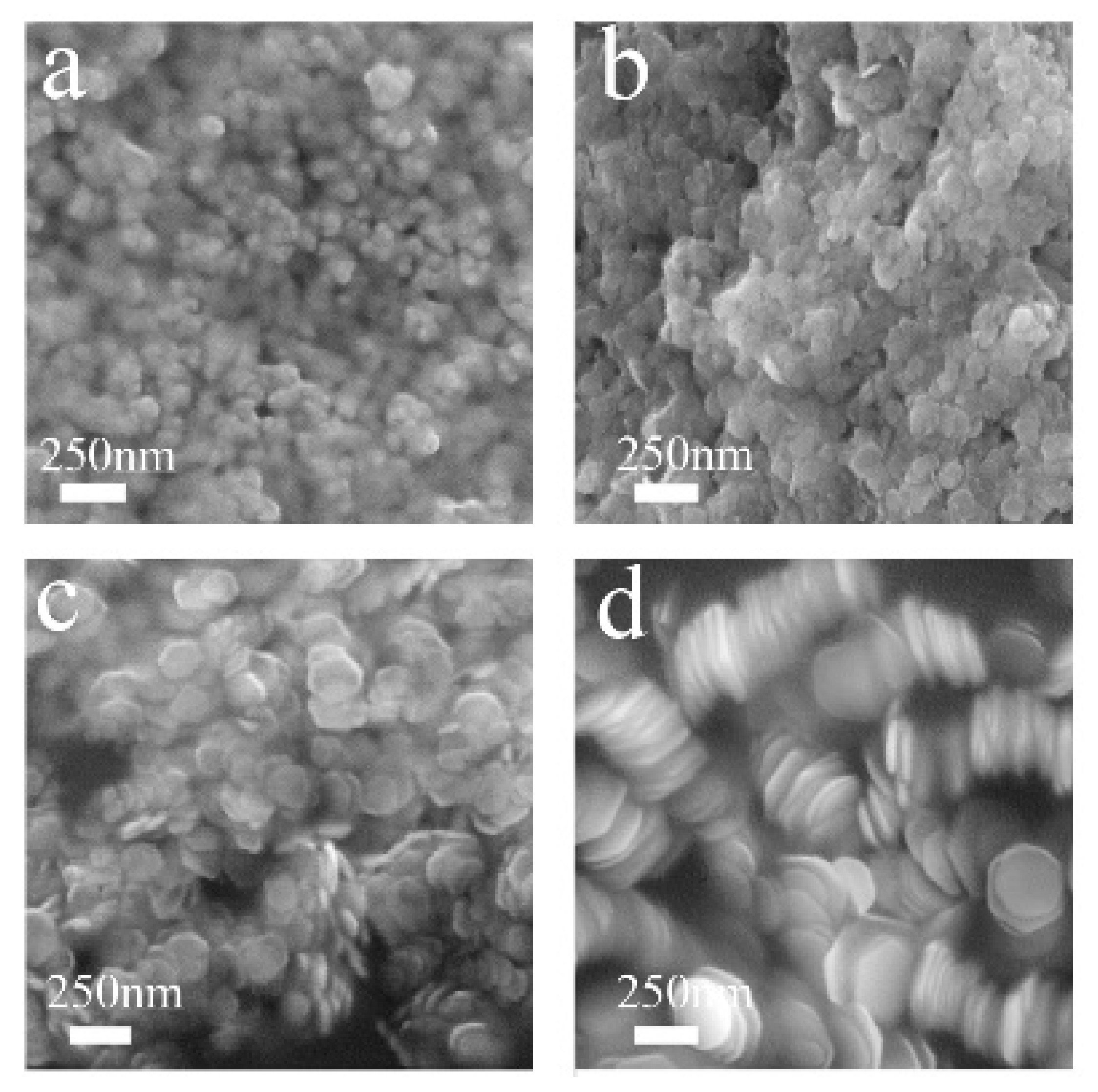
The reaction temperature generally plays a key role in preparing Ni(OH)2 hexagonal nanoplates. Figure 4a shows the SEM image of the sample prepared at room temperature; the obtained Ni(OH)2 has spherical morphology. Under 80 °C, Ni(OH)2 nanoplates with an average diameter of 86 nm are shown in Figure 4b. When the reaction temperature was increased to 140 (Figure 4c) and 180 °C (Figure 4d), the average edge lengths of the Ni(OH)2 hexagonal nanoplates were 141 and 259 nm, respectively. With the increase in the reaction temperature from 80 to 180 °C, the edge length of the Ni(OH)2 normal to (001) direction increased.
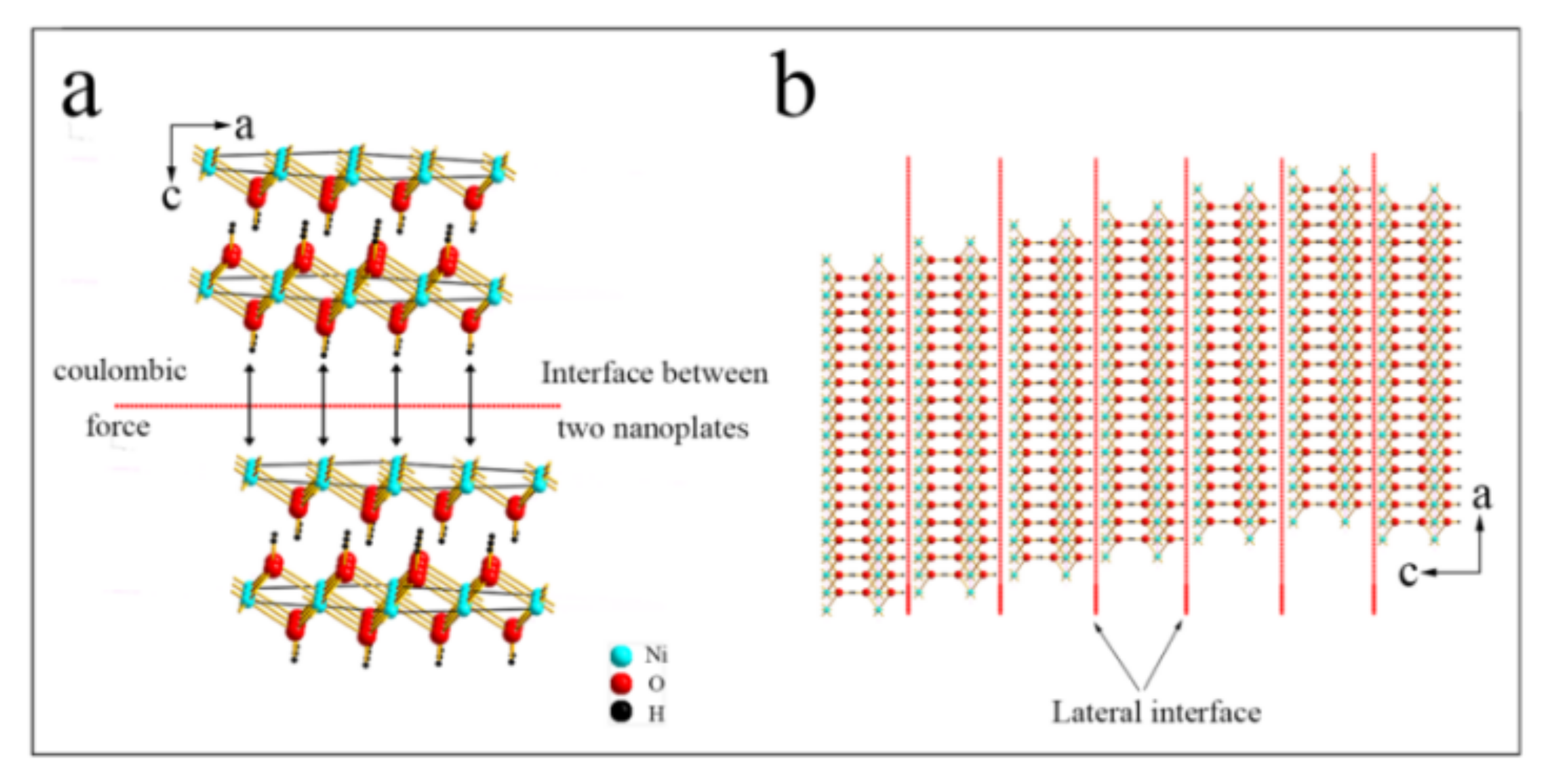
To further understand the formation mechanism of the stacked structures, a formation mechanism on the basis of the Ni(OH)2 crystal structure is proposed. Figure 5a schematically shows the interaction force between two adjacent Ni(OH)2 nanoplates. The Ni(OH)2 lattice cell shows that the saturated Ni atoms could be balanced by O atoms located between the upper and lower Ni layers. The two-layer O atoms are not in a similar position. However, the Ni atoms on the top surface of the Ni(OH)2 hexagonal nanoplates are in an unsaturated state, which easily attracts the O atoms of the neighboring hexagonal nanoplate surface by Coulombic forces. Thus, the Ni and O atoms interact with a dislocation, resulting in the misplaced formation of the stacked structures without overlapping. Figure 5b displays the lateral formation mechanism of the stacked structures. Ni(OH)2 hexagonal nanoplates are stacked by Coulombic forces along the c-axis, which is beneficial for lowering the surface energy of the samples [13,14]. In addition, hexagonal Ni(OH)2 crystal generally grows along the (001) direction due to its intrinsically anisotropic character.

4. Conclusions

The stacked Ni(OH)2 hexagonal nanoplates, with uniform sizes and orientated along the (001) direction, were synthesized in a large scale in this work. The edge length normal to (001) direction can be effectively controlled by the hydrothermal temperature. Based on the analysis of the Ni(OH)2 crystal structure, the Coulombic forces (Ni2+ and O2−) between the top and the bottom surfaces of Ni(OH)2 hexagonal nanoplates are suggested to be responsible for the formation of the dislocated stacked structure.

Author Contributions

Writing—original draft and investigation, Y.D.; data curation, G.L.; supervision, Z.X. and T.L. All authors have read and agreed to the published version of the manuscript.

Funding

This research was funded by the National Natural Science Foundation of China (NSFC) (No. 52075503). The APC was funded by the Shanxi Provincial Youth Science and Technology Fund (No. 201901D211266).

Institutional Review Board Statement

Not applicable.

Informed Consent Statement

Not applicable.

Data Availability Statement

Not applicable.

Acknowledgments

This work was supported by the project of the National Natural Science Foundation of China (NSFC) (No. 52075503) and the Shanxi Provincial Youth Science and Technology Fund (No. 201901D211266).

Conflicts of Interest

The authors declare no conflict of interest.

References

  1. Ede, S.R.; Anantharaj, S.; Kumaran, K.T.; Mishra, S.; Kundu, S. One step synthesis of Ni/Ni(OH)2 nano sheets (NSs) and their application in asymmetric supercapacitors. RSC Adv. 2017, 7, 5898–5911. [Google Scholar] [CrossRef] [Green Version]
  2. Taniguchi, A.; Fujioka, N.; Ikoma, M.; Ohta, A. Development of nickel/metal-hydride batteries for EVs and HEVs. J. Power Sources 2001, 100, 117–124. [Google Scholar] [CrossRef]
  3. Cao, M.; He, X.; Chen, J.; Hu, C. Self-assembled nickel hydroxide three-dimensional nanostructures: A nanomaterial for alkaline rechargeable. Cryst. Growth Des. 2007, 7, 170–174. [Google Scholar] [CrossRef]
  4. Zhang, W.; Jiang, W.; Yu, L.; Fu, Z.; Xia, W.; Yang, M. Effect of nickel hydroxide composition on the electrochemical performance of spherical Ni(OH)2 positive materials for Ni-MH batteries. Int. J. Hydrogen Energy 2009, 34, 473–480. [Google Scholar] [CrossRef]
  5. Chen, X.; Chen, X.; Zhang, F.; Yang, Z.; Huang, S. One-pot hydrothermal synthesis of reduced graphene oxide/carbon nanotube/α-Ni(OH)2 composites for high performance electrochemical supercapacitor. J. Power Sources 2013, 243, 555–561. [Google Scholar] [CrossRef]
  6. Dubal, D.; Fulari, V.; Lokhande, C. Effect of morphology on supercapacitive properties of chemically grown β-Ni(OH)2 thin films. Microporous Mesoporous Mater. 2012, 151, 511–516. [Google Scholar] [CrossRef]
  7. Devaraj, S.; Munichandraiah, N. Effect of Crystallographic Structure of MnO2 on Its Electrochemical Capacitance Properties. J. Phys. Chem. C 2008, 112, 4406–4417. [Google Scholar] [CrossRef]
  8. Chmiola, J.; Yushin, G.; Dash, R.; Gogotsi, Y. Effect of pore size and surface area of carbide derived carbons on specific capacitance. J. Power Sources 2006, 158, 765–772. [Google Scholar] [CrossRef]
  9. Ren, X.; Guo, C.; Xu, L.; Li, T.; Hou, L.; Wei, Y. Facile Synthesis of Hierarchical Mesoporous Honeycomb-like NiO for Aqueous Asymmetric Supercapacitors. ACS Appl. Mater. Interfaces 2015, 7, 19930–19940. [Google Scholar] [CrossRef]
  10. Wu, M.S.; Huang, K.C. Fabrication of nickel hydroxide electrodes with open-ended hexagonal nanotube arrays for high capacitance supercapacitors. Chem. Commun. 2011, 47, 12122–12124. [Google Scholar] [CrossRef]
  11. Mao, Y.; Li, T.; Guo, C.; Zhu, F.; Zhang, C.; Wei, Y.; Hou, L. Cycling stability of ultrafine β-Ni(OH)2 nanosheets for high capacity energy storage device via a multilayer nickel foam electrode. Electrochim. Acta 2016, 211, 44–51. [Google Scholar] [CrossRef] [Green Version]
  12. Qi, Y.J.; Qi, H.Y.; Li, J.H.; Lu, C.J. Synthesis, microstructures and UV-vis absorption properties of β-Ni(OH)2 nanoplates and NiO nanostructures. J. Cryst. Growth 2008, 310, 4221–4225. [Google Scholar] [CrossRef]
  13. Li, X.; Shen, H.; Niu, J.; Li, S.; Zhang, Y.; Wang, H.; Li, L.S. Columnar Self-Assembly of Cu2S Hexagonal Nanoplates Induced by Tin(IV)−X Complex as Inorganic Surface Ligand. J. Am. Chem. Soc. 2010, 132, 12778–12779. [Google Scholar] [CrossRef] [PubMed]
  14. Wang, G.; Ma, Z.; Fan, Y.; Shao, G.; Kong, L.; Gao, W. Preparation of size-selective Mn3O4 hexagonal nanoplates with superior electrochemical properties for pseudocapacitors. Phys. Chem. Chem. Phys. 2015, 17, 23017–23025. [Google Scholar] [CrossRef] [PubMed]
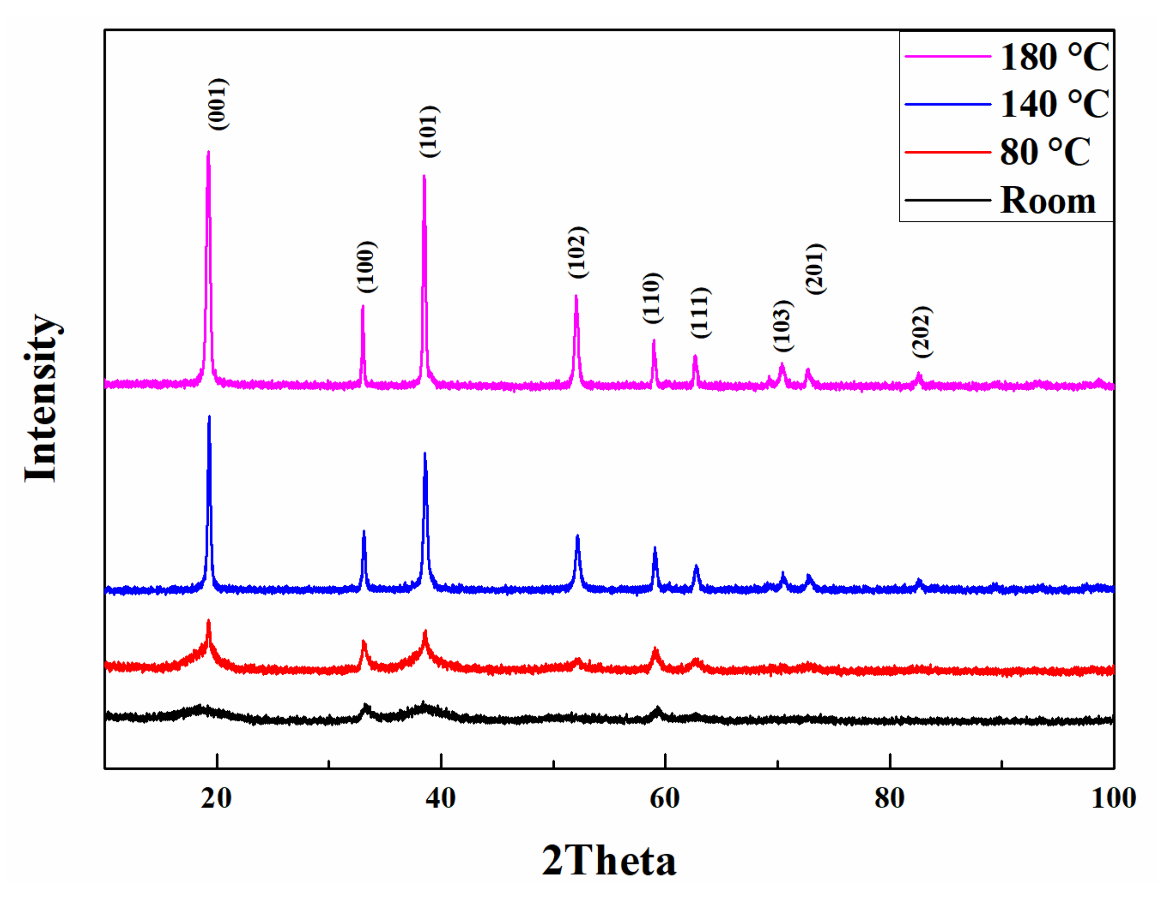
Figure 1. XRD profiles of the samples prepared at room temperature, 80, 140, and 180 °C.
Crystals 11 01407 g001
Figure 2. The SEM images of the sample prepared at 180 °C under (a) low and (b) high magnifications; TEM images viewed from different directions: (c) frontal and (d) lateral.
Crystals 11 01407 g002
Figure 3. (a) The simulated image of Ni(OH)2 hexagonal nanoplate; (b) The frontal TEM image of the hexagonal nanoplate, (c) the corresponding SAED of (b); (d) an HRTEM image of the white rectangle in (b); (e) TEM image of the lateral stacked structures; (f) corresponding SAED of the area in (e).
Crystals 11 01407 g003
Figure 4. SEM images of the samples prepared at different temperatures: (a) room temperature; (b) 80 °C; (c) 140 °C; (d) 180 °C.
Crystals 11 01407 g004
Figure 5. The formation mechanism of the stacked structures: (a) interaction force between two neighboring nanoplates; (b) lateral view of the stacked structure.
Crystals 11 01407 g005
Publisher’s Note: MDPI stays neutral with regard to jurisdictional claims in published maps and institutional affiliations.