Article

New Mononuclear Complex of Europium(III) and Benzoic Acid: From Synthesis and Crystal Structure Solution to Luminescence Emission

by
Carlos Hernández-Fuentes
1,
Rosario Ruiz-Guerrero
1,*,
Angel de Jesús Morales-Ramírez
1,2,
Paulina Molina-Maldonado
1 and
Dulce Y. Medina-Velazquez
3
1
Centro de Investigación e Innovación Tecnológica, Instituto Politécnico Nacional, cerrada de Cecati Sta. Catarina Azcapotzalco CP. 02250 CDMX, Mexico
2
Escuela Superior de Ingeniería Química e Industrias Extractivas, Instituto Politécnico Nacional, Av. Luis Enrique Erro S/N, Unidad Profesional Adolfo López Mateos, Zacatenco, GAM, C.P. 07738 CDMX, Mexico
3
División de Ciencias Básicas e Ingeniería, Universidad Autónoma Metropolitana-Azcapotzalco, C.P. 02200 CDMX, Mexico
*
Author to whom correspondence should be addressed.
Crystals 2020, 10(8), 674; https://doi.org/10.3390/cryst10080674
Submission received: 7 July 2020 / Revised: 31 July 2020 / Accepted: 3 August 2020 / Published: 4 August 2020
(This article belongs to the Section Crystal Engineering)

Abstract

:
This study presents a general method which can be used for the synthesis of mononuclear complexes with europium(III) and organic ligands with carboxylic groups. It describes the procedure for preparing a new mononuclear coordination complex with europium(III) and carboxylate ligands sourced from benzoic acid. The construction of mononuclear complexes with a coordination sphere saturated in carboxylic ligands must go through the preparation and purification of a europium(III) intermediate complex that presents a coordination sphere with anions that will be later exchanged for carboxylic groups and finally precipitated as a solvent-free or anion-free complex within the coordination sphere. The detailed synthesis procedure for powders of a new complex, as well as studies of its structural composition at each phase and luminescent properties, are detailed in this study. Analytical and spectroscopic data reveal the formation of a new mononuclear complex of the general formula [Eu(OOCC6H5)3·(HOOCC6H5)2]. The crystal structure of the Eu(III) complex was solved using X-ray powder diffraction data and EXPO2014 software, and the crystal structure result was deposited in the CCDC service with number 19771999.

Graphical Abstract

1. Introduction

In recent decades, there has been scientific and technological development regarding the use of lanthanides due to their versatility in different areas, including chemical analysis [1], catalysis [2] and biologic applications [3,4]. In particular, they are used for their luminescence properties, including highly pure color [5,6] and short radiative lifetimes [7]. Among the lanthanides, europium(III) stands out for its wide range of optical applications; however, the use of pure lanthanides is uncommon due to their low efficiency of direct absorption of the 4f–4f states [8]. This problem can be solved with the use of ligands that provide an antenna effect, which is defined as a light conversion process via an absorption-energy transfer–emission sequence, where the ligand functions as a light collector and the metal ion emits energy [9,10,11].
Moreover, there is new interest in the use of organic ligands, especially carboxylic compounds [12,13,14]. These ligands are extensively studied because of the high kinetic and thermodynamic stability of their corresponding lanthanide chelates [15], which have demonstrated to be efficient sensitizers when modified with light-harvesting moieties [16,17].
However, the most common use of carboxylic acids as ligands is as bidentate molecules, (dicarboxylic acids), which results in binuclear compounds, or even more lanthanide cations in its structure, [18,19,20] in which an indirect interaction between the metal centers through the ligands causes energy losses through non-radiative pathways [21]. It is also common to find in these complexes the presence of solvent in the coordination sphere of the final product, which does not favor luminescent emission mechanisms [22,23,24,25].
Of course, there are reports of mononuclear complexes [26,27], but it is not easy to get solvent-free compounds in the coordination sphere. In this study, we report a method for obtaining mononuclear complexes (free of solvents in their sphere of coordination) with organic ligands, specifically carboxylic ligands. The present work presents a newly developed method for the synthesis of mononuclear compounds that can easily absorb and conduct energy, avoiding the quenching effect caused by subtractor molecules of energy [28,29] and direct interactions between metallic centers [30].
For the method described in this paper, benzoic acid was used as organic ligand capable of coordinating with europium(III) to form a mononuclear species with this cation. This acid (Figure 1) presents in its structure an aromatic ring formed by conjugated bonds that facilitate the absorption of photons [31,32] as well as a carboxylic group (COOH) capable of chelating cations and facilitate the charge transfer to the metal ion. Overall, it is a molecule of relatively simple structure with the ability to absorb and transport UV radiation to the central ion.
This study presents the synthesis, structural composition and luminescence properties of new europium(III) complex. The lanthanide is used as the central cation and benzoic acids are used exclusively as ligands. Detailed synthesis and characterization of the intermediate complex are presented due to the importance it represents in the construction of the final complex. The sum of the total procedure allows us to have a complete description of the methodology that can be followed for the construction of mononuclear complexes that are solvent-free in their coordination sphere and include lanthanide cations and carboxylic acids.

2. Experimental Section

Molecules of organic solvent in the coordination sphere pose an issue for practical luminescent devices because they act as load subtraction groups with low quantum efficiency [33]. Water was used as a solvent in the reaction because Europium chloride and benzoic acid have high solubility in this medium. In addition, water is an easy replacement molecule when it is in the coordination sphere [34]. The precipitation of the final complex was conducted in an ionic reaction medium favored by the presence of Na+ and Cl ions.

2.1. Materials and Methods

Reagents: The benzoic acid (99%), sodium hydroxide (99.9%) and europium(III) chloride (99%) used were supplied by Sigma-Aldrich (St. Louis, MI, USA). Deionized water was used as the only solvent.
Methods: The Fourier-transform infrared spectroscopy (FTIR) was performed on a Perkin Elmer Spectrum 65 FT-IR spectrometer using KBr pellets as support. The elemental analyses for C and H were carried out using a Thermo Scientific/Flash 2000 (Thermo Fisher Scientific Inc, Waltham, MA, USA) elemental analyzer. A quantitative analysis was also conducted by atomic absorption spectrometry on a spectrometer AAnalyst 300 (Perkin Elmer, Waltham, MA, USA) to evaluate accurate percentages of europium(III) in synthesized europium(III) complexes. The mass spectrometry was performed on a MicrOTOF-Q II mass spectrometer (Bruker corporation, Billerica, MA, USA) using methanol as a solvent. The thermogravimetric analysis was obtained on STA 2500 equipment (NETZSCH-Gruppe, Selb, Germany) where about 10 mg of samples were heated at 5 °C/min from room temperature to 300 °C in a dynamic nitrogen atmosphere (flow rate = 50 mL/min). The melting point was determined by an IA 9000 (Electrothermal, Staffordshire, UK) series digital melting point apparatus. Scanning electron microscopy (SEM) was conducted on a JSM-7800 F Field Emission Scanning Electron microscope (Hitachi High-tech, Tokyo, Japan) operating at 10 keV. X-ray diffraction patterns were collected using a D8 Advance Eco powder diffractometer (Bruker corporation, Billerica, MA, USA) with Cu Kα (1.54), with an angular range of 5° to 90° and a step size of 0.02. The determination of the unit cell parameters, identification of the space group, structure solution and model refinement via the Rietveld method, were carried out by EXPO2014 software (2014, Institute of Crystallography, Bari, Italy): a package able to automatically execute the full pathway of the powder solution process. The photoluminescence studies of the synthesized complex powders were recorded on an F-7000 FL spectrophotometer (Hitachi High-tech, Tokyo, Japan) equipped with a 150 W xenon lamp, at room temperature.

2.2. Synthesis

The first step gave rise to the complex 2 [Eu(OOCC6H5)3·(H2O)3], in which the bidentate interaction of three benzoate molecules with the metal ion results in the displacement of three chlorine molecules and three water molecules. In the second step, the water molecules still present in complex 2 were displaced by two benzoic acid molecules linked by monodentate coordinated bonds, to form complex 3 [Eu(OOCC6H5)3·(HOOCC6H5)2], see Figure 2.

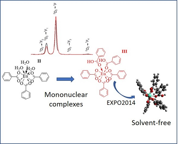
2.2.1. Synthesis of Complex 2: [Eu(OOCC6H5)3·(H2O)3]

A sodium benzoate solution prepared from sodium hydroxide (3 mL, 1 mol L−1) and benzoic acid (1) solution (30 mL, 0.1 mol L−1) was reacted with europium(III) chloride (10 mL, 0.1 mol L−1) in an aqueous solution. The mixture was stirred for 6 h at room temperature to obtain complex 2. The precipitate was washed with deionized water and allowed to air dry (Figure 2).
Eu(OOCC6H5)3·(H2O)3 (2): Yield: 89%. Theoretical elemental analysis for C21H21O9Eu: C: 44.30%, H: 3.72%, Eu: 26.69%; Experimental: C: 44.61%, H: 3.68%, Eu: 27.12%.

2.2.2. Synthesis of Complex 3: [Eu(OOCC6H5)3·(HOOCC6H5)2]

First, 0.7 mmol of complex 2 dissolved in deionized water (30 mL) was added to an aqueous solution of benzoic acid (1.4 mmol in 30 mL) and stirred for 8 h. The precipitate obtained was washed with deionized water then allowed to dry at room temperature.
Eu(OOCC6H5)3·(HOOCC6H5)2 (3): Yield: 96%. Theoretical elemental analysis for C35H27O10Eu: C: 55.35%, H: 3.58%, Eu: 20.01%; Experimental: C: 55.64%, H: 3.52%, Eu: 19.60%.

3. Results and Discussion

The reaction was conducted to obtain the complexes 2 and 3 sequentially. Both complexes 2 and 3 were isolated and characterized by FTIR vibrational spectroscopy, thermogravimetric analysis, excitation, emission and luminescent decay curves. Complex 2 was characterized by mass spectrometry (ms/ms) and the X-ray diffraction pattern is shown for the complex 3.

3.1. Infrared Spectroscopy

Figure 3 shows the FTIR spectra of the benzoic acid used as precursor (1) and the complexes 2 and 3 are shown in Figure 2.
The nature of the obtained complexes was corroborated by infrared spectra. Figure 3 shows a shift of the carbonyl bond frequencies v (COO) for the blank sample (1) and both complexes (2 and 3). The variations in the frequencies, both symmetric as asymmetric, of the COOEu bond-stretching band for complexes 2 (vsym = 1429 cm−1 and vasym = 1538 cm−1) and 3 (vsym = 1406 cm−1 and vasym = 1525 cm−1) were lower with respect to 1. The free coordination ligand is associated with the coordination of benzoyl to europium(III) [35].
The magnitude of the Δv value (Table 1) produced by each complex is related to the type of COO coordination character of the ligands associated with the Eu cation. Complex 2 presents the smallest value of this magnitude (109), which is lower than that presented by the benzoate 1 (132); this reflects the coordination to three benzoates as chelating ligands. An increase in this value to 119 for 3 indicates the additional coordination of two benzoic acid molecules in a monodentate form [36].
According to previously indicated in the spectra of Figure 3, in complex 3, the band corresponding to the HO–H group of the coordinated water is no longer present, confirming its absence in the coordination sphere. By contrast, the spectrum on complex 2 shows a tension band corresponding to the HO–H of the three coordinated molecules at 3420 cm−1. For its part, complex 2 presents a clear water signal in its sphere of coordination with a band at 3400 cm−1.
In addition, mass spectroscopy studies of complex 2 show the characteristic losses of two molecules of water. The record of mass/mass technique of molecule 2 shows the fragmentation of the molecule and the molecular ion m/z = 551.03. The first loss of a water molecule is observed with the peak m/z = 533.02 and the second loss of water at m/z = 515.01. It is worth mentioning that the same technique of characterization was not possible for complex 3 since the powder is insoluble in solvents that could be useful for this.

3.2. Thermogravimetric Analysis

In Figure 4a, the number of water molecules present in the powders of complex 2 was determined by the data obtained from the thermogravimetric analysis, which showed a 9.5% loss by weight at the temperature of 100 °C as a result of the elimination of three water molecules (54.06 of 570.36), then continued stability until the temperature of 300 °C. The inflexion point at 241 °C corresponds to the phase change from a solid to a liquid of the compound, which is very close to the experimentally determined of 242 °C.
The powders of complex 3 were analyzed by thermogravimetry from 24 °C to 300 °C (Figure 4b) without weight loss, indicating the absence of water molecules. The data were corroborated with the FTIR results. The inflexion point at 245 °C corresponds to the phase change from solid to liquid, which is very close to the experimentally determined of 246 °C.

3.3. Morphologic Studies

3.3.1. Scanning Electron Microscopy

Figure 5a,b shows the particles of complex 2 and complex 3 by scanning electron microscopy (SEM), with a whiskers-like morphology and crystal shape characteristic typical of the triclinic space group [37]. For both cases, the SEM images and the non-symmetric unimodal graphics for the dispersion of sizes, revealed that the whiskers tend to be smaller in the longitudinal direction, varying from 1.0 µm to 2 µm with a median longitudinal particle size (d50) of 6.7 µm and longitudinal particle size distribution d80 of 11.1 µm for the complex 2. For complex 3, the whisker sizes ranged from 4.0 µm to 8.1 µm with longitudinal particle size distributions d50 of 1.6 µm and d80 of 2.9 µm. The radial particle size presented an average size of 0.5 µm for complex 2 and 0.6 µm for complex 3.

3.3.2. X-ray Powder Diffraction Data

Various crystallization systems were used to obtain solvent-free monocrystals in the coordination sphere without any success. Therefore, the X-ray powder diffraction was used.

3.4. Crystal Structure Solution

EXPO2014 [38] was used in the solution and refined process of the structure. EXPO2014 integrates a package to index a powder diffraction pattern, extraction of integrated intensities, determination of the space group, the solution of crystal structure through direct methods and/or direct space approach and refinement of the structure using the Rietveld method.
The indexing of the X-ray diffraction pattern of 3 was obtained through the N-TREOR09 computer program integrated through EXPO2014. The approximate unit cell parameters were calculated from the 31 major peaks. The space group determination step gave triclinic P-1 space group with two molecules in the unit cell. The solution was carried out by the direct space method, which involved the generation of a random sequence of test structures from an appropriate 3D model (drawn by ChemBioDraw Ultra 13.0 and optimized in MOPAC [39] by the RM1 model, which was moved until a good match was found between the calculated and observed pattern (X2 = 1.564) (Figure 6).
The model was refined by the Rietveld method and the optimized crystal structure is shown in Figure 7.
In the crystal structure, each atom of Europium, independent of each other, is bidentaly linked to three benzoate anions and connected through monodentate to two carboxylic acid molecules. The packing seems to be influenced by π···π interactions (Figure 8). Neighboring benzoic acid rings are at 3.471 Å distances (centroid-to-centroid) with slippage of 0.41 Å.
Table 2 shown the Eu–O distances, the range from 2.3001 Å (Eu–O2) to 2.3895 Å (Eu–O3) are in good agreement with similar Eu–O bond distances as found in powder X-ray structures from the Cambridge Crystallographic Data Center [40,41].
The resolution parameters for complex 3 are shown in Table 3 and CCDC 19771999 contains the supplementary crystallographic data for this study. These data can be obtained free of charge from the Cambridge Crystallographic Data Center (CCDC) via www.ccdc.cam.ac.uk/structures.

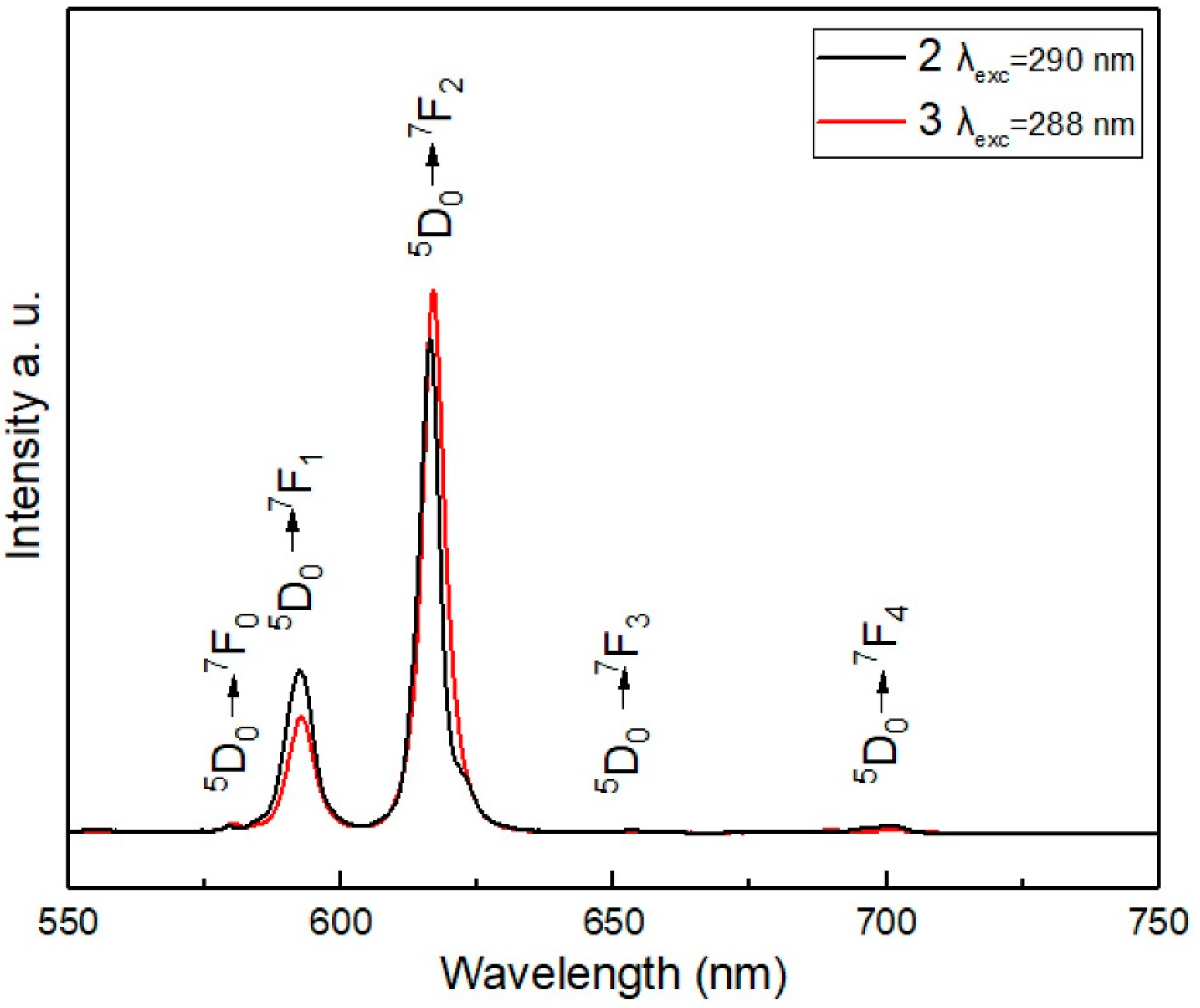
3.5. Luminescence Studies: Excitation and Emission Spectra

3.5.1. Excitation and Emission Spectra

The excitation spectra of the complexes are shown in Figure 9, which were performed with λem = 616 nm. It includes a broad band assigned to the S0 → S1 (π, π*) transition centered at 290 nm in complex 2 and 288 nm in complex 3, and on the right side, excitations correspond to the absorption bands that originated from the 7F0 ground state to the excited level 5L7 centered at 393 nm. In complex 3, this last excited level is, in terms of intensity, higher than in complex 2 (i.e., the area under the curve shows 70% more energy absorption in complex 3 than in complex 2), indicating the potential contribution of an energy ligand to the metal charge transfer states (LMCTS) of the two additional ligands. This corresponds to the antenna effect; at this wavelength, it absorbs the ligand and subsequently transfers the energy to the Eu3+ ion.
Figure 10 shows the emission spectra of both compounds of europium(III). The compound containing five carboxylic groups presents higher emission, even when it has the same europium(III) (5 × 10⁻³ mmol). This effect can be explained by the fact that by presenting a greater number of ligands, complex 3 allows a greater transfer of energy towards the luminescent center, as observed in the excitation spectrum. Conversely, the intensity of the 5D07F1 magnetic dipole transition (centered at 592 nm) is independent of the surroundings of the Eu3+, whereas the intensity of the 5D07F2 electric dipole transition (centered at 616 nm) is very sensitive to site symmetry [42]. In both complexes, the area under the curve was calculated using integration for the 5D0-7F2 transition. It was determined that complex 3 presents 15% more energy emission than complex 2.
Moreover, it is possible to set the ratio R = I (5D07F2)/I (5D07F1) as a reference to the coordination state and symmetry. Since the intensity 5D07F2 increases with the decreasing of the site symmetry. If the R value is low, the Eu3+ cation tends to localize in a centrosymmetric site or at a high symmetry site. In contrast, when the R value is high, the Eu3+ cation tends to localize in non-centrosymmetric site or at a low symmetry site [43,44]). The R factor increases from 2.41 in complex 2 to 3.89 in complex 3, which shows that the Eu3+ cation is in a position of less symmetry in complex 3. Then, the energy in complex 3 prefers the 5D07F2 transition to the 5D07F1 transition because of the influence of the local symmetry, effect of the two extra ligands.
Moreover, the observed broadening of the 5D07F2 band on complex 3 indicates that Eu3+ cations are located inhomogeneously at those sites without inversion symmetry [45,46]. Another important factor is the absence of water molecules, which acts as energy subtractors since the emission level decreases strongly through coupling with the excited vibratory levels of the OH oscillator of the coordinated water molecules [33].
Figure 11 shows the Jablonski partial energy diagram of complex 3 excited at 290 nm. The emission peaks, observed in Figure 10, appearing as the five typical bands at approximately 580 nm, 592 nm, 616 nm, 653 nm and 700 nm correspond to the characteristic emission transitions 5D07FJ (J = 0, 1, 2, 3, 4) [47]. The intramolecular energy transfer from the S0 → S1 (π, π*) state of the ligand to the excited state of Eu(III) is promoted by the excitation energy of the ligand during the photoluminescence.

3.5.2. Luminescence Decay Profiles

The decay lifetime curves of the Eu(III) for both complexes are shown in Figure 12, with emission monitored at 616 nm with a 290 nm excitation energy. The observed mono-exponential fitting (goodness of fit, R2 = 0.99991 and 0.9996 for 2 and 3, respectively) indicates that both complexes present a homogeneous composition [48]. The average life (τ av) increases from 0.42 ms in 2 to 0.53 ms 3. The change of the symmetry from higher to lower modifies the luminescence spectrum and shortens the radiative lifetime [49]. This effect can be explained since the two extra carboxylic acids in the coordination sphere provide the function of a chelating agent, prevent nonradiative transitions and produce better use of energy in the form of light, thereby increasing decay time. In addition, is well known that water molecules serve as quenching sites in many Eu(III) carboxylate complexes when they are at the coordination sphere due to can efficiently non-radiatively depopulate the 5D0 excited state of Eu3+ [50,51].

4. Conclusions

In this study, a new mononuclear compound of europium(III) with benzoic acid as the ligand and a full, solvent-free coordination sphere as the main feature was synthesized: [Eu(OOCC6H5)3·(HOOCC6H5)2].
The synthesis of this complex was conducted in two stages in which the formation of an intermediate compound [Eu(OOCC6H5)3·(H2O)3] is necessary to achieve the purity of the compound and a solvent-free structure. Both compounds were fully characterized and their photophysical properties compared. The main compound and the intermediate complex were widely characterized to corroborate the formation of the structure and mainly to see how the full sphere of coordination and the absence of water as a solvent with charge subtractor characteristics influenced the luminescent properties of the compound. Both compounds showed luminescent absorption in the region of 290 nm (290 in complex 2 and 288 in complex 3) assigned to the S0–S1 (π, π*) transition and an emission centered on 616 nm (corresponding to transition 5D07F2) with red light emission characteristics. The 5D07F2 the hypersensitive transition band and its intensity are much more influenced by the local symmetry of the Eu3+ ion due to the interactions of the ligand with the ion. This interaction is greater in complex 3 than in complex 2 due to the apparent chelating action of the two additional ligands that promote better use of energy and prevent energy losses by non-radiative routes. The complete coordination compound showed a 15% increase in the emission, showing greater luminescent capacities. Decay times suggest a better use of absorbed light, as the time in the main compound is longer. In some way, the chelating action ensures a greater transfer of the ligands to the central ion, avoiding the loss of energy by non-radiative transitions. A structure with a coordination sphere full of carboxylic acids and free of water has better use of energy in the form of light. The two-stage method described in this study has the potential to be used for the synthesis of mononuclear europium(III) complexes with different carboxylic acids as ligands.

Author Contributions

Conceptualization, R.R.-G.; Formal Analysis, A.d.J.M.-R. and D.Y.M.-V.; Investigation, P.M.-M.; Methodology, C.H.-F. All authors have read and agreed to the published version of the manuscript.

Funding

This research was funded by the National Polytechnic Institute, SIP projects 20201463 and 20201512.

Conflicts of Interest

The authors declare no conflict of interest.

References

  1. Doat, A.; Fanjul, M.; Pellé, F.; Hollande, E.; Lebugle, A. Europium-doped bioapatite: A new photostable biological probe, internalizable by human cells. Biomaterials 2003, 24, 3365–3371. [Google Scholar] [CrossRef]
  2. Lai, J.; Wang, T.; Zhang, H.; Ye, L.; Yan, C.; Gu, W. Modulating the photoluminescence of europium oxide nanoparticles by controlling thermal decomposition conditions. J. Lumin. 2019, 214, 116534. [Google Scholar] [CrossRef]
  3. Zou, Y.; Sun, F.; Liu, C.; Yu, C.; Zhang, M.; He, Q.; Xiong, Y.; Xu, Z.; Yang, S.; Liao, G. A novel nanotheranostic agent for dual-mode imaging-guided cancer therapy based on europium complexes-grafted-oxidative dopamine. Chem. Eng. J. 2019, 357, 237–247. [Google Scholar] [CrossRef]
  4. Xuan, Y.; Song, X.L.; Yang, X.Q.; Zhang, R.Y.; Song, Z.Y.; Zhao, D.H.; Hou, X.L.; An, J.; Zhang, X.S.; Zhao, Y. Di Bismuth particles imbedded degradable nanohydrogel prepared by one-step method for tumor dual-mode imaging and chemo-photothermal combined therapy. Chem. Eng. J. 2019, 375, 122000. [Google Scholar] [CrossRef]
  5. Decadt, R.; Van Hecke, K.; Depla, D.; Leus, K.; Weinberger, D.; Van Driessche, I.; Van Der Voort, P.; Van Deun, R. Synthesis, Crystal Structures, and Luminescence Properties of Carboxylate Based Rare-Earth Coordination Polymers. Inorg. Chem. 2012, 51, 11623–11634. [Google Scholar] [CrossRef]
  6. Singh, R.; King, A.; Nayak, B.B. Reddish emission of europium doped zinc oxide nanophosphor prepared through precipitation route using sodium borohydride. J. Alloys Compd. 2019, 792, 1191–1199. [Google Scholar] [CrossRef]
  7. Xu, H.; Sun, Q.; An, Z.; Wei, Y.; Liu, X. Electroluminescence from europium(III) complexes. Coord. Chem. Rev. 2015, 293–294, 228–249. [Google Scholar] [CrossRef]
  8. Binnemans, K. Interpretation of europium(III) spectra. Coord. Chem. Rev. 2015, 295, 1–45. [Google Scholar] [CrossRef] [Green Version]
  9. Mech, A.; Karbowiak, M.; Görller-Walrand, C.; Van Deun, R. The luminescence properties of three tetrakis dibenzoylmethane europium(III) complexes with different counter ions. J. Alloys Compd. 2008, 451, 215–219. [Google Scholar] [CrossRef]
  10. Pham, Y.H.; Trush, V.A.; Amirkhanov, V.M.; Gawryszewska, P. Structural and spectroscopic study of the europium complex with N-(diphenylphosphoryl)pyrazine-2-carboxamide. Opt. Mater. 2017, 74, 197–200. [Google Scholar] [CrossRef]
  11. Akerboom, S.; Meijer, M.S.; Siegler, M.A.; Fu, W.T.; Bouwman, E. Structure, Photo-and triboluminescence of the lanthanoid dibenzoylmethanates: HNEt3[Ln(dbm)4]. J. Lumin. 2014, 145, 278–282. [Google Scholar] [CrossRef]
  12. Hilder, M.; Lezhnina, M.; Junk, P.C.; Kynast, U.H. Spectroscopic properties of lanthanoid benzene carboxylates in the solid state: Part 3. N-heteroaromatic benzoates and 2-furanates. Polyhedron 2013, 52, 804–809. [Google Scholar] [CrossRef]
  13. Lee, B.H.; Chung, K.H.; Shin, H.S.; Park, Y.J.; Moon, H. Europium(III) complexes of polyfunctional carboxylic acids: Luminescence probes of possible binding sites in fulvic acid. J. Colloid Interface Sci. 1997, 188, 439–443. [Google Scholar] [CrossRef]
  14. Bergbreiter, D.E.; Poteat, J.L.; Zhang, L. Complexation of europium using polyethylene carboxylic acid. React. Polym. 1993, 20, 99–109. [Google Scholar] [CrossRef]
  15. Feng, J.; Zhang, H. Hybrid materials based on lanthanide organic complexes: A review. Chem. Soc. Rev. 2013, 42, 387–410. [Google Scholar] [CrossRef] [PubMed]
  16. Reddy, M.L.P.; Sivakumar, S. Lanthanide benzoates: A versatile building block for the construction of efficient light emitting materials. Dalt. Trans. 2013, 42, 2663–2678. [Google Scholar] [CrossRef]
  17. Nakajima, A.; Nakanishi, T.; Kitagawa, Y.; Seki, T.; Ito, H.; Fushimi, K.; Hasegawa, Y. Hyper-stable organo-Eu III luminophore under high temperature for photo-industrial application. Sci. Rep. 2016, 6, 1–9. [Google Scholar] [CrossRef] [Green Version]
  18. Ramya, A.R.; Reddy, M.L.P.; Cowley, A.H.; Vasudevan, K.V. Synthesis, crystal structure, and photoluminescence of homodinuclear lanthanide 4-(dibenzylamino)benzoate complexes. Inorg. Chem. 2010, 49, 2407–2415. [Google Scholar] [CrossRef]
  19. Carter, K.P.; Thomas, K.E.; Pope, S.J.A.; Holmberg, R.J.; Butcher, R.; Murugesu, M.; Cahill, C.L. Supramolecular Assembly of Molecular Rare-Earth-3,5-Dichlorobenzoic Acid-2,2′:6′,2″-Terpyridine Materials: Structural Systematics, Luminescence Properties, and Magnetic Behavior. Inorg. Chem. 2016, 55, 6902–6915. [Google Scholar] [CrossRef]
  20. Carter, K.P.; Pope, S.J.A.; Cahill, C.L. A series of Ln-p-chlorobenzoic acid-terpyridine complexes: Lanthanide contraction effects, supramolecular interactions and luminescent behavior. CrystEngComm 2014, 16, 1873–1884. [Google Scholar] [CrossRef]
  21. Allendorf, M.D.; Bauer, C.A.; Bhakta, R.K.; Houk, R.J.T. Luminescent metal-organic frameworks. Chem. Soc. Rev. 2009, 38, 1330–1352. [Google Scholar] [CrossRef] [PubMed]
  22. Xiao, P.; Zhang, J.J.; Wang, Z.Q.; Lin, H. Photon Conversion and Radiation Synergism in Eu/Tb Complexes Incorporated Poly Methyl Methacrylate. Adv. Mater. Sci. Eng. 2016, 2016, 2618253. [Google Scholar] [CrossRef] [Green Version]
  23. Wang, Z.M.; Van De Burgt, L.J.; Choppin, G.R. Spectroscopic study of lanthanide(III) complexes with carboxylic acids. Inorg. Chim. Acta 1999, 293, 167–177. [Google Scholar] [CrossRef]
  24. Champness, N.R. The future of metal-organic frameworks. Dalt. Trans. 2011, 40, 10311–10315. [Google Scholar] [CrossRef] [PubMed]
  25. Utochnikova, V.V.; Solodukhin, N.N.; Aslandukov, A.N.; Marciniak, L.; Bushmarinov, I.S.; Vashchenko, A.A.; Kuzmina, N.P. Lanthanide tetrafluorobenzoates as emitters for OLEDs: New approach for host selection. Org. Electron. Phys. Mater. Appl. 2017, 44, 85–93. [Google Scholar] [CrossRef]
  26. Fiedler, T.; Hilder, M.; Junk, P.C.; Kynast, U.H.; Lezhnina, M.M.; Warzala, M. Synthesis, structural and spectroscopic studies on the lanthanoid p-aminobenzoates and derived optically functional polyurethane composites. Eur. J. Inorg. Chem. 2007, 2007, 291–301. [Google Scholar] [CrossRef]
  27. Chen, X.-F.; Duan, C.-Y.; Zhu, X.-H.; You, X.-Z.; Raj, S.S.S.; Fun, H.-K.; Wu, J. Triboluminescence and crystal structures of europium(III) complexes. Mater. Chem. Phys. 2001, 72, 11–15. [Google Scholar] [CrossRef]
  28. Zhang, J.; Wang, L.X.; Zhang, L.; Chen, Y.; Zhang, Q.T. Co-luminescence properties of terbium ions-benzoic acid-phen complexes doped with europium ions. Rare Met. 2013, 32, 599–604. [Google Scholar] [CrossRef]
  29. Borges, A.S.; Dutra, J.D.L.; Freire, R.O.; Moura, R.T.; Da Silva, J.G.; Malta, O.L.; Araujo, M.H.; Brito, H.F. Synthesis and characterization of the europium(III) pentakis(picrate) complexes with imidazolium countercations: Structural and photoluminescence study. Inorg. Chem. 2012, 51, 12867–12878. [Google Scholar] [CrossRef]
  30. Baldo, M.A.; O’Brien, D.F.; You, Y.; Shoustikov, A.; Sibley, S.; Thompson, M.E.; Forrest, S.R. Highly efficient phosphorescent emission from organic electroluminescent devices. Nature 1998, 395, 151–154. [Google Scholar] [CrossRef]
  31. Hilder, M.; Junk, P.C.; Kynast, U.H.; Lezhnina, M.M. Spectroscopic properties of lanthanoid benzene carboxylates in the solid state: Part 1. J. Photochem. Photobiol. A Chem. 2009, 202, 10–20. [Google Scholar] [CrossRef]
  32. de Oliveira, T.C.; Santos, H.P.; Lahoud, M.G.; Franco, D.F.; Freire, R.O.; Dutra, J.D.L.; Cuin, A.; de Lima, J.F.; Marques, L.F. Elucidating the energy transfer process in mononuclear and binuclear lanthanide complexes of the anti-inflammatory drug ibuprofen: From synthesis to high luminescence emission. J. Lumin. 2017, 181, 196–210. [Google Scholar] [CrossRef]
  33. Albuquerque, R.Q.; Freire, R.O.; Malta, O.L. On the use of ligand field parameters in the study of coordinated water molecules in Eu3+ complexes. J. Phys. Chem. A 2005, 109, 4607–4610. [Google Scholar] [CrossRef] [PubMed]
  34. Abdel-Mottaleb, M.S.A.; Galal, H.R.; Dessouky, A.F.M.; El-Naggar, M.; Mekkawi, D.; Ali, S.S.; Attya, G.M. Fluorescence and photostability studies of anthracene-9-carboxylic acid in different media. Int. J. Photoenergy 2000, 2, 47–53. [Google Scholar] [CrossRef]
  35. Yu, Z.; Guo, J.; Li, D.; Lin, H. Multiligand Europium Complexes Incorporated Polyvinylpyrrolidone for Enhanced Solar Cell. Adv. Mater. Sci. Eng. 2019, 2019, 6180656. [Google Scholar] [CrossRef] [Green Version]
  36. Deacon, G.B.; Phillips, R.J. Relationships between the carbon-oxygen stretching frequencies of carboxylato complexes and the type of carboxylate coordination. Coord. Chem. Rev. 1980, 33, 227–250. [Google Scholar] [CrossRef]
  37. Strelcov, E.; Kolmakov, A. Chemical sensors from lead metallophthalocyanine whiskers. Proc. IEEE Sensors 2007, 2, 636–639. [Google Scholar] [CrossRef]
  38. Altomare, A.; Cuocci, C.; Giacovazzo, C.; Moliterni, A.; Rizzi, R.; Corriero, N.; Falcicchio, A. EXPO2013: A kit of tools for phasing crystal structures from powder data. J. Appl. Crystallogr. 2013, 46, 1231–1235. [Google Scholar] [CrossRef]
  39. Stewart Computational Chemistry—MOPAC Home Page. Available online: http://www.openmopac.net/index.html (accessed on 20 May 2020).
  40. Jiang, W.; Hong, C.; Wei, H.; Wu, Z.; Ye, S.; Huang, C.-H. A green-emitting iridium complex used for sensitizing europium ion with high quantum yield. Inorg. Chim. Acta 2017, 459, 124–130. [Google Scholar] [CrossRef]
  41. Wang, A.-L.; Zhou, D.; Chen, Y.-N.; Li, J.J.; Zhang, H.-X.; Zhao, Y.-L.; Chu, H. Crystal structure and photoluminescence of europium, terbium and samarium compounds with halogen-benzoate and 2,4,6-tri(2-pyridyl)-s-triazine. J. Lumin. 2016, 177, 22–30. [Google Scholar] [CrossRef]
  42. Hreniak, D.; Speghini, A.; Bettinelli, M.; Strek, W. Spectroscopic investigations of nanostructured LiNbO3 doped with Eu3+. J. Lumin. 2006, 119–120, 219–223. [Google Scholar] [CrossRef]
  43. Medina, D.Y.; Orozco, S.; Hernandez, I.; Hernandez, R.T.; Falcony, C. Characterization of europium doped lanthanum oxide films prepared by spray pyrolysis. J. Non-Cryst. Solids 2011, 357, 3740–3743. [Google Scholar] [CrossRef]
  44. García, R.N.; Desirena, H.; López-Luke, T.; Guerrero-Contreras, J.; Jayasankar, C.; Quintero-Torres, R.; De La Rosa, E. Spectroscopic properties of Eu3+/Nd3+ co-doped phosphate glasses and opaque glass-ceramics. Opt. Mater. 2015, 46, 34–39. [Google Scholar] [CrossRef]
  45. Crystal, P.H.; Fujihara, S.; Tokumo, K. Multiband Orange-Red Luminescence of Eu3+ Ions Based on the Pyrochlore-Structured Host Crystal. Chem. Mater. 2005, 17, 5587–5593. [Google Scholar]
  46. Yang, C.; Fu, L.-M.; Wang, Y.; Zhang, J.-P.; Wong, W.-T.; Ai, X.-C.; Qiao, Y.-F.; Zou, B.-S.; Gui, L.-L. A Highly Luminescent Europium Complex Showing Visible-Light-Sensitized Red Emission: Direct Observation of the Singlet Pathway. Angew. Chem. 2004, 116, 5120–5123. [Google Scholar] [CrossRef]
  47. Yan, B.; Song, Y.S. Spectroscopic study on the photophysical properties of lanthanide complexes with 2,2′-bipyridine-N, N′-dioxide. J. Fluoresc. 2004, 14, 289–294. [Google Scholar] [CrossRef]
  48. Wu, M.; You, J.; Song, Z. Luminescence Study of the Interaction of Carboxymethyl Chitosan with Metal Ions. J. Appl. Spectrosc. 2014, 81, 602–606. [Google Scholar] [CrossRef]
  49. Shavaleev, N.M.; Eliseeva, S.V.; Scopelliti, R.; Bünzli, J.-C.G. Influence of Symmetry on the Luminescence and Radiative Lifetime of Nine-Coordinate Europium Complexes. Inorg. Chem. 2015, 54, 9166–9173. [Google Scholar] [CrossRef]
  50. Moreau, P.; Colette-Maatouk, S.; Vitorge, P.; Gareil, P.; Reiller, P.E. Complexation of europium(III) by hydroxybenzoic acids: A time-resolved luminescence spectroscopy study. Inorganica Chim. Acta 2015, 432, 81–88. [Google Scholar] [CrossRef] [Green Version]
  51. Barkleit, A.; Kretzschmar, J.; Tsushima, S.; Acker, M. Americium(iii) and europium(iii) complex formation with lactate at elevated temperatures studied by spectroscopy and quantum chemical calculations. Dalt. Trans. 2014, 43, 11221–11232. [Google Scholar] [CrossRef] [Green Version]
Figure 1. Chemical structure of benzoic acid: (1).
Crystals 10 00674 g001
Figure 2. Synthesis of 2: [Eu(OOCC6H5)3·(H2O)3] and 3: [Eu(OOCC6H5)3·(HOOCC6H5)2]. In both complexes, the oxidation state of europium is 3+.
Crystals 10 00674 g002
Figure 3. FTIR spectroscopy for 1: benzoic acid; 2: [Eu(OOCC6H5)3·(H2O)3]; 3: [Eu(OOCC6H5)3·(HOOCC6H5)2].
Crystals 10 00674 g003
Figure 4. Thermogravimetric analysis. (a) 2: [Eu(OOCC6H5)3·(H2O)3]; (b) 3: [Eu(OOCC6H5)3· (HOOCC6H5)2].
Crystals 10 00674 g004
Figure 5. Scanning electron microscopy. (a) 2: [Eu(OOCC6H5)3·(H2O)3]; (b) 3: [Eu(OOCC6H5)3·(HOOCC6H5)2].
Crystals 10 00674 g005
Figure 6. Rietveld plot of 3 [Eu(OOCC6H5)3·(HOOCC6H5)2]. (blue line) Observed diffraction profile; (red line) calculated profile; (green line) background and (violet line) difference profile.
Crystals 10 00674 g006
Figure 7. Crystal structure of complex 3: [Eu(OOCC6H5)3·(HOOCC6H5)2. Hydrogen atoms are omitted for clarity.
Crystals 10 00674 g007
Figure 8. Crystal packing of complex 2, showing π···π interactions.
Crystals 10 00674 g008
Figure 9. Excitation spectrum with λem = 616 nm, for 2: [Eu(OOCC6H5)3·(H2O)3] and 3: [Eu(OOCC6H5)3·(HOOCC6H5)2].
Crystals 10 00674 g009
Figure 10. Emission spectrum, with λex = 290 nm, for 2: [Eu(OOCC6H5)3·(H2O)3] and 3: [Eu(OOCC6H5)3·(HOOCC6H5)2].
Crystals 10 00674 g010
Figure 11. Partial energy level diagram of 3: [Eu(OOCC6H5)3·(HOOCC6H5)2].
Crystals 10 00674 g011
Figure 12. 5D07F2 level decay time profile at 393 nm of complex 2: [Eu(OOCC6H5)3·(H2O)3] and complex 3: [Eu(OOCC6H5)3·(HOOCC6H5)2].
Crystals 10 00674 g012
Table 1. Overview of synthesis identification.
ComplexIdentificationVibrations (cm−1)Δv (cm−1)
v (HO–H)vasym (COO)vsym (COO)asym-sym
Benzoic acid a114241292132
[Eu(OOCC6H5)3·(H2O)3]2343815381429109
Eu(OOCC6H5)3·(HOOCC6H5)2315251406119
a precursor material evaluated as a blank.
Table 2. Bond distances europium–oxygen (Å) for complex 3.
Bond Distances of Europium–Oxygen(Å)
Eu–O1 2.3637(3)Eu–O3 2.3895(2)Eu–O5 2.3866(3)Eu–O7 2.3409(2)
Eu–O2 2.3001(2)Eu–O4 2.3324(3)Eu–O6 2.3236(2)Eu–O9 2.3347(2)
Table 3. Crystal data, Rietveld refinement parameters and Cambridge Crystallographic Data Center (CCDC) depository number for complex 3: [Eu(OOCC6H5)3·(HOOCC6H5)2].
Formula, molecular weightEu C35H27O10, 760.56
Temperature (K), (Å)293, 1.54056
System, space groupTriclinic, P-1
a, b, c (Å); α, β, γ (°)24.496(3), 15.166(2), 5.0934(8), 92.408(1), 91.007(3), 74.814(6),
V (Å3), Z1824.42, 2
2θ range, step (°)4–40, 0.02
Nr. of data points3701
Nr. of Bragg refl., parameters884, 112
Rp (%), Rwp (%), X23.876, 5.032, 1.564
Rexp (%), RBragg (%)4.024, 1.397
CCDC depository nr.CCDC 19771999

