Article

Preparation of Few-Layered Wide Bandgap MoS2 with Nanometer Lateral Dimensions by Applying Laser Irradiation

1
Department of Physics, Central Tehran Branch, Islamic Azad University, Tehran 1584743311, Iran
2
Nano Research Lab (NRL), Department of Physics, Central Tehran Branch, Islamic Azad University, Tehran 1584743311, Iran
3
Department of Physics, East Tehran Branch, Islamic Azad University, Tehran 1584743311, Iran
*
Author to whom correspondence should be addressed.
Crystals 2020, 10(3), 164; https://doi.org/10.3390/cryst10030164
Submission received: 29 January 2020 / Revised: 19 February 2020 / Accepted: 20 February 2020 / Published: 2 March 2020
(This article belongs to the Special Issue 2D Materials: From Structures to Functions)

Abstract

:
In this study, we report a new method for the quick, green, and one-step preparation of few-layered molybdenum disulfide (MoS2) nanosheets with wide bandgap. MoS2 nanosheets with small lateral dimension and uniform size distribution were synthesized for various applications. MoS2 powder was synthesized using the hydrothermal method; then, thinned by applying laser irradiation with different energies from 40 to 80 mJ. Transmission electron microscopy (TEM), X-ray diffraction (XRD), Fourier transform infrared spectroscopy (FTIR), UV-Vis absorption spectra, and photoluminescence (PL) spectra were applied for the characterization of the MoS2 nanosheets in terms of morphology, crystal structures, and optical properties. The widest calculated bandgap 4.7 eV was for the sample under 80 mJ laser energy. The results confirmed the successful preparation of highly pure, uniform, and few-layered MoS2 nanosheets. Furthermore, it was possible to enhance the production rate of MoS2 nanosheets (including nanosheets and nanoparticles) through laser irradiation. Thus, the present paper introduces a simple and green alternative approach for preparing few-layered MoS2 nanosheets of transition metal dichalcogenides or other layered materials.

1. Introduction

Lately, few-layered materials along with two-dimensional materials have attracted the most attention due to their diverse properties and applications [1,2,3]. These materials exhibit completely different properties as compared with their bulk counterparts making them one of the best candidates for new generations of electronic and optoelectronic devices [4,5,6]. The application of few-layered materials has contributed to advances in the fabrication of ultrathin layered materials with high surface-to-volume ratio [7,8,9]. Transition metal dichalcogenides (TMDs) are in a form of MX2, where M and X denote transition metal and chalcogen atoms, respectively. The TMDs are important two-dimensional (2D) materials, which include about 60 members. The structures of these layered materials are hexagonally packed. Due to this property, the TMDs, such as MoS2, WS2, WTe2, TiS2, TaS2, ZrS2, or graphite are exfoliated to prepare the 2D materials. Unlike graphene which has no bandgap, the TMDs are natural semiconductors. Therefore, they are applied in electronic devices and optical devices. The mobility of 2D TMDs is much lower than graphene, which limits their applications in devices. Zhang’s group prepared few-layer black phosphorus and characterized a few-layer black phosphorus field-effect transistor [10]. A wafer-scale MoS2 was prepared by Yu et al. [11]. Novoselov et al. predicted some materials including boron nitride which could be exfoliated into a single layer [12]. Coleman et al. sonicated commercial molybdenum disulfide (MoS2) and WS2 [13]. May et al. tried several polymers as stabilizers to exfoliate and disperse MoS2 and boron nitride [14]. Varrla et al. exfoliated molybdenum disulfide in aqueous surfactant solutions under shearing by a kitchen blender [15]. Zhang’s group prepared MoS2/polyvinylpyrrolidone (PVP) nanocomposites by sonication of MoS2 powder in ethanol with PVP [16]. So far, the practical application of g-CN has been limited to photocatalytic water splitting, photodegradation of dyes, bioimaging, photocatalyst, and fluorescence applications [17,18,19].
Molybdenum disulfide (MoS2) and tungsten disulfide (WS2) are layered materials that show great potential to extend to few-layered sheets paving the way for the new physical phenomena [20,21]. MoS2 has been favored greatly due to its inherent structural characteristics that are composed of strong covalently S-Mo-S sheets detained by van der Waals interactions among the layered structures [22]. These weak interlayer interactions make the reduction of MoS2 layers possible by micromechanical exfoliation from bulk crystalline [23]. It is known that electronic properties, including the energy gap of MoS2, are strongly related to the number of atomic layers. Researches have shown that few-layered MoS2 turns into a direct bandgap semiconductor if thinned, while bulk MoS2 has an indirect bandgap, which is favorable for optoelectronics [24,25,26]. Due to this distinctive structure and features, MoS2 can be used in numerous fields such as biomedicine, energy storage, gas sensing, catalysis, and electronics engineering [27]. MoS2 has become an efficient electrocatalyst for hydrogen evolution reactions due to its abundant active edge sites, high specific surface area, and excellent electrical conductivity, together with strong hydrogen adsorption properties [28,29]. The small size particles show strong optical nonlinearity features [30]. MoS2 in both bulk and few-layered form has been applied in photovoltaics [31,32] and photocatalysis [33] applications as they are capable of efficiently absorbing a broad region of solar spectrum. Furthermore, more stable quantum size confinement effects are expected because of the even size distribution of the monolayer nanosheets. The quantum confinements in layered d-electron materials such as MoS2 pave the way to engineer the electronic structure of matter at the nanoscale. Consequently, finding an effective method to prepare uniform-size monolayer MoS2 nanosheets seems essential. The literature review shows that the current research on two-dimension (2D) MoS2 nanosheets is principally based on the exfoliation technique, chemical vapor deposition method, or solvothermal route [34,35,36]. The chemical vapor deposition method requires laborious experimental conditions including high temperature, high vacuum, and special substrates that probably would restrict the application of 2D MoS2 nanosheets in practice. Simple and low-cost methods are needed for large-scale fabrication and commercial application of MoS2-based devices.
In this study, we report a new method for the quick, green, and one-step preparation of even monolayer MoS2 nanosheets. First MoS2 powder was synthesized using the hydrothermal method, and then they were thinned by reduction layers using laser irradiation. It is the first time to apply this method to exfoliate the MoS2 sheets. Next, we study the characteristic of MoS2 nanosheets.

2. Experimental Details

Materials for these experiments were acquired from Merck Company. To synthesize MoS2 powder, a solution of 0.1 M ammonium heptamolybdate (NH4)6Mo7O24 and 0.2 M thiourea CH4N2S in ethanol was made while stirred for 30 min at 60 °C to obtain a milky homogeneous solution. Then, the solution was filled into a 100 mL Teflon lined stainless steel autoclave. The reaction was performed for 12 h at 220 °C. After cooling down to room temperature, the achieved black powder was washed with ethanol (twice) and dried in a vacuum oven at 80 °C. In the next step, 0.5 g of obtained MoS2 powder was dispersed in 20 mL ethanol and stirred for 20 min. Three suspensions individually were prepared and separately exposed under 532 nm Nd:YAG pulsed laser irradiation (5 ns pulse duration and 7 mm beam diameter) for 15 min with different energies 40, 60, and 80 mJ. After laser irradiation, three suspensions were dried in a vacuum oven at 40 °C. The analysis was applied to the resulted powder. Figure 1 shows a flow chart of the synthesis process.

Characterization

X-Ray diffraction (XRD) was utilized to study the structure of fabricated samples characterized by using X’Pert PRO (Malven Panalytical, Almelo (Netherlands)), with Cu-Kα radiation, (λ = 0.154 nm). Fourier transforms infrared (FTIR) spectra were applied using a spectrophotometer (FTIR-410) (Jasco, Toku, Japan), from 500 to 4000 cm−1. To determine the morphology and structure of the samples, transmission Electron microscopy (TEM) (Philips XL, Netherlands), was done. The bandgap of the samples calculated from UV-Vis optical absorption and transmission spectra (Lambda 750, PerkinElmer, Germany). The photoluminescence spectrum was recorded by a fluorescence spectrometer (PL, PerkinElmer LS 45, Germany).

3. Results and Discussion

The XRD of the MoS2 powder and few-layered MoS2 are shown in Figure 2. The existence peaks at 14.2°, 32.6°, 39.5°, 44.2°, 49.8°, and 58.3° are consistent with the (002), (100), (103), (006), (105), and (110) planes of hexagonal MoS2 structure (JCPDS NO. 37-1492). By increasing laser energy, the intensity of these peaks is reduced. The calculated lattice constants a = b = 0.315 nm, and c = 1.229 nm represent that all the samples have the crystallite nature of MoS2 materials. The (002) peak ascribing to a d-spacing of 0.62 nm illustrates the stacking of single layers, whereas peaks at higher angle are attributes of the crystallinity of the samples [37]. After laser irradiation, the intensity reduction of (002) peak proposed more discount in the thickness of MoS2 nanosheets [38,39]. It is considered that the (002) diffraction peak of the MoS2 powder, after laser irradiation, involves a shift to the lower angles and as a result, an increase in d-spacing indicating the destacking of MoS2 layers. It can be deduced with the enhancement in the laser energy the average grain sizes of the MoS2 nanosheets decrease. Therefore, the broadened diffraction peaks correspondingly confirm the decrease of the size of the MoS2 nanosheets, which is initiated by the laser irradiation.
The FTIR spectra of the samples are shown in Figure 3. The peaks below 700 cm−1 reveal the (Mo-O) deformation mode possibly caused by the sulfidation vibrations. The peaks between 750 and 1000 cm−1 indicate the most definite information on Mo-O coordination [40]. By increasing laser energy these peaks are more potent indicating stronger bonds of Mo-O. The absorption band between 1100 and 1650 cm−1 is ascribed to the stretching vibrations of the hydroxyl group and Mo-O vibrations. By reduction of MoS2 layers after laser irradiation, these peaks are stronger. The Mo-S vibration peak is around 600 cm−1, which is faded by increasing laser energy, meaning weaker bond of Mo-S. The C = C symmetric and asymmetric stretching vibrations are at 2890 and 2970 cm−1, which show small changes by different laser energies [41]. After laser irradiation, the peak at 1600 becomes stronger, which means stronger Mo-S bound, which is the result of monolayer MoS2 [42].
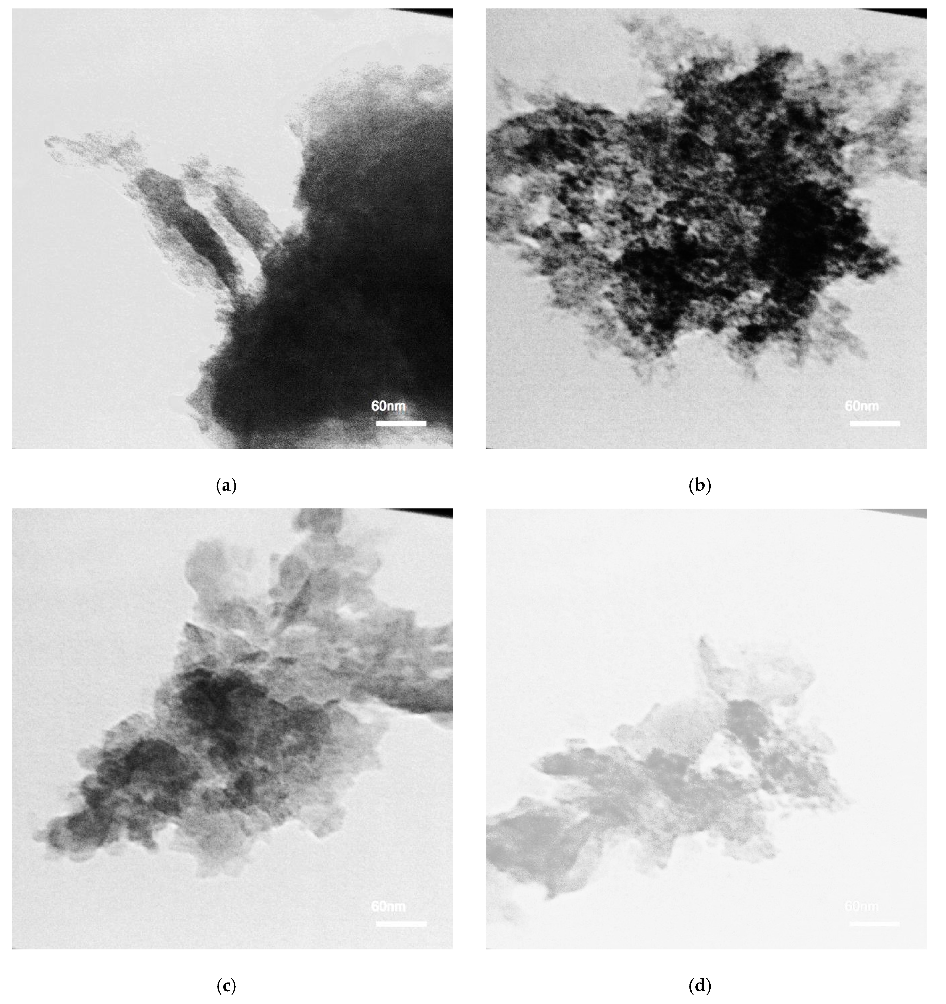
The TEM images of the MoS2 samples were performed to investigate the structural deviations that occur after various laser irradiation energies (Figure 4). Figure 4a is the image of MoS2 powder before laser irradiation. In Figure 4b, the large area could be distinguished for 40 mJ laser energy. By increasing laser irradiation energy, decreasing of the number of layers is obvious. Figure 4c shows the image after 60 mJ laser energy. Figure 4d shows few-layered MoS2 nanosheets with folding at the edge, which is a phenomenon that occurs in two-dimensional materials. The number of layers could be determined using this morphological deviation [43]. A single dark line at the folded edge could be related to a monolayer [44]. The TEM results, in this study, indicate that the prepared MoS2 is few-layered with uniform size distribution.
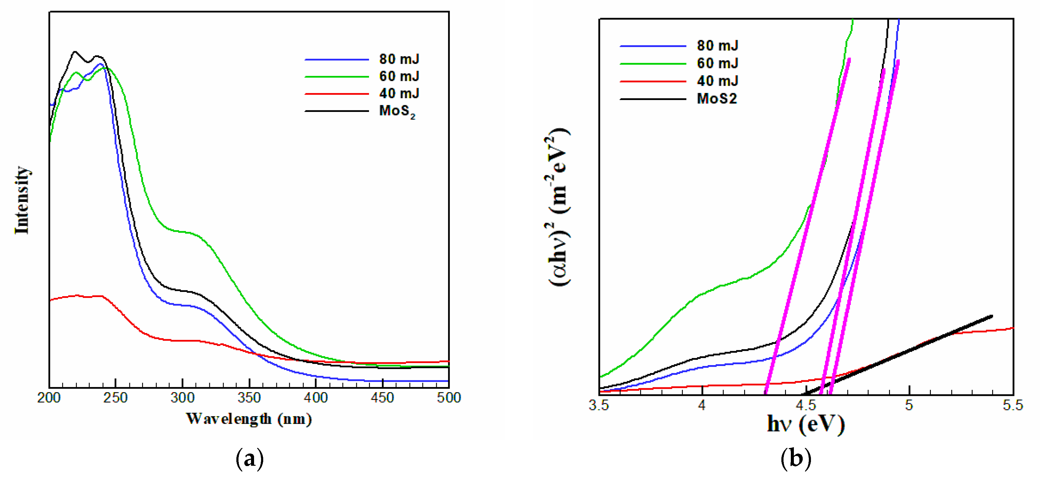
To investigate the effect of laser energy on the absorption evaluation of MoS2, the few-layered MoS2 nanosheets were examined via UV-Vis spectroscopic technique by dispersing in water as a reference. Figure 5a shows the room temperature UV-Vis absorption spectra of the few-layered MoS2 nanosheets before laser irradiation and after 15 min laser irradiation under 40, 60, and 80 mJ energies. The UV-Vis absorption spectrum of MoS2 solution displays a clear broad peak located at 250 nm ascribed to the excitonic property of MoS2 nanosheets [45]. It is different from the 2D MoS2 counterpart with large lateral dimensions, which display four peaks at 340, 430, 590, and 650 nm as a characteristic absorption band of the original bulk MoS2. For bulk MoS2, the peaks at 590 and 650 nm are ascribed to the K point of the Brillouin zone, and the peaks at 340 and 430 nm are ascribed to the direct transition from the deep valence band to the conduction band [26,46]. Nonetheless, these four characteristic absorption bands fade in the spectra of the as-prepared MoS2 nanosheets. Only one peak is detected in the near-UV region (λ < 300 nm) ascribed to the excitonic property of MoS2 [47]. The strong blue shift in the optical absorption is ascribed to the quantum confinement and edge effects [47,48] upon the reduction of the lateral size of the MoS2 nanosheets to <50 nm. Thus, quantum confinement in layered d-electron materials such as MoS2 paves the way to engineer the electronic structure of matter at the nanoscale. As the TEM results show, most of the as-prepared MoS2 nanosheets are ~30 nm and a strong blue shift is detected; this finding is in a good agreement with the results reported elsewhere [49,50].
The absorption spectra of all four samples exhibit shoulders ranging from about 280 nm to 340 nm attributed to the interband transitions from the occupied dz2 orbital to unoccupied dxy, dyz, dxz, and dx2–y2 orbitals [51]. From the UV-Vis data, it is possible to calculate the optical energy bandgap values of materials with a direct allowed bandgap by plotting the graph of (αhν)2 versus hν, according to the Tauc relation, (αhν)n = A (hν − Eg). In the Tauc equation, α is the absorption coefficient calculating from UV-Vis data according to the Beer–Lambert formula, hν is the photon energy in eV, Eg is the bandgap energy, and A is a constant. The symbol “n” indicates the nature of the electronic transition causing the absorption and can have the values 2, 1/2, 2/3, and 1/3 if the electronic transition is direct allowed, indirect allowed, direct forbidden, and indirect forbidden transitions, respectively. The extrapolation of the linear part of the UV-Vis spectrum to the point in which (αhν)2 = 0, gives the optical energy bandgap of the samples (Figure 5b). The calculated bandgap energies are greater than that of monolayer MoS2 (1.9 eV) and bulk MoS2 (1.2 eV) [26,52,53] and comparable to that of described monolayer MoS2 quantum dots (4.96 eV) [54]. The data are collected in Table 1. The slight difference in bandgap is ascribed to the size of nanosheets.
Conventional semiconductors like silicon have a bandgap in the range 1−1.5 eV, whereas wide bandgap (WBG) materials have bandgaps in the range 2−6 eV such as SiC, GaN, and diamond [55,56]. Among them, gallium oxide (Ga2O3) has the largest band gap (~4.66 eV) showing the widest tunable spectral range as compared with those of the other WBG semiconductors. The MoS2 nanosheets prepared in this study, are in the range of WBG semiconductors. Among the samples, the one under 80 mJ laser energy has the widest bandgap (4.7 eV). Therefore, it is suitable to use in optoelectronic and electronic devices.
The MoS2 nanostructure has specific photoluminescence capabilities due to the quantum size effect [57,58]. By thinning the MoS2 crystal to a monolayer, strong photoluminescence appears showing an indirect to direct bandgap transition in this d-electron system [59]. Figure 6 shows the PL spectrum of MoS2 nanosheets solution excited at 400 nm. A strong sharp peak at 530 nm as a typical fluorescence spectrum of MoS2 particles with nanometer scale lateral dimensions is observed [60]. The PL peak at 530 nm can be attributed to MoS2 nanosheets with a lateral dimension up to a few tens of nm [61]. This is also supported by the TEM images showing the formation of few-layered MoS2 nanosheets. The PL spectrum is different from the two distinguished peaks positioned at around 610 and 661 nm in 2D MoS2 flakes with large lateral dimensions from the direct bandgap of the K point [26]. It is remarkably hard to explain a shift in the PL of the samples because many factors such as strain [62] or quantum confinement [63] have an effect on the shift. This phenomenon could also be caused by the presence of several trap states [52,64]. It is noticeable, after laser irradiation, there is a redshift in the PL peak of the samples and its intensity decreases. This property is attributed to the existence of polydispersity of MoS2 nanosheets [46,65], the hot PL from Brillouin zone’s K point, and many trap states in MoS2 nanosheets as detected in graphene quantum dots [65]. Reduction of the intensity means a reduction in electron-hole recombination.
The bandgap calculated by PL emission is always less than the original bandgap and is not the exact bandgap. The PL emission is mostly affected by the polarity of the solvent system and the changing solvent polarity causes a shift for the peaks. However, the absorption spectrum is much less affected by change in solvent polarity. Therefore, the bandgap is calculated from the absorbance spectrum. Bao et al. reported the change in PL peak position by the changing in excitation wavelength [66]. The PL spectra of MoS2 were measured at various excitation wavelengths. By increasing the excitation wavelength, the PL emission peaks shift to longer wavelengths. This phenomenon can also be due to the presence of several trap states, defects, and quantum confinements in layered material such as MoS2 [67].
Researches on semiconductors, especially WBG semiconductors, have fabricated various power devices, which are reliable and performant enough to design high-efficiency level converters to match application requirements. The WBG semiconductors are significant scientifically and technologically, and they have become the main materials to be applied in high-performance optoelectronic and electronic devices. The WBG semiconductors have many benefits for optoelectronic and electronic purposes because of their large bandgap energy and they help devices operate at much higher temperatures, voltages, and frequencies [68]. The power electronic modules make by these materials are considerably more powerful and energy efficient as compared with those made from conventional semiconductor materials [69]. They are becoming popular in high voltage and high power applications such as the traction motors and inverters found in the automotive sector [70]. The nature of WBG energy is appropriate for absorbing or emitting ultraviolet (UV) light in optoelectronic devices. For optical absorption and emission of the UV lights, WBG materials are significantly involved. The prepared WBG few-layered MoS2 nanosheets, in this study, are suitable for the mentioned applications.

4. Conclusions

A new, quick, green, and one-step method was introduced for preparing few-layered MoS2 nanosheets by applying the laser irradiation. Uniform size distribution and a high production rate of few-layered MoS2 nanosheets were achieved by solely adjusting the laser energy. Samples under 40, 60, and 80 mJ laser energy were fabricated. The calculation displayed the widest bandgap, i.e., 4.7 eV, was for the sample under 80 mJ laser energy. The as-prepared MoS2 nanosheets exert strong quantum confinement and edge effects. MoS2 nanosheets are a type of WBG semiconductor having minor, quicker, more reliable power electronic components with high efficiency. Increasing the temperature reduces the bandgap of a semiconductor, thereby affecting most of the semiconductor material parameters. The spacing of the electronic levels and the bandgap increases with decreasing particle size, therefore, they are more compact in size and, as a result, less design margin is required to allow for temperature variation, allowing designers to achieve better performance from these devices. To the best of our knowledge, this is the first time to apply this method to exfoliate the MoS2 sheets.

Author Contributions

Conceptualization, S.K.; methodology, S.K. and M.M.; Validation, S.K. and M.M.; formal analysis, M.M.; investigation, S.K. and M.M.; Resources, M.M.; writing—original draft preparation, S.K. and F.A.; writing—review and editing, S.K. and F.A.; Visualization, S.K. and F.A.; supervision, S.K.; project administration, S.K. All authors have read and agreed to the published version of the manuscript.

Funding

This research received no external funding.

Conflicts of Interest

The authors declare no conflict of interest.

References

  1. Néstor, P.; Elías, A.L.; Berkdemir, A.; Castro-Beltran, A.; Gutiérrez, H.R.; Feng, S.; Lv, R.; Hayashi, T.; López-Urías, F.; Ghosh, S.; et al. Photosensor device based on few-layered WS2 films. Adv. Funct. Mater. 2013, 23, 5511–5517. [Google Scholar]
  2. Zhao, G.; Li, J.; Ren, X.; Chen, C.; Wang, X. Few-layered graphene oxide nanosheets as superior sorbents for heavy metal ion pollution management. Environ. Sci. Technol. 2011, 45, 10454–10462. [Google Scholar]
  3. Jun, F.; Sun, X.; Wu, C.; Peng, L.; Lin, C.; Hu, S.; Yang, J.; Xie, Y. Metallic few-layered VS2 ultrathin nanosheets: High two-dimensional conductivity for in-plane supercapacitors. J. Am. Chem. Soc. 2011, 133, 17832–17838. [Google Scholar]
  4. Jafari, M.; Asadpour, M.; Majelan, N.A.; Faghihnasiri, M. Effect of boron and nitrogen doping on electro-optical properties of armchair and zigzag graphyne nanoribbons. Comput. Mater. Sci. 2014, 82, 391–398. [Google Scholar] [CrossRef]
  5. Ke, Q.; Wang, J. Graphene-based materials for supercapacitor electrodes—A review. J. Mater. 2016, 2, 37–54. [Google Scholar]
  6. Francesco, B.; Bartolotta, A.; Coleman, J.N.; Backes, C. 2D-crystal-based functional inks. Adv. Mater. 2016, 28, 6136–6166. [Google Scholar]
  7. Bo, L.; Zhou, L.; Wu, D.; Peng, H.; Yan, K.; Zhou, Y.; Liu, Z. Photochemical chlorination of graphene. ACS Nano 2011, 5, 5957–5961. [Google Scholar]
  8. Peng, G.; Song, H.; Chen, X. Electrochemical performance of graphene nanosheets as anode material for lithium-ion batteries. Electrochem. Commun. 2009, 11, 1320–1324. [Google Scholar]
  9. Yuan, H.; Wu, J.; Xu, X.; Ho, Y.; Ni, G.; Zou, Q.; Koon, G.K.W.; Zhao, W.; Neto, A.H.C.; Eda, G.; et al. An innovative way of etching MoS2: Characterization and mechanistic investigation. Nano Res. 2013, 6, 200–207. [Google Scholar]
  10. Li, L.; Yu, Y.; Ye, G.J.; Ge, Q.; Ou, X.; Wu, H.; Feng, D.; Chen, X.H.; Zhang, Y. Black phosphorus field-effect transistors. Nat. Nanotechnol. 2014, 9, 372. [Google Scholar]
  11. Yu, Y.; Li, C.; Liu, Y.; Su, L.; Zhang, Y.; Cao, L. Controlled scalable synthesis of uniform, high-quality monolayer and few-layer MoS2 films. Sci. Rep. 2013, 3, 1866. [Google Scholar]
  12. Novoselov Kostya, S.; Jiang, D.; Schedin, F.; Booth, T.J.; Khotkevich, V.V.; Morozov, S.V.; Geim, A.K. Two-dimensional atomic crystals. Proc. Nat. Acad. Sci. USA 2005, 102, 10451–10453. [Google Scholar] [CrossRef] [PubMed] [Green Version]
  13. Coleman Jonathan, N.; Lotya, M.; O’Neill, A.; Bergin, S.D.; King, P.J.; Khan, U.; Young, K.; Gaucher, A.; De, S.; Smith, R.J.; et al. Two-dimensional nanosheets produced by liquid exfoliation of layered materials. Science 2011, 331, 568–571. [Google Scholar] [CrossRef] [PubMed] [Green Version]
  14. Peter, M.; Khan, U.; Hughes, J.M.; Coleman, N.J. Role of solubility parameters in understanding the steric stabilization of exfoliated two-dimensional nanosheets by adsorbed polymers. J. Phys. Chem. C 2012, 116, 11393–11400. [Google Scholar]
  15. Eswaraiah, V.; Backes, C.; Paton, K.R.; Harvey, A.; Gholamvand, Z.; McCauley, J.; Coleman, J.N. Large-scale production of size-controlled MoS2 nanosheets by shear exfoliation. Chem. Mater. 2015, 27, 1129–1139. [Google Scholar]
  16. Liu, J.; Zeng, Z.; Cao, X.; Lu, G.; Wang, L.H.; Fan, Q.L.; Huang, W.; Zhang, H. Preparation of MoS2-polyvinylpyrrolidone nanocomposites for flexible nonvolatile rewritable memory devices with reduced graphene oxide electrodes. Small 2012, 8, 3517–3522. [Google Scholar]
  17. Chouhan Raghuraj, S.; Žitko, G.; Fajon, V.; Živković, I.; Pavlin, M.; Berisha, S.; Jerman, I.; Vesel, A.; Horvat, M. A Unique Interactive Nanostructure Knitting based Passive Sampler Adsorbent for Monitoring of Hg2+ in Water. Sensors 2019, 19, 3432. [Google Scholar] [CrossRef] [Green Version]
  18. Dong, Y.; Wang, Q.; Wu, H.; Chen, Y.; Lu, C.H.; Chi, Y.; Yang, H.H. Graphitic carbon nitride materials: Sensing, imaging and therapy. Small 2016, 12, 5376–5393. [Google Scholar]
  19. Tian, J.; Liu, Q.; Asiri, A.M.; Qusti, A.H.; Al-Youbi, A.O.; Sun, X. Ultrathin graphitic carbon nitride nanosheets: A novel peroxidase mimetic, Fe doping-mediated catalytic performance enhancement and application to rapid, highly sensitive optical detection of glucose. Nanoscale 2013, 5, 11604–11609. [Google Scholar]
  20. Manish, C.; Shin, H.S.; Eda, G.; Li, L.; Loh, K.P.; Zhang, H. The chemistry of two-dimensional layered transition metal dichalcogenide nanosheets. Nat. Chem. 2013, 5, 263. [Google Scholar]
  21. Fai, M.K.; Shan, J. Photonics and optoelectronics of 2D semiconductor transition metal dichalcogenides. Nat. Photonics 2016, 10, 216. [Google Scholar]
  22. Hua, W.Q.; Kalantar-Zadeh, K.; Kis, A.; Coleman, J.N.; Strano, M.S. Electronics and optoelectronics of two-dimensional transition metal dichalcogenides. Nat. Nanotechnol. 2012, 7, 699. [Google Scholar]
  23. Shi, H.; Yan, R.; Bertolazzi, S.; Brivio, J.; Gao, B.; Kis, A.; Jena, D.; Xing, H.G.; Huang, L. Exciton dynamics in suspended monolayer and few-layer MoS2 2D crystals. ACS Nano 2013, 7, 1072–1080. [Google Scholar]
  24. Erwin, P.; Gelato, L.; Chabot, B.; Penzo, M.; Cenzual, K.; Gladyshevskii, R. TYPIX Standardized Data and Crystal Chemical Characterization of Inorganic Structure Types; Springer: Berlin/Heidelberg, Germany, 2013. [Google Scholar]
  25. Lebegue, S.; Eriksson, O. Electronic structure of two-dimensional crystals from ab initio theory. Phys. Rev. B 2009, 79, 115409. [Google Scholar] [CrossRef] [Green Version]
  26. Fai, M.K.; Lee, C.; Hone, J.; Shan, J.; Heinz, T.F. Atomically thin MoS2: A new direct-gap semiconductor. Phys. Rev. Lett. 2010, 105, 136805. [Google Scholar]
  27. Shi, Y.; Zhou, W.; Lu, A.; Fang, W.; Lee, Y.; Hsu, A.L.; Kim, S.M.; Kim, K.K.; Yang, H.Y.; Li, L.; et al. van der Waals epitaxy of MoS2 layers using graphene as growth templates. Nano Lett. 2012, 12, 2784–2791. [Google Scholar]
  28. John, B.; Li, M.; Wang, S.; Wang, P.; Papakonstantinou, P. Electrocatalytic hydrogen evolution reaction on edges of a few layer molybdenum disulfide nanodots. ACS Appl. Mater. Interfaces 2015, 7, 14113–14122. [Google Scholar]
  29. Jacob, B.; Moses, P.G.; Jaramillo, T.F.; Nørskov, J.K.; Chorkendorff, I. Hydrogen evolution on nano-particulate transition metal sulfides. Faraday Discuss. 2009, 140, 219–231. [Google Scholar]
  30. Chen, L.; Zheng, X.; Du, Z.; Jia, B.; Gu, M.; Hong, M. A frozen matrix hybrid optical nonlinear system enhanced by a particle lens. Nanoscale 2015, 7, 14982–14988. [Google Scholar]
  31. Tsai, M.L.; Su, S.H.; Chang, J.K.; Tsai, D.S.; Chen, C.H.; Wu, C.I.; Li, L.J.; Chen, L.J.; He, J.H. Monolayer MoS2 heterojunction solar cells. ACS Nano 2014, 8, 8317–8322. [Google Scholar]
  32. Mariyappan, S.; Bansal, T.; Durcan, C.A.; Yu, B. Molybdenum disulphide/titanium dioxide nanocomposite-poly 3-hexylthiophene bulk heterojunction solar cell. Appl. Phys. Lett. 2012, 100, 153901. [Google Scholar]
  33. Ho, W.; Yu, J.C.; Lin, J.; Yu, J.; Li, P. Preparation and photocatalytic behavior of MoS2 and WS2 nanocluster sensitized TiO2. Langmuir 2004, 20, 5865–5869. [Google Scholar]
  34. Benavente, E.; Ana, M.A.S.; Mendizábal, F.; González, G. Intercalation chemistry of molybdenum disulfide. Coord. Chem. Rev. 2002, 224, 87–109. [Google Scholar] [CrossRef]
  35. Lee, Y.S.; Zhang, X.Q.; Zhang, W.; Chang, M.T.; Lin, C.T.; Chang, K.D.; Yu, Y.C.; Wang, J.T.; Chang, C.S.; Li, L.J.; et al. Synthesis of large-area MoS2 atomic layers with chemical vapor deposition. Adv. Mater. 2012, 24, 2320–2325. [Google Scholar]
  36. Xie, J.; Zhang, H.; Li, S.; Wang, R.; Sun, X.; Zhou, M.; Zhou, J.; Lou, X.W.; Xie, Y. Defect-rich MoS2 ultrathin nanosheets with additional active edge sites for enhanced electrocatalytic hydrogen evolution. Adv. Mater. 2013, 25, 5807–5813. [Google Scholar]
  37. Afanasiev, P. Synthetic approaches to the molybdenum sulfide materials. Comptes Rendus Chim. 2008, 11, 159–182. [Google Scholar] [CrossRef]
  38. Bindhu, B.; Sharu, B.K.; Gopika, M.S.; Praseetha, P.K.; Veluraja, K. Molybdenum disulfide nanoflakes through Li-AHA assisted exfoliation in an aqueous medium. RSC Adv. 2016, 6, 22026–22033. [Google Scholar] [CrossRef]
  39. Hung, P.V.; Kim, K.; Jung, D.; Singh, K.; Oh, E.; Chung, J.S. Liquid phase co-exfoliated MoS2–graphene composites as anode materials for lithium ion batteries. J. Power Sources 2013, 244, 280–286. [Google Scholar]
  40. Kumar, K.; Santhana, A.; Jiang, S.; Warchoł, J.K. Synthesis and characterization of two-dimensional transition metal dichalcogenide magnetic MoS2@ Fe3O4 nanoparticles for adsorption of Cr (VI)/Cr (III). ACS Omega 2017, 2, 6187–6200. [Google Scholar] [CrossRef]
  41. Khawula Tobile, N.Y.; Raju, K.; Franklyn, P.J.; Sigalas, I.; Ozoemena, K.I. The effects of morphology re-arrangements on the pseudocapacitive properties of mesoporous molybdenum disulfide (MoS2) nanoflakes. J. Electrochem. Soc. 2016, 163, A1927–A1935. [Google Scholar] [CrossRef] [Green Version]
  42. Tobias, K.; Heydrich, S.; Hirmer, M.; Schmutzler, J.; Schüller, C. Low-temperature photocarrier dynamics in monolayer MoS2. Appl. Phys. Lett. 2011, 99, 102109. [Google Scholar]
  43. Meyer Jannik, C.; Geim, A.K.; Katsnelson, M.I.; Novoselov, K.S.; Booth, T.J.; Roth, S. The structure of suspended graphene sheets. Nature 2007, 446, 60. [Google Scholar] [CrossRef] [PubMed]
  44. Sagar, P.; Harle, A.; Sathaye, S.; Patil, K. Development of a novel method to grow mono-/few-layered MoS 2 films and MoS2—Graphene hybrid films for supercapacitor applications. CrystEngComm 2014, 16, 10845–10855. [Google Scholar]
  45. Dong, H.H.; Han, D.J.; Choi, J.S.; Park, M.; Seo, T.S. Dual role of blue luminescent MoS2 quantum dots in fluorescence resonance energy transfer phenomenon. Small 2014, 10, 3858–3862. [Google Scholar]
  46. Deepesh, G.; Damien, D.; Shaijumon, M.M. MoS2 quantum dot-interspersed exfoliated MoS2 nanosheets. ACS Nano 2014, 8, 5297–5303. [Google Scholar]
  47. Chikan, V.; Kelley, D.F. Size-dependent spectroscopy of MoS2 nanoclusters. J. Phys. Chem. B 2002, 106, 3794–3804. [Google Scholar] [CrossRef]
  48. Jaramillo Thomas, F.; Jørgensen, K.P.; Bonde, J.; Nielsen, J.H.; Horch, S.; Chorkendorff, I. Identification of active edge sites for electrochemical H2 evolution from MoS2 nanocatalysts. Science 2007, 317, 100–102. [Google Scholar] [CrossRef] [Green Version]
  49. Xi, S.; Li, D.; Wu, P. One-pot, facile, and versatile synthesis of monolayer MoS2/WS2 quantum dots as bioimaging probes and efficient electrocatalysts for hydrogen evolution reaction. Adv. Funct. Mater. 2015, 25, 1127–1136. [Google Scholar]
  50. Dai, W.; Dong, H.; Fugetsu, B.; Cao, Y.; Lu, H.; Ma, X.; Zhang, X. Tunable fabrication of molybdenum disulfide quantum dots for intracellular microRNA detection and multiphoton bioimaging. Small 2015, 11, 4158–4164. [Google Scholar]
  51. Bojana, V.; Dominko, R.; Gunde, M.K.; Hauptman, N.; Skapin, S.D.; Remskar, M. Optical properties of exfoliated MoS2 coaxial nanotubes-analogues of graphene. Nanoscale Res. Lett. 2011, 6, 593. [Google Scholar]
  52. Václav, Š.; Henych, J. Strongly luminescent monolayered MoS2 prepared by effective ultrasound exfoliation. Nanoscale 2013, 5, 3387–3394. [Google Scholar]
  53. Hao, H.; Du, C.; Shi, H.; Feng, X.; Li, J.; Tan, Y.; Song, W. Water-Soluble Monolayer Molybdenum Disulfide Quantum Dots with Upconversion Fluorescence. Part. Part. Syst. Charact. 2015, 32, 72–79. [Google Scholar]
  54. Ren, X.; Pang, L.; Zhang, Y.; Ren, X.; Fan, H.; Liu, S.F. One-step hydrothermal synthesis of monolayer MoS2 quantum dots for highly efficient electrocatalytic hydrogen evolution. J. Mater. Chem. A 2015, 3, 10693–10697. [Google Scholar]
  55. Prestopino, G.; Marinelli, M.; Milani, E.; Verona, C.; Verona-Rinati, G. Transient lateral photovoltaic effect in synthetic single crystal diamond. Appl. Phys. Lett. 2017, 111, 143504. [Google Scholar] [CrossRef]
  56. Eva, M.; Omnès, F.; Calle, F. Wide-bandgap semiconductor ultraviolet photodetectors. Semicond. Sci. Technol. 2003, 18, R33. [Google Scholar]
  57. Andrea, S.; Sun, L.; Zhang, Y.; Li, T.; Kim, J.; Chim, C.; Galli, G.; Wang, F. Emerging photoluminescence in monolayer MoS2. Nano Lett. 2010, 10, 1271–1275. [Google Scholar]
  58. Yafei, L.; Zhou, Z.; Zhang, S.; Chen, Z. MoS2 nanoribbons: High stability and unusual electronic and magnetic properties. J. Am. Chem. Soc. 2008, 130, 16739–16744. [Google Scholar]
  59. Wei, L.; Birdwell, A.G.; Amani, M.; Burke, R.A.; Ling, X.; Lee, Y.; Liang, X.; Peng, L.; Richter, C.A.; Kong, J.; et al. Broadband optical properties of large-area monolayer CVD molybdenum disulfide. Phys. Rev. B 2014, 90, 195434. [Google Scholar]
  60. Subhrajit, M.; Maiti, R.; Midya, A.; Das, S.; Ray, S.K. Tunable direct bandgap optical transitions in MoS2 nanocrystals for photonic devices. ACS Photonics 2015, 2, 760–768. [Google Scholar]
  61. Wu, J.Y.; Zhang, X.Y.; Ma, X.D.; Qiu, Y.P.; Zhang, T. High quantum-yield luminescent MoS2 quantum dots with variable light emission created via direct ultrasonic exfoliation of MoS2 nanosheets. RSC Adv. 2015, 5, 95178–95182. [Google Scholar]
  62. Shi, H.; Pan, H.; Zhang, Y.W.; Yakobson, B.I. Quasiparticle band structures and optical properties of strained monolayer MoS2 and WS2. Phys. Rev. B 2013, 87, 155304. [Google Scholar]
  63. Miller, D.A.B.; Chemla, D.S.; Schmitt-Rink, S. Relation between electroabsorption in bulk semiconductors and in quantum wells: The quantum-confined Franz-Keldysh effect. Phys. Rev. B 1986, 33, 6976. [Google Scholar] [CrossRef] [PubMed]
  64. Shinde Dhanraj, B.; Vijayamohanan, K.P. Electrochemical preparation of luminescent graphene quantum dots from multiwalled carbon nanotubes. Chem. Eur. J. 2012, 18, 12522–12528. [Google Scholar] [CrossRef]
  65. Deepesh, G.; Damien, D.; Li, B.; Gullappalli, H.; Pillai, V.K.; Ajayan, P.M.; Shaijumon, M.M. Electrochemical synthesis of luminescent MoS2 quantum dots. Chem. Commun. 2015, 51, 6293–6296. [Google Scholar]
  66. Bao, L.; Zhang, Z.; Tian, Z.; Zhang, L.; Liu, C.; Lin, Y.; Qi, B.; Pang, D. Electrochemical tuning of luminescent carbon nanodots: From preparation to luminescence mechanism. Adv. Mater. 2011, 23, 5801–5806. [Google Scholar] [CrossRef]
  67. Bo, L.; Li, X.; Ran, P.; Zuo, P.; Wang, A.; Qu, L.; Zhao, Y.; Cheng, Z.; Lu, Y. Preparation of monolayer MoS2 quantum dots using temporally shaped femtosecond laser ablation of bulk MoS2 targets in water. Sci. Rep. 2017, 7, 1–12. [Google Scholar]
  68. Ahmed, E.; Chow, T.P. Silicon carbide benefits and advantages for power electronics circuits and systems. Proc. IEEE 2002, 90, 969–986. [Google Scholar]
  69. Burak, O.; Tolbert, L.M. Comparison of Wide-Bandgap Semiconductors for Power Electronics Applications; Department of Energy: Washington, DC, USA, 2004. [Google Scholar] [CrossRef] [Green Version]
  70. John, R.; Dorn-Gomba, L.; Mak, C.; Emadi, A. Automotive traction inverters: Current status and future trends. IEEE Trans. Veh. Technol. 2019, 68, 3337–3350. [Google Scholar]
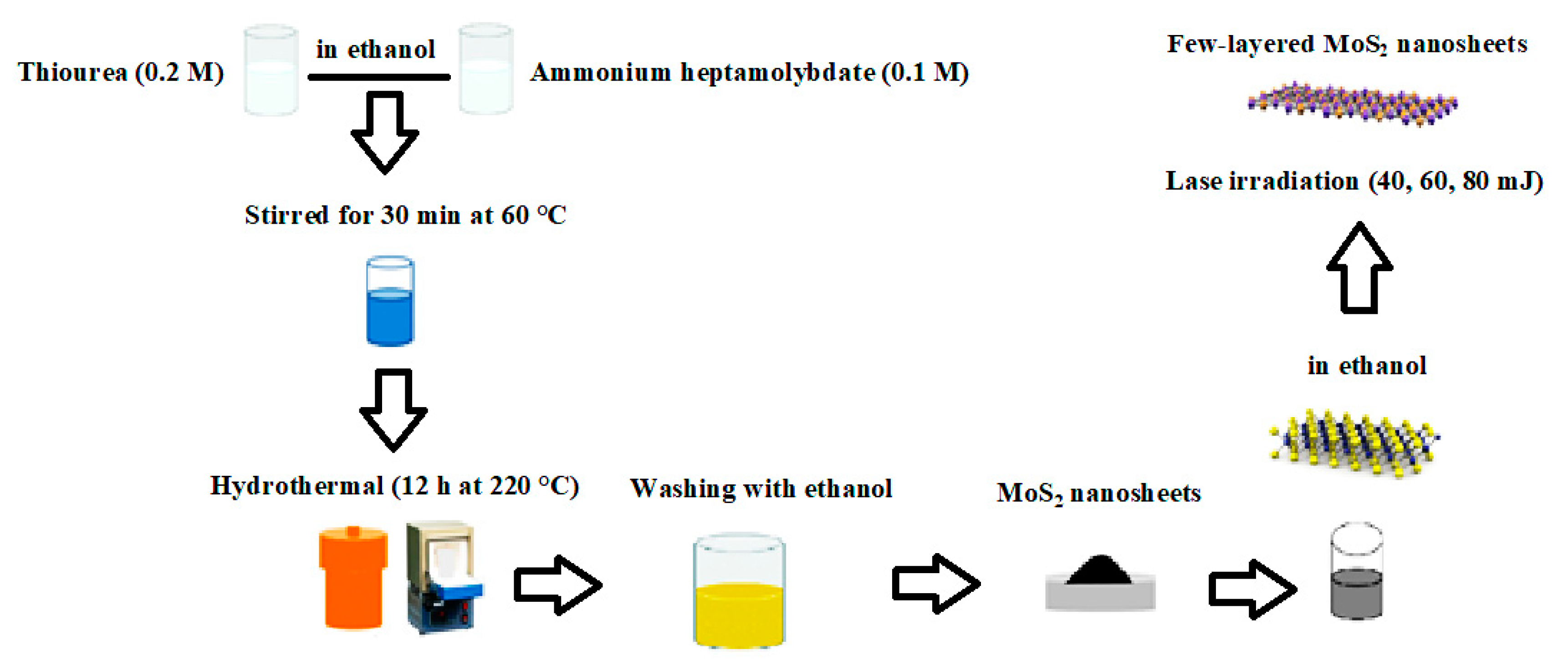
Figure 1. Flowchart of the synthesis process of the few-layered molybdenum disulfide (MoS2) nanosheets.
Crystals 10 00164 g001
Figure 2. X-ray diffraction (XRD) pattern of the MoS2 structures under various laser energies from 40 to 80 mJ.
Crystals 10 00164 g002
Figure 3. Fourier transform infrared spectroscopy (FTIR) spectrum of the MoS2 structures under various laser energies from 40 to 80 mJ.
Crystals 10 00164 g003
Figure 4. Transmission electron microscopy (TEM) images of (a) MoS2 powder, few-layered MoS2 nanosheets after (b) 40 mJ; (c) 60 mJ; and (d) 80 mJ laser energy (the scale for all images is 60 nm).
Crystals 10 00164 g004
Figure 5. UV-Vis absorption spectrum of few-layered MoS2 nanosheets under various laser energies from 40 to 80 mJ.
Crystals 10 00164 g005
Figure 6. Room temperature photoluminescence spectra of the few-layered MoS2 under various laser energies from 40 to 80 mJ.
Crystals 10 00164 g006
Table 1. Bandgap calculation from Figure 6 (error 0.05 eV).
SampleMoS240 mJ60 mJ80 mJ
Bandgap (eV)4.64.54.34.7