Article

Crystal Structure, Synthesis and Luminescence Sensing of a Zn(II) Coordination Polymer with 2,5-Dihydroxy-1,4-Terephthalic Acid and 2,2′-Bipyridine as Ligands

1
Department of Chemistry, Jilin Normal University, Siping 136000, China
2
Key Laboratory of Preparation and Application of Environmental Friendly Materials, Ministry of Education, Jilin Normal University, Changchun 130103, China
*
Author to whom correspondence should be addressed.
Crystals 2020, 10(12), 1105; https://doi.org/10.3390/cryst10121105
Submission received: 23 October 2020 / Revised: 21 November 2020 / Accepted: 28 November 2020 / Published: 3 December 2020

Abstract

:
A new coordination polymer was synthesized under hydrothermal conditions, namely [Zn(Bpy)(DHTA)0.5]n (DHTA = 2,5-dihydroxy-1,4-terephthalic acid tetra-anion, Bpy = 2,2’-bipyridine). There is one Zn(II) ion in [Zn(Bpy)(DHTA)0.5]n, the metal center Zn(II) is five-coordinated and forms a slightly twisted square pyramidal geometry. It remains stable at temperatures as high as 380 °C, and a sharp drop in weight is found in the temperature range of 380–570 °C. The calculation results show that the residual is zinc(II) oxide (ZnO). The emission peak of the complex appears at 365 nm (λex = 366 nm). The photoluminescence results suggest that the complex has potential as a new luminescence material. At the same time, in luminescence explorations experiments, the complex showed a high degree of selectivity and sensitivity to organic solvents, acetone (DMK) and metal ions (Fe3+).

Graphical Abstract

1. Introduction

In recent decades, metal organic frameworks (MOF) and coordination polymers (CPs) have become attractive for academic and industrial fields, due to their luminescence [1,2,3,4,5,6], electrical conductivity [7], chemical sensing [8,9,10,11,12], magnetic [13], catalytic activity [14,15], gas storage or separation [16,17,18,19,20] and other applications leading to them having different functional characteristics in economic, technical and environmental aspects. In particular, the luminescent MOF has proved its ability to detect metal ions and organic pollutants efficiently and sensitively. For example, two zinc(II) coordination complexes with excellent water stability and strong luminescence were reported by Zhang et al., and their application in sensing/detection in water systems was further explored [21]. In recent years, Zhang and co-workers have synthesized a good luminescent zinc(II) coordination complex sensor that is beneficial to the environment and has a wide range of applications for detecting environmental pollutants in organic solutions or water [22]. Rosales-Vázquez et al. have successfully synthesized a new type of luminescent water-stabilized zinc complex, which has excellent photoluminescence chemical sensing ability for cyanide ions in sewage [23].
Nowadays, a host of MOFs are widely known for their applications in the examination of organic and inorganic molecules and metal ions, and their excellent performance has also attracted widespread attention [24,25]. The content of iron in the Earth’s crust ranks second, it is a critical trace element that exists in biological systems. Supposing that the human body lacks iron, this may cause abnormalities in the nervous system, digestive system and immune system. By contrast, excessive iron can cause tumors and heart arteriosclerosis. Therefore, the detection of iron ions is particularly important [26]. In recent years, as a global environmental problem, water pollution has attracted wide attention, and organic pollution is one of the most serious aspects [27,28,29]. Among them, the detection of acetone is particularly important. Therefore, it is very important to detect the organic pollutants selectively [30]. Transition metal ions, a typical example of which is Zn(II), can not only produce novel structures, but also various complexes capable of detecting and identifying inorganic metal cation and organic solvent molecules. A typical example was reported by Jin and Jin and colleagues, a Zn(II) luminescent MOF with double detecting capabilities for Fe2+, Ag+, Cu2+ and Fe3+ ions and nitro-aromatic compounds [31]. Lv et al. constructed an amino-modified MOF (NH2-Zn-MOF) with excellent sensitivity and selectivity to Cr(VI), Cr(III) ions and nitro-aromatic complexes [32].
Even if a host of Zn complexes have been reported, but there are still great difficulties in designing and synthesizing novel structures. 2,2’-bipyridine is the most common chelated nitrogen-containing ligand, 2,5-dihydroxyterephthalic acid has several coordination modes (in Scheme 1). In mode A, possessing an ortho-carboxy-phenol motif, it adopts single dentate bridging mode and bidentate chelate manner (of the carboxylate and hydroxy); in this case, the monodentate carboxylate oxygen atom in the motif is connected to the zinc center in the bridging mode, and it can form the pentacoordinate (ZnN2O3) geometry configuration. Zhu et al. researched a Zn(II) 2,5-dihydroxyterephthalic acid complex and discussed the emission of the Zn(II) complex [33]. Our previous work also investigated the luminescent property [34], and in this work we will further intensively study the luminescence of zinc complex.
In this paper, we use the hydrothermal method to conduct experiments, we designed and synthesized a new type of Zn(II) complex. The Fe3+ ions and acetone (DMK) molecules was detected by the complex and displayed high selectivity and sensitivity in a luminescence investigation, which demonstrates that the application of complex 1 in a luminescent sensor has great potential. Through overall observation, complex 1 has an attractive prospect for luminescent material in the chemical industry.

2. Experimental

2.1. Materials and Method

The chemicals and solvents used in this experiment were of analytical purity, and no further purification was performed before use.

2.2. Characterization

An Rigaku AFC12 X-ray diffractometer (RIGAKu, Tokyo, Japan) was used to collect the diffraction data, with graphite monochromatic Mo Kα (λ = 0.71073 nm) radiation at 22.85 °C in the phi and Ω scanning modes. The C, O, N, H elemental analyses were preformed on a Perkin-Elmer 2400 elemental analyzer. The luminescence spectrum in the solid state at 25 °C were recorded on a Hitachi F-4600 luminescence spectrophotometer (Hitachi High-Tech Corporation, Tokyo, Japan). The sample of the complex was locked between two glass slides, and the scanning wavelength during measurement was 20 nm/s. The infrared (IR) spectrum of the complex was collected by a Shimadzu IR-408 spectrophotometer (Shimadzu Corporation, Kyoto, Japan) in the range of 4000–400 cm−1 (using KBr particles). The thermal stability experiment was conducted on a TG SDT2960 (TA Instruments, New Castle, DE, USA) thermal analyzer at a rate of 10 °C per minute under the action of nitrogen from 50 °C to 800 °C. The Rigaku model RINT Ultima III diffractometer (RIGAKu, Tokyo, Japan) was used for powder X-ray diffraction (PXRD) data collection.

2.3. Synthesis of [Zn(Bpy)(DHTA)0.5]n

The [Zn(Bpy)(DHTA)0.5]n was synthesized by a hydrothermal method: a mixture of Zn(CH3COO)2·2H2O (110 mg, 1 mmol), Bpy (156 mg, 1 mmol), DHTA (99 mg, 0.5 mmol) and 10 mL ultra-pure water and stirred for 0.5 hour to obtain a homogeneous system at 25 °C. Then 5 mL of NaOH solution (2.5 M) was added to the solution and agitated for 0.5 hour. The resulted solution was poured into a 25 mL autoclave. The container was pressure-tight and heated to 145 °C for lasted 96 h, and the reaction mixture is slowly cooled to 25 °C at a cooling rate of 5 °C per hour and the resulted product was washed with distilled water for 3 times, obtained dark red block-shaped crystals. Yield 67.3% (based on Zn(CH3COO)2·2H2O). Anal. Calcd for C14H9N2O3Zn (%): C, 52.78; H, 2.85; N, 8.79; O, 15.06. Found: C, 52.69; H, 2.99; N, 8.72; O, 15.13. Fourier transform infrared (FT-IR, KBr pellets, cm−1): 1746(w), 1609(s), 1590(s), 1500(w), 1450(m), 1408(s), 1398(m), 1253(w), 1206(m), 808(m), 480(w), as shown in Figure S1 (in Supplementary Materials).

2.4. Luminescence Sensing Experiments

The complex power (3.0 mg) was soaked in a 10−2 M M(NO3)x H2O solution, then sonicated for 3 h to become inorganic metal cation loaded 1@M (1 = the complex) solid powder. Then the complex was immersed in different concentrations of Fe3+ in aqueous solution. The aqueous solutions of M(NO3)x were stood at 25 °C for 72 hours were prepared for luminescence measurements (Mx+ = Na+, Mg2+, Cd2+, Ni2+, Hg2+, Co2+, K+, Zn2+, Li+, Pb2+, Al3+, Fe3+).
The complex powder (3.0 mg) was soaked in different kinds of pure organic solvent, such as acetonitrile (ACE), petroleum ether (PE), isopropanol (IPA), ethyl acetate (EAC), dimethylacetamide (DMA), dimethyl sulfoxide (DMSO), dimethylformamide (DMF), ethanol (ET), tetrahydrofuran (THF), and dimethyl ketone (DMK) (5 mL), respectively. Then the sample was treated with ultrasonication for 30 min and kept for 72 h to form a uniform liquid and, next, the luminescence research was carried out.

2.5. X-ray Crystallography

A dark red crystal with dimensions of 0.27 × 0.25 × 0.20 mm3 was mounted on an X-ray diffractometer. The crystal data collection was performed at room temperature. The total number of 7960 reflections were collected in the range of 2.399 to 27.116° using ω scans, of which 2636 were unique with Rint = 0.0150. The crystal structure was solved using the SHELXL-2018 (Sheldrick, 2018) software by direct methods, and then refined with full matrix least-square methods implemented in the WINGX package based on F2 (SHELX-2018 program) [35]. All non-hydrogen atoms were refined anisotropically, and all the hydrogen atoms were located from Fourier maps. The crystal data for the complex are listed in Table S1, and selected bond lengths and angles of the complex are given in Table S2. (CCDC (Cambridge Crystallographic Data Centre) no.: 2008409).

3. Results and Discussions

3.1. Structure Description

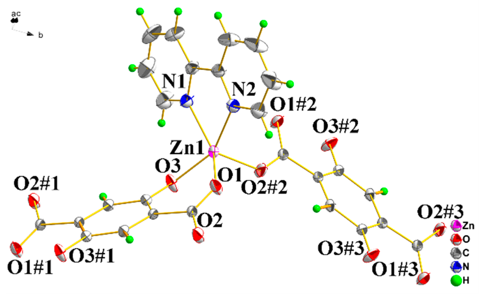
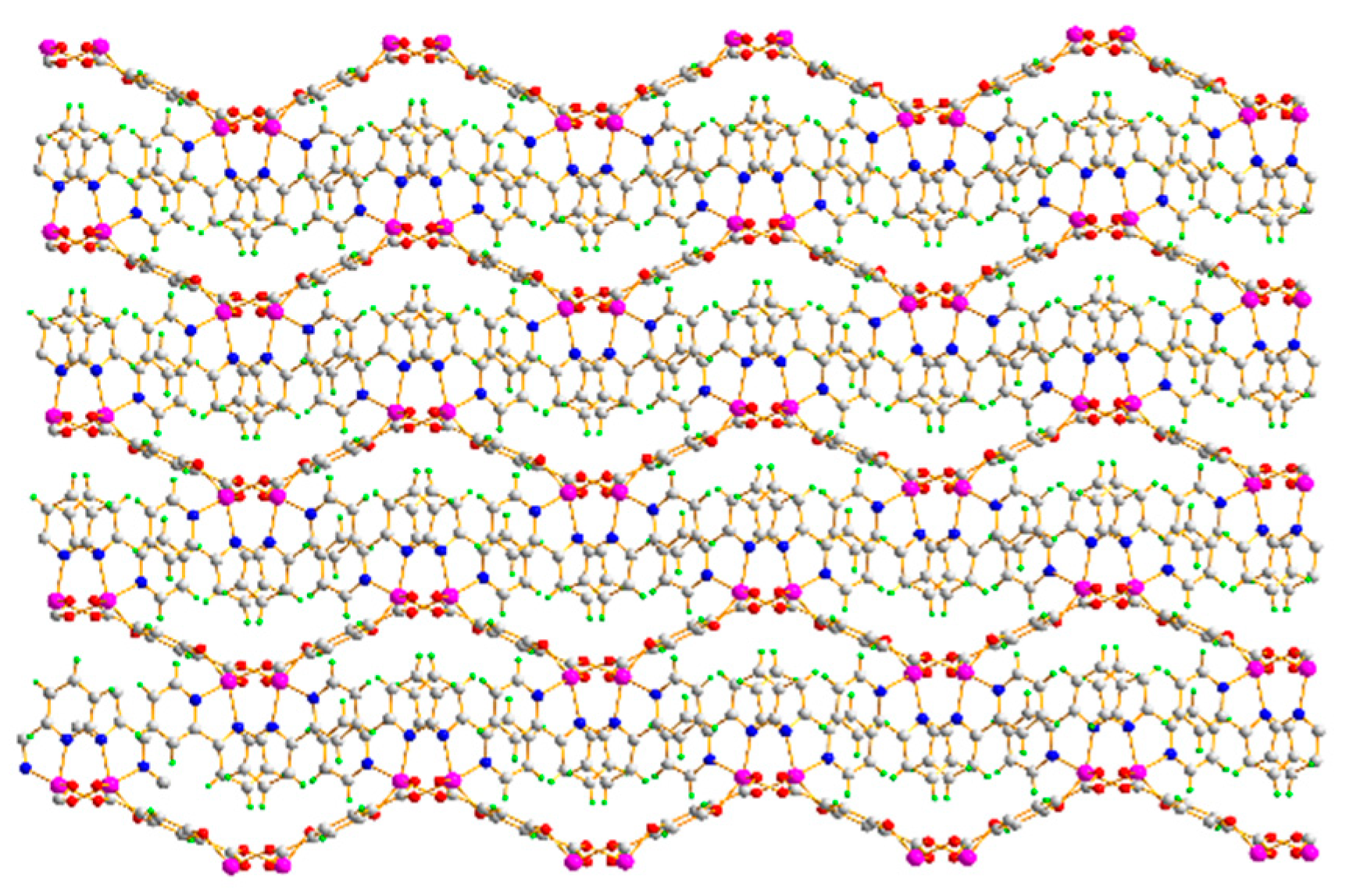
Single-crystal X-ray diffraction analysis demonstrates that the complex is a mononuclear species, and it belongs to the monoclinic crystal system and the space group is P21/c. As displayed in Figure 1, the metal ion Zn(II) is coordinated by two DHTA anions and one Bpy ligand in a highly distorted square planar pyramidal geometry (Figure S2). The metal is chelated by one carboxylate oxygen atom (O1) and one hydroxy oxygen atom (O3) from the deprotonated DHTA, and coordinated by a monodentate carboxylate oxygen atom (O2#2) of a symmetry related DHTA, as well as by the Bpy nitrogen atoms (N1, N2) to give a {O3N2} donor set. Bpy is coordinated with the metal ion Zn(II) in a chelating manner to form a five-membered ring, as commonly observed. Data of Table S2 indicate that the Zn-O bond lengths range from 1.9508(11) to 2.0761(12) Å. The Zn-N bond lengths are of 2.357(14) Å and 2.1564(14) Å. The coordination bond angles indicate large distortions from the ideal square planar pyramidal geometry, the values in the O(1)-Zn(1)-N(1) and O(3)-Zn(1)-N(2) values in the basal plane being of 143.91(5) and 150.67(5). Since the DHTA anions act as chelating and bridging ligands, they form a polymeric chain through weak hydrogen bonds C(2)-H···O(3)#4 (at -x, -y, -z+2) (Figure 2, Table S3) to form a two-dimensional layer (Figure 3).

3.2. Powder X-ray Diffraction (PXRD) Analysis

First, the powder X-ray diffraction (PXRD) mode of the complex was performed at 25 °C (Figure S3) to examine the phase purity of the complex for the purpose of luminescence measurement. The single phase of the corresponding material can be seen from matching the peak position of the as-synthesized PXRD pattern with the simulated peak pattern.

3.3. Stability Properties of the Complex

A thermal gravimetric analysis (TGA) of the complex was performed to verify its thermal stability. In a N2 atmosphere, we slowly increased the temperature at a heating rate of 10 °C per minute to test the sample. It can be observed in Figure S3, that the complex can remain stable at a temperature as high as 380 °C, and the weight of the complex suddenly decreases in the temperature range of 380–570 °C. When the temperature is between 50–800 °C, this can be attributable to the decomposition of organic ligands. Except for the ZnO residues, only a part of the complex lost weight. The weightlessness rate throughout the experiment was 75.67%, which was without obvious difference from the theoretical estimate of 74.58%. In summary, the complex exhibited excellent thermal stabilities. To prove the water stability of the complex, an immersion experiment was carried out. After soaked 1 in water for three days and dried it. Performed at the the powder X-ray diffraction (PXRD) mode of the complex at 25 °C, the result shows that the PXRD of the complex is not significantly different before and after the experiment (Figure S4), and it also shows that 1 has excellent water stability.

3.4. Luminescent Property of the Complex with High Sensitivity and Selectivity for Acetone (DMK) and Fe3+

3.4.1. Luminescent Property of the Complex

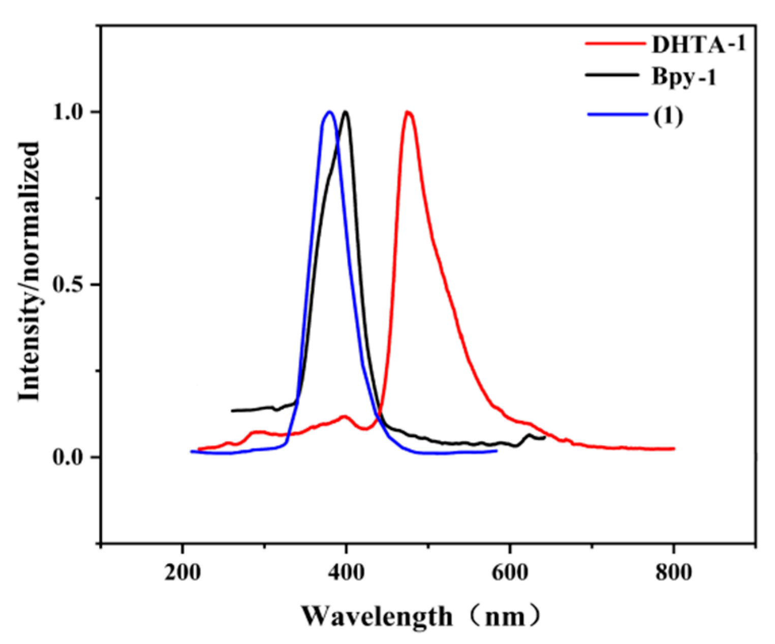
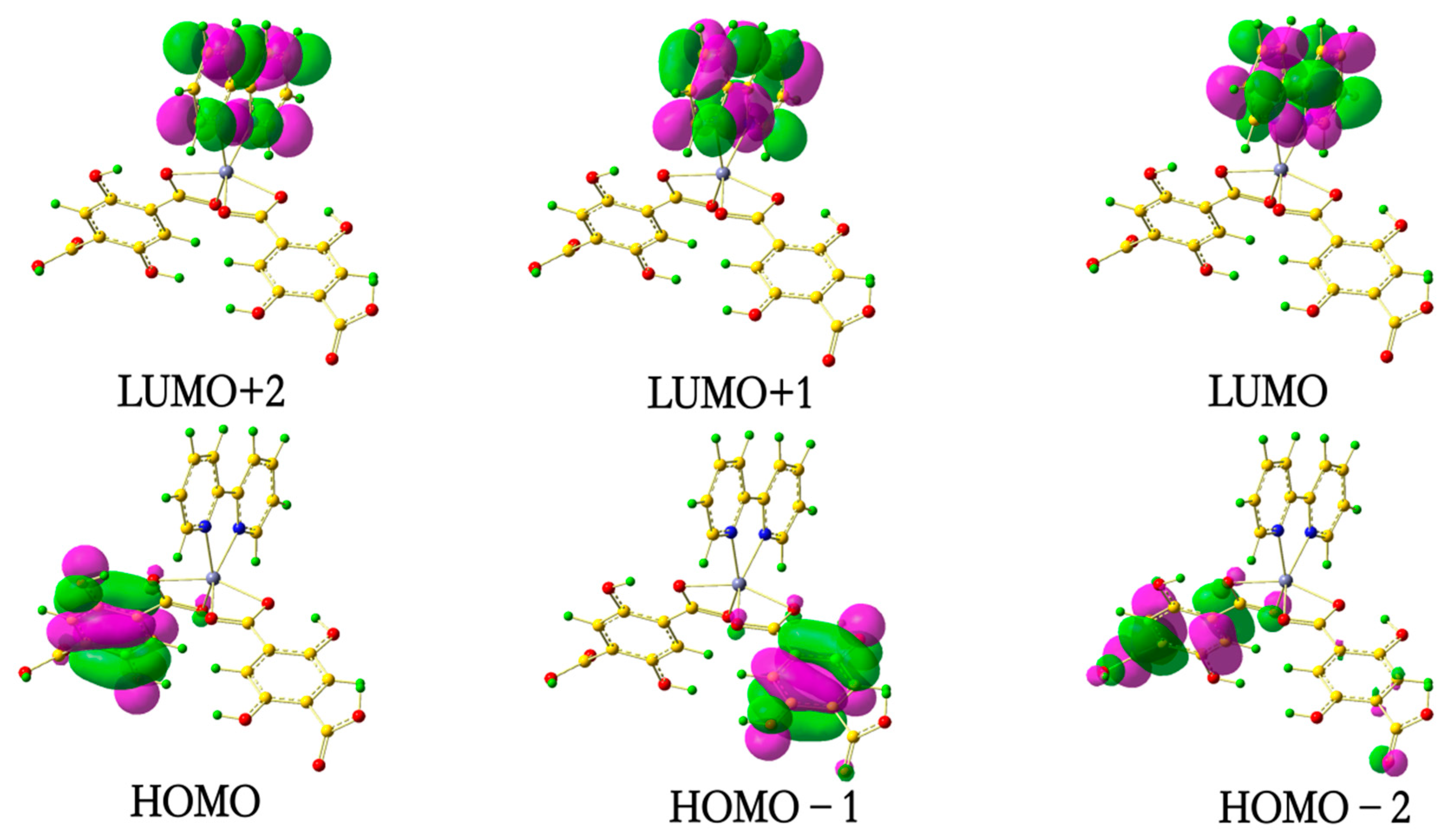
Because of the potential applications of coordination polymers with transition metal centers in many fields such as chemical sensors, electroluminescence and display photochemistry, they have attracted the interest of a wide range of researchers [36,37,38,39]; thus the luminescence of the complex was detected in a solid state at 25 °C (Figure 4), and the luminescence spectra of free 2,5-dihydroxy-1,4-terephthalic acid ligand (DHTA-1) and free 2,2′-bipyridine ligand (Bpy-1) were also obtained under the same condition. As we know, the emission peak for DHTA-1 is located at 476 nm (λex = 366 nm) [40]. The Bpy-1 that is excited near 398 nm exhibits an obvious emission peak (λex = 366 nm). The complex shows a significant emission peak at 365 nm (λex = 366 nm). Compared with the Bpy-1 and DHTA-1, the emission peak of the complex is blue-shifted. The shift may be caused by the n→π* transition of Bpy and DHTA which is caused by the ligand-to-ligand (DHTA-to-Bpy) charge transfer transition (LLCT). The HOMO orbital electron cloud is provided by C and O atoms of the DHTA ligand, and after emitting light energy it transfers to the Bpy ligand on the LUMO + 2 orbital.

3.4.2. High Sensitivity and Selectivity for Fe3+

Based on the excellent performance of the complex in water stability and luminescence, it was decided to study its application in inorganic metal cation sensing/detection. In order to explore its ability to sense inorganic metal cation selectively, different cations were investigated by adding the aqueous solutions of Na+, Mg2+, Cd2+, Ni2+, Hg2+, Co2+, K+, Zn2+, Li+, Pb2+ and Fe3+ with NO3 as counter anions into the suspension of the complex for luminescence research. The type of metal ions has a dramatic effect on the emission intensity of various suspensions. Interestingly, the presence of cations such as Na+, Mg2+, Cd2+, Ni2+, Hg2+, Co2+, K+, Zn2+, Li+ and Pb2+ has an insignificant effect on the luminescence intensity, while the emission intensity of the dispersion of the complex was obviously quenched by the trivalent iron cation (Fe3+) when excited (Figure 5a and Figure S6). In order to explore the influence of different concentrations of Fe3+ ions on the quenching of luminescence, the luminescence spectrum of the complex in the presence of increasing concentrations of iron cations was explored. It is observed that the luminescence intensity of the complex is inversely proportional to the concentration of Fe3+ cations. As depicted in Figure 6a,b, with the continuous increase of Fe3+ ion concentration, the luminescence intensity of the aqueous dispersions of the complex drops sharply (with the peaks at 360 nm) [quenching percentage = (I0 − I)/I0 × 100%, where I0 and I are luminescent intensities of the complex before and after addition of metal ions]. The small blue shift of the complex in aqueous suspension relative to that of the solid state may be due to the solvation effect of aqueous [41]. It is apparent that the concentration of iron ions is directly proportional to the degree of luminescence quenching. The experimental results show that the complex is efficient in detecting Fe3+ ions. The Fe3+ detection limit is calculated to be 1.241 × 10⁻⁹ M [LOD = 3σ/m, where σ = standard deviation from 10 measurements for each Fe3+ concentration and m = slope of the linear curve plotted at lower concentration for LOD measurements], which is relatively high compared to previously reported complexes (Table S5) [42]. Therefore, the complex can be used as a luminescent probe for detecting Fe3+ with high sensitivity.
In order to study the selectivity of the complex to Fe3+ ions relative to other metal ions, a mixture of Fe3+ ions and other competing ions was added to the suspension in water of the complex (Figure 5c). When Fe3+ ions were added, the luminescence intensity of the complex was completely quenched. These results demonstrate that even if it coexists with other common metal ions, the complex can also be used to detect Fe3+, further confirming the selectivity and sensitivity quenching of the complex in the case of Fe3+ ions. Because wastewater contains a variety of interfering ions, it is necessary to develop a new complex for selective detection of harmful iron ions in industrial waste.
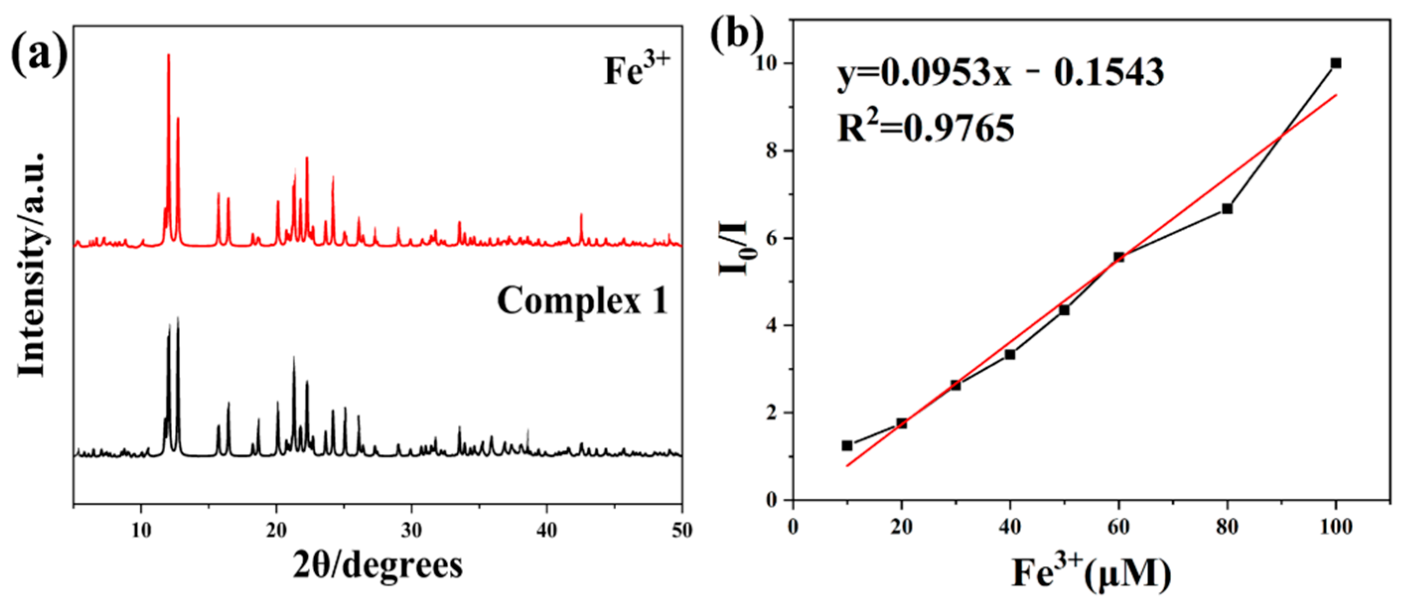
We assessed the recycling performance of the complex as a luminescence sensor. We used a centrifuge to obtain a solid sample from the solution and washed it three times with deionized water. After the second sensing test, a similar luminescence measurement using the obtained solid and the same amount of Fe3+ ions showed that the maximum emission at 360 nm did not change and possessed similar luminescence intensity. From the second to the fifth cycles, the degree of luminescence quenching is almost unchanged after adding Fe3+ ions, which proves that the complex has excellent recyclability as a luminescence sensor (Figure 6c). Compared with the PXRD pattern of the original sample, the PXRD pattern of the recycled sample has no obvious change (Figure 7a), which indicates that fixing the metal ions in the aqueous solution has almost no effect on the integrity of the framework structure.
Based on the above research, we have conducted a detailed study on the underlying mechanism of luminescence quenched of Fe3+ ion quenching. Chen and his colleagues summarized the factors [43,44,45,46,47,48,49,50] that cause Fe3+ ion to exhibit a quenching effect on luminescence of MOFs as follows: (i) changes in the backbone network (ii) resonance energy transfer. Firstly, we consider the changes in the backbone network. As confirmed by the PXRD pattern (Figure 7a), the crystal structure is mostly consistent with the complex, indicating that this quenching phenomenon has nothing to do with the backbone network of the complex, so the first factor was eliminated. On the other hand, the resonance energy transfer may be another factor leading to the quenching phenomenon [51,52]. It can be found from the ultraviolet–visible (UV-vis) spectrum that the absorption peak of Fe3+ appears in the range of 275–400 nm, while the absorption peaks of other Mx+ do not appear in this range. (Figure S7). Therefore, competitive absorption occurs between 1@Fe3+ and 1, which may be the main reason for the quenching effect. In fact, the XPS (X-ray Photoelectron Spectroscopy) spectrum displays that the typical energy of the Fe 2p1 is 727.32 eV, which is about 50.0 eV more than the standard value (Figure 7a) [53]. In addition, after adding Fe3+, the O1s peak of the free oxygen atoms in the coordination carboxylate observed at 535.5 eV in 1 shifted to 540.1 eV (Figure 8b). The shift of O1s peak indicates that the oxygen atom in 1@ Fe3+ has a weak binding with Fe3+.
Usually, we take the Stern–Volmer equation as the standard for quantitatively evaluating the luminescence quenching efficiency, I0/I = Ksv[M] + 1, where I0 and I are the maximum luminescent intensities before and after the addition of the complex, [M] is the molar concentration of the complex solution, and Ksv is the quenching constant (M−1) [54,55,56]. As displayed in Figure 7b, the luminescence intensity of the suspension of the complex showed an approximately linear correlation when using Fe3+ quenching, and perfectly followed the equation in the lower concentration range. The complex presents the higher Ksv values (the complex: Ksv(Fe3+) = 9.53 × 10⁴ M−1, R2 = 0.9765.) suggesting high quenching efficiency. As a Fe3+ luminescence sensor, complex 1 exhibited high sensitivity and selectivity in the results.

3.4.3. High Sensitivity for DMK

The test of the luminescence response of the complex in the aqueous solution suspensions was conducted by adding different kinds of organic solvents [57]. In this experiment, distilled water was chosen as a blank control experiment. The complex powder was submerged in diverse pure organic solvents, then the sample was ultrasonicated for 45 minutes and aged for 72 hours to form a stable suspension used for luminescence research. The luminescence spectra of the complex in different solvents were recorded. The result shows exist DMK has a meaningful luminescence quenching effect and the DMK’s quenching percentage was 97.56% (Figure 5b and Figure S9). By contrast, other organic small molecules had no significant effect on the luminescence intensity of the the complex suspension. Therefore, the aqueous solution suspension of the complex was chosen to detect various organic solvents. The PXRD spectrum of the complex was recorded again (Figure S10). The complex was characterized by PXRD which indicated that it still maintained crystallinity in DMK solvent.

3.5. Theoretical Calculation

The calculation of the complex was performed using Gaussian09 software [58] at the B3LYP/GenECP level, the for C, O, N, H using 6-31 + G(d) basis set and Zn using LANL2DZ [59]. The initial structure was obtained from X-ray refinement data (cif). For reducing the computation time, we adopted the minimum approximate structure model and the structure was optimized in the calculation. We used DHTA containing one deprotonated uncoordinated carboxyl group instead of the bridged DHTA dianion in the complex.
The calculation covered 59 atoms, 774 basis functions, 1391 primitive Gaussians, 148 α electrons and 148 β electrons. The molecular energy was 2079.619 a.u., the HOMO (Lowest Unoccupied Molecular Orbital) and LUMO (Highest Occupied Molecular Orbital) energies were −0.222 and −0.119 a.u., while the ΔE (ELUMO - EHOMO) value is −0.103 a.u. The HOMO, LUMO are shown in Figure 8; the electron of LUMO, LUMO+1 and LUMO + 2 is mostly sited at Bpy-1 and the HOMO, HOMO-1 and HOMO-2 electron clouds are mainly at the DHTA-1. The net charge of the selected atom and the electronic configuration at the B3LYP/(6-31 + G(d) (for C, H, N and O) and Lanl2dz (for Zn) levels are displayed in Table S4. The results indicated that the configurations of zinc atom is 4S0.303d9.984p0.34, and O is 2s1.65~1.712p5.03~5.063p0~0.013d0~0.014p0~0.01 and N atoms is 2s1.332p4.174p0.01. The valence is positive bivalent for Zn atom. The Zn picked up electrons from DHTA and Bpy, indicating that the charge of the central Zn has a positive charge, and it is 1.37312 for Zn1. In view of the former data, we can deem the Zn(II) ion coordinated to N and O atoms is mostly using the 4s and 3d orbitals. The O atoms donate electrons of 2s and 2p to the Zn(II) ion and then the coordination bonds are found. The coordination bonds were of N atoms and the Zn(II) ion, in it the Zn used the 2s and 2p orbitals. Therefore, the Zn(II) ion pick up electrons from O atoms of DTHA and N atoms of Bpy species. In summary, based on the valence-bond theory, the net charge distribution in the complex indicates that there are evidently covalent interactions between the coordinated atoms and central metal ion.
For the emission spectrum, we optimized the singlet state (S1 state) from ground state (S0) of the model, and the calculated emission peak is located at 355.2 nm, a value close to the experimental value (365.0 nm). The calculated peak is totally due to the transition of electrons from the HOMO orbital to the LUMO + 2 orbital (there is only one transition and it contributed 100% to the emission peak.) From the MO diagram we have identified that the orbitals correspond to the n-pi* transition, which is caused by the ligand-to-ligand (DHTA-to-Bpy) charge transfer transition (LLCT). The LUMO + 2 orbital electron cloud is provided by C and N atoms of the Bpy ligand, and after emitting light energy it fell down to the DHTA ligand on the HOMO orbital.

4. Conclusions

A new zinc coordination polymer has been successfully synthesized by hydrothermal methods with an aromatic dicarboxylate anion (DHTA) and Bpy ligand. The complex possesses a photoluminescent property, and the emission peak is located at 365 nm. The emission band of the complex exhibits blue shifts compared with the free Bpy ligand. The results indicate that the complex has the potential to be a remarkable luminescent material. The luminescence property of the complex displays excellent sensing ability for organic solvents and inorganic metal cations, especially for DMK and Fe3+ ions. It can be seen that the complex has great potential as a luminescent probe. The preparation of other novel coordination polymers with adjustable structures and functional properties based on multicarboxylate and N-donor auxiliary ligands is our next goal.

Supplementary Materials

The following are available online at https://www.mdpi.com/2073-4352/10/12/1105/s1, Figure S1: The IR spectrum of the complex; Figure S2: The geometric configuration of the complex; Figure S3: The thermal analysis of the complex; Figure S4: PXRD patterns of the complex: the simulated pattern from single crystal; Figure S5: PXRD patterns of the complex (for immersion experiment): the red is before and the black is after; Figure S6: Emission spectrum of the complex at different metal ions in deionized water; Figure S7: The UV-vis absorption spectra of metal cations aqueous solution with the same concentration of analytes (10−2 M) and the excitation spectra of the complex; Figure S8: (a) XPS spectra of the 1@Fe (red) and original 1 (black); (b) O1s XPS spectra of the original 1 (black) and 1@Fe3+ (red); Figure S9: Emission spectra of the complex at different solvents concentrations in deionized; Figure S10: PXRD patterns of the complex: the red is DMK and the black is the complex. Table S1: Crystal data and structure refinement for the complex; Table S2: Selected bond lengths (Å) and bond angles (°) for the complex; Table S3: Hydrogen bond length (Å) and bond angle (°) for the complex; Table S4: The LOD compared to previously reported complexes; Table S5: Selected atom net charges and electronic configuration of the complex at the B3LYP/(6-31 + G(d) (for C, H, N and O) and Lanl2dz (for Zn) levels.

Author Contributions

L.D. performed an experiment and wrote the article. B.Z. conducted date processing and the analysis. J.W. carried out the quantum chemical calculations. H.W., X.C. undertook the structural characterization. C.L. designed the experiments and conceived the project. All authors have read and agreed to the published version of the manuscript.

Funding

This work was supported by the Jilin Province Science and Technology Development Plan Item (No. 20140204080GX), the Project of the Education Department of Jilin Province, China (No. JJKH20180777KJ) and the Science and Technology Development Projects of Siping City (No. 2017057).

Acknowledgments

The authors thank Jilin Normal University for supporting this work.

Conflicts of Interest

The authors declare no conflict of interest.

References

  1. Fromm, K.M.; Batten, V.S.S.; Neville, S.M.; Turner, D.R. Coordination polymers design, analysis and application. Angew. Chem. 2009, 121, 4986–4987. [Google Scholar] [CrossRef]
  2. Rowsell, J.L.C.; Yaghi, O.M. Metal–organic frameworks: A new class of porous materials. Microporous Mesoporous Mater. 2004, 73, 3–14. [Google Scholar] [CrossRef]
  3. Long, J.R.; Yaghi, O.M. The pervasive chemistry of metal–organic frameworks. Chem. Soc. Rev. 2009, 38, 1213–1214. [Google Scholar] [CrossRef]
  4. Heine, J.; Müller-Buschbaum, K. Engineering metal-based luminescence in coordination polymers and metal–organic frameworks. Chem. Soc. Rev. 2013, 42, 9232–9242. [Google Scholar] [CrossRef]
  5. Koen, B. Lanthanide-based luminescent hybrid materials. Chem. Rev. 2009, 109, 4283–4374. [Google Scholar]
  6. Chen, L.; Ye, J.W.; Wang, H.P.; Pan, M.; Yin, S.Y.; Zhang, W.W.; Zhang, L.Y.; Wu, K. Ultrafast water sensing and thermal imaging by a metal-organic framework with switchable luminescence. Nat. Commun. 2017, 8, 15985–15994. [Google Scholar] [CrossRef] [Green Version]
  7. Scheurle, P.I.; Mähringer, A.; Jakowetz, A.C.; Hosseini, P.; Richter, A.F.; Wittstock, G.; Medina, D.D.; Bein, T. A highly crystalline anthracene-based MOF-74 series featuring electrical conductivity and luminescence. Chem. Sci. 2020, 10, 1039–1043. [Google Scholar] [CrossRef]
  8. Mark, D.A.; Ronald, J.T.H.; Leanne, A.; Talin, A.A.; Pikarsky, J.; Choudhury, A.; Gall, K.A.; Hesketh, P.J. Stress-induced chemical detection using flexible metal−organic frameworks. J. Am. Chem. Soc. 2008, 130, 14404–14405. [Google Scholar]
  9. Chen, B.L.; Wang, L.B.; Xiao, Y.; Fronczek, F.R.; Xue, M.; Cui, Y.; Qian, G.D. A luminescent metal-organic framework with lewis basic pyridyl sites for the sensing of metal ions. Angew. Chem. Int. Ed. 2009, 48, 508–511. [Google Scholar] [CrossRef]
  10. Lan, A.J.; Li, K.H.; Wu, H.H.; Olson, D.H.; Thomas, J.E.; Woosoek, K.C.; Mao, H.; Li, J. A luminescent microporous metal–organic framework for the fast and reversible detection of high explosives. J. Angew. Chem. Int. Ed. 2009, 48, 2370–2374. [Google Scholar] [CrossRef]
  11. Xie, Z.G.; Ma, L.Q.; DeKrafft, K.E.; Athena, J.; Lin, W.B. Porous phosphorescent coordination polymers for oxygen sensing. J. Am. Chem. Soc. 2010, 132, 922–923. [Google Scholar] [CrossRef] [PubMed]
  12. White, K.A.; Chengelis, D.A.; Gogick, K.A.; Stehman, J.; Rosi, N.L.; Petoud, S. Near-infrared luminescent lanthanide MOF barcodes. J. Am. Chem. Soc. 2009, 131, 18069–18071. [Google Scholar] [CrossRef]
  13. Yan, Y.T.; Zhang, W.Y.; Zhang, F.; Cao, F.; Yang, R.F.; Wang, Y.Y.; Hou, L. Four new metal–organic frameworks based on diverse secondary building units: Sensing and magnetic properties. Dalton Trans. 2018, 47, 1682–1692. [Google Scholar] [CrossRef]
  14. Chen, Y.Z.; Wang, C.M.; Wu, Z.Y.; Xiong, Y.J.; Xu, Q.; Yu, S.H.; Jiang, H.L. From bimetallic metal-organic framework to porous carbon: High surface area and multicomponent active dopants for excellent electrocatalysis. Adv. Mater. 2015, 27, 5010–5016. [Google Scholar] [CrossRef]
  15. Ni, D.; Chen, Y.X.; Yang, X.W.; Liu, C.C.; Cai, K.F. Microwave-assisted synthesis method for rapid synthesis of tinselenide electrode material for supercapacitors. J. Alloys Compd. 2018, 737, 623–629. [Google Scholar] [CrossRef]
  16. Rowsell, J.L.; Yaghi, O.M. Strategies for hydrogen storage in metal–organic frameworks. Angew. Chem. Int. Ed. 2005, 44, 4670–4679. [Google Scholar] [CrossRef]
  17. Kesanli, B.; Cui, Y.; Smith, M.R.; Bittner, E.W.; Bockrath, B.C.; Lin, W.B. Highly interpenetrated metal–organic frameworks for hydrogen storage. Angew. Chem. Int. Ed. 2005, 117, 74–77. [Google Scholar] [CrossRef]
  18. Dinca, M.; Long, J.R. Hydrogen storage in microporous metal–organic frameworks with exposed metal sites. Angew. Chem. Int. Ed. 2008, 47, 6766–6779. [Google Scholar] [CrossRef]
  19. Wu, H.H.; Gong, Q.H.; Olson, D.H.; Li, J. Commensurate adsorption of hydrocarbons and alcohols in microporous metal organic frameworks. Chem. Rev. 2012, 112, 836–868. [Google Scholar] [CrossRef]
  20. Li, J.R.; Kuppler, R.J.; Zhou, H.C. Selective gas adsorption and separation in metal–organic frameworks. Chem. Soc. Rev. 2009, 38, 1477–1504. [Google Scholar] [CrossRef]
  21. Zhang, M.L.; Zheng, Y.J.; Liu, M.; Ren, Y.X.; Wang, Z.X.; Cao, J.; Wang, J.J. Two zinc(II) coordination polymers based on flexible co-ligands featuring assembly imparted sensing abilities for Cr2O72- and o-NP. RSC Adv. 2019, 9, 21086–21094. [Google Scholar] [CrossRef] [Green Version]
  22. Zhang, Y.Q.; Vladislav, A.B.; Zheng, T.R.; Yang, C.H.; Qian, L.L.; Li, K.; Li, B.L.; Zhang, B.W.; Blatov, Y.Q.; Zheng, V.A.; et al. A luminescent zinc(II) coordination polymer with unusual (3,4,4)-coordinated self-catenated 3D network for selective detection of nitroaromatics and ferric and chromate ions: A versatile luminescent sensor. Dalton Trans. 2018, 47, 6189–6198. [Google Scholar] [CrossRef] [PubMed]
  23. Rosales-Vázquez, L.D.; Valdes-García, J.; Bazany-Rodríguez, I.; Germán-Acacio, J.M.; Martínez-Otero, D.; Vilchis-Néstor, A.; Morales-Luckie, R.; Sánchez-Mendieta, V.; Dorazco-González, A. Sensitive photoluminescent chemosensor for cyanide in water based on a zinc coordination polymer bearing ditert-butyl-bipyridine. Dalton Trans. 2019, 48, 12407–12420. [Google Scholar] [CrossRef]
  24. Kreno, L.E.; Leong, K.; Farha, O.K.; Allendorf, M.; Van Duyne, R.P.; Hupp, J.T. Metal–organic framework materials as chemical sensors. Chem. Rev. 2012, 112, 1105–1125. [Google Scholar] [CrossRef]
  25. Que, E.L.; Domaille, D.W.; Chang, C.J. Metals in neurobiology: Probing their chemistry and biology with molecular imaging. Chem. Rev. 2008, 108, 1517–1549. [Google Scholar] [CrossRef]
  26. Chen, W.M.; Meng, X.L.; Zhuang, G.L.; Wang, Z.; Kurmoo, M.; Zhao, Q.Q.; Wang, X.P.; Shan, B.R.; Tung, C.H.; Sun, D. A superior fluorescent sensor for Al3+ and UO22+ based on a Co(II) metal–organic framework with exposed pyrimidyl Lewis base sites. J. Mater. Chem. A 2017, 5, 13079–13085. [Google Scholar] [CrossRef]
  27. Carter, K.P.; Young, A.M.; Palmer, A.E. Fluorescent sensors for measuring metal ions in living systems. Chem. Rev. 2014, 114, 4564–4601. [Google Scholar] [CrossRef]
  28. Xu, H.B.; Zhou, S.H.; Xiao, L.L.; Wang, H.H.; Li, S.Z.; Yuan, Q.H. Fabrication of a nitrogen-doped graphene quantum dot from MOF-derived porous carbon and its application for highly selective fluorescence detection of Fe3+. J. Mater. Chem. C 2015, 3, 291–297. [Google Scholar] [CrossRef]
  29. Tang, S.F.; Li, L.J.; Lv, X.X.; Wang, C.; Zhao, X.B. Tuning the structure of metal phosphonates using uncoordinating methyl group: Syntheses, structures and properties of a series of metal diphosphonates. CrystEngComm 2014, 16, 7043–7052. [Google Scholar] [CrossRef] [Green Version]
  30. Zhai, F.; Zheng, Q.; Chen, Z.; Ling, Y.; Liu, X.; Weng, L.; Zhou, Y. Crystal transformation synthesis of a highly stable phosphonate MOF for selective adsorption of CO2. CrystEngComm 2013, 15, 2040–2043. [Google Scholar] [CrossRef]
  31. Xu, H.; Gao, J.K.; Qian, X.F.; Wang, J.P.; He, H.J.; Cui, Y.J.; Yang, Y.; Wang, Z.Y.; Qian, G.D. Metal–organic framework nanosheets for fast-response and highly sensitive luminescent sensing of Fe3+. J. Mater. Chem. A 2016, 16, 4036–4044. [Google Scholar] [CrossRef]
  32. Lv, R.; Wang, J.Y.; Zhang, Y.P.; Li, H.; Yang, L.Y.; Liao, S.Y.; Gu, W.; Liu, X. An amino-decorated dual-functional metal–organic framework for highly selective sensing of Cr(III) and Cr(VI) ions and detection of nitroaromatic explosives. J. Mater. Chem. A 2016, 4, 15494–15500. [Google Scholar] [CrossRef]
  33. Zhu, W.Q.; Fan, P.; Li, Y.; Zhu, Y.M.C.; Zhang, J. The Constuction of Double Ligands Zn (II) MOFs and Its Photoluminescence Properties. Imaging Sci. Photochem. 2020, 38, 159–166. [Google Scholar]
  34. Sun, H.Y.; Li, X.; Wang, Z.R.; Sun, S.Q.; Li, C.B.; Wang, J.J. Synthesis, Crystal Structure and Theoretical Calculations of Two Zn(II) Coordination Polymers Based on 2,5-Dihydroxyterephthalic Acid. J. Clust. Sci. 2018, 29, 1275–1283. [Google Scholar] [CrossRef]
  35. Altomare, A.; Burla, M.C.; Camalli, M.; Cascarano, G.; Giacovazzo, C.; Guagliardi, A.; Moliterni, G.G.A.; Polidori, G.; Spagna, R. SIR97: A new tool for crystal structure determination and refinement. J. Appl. Crystallogr. 1999, 32, 115–119. [Google Scholar] [CrossRef]
  36. Guo, F.B.; Zhu, Y.; Xu, G.L.; Zhang, M.M.; Zhang, X.L.; Zhang, J. Tuning structural topologies of five photoluminescent Cd(II) coordination polymers through modifying the substitute group of organic ligand. J. Solid. State Chem. 2013, 199, 42–48. [Google Scholar] [CrossRef]
  37. Man, Z.M.; Guo, F. Coordination polymers constructed from an asymmetric biphenyl-dicarboxylate and N-donor ligands: Structures and photoluminescent properties. J. Coord. Chem. 2013, 66, 1–8. [Google Scholar] [CrossRef]
  38. Xiang, J.; Luo, Y.; Zhao, L.L.; Wang, C.H.; Wu, J.S. Ancillary ligands assisted self-assembly of metal organic frameworks: Synthesis, crystal structures and photophysical properties of two Zn(II) complexes containing in-situ formed tetrazole ligands. Inorg. Chem. Commun. 2013, 31, 23–28. [Google Scholar] [CrossRef]
  39. Wang, X.J.; Liu, Y.H.; Xu, C.Y.; Guo, Q.Q.; Huo, H.W.; Fan, Y.T. Series of Cd(II) metal–organic frameworks based on a flexible tripodal ligand and polycarboxylate acids: Syntheses, structures, and photoluminescent properties. Cryst. Growth Des. 2012, 12, 2435–2444. [Google Scholar] [CrossRef]
  40. Wang, H.; Han, S.; Wang, J.J.; Dun, L.N.; Zhang, B.S.; Chen, X.; Li, W.F.; Li, C.B. Crystal Structure, Thermal Behavior and Luminescence of a new Manganese(II) coordination polymer constructed with 1, 10-phenanthroline-5, 6-dione and 2, 5-dihydroxyl-1, 4-terephthalic acid. J. Mol. Struct. 2019, 1204, 127466–127479. [Google Scholar] [CrossRef]
  41. Parmar, B.; Rachuri, Y.; Bisht, K.K.; Laiya, R.; Suresh, E. Mechanochemical and conventional synthesis of Zn(II)/Cd(II) luminescent coordination polymers: Dual sensing probe for selective detection of chromate anions and TNP in aqueous phase. Inorg. Chem. 2017, 56, 2627–2638. [Google Scholar] [CrossRef]
  42. He, H.M.; Zhang, D.Y.; Guo, F.; Sun, F.X. A versatile microporous Zinc(II) metal–organic framework for selective gas adsorption, cooperative catalysis, and luminescent sensing. Inorg. Chem. 2018, 10, 1021–1030. [Google Scholar] [CrossRef]
  43. Hu, Z.C.; Deibert, B.J.; Li, J. Luminescent metal–organic frameworks for chemical sensing and explosive detection. Chem. Soc. Rev. 2014, 43, 5815–5840. [Google Scholar] [CrossRef] [Green Version]
  44. Nagarkar, S.S.; Desai, A.V.; Samanta, P.; Ghosh, S.K. Aqueous phase selective detection of 2,4,6-trinitrophenol using a fluorescent metal–organic framework with a pendant recognition site. Dalton Trans. 2015, 44, 15175–15180. [Google Scholar] [CrossRef]
  45. Wang, J.; Jiang, M.; Yan, L.; Ren, P.; Meng, J.; Huang, F.; Guo, X.X.; Li, Y.; Wu, P.Y. Multifunctional luminescent Eu(III)-based metal–organic framework for sensing methanol and detection and adsorption of Fe(III) ions in aqueous solution. Inorg. Chem. 2016, 55, 12660–12668. [Google Scholar] [CrossRef]
  46. Wang, X.; Han, Y.; Han, X.X.; Hou, X.Y.; Wang, J.J.; Fu, F. Highly selective and sensitive detection of Hg2+, Cr2O72-,and nitrobenzene/2,4-dinitrophenol in water via two fluorescent Cd-CPs. New J. Chem. 2018, 42, 19844–19852. [Google Scholar] [CrossRef]
  47. Pramanik, S.; Zheng, C.; Zhang, X.; Emge, T.J.; Li, J. New microporous metal−organic framework demonstrating unique selectivity for detection of high explosives and aromatic compounds. J. Am. Chem. Soc. 2011, 133, 4153–4155. [Google Scholar] [CrossRef]
  48. Xu, H.; Liu, F.; Cui, Y.J.; Chen, B.L.; Qian, G.D. A luminescent nanoscale metal–organic framework for sensing of nitroaromatic explosives. Chem. Commun. 2011, 47, 3153–3155. [Google Scholar] [CrossRef]
  49. Zhang, J.; Zheng, B.; Zhao, T.T.; Li, G.H.; Huo, Q.S.; Liu, Y.L. Topological diversities and luminescent properties of lanthanide metal–organic frameworks based on a tetracarboxylate ligand. Cryst. Growth Des. 2014, 14, 2394–2400. [Google Scholar] [CrossRef]
  50. Jin, J.C.; Wu, X.R.; Luo, Z.D.; Deng, F.Y.; Liu, J.Q.; Singh, A.; Kumar, A. Luminescent sensing and photocatalytic degradation properties of an uncommon (4,5,5)-connected 3D MOF based on 3,5-di(3′,5′-dicarboxylphenyl)benzoic acid. CrystEngComm 2017, 19, 4368–4377. [Google Scholar] [CrossRef]
  51. Zhou, J.M.; Shi, W.; Li, H.M.; Li, H.; Cheng, P. Experimental studies and mechanism analysis of high-sensitivity luminescent sensing of pollutional small molecules and ions in Ln4O4 cluster based microporous metal–organic frameworks. J. Phys. Chem. C 2014, 118, 416–426. [Google Scholar] [CrossRef]
  52. Wu, Y.L.; Yang, G.P.; Zhang, Y.D.; Shi, N.N.; Han, J.; Wang, Y.Y. A new luminescent Cd(II)-MOF as a highly selective chemical probe for Fe3+ in aqueous solution with mixed metal ions. RSC Adv. 2015, 5, 90772–90777. [Google Scholar] [CrossRef]
  53. Zhou, Y.; Chen, H.H.; Yan, B. An Eu3+ post-functionalized nanosized metal–organic framework for cation exchange-based Fe3+-sensing in an aqueous environment. J. Mater. Chem. A 2014, 2, 13691–13697. [Google Scholar] [CrossRef]
  54. Ju, P.; Zhang, E.S.; Jiang, L.; Zhang, Z.; Hou, X.Y.; Zhang, Y.Q.; Yang, H.; Wang, J.J. A novel microporous Tb-MOF fluorescent sensor for highly selective and sensitive detection of picric acid. RSC Adv. 2018, 8, 21671–21678. [Google Scholar] [CrossRef] [Green Version]
  55. Hao, J.N.; Yan, B. Ln3+ post-functionalized metal–organic frameworks for color tunable emission and highly sensitive sensing of toxic anions and small molecules. New J. Chem. 2016, 40, 4654–4661. [Google Scholar] [CrossRef]
  56. Gong, W.J.; Yao, R.; Li, H.X.; Ren, Z.G.; Zhang, J.G.; Lang, J.P. Luminescent cadmium(II) coordination polymers of 1,2,4,5-tetrakis(4-pyridylvinyl)benzene used as efficient multi-responsive sensors for toxic metal ions in water. Dalton Trans. 2017, 46, 16861–16871. [Google Scholar] [CrossRef]
  57. Li, Y.; Song, H.; Chen, Q.; Liu, K.; Zhao, F.Y.; Ruan, W.J.; Chang, Z. Two coordination polymers with enhanced ligand-centered luminescence and assembly imparted sensing ability for acetone. J. Mater. Chem. A 2014, 2, 9469–9473. [Google Scholar] [CrossRef]
  58. Frisch, M.J.; Trucks, G.W.; Schlegel, H.B.; Scuseria, G.E.; Robb, M.A.; Cheeseman, J.R.; Scalmani, G.; Barone, V.; Mennucci, B.; Petersson, G.A.; et al. Gaussian 09; Gaussian, Inc.: Wallingford, CT, USA, 2009. [Google Scholar]
  59. Wu, H.; Dong, X.W.; Liu, H.Y.; Ma, J.F.; Li, S.L.; Yang, J.; Liu, Y.Y.; Su, Z.M. Syntheses, X-ray structures and CVD of titanium(IV) arsine complexes. Dalton Trans. 2008, 39, 5331–5341. [Google Scholar] [CrossRef]
Scheme 1. The coordination modes of 2,5-dihydroxyterephthalic acid [(A) {μ4-bis[(μ1-η1:η1-bidentate chelating), (μ2-η1:η1-double monodentate bridging)]} mode, (B) [μ2-bis(monodentate, μ1-η1:η0) bridging] mode, (C) {μ4-bis[(μ2-η2:η1-chelating), (μ2-η2:η1-bidentate chelating)]} mode, (D) {μ2-[((μ1-η1:η1-bidentate chelating), (μ1-η1:η0-monodentate bridging)])} mode, (E) {μ6-bis[((μ2-η2:η1-bidentate chelating), (μ3-η2:η2-bridging/bidentate chelating/bridging)]} mode] (in this work the acid adopts mode A connected to zinc).
Crystals 10 01105 sch001
Figure 1. The coordination environment of the complex (Symmetry codes: #1 = −x, −y, 1 − z; #2 = x, 0.5 − y, 0.5 + z; #3 = −x, 0.5 + y, 1.5 − z.).
Crystals 10 01105 g001
Figure 2. The hydrogen bonds (green dotted line).
Crystals 10 01105 g002
Figure 3. The 3D network structure of the complex.
Crystals 10 01105 g003
Figure 4. Solid-state emission spectrum of the complex, DHTA (2,5-dihydroxy-1,4-terephthalic acid) and Bpy (2,2′-bipyridine) at room temperature.
Crystals 10 01105 g004
Figure 5. (a) The luminescence quenching exhibition of the complex dispersed in metal cations (the solution with a concentration: 1.0 mmol·L−1). (b) The luminescence quenching of the complex dispersed in organic small molecules. (c) Comparison of the luminescence intensity of the complex along with the mixed metal ions.
Crystals 10 01105 g005
Figure 6. (a) The luminescence quenching experiments of the complex dispersed in Fe3+ solution with different concentration gradients (0–150.0 μM), respectively. (b) Luminescence quenching efficiency of Fe3+ solution with different concentration gradients (0–150.0 μM) of the complex. (c) Recycling tests by monitoring the luminescence intensity before (blue) and after (red) loading Fe3+ ions.
Crystals 10 01105 g006
Figure 7. (a) The powder X-ray diffraction (PXRD) patterns of the complex before and after adsorption of Fe3+ ions. (b) Stern–Volmer plot of the complex suspension quenched by Fe3+.
Crystals 10 01105 g007
Figure 8. The molecular orbitals of the complex.
Crystals 10 01105 g008
Publisher’s Note: MDPI stays neutral with regard to jurisdictional claims in published maps and institutional affiliations.