Article

Production of 4-Ene-3-ketosteroids in Corynebacterium glutamicum

by
Julia García-Fernández
1,
Beatriz Galán
1,
Carmen Felpeto-Santero
1,
José L. Barredo
2 and
José L. García
1,*
1
Department of Environmental Biotechnology, Centro de Investigaciones Biológicas (CSIC), Ramiro de Maeztu 9, 28040 Madrid, Spain
2
Department of Biotechnology, Crystal Pharma, A Division of Albany Molecular Research Inc. (AMRI), 47151 Boecillo Valladolid, Spain
*
Author to whom correspondence should be addressed.
Catalysts 2017, 7(11), 316; https://doi.org/10.3390/catal7110316
Submission received: 29 September 2017 / Revised: 19 October 2017 / Accepted: 20 October 2017 / Published: 27 October 2017
(This article belongs to the Special Issue Biocatalysis and Biotransformations)

Abstract

:
Corynebacterium glutamicum has been widely used for the industrial production of amino acids and many value-added chemicals; however, it has not been exploited for the production of steroids. Using C. glutamicum as a cellular biocatalyst we have expressed the 3β-hydroxysteroid dehydrogenase/isomerase MSMEG_5228 from Mycobacterium smegmatis to demonstrate that the resulting recombinant strain is able to oxidize in vivo C19 and C21 3-OH-steroids to their corresponding keto-derivatives. This new approach constitutes a proof of concept of a biotechnological process for producing value-added intermediates such as 4-pregnen-16α,17α-epoxy-16β-methyl-3,20-dione.

1. Introduction

Corynebacterium glutamicum has been widely used for the industrial production of amino acids, but the publication of its complete genome [1,2] has provided the basis for an enormous progress in the use of this microorganism for other biotechnological applications placing it as an ideal chassis within the top ranking of cell factories [3]. In this sense, a massive deletion of the majority of the non-essential DNA sequences has been recently carried out in order to improve its properties [4].
Although C. glutamicum has been used for the production of several value-added chemicals [5], it has not been used for steroid production so far. The only reports related to this issue are the work of Charney et al. [6] concerning biotransformations of steroid substrates using Corynebacterium simplex (previously classified as Arthrobacter simplex and Pimelobacter simplex, and currently known as Nocardioides simplex) and the recent work of Zhang et al. [7] who used Corynebacterium crenatum as host to express the 3-ketosteroid-Δ1-dehydrogenase from Mycobacterium neoaurum to catalyze the bioconversion of 4-androstene-3,17-dione (AD) to androst-1,4-diene-3,17-dione (ADD).
In the steroid industry, the biotransformations have been generally performed by fungi and actinomycetes taking advantage of the regio- and stereo-specificity of their enzymes [8,9,10,11]. The aim of this work is to demonstrate that C. glutamicum can be an ideal chassis (cellular biocatalyst) for being engineered for such purposes. To probe this hypothesis, we have selected the enzyme responsible for the first reaction of the bacterial cholesterol catabolism that renders its keto-derivative choles-4-ene-3-one (cholestenone). In bacteria, this reaction can be catalyzed by cholesterol oxidases (ChOx) or by 3β-hydroxysteroid dehydrogenases (3β-HSD) depending on the microorganism [9,12]. In the case of Mycobacterium smegmatis, the reaction is carried out by a 3β-HSD (MSMEG_5228), a member of the short chain dehydrogenase superfamily [13]. In this work, we have expressed the MSMEG_5228 gene encoding the 3β-HSD from M. smegmatis in C. glutamicum to explore the biotechnological potential of the recombinant chassis to produce steroids of industrial interest.

2. Results

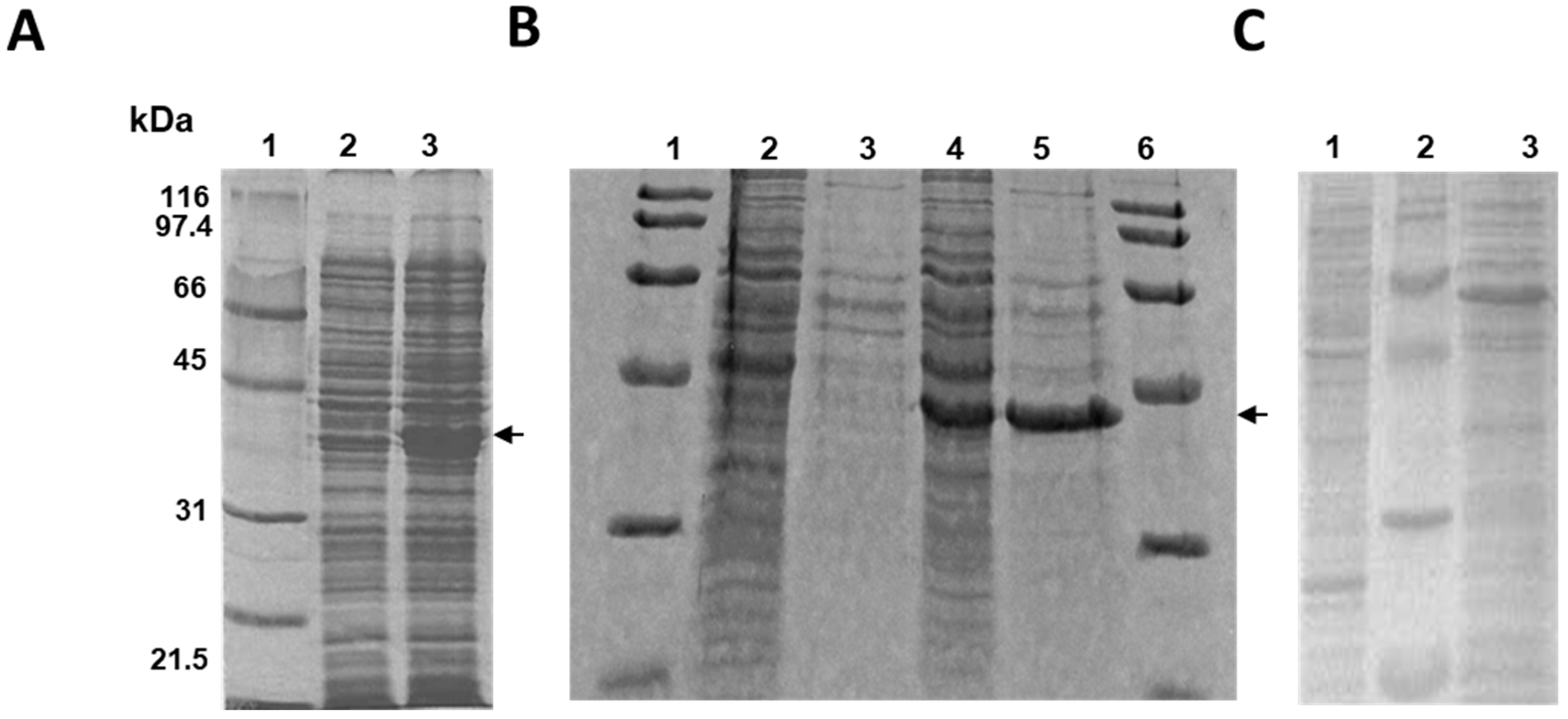
2.1. Cloning and Expression of the MSMEG_5228 Gene in Different Bacteria

The initial attempts to produce highly active 3β-HSD in Escherichia coli (pET5228) cells were unsuccessful due to a low soluble expression caused most probably by the high GC content of the gene and the different codon usage of Mycobacterium and E. coli (Figure 1A). Therefore, to test the portability of the mycobacterial 3β-HSD enzyme, we decided to use a heterologous host most closely related to Mycobacterium such as Rhodococcus erythropolis. The protein extracts obtained from the recombinant strain R. erythropolis IGTS8 (pTip5228) showed high levels of soluble and active enzyme (Figure 1B). The soluble enzyme showed a wide substrate versatility in vitro since the cell extract was able to oxidize cholesterol (C27), pregnenolone (C21), and dehydroepiandrosterone (DHEA) (C19) to their corresponding keto-derivatives, cholestenone, progesterone, and AD, respectively, in the presence of NAD (Figure S1). These results demonstrate that the active enzyme can be produced in a very high yield in R. erythropolis and therefore, can be used as an in vitro biocatalyst. However, the requirement of NAD as a cofactor precludes the in vitro use for industrial purposes.
In addition and taking into account that R. erythropolis is able to naturally degrade and modify steroids, its use as cellular biocatalyst (e.g., under growing or resting cells conditions) might compromise the final yield of the biotransformation process. In order to bypass this metabolic inconvenient, we looked for other non-steroid degrading actinomycetes that might act as a clean microbial chassis where an optimal in vivo transformation of steroids could be achieved. This search led us to implement the system in C. glutamicum that, in contrast to R. erythropolis, does not contain the genes for steroid catabolism. In addition, this host is closely related to Mycobacterium and Rhodococcus, which could allow us to abrogate possible protein expression problems. To prove this hypothesis, we cloned the MSMEG_5228 gene into pCGL0482, an expression plasmid able to replicate in C. glutamicum, generating the recombinant strain C. glutamicum R31 (pCGL5228). Nevertheless, the protein was not overproduced (Figure 1C).

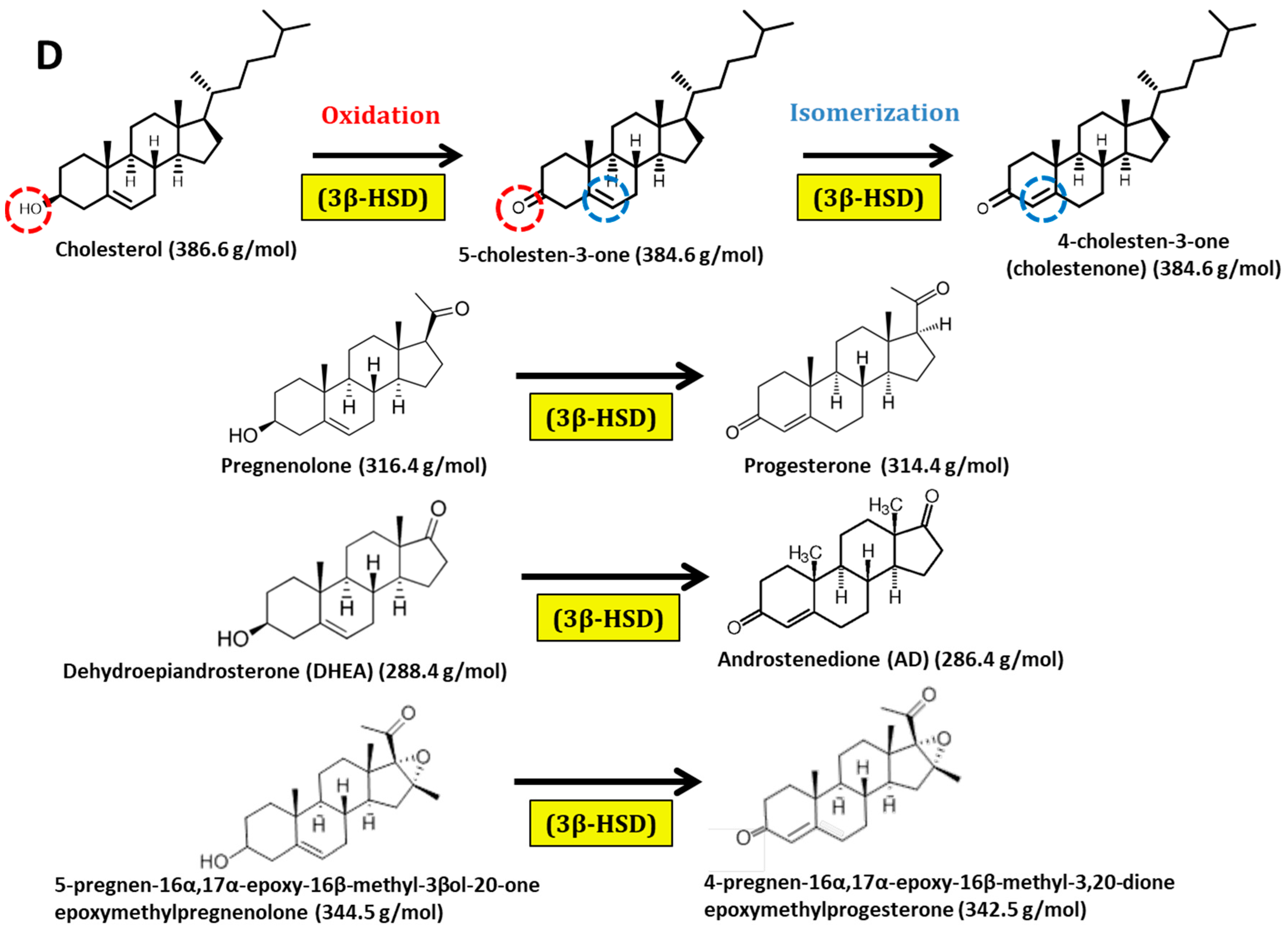
2.2. Biotransformation of Different Steroids by C. glutamicum R31 (pCGL5228)

To test the ability of C. glutamicum R31 (pCGL5228) to perform 3OH-dehydrogenations of steroids in vivo, we carried out a resting cell assay using DHEA as substrate. Figure 2 shows that C. glutamicum R31 (pCGL5228) was able to completely transform DHEA into AD with a conversion yield close to 85% plus some minor contaminants. The main contaminant was characterized by mass spectrometry as 6-OH-AD (data not shown). The 4-cholesten-6-ol-3-one has been detected as a lateral product in the transformations of cholesterol performed by ChOx [14]. In fact, this 6-hydroxylation has been demonstrated to be an auto-oxidation from the 5-cholesten-3-one intermediate accumulated in the first reaction step [15]. In addition, control experiments using AD as a substrate confirmed that C. glutamicum endogenous enzymes are not able to metabolize or modify the product of the reaction (data not shown). Therefore, although a 6-hydroxylation has not been described before as a 3β-HSD reaction, it seems reasonable to assume that in our case the 6-OH-AD might result from the same auto-oxidation mechanism.
Interestingly, when we tested cholesterol as a substrate of the biotransformation, no cholestenone was detected even after 28 h of incubation (data not shown), suggesting that cholesterol is not able to diffuse to the cytoplasm of C. glutamicum. This result would confirm studies previously described by us and other authors showing that steroids with long side-chain as cholesterol need to be transported by a specific Mce4 uptake system that contains Mycobacterium and Rhodococcus [16,17] but is absent in C. glutamicum.

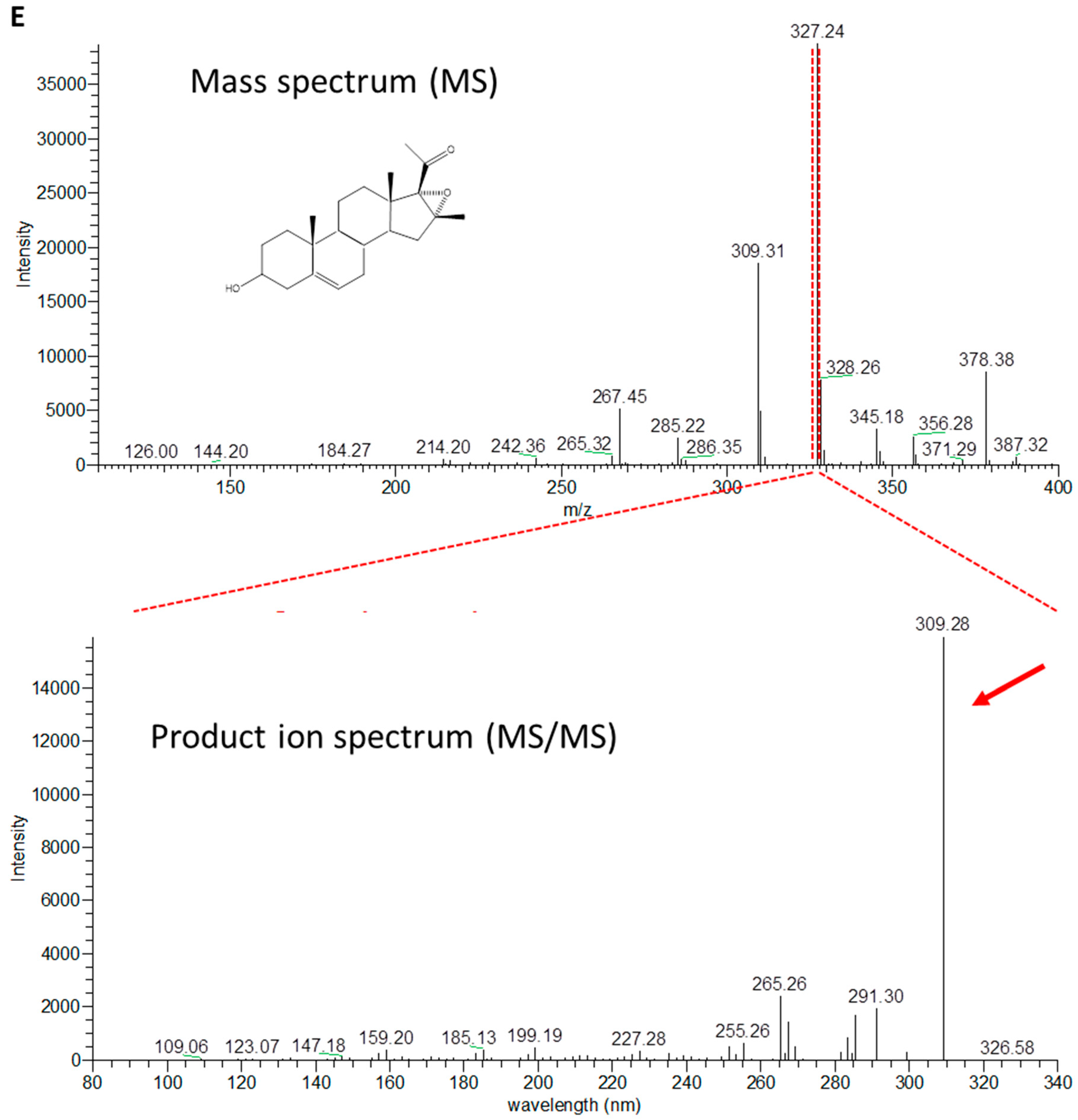
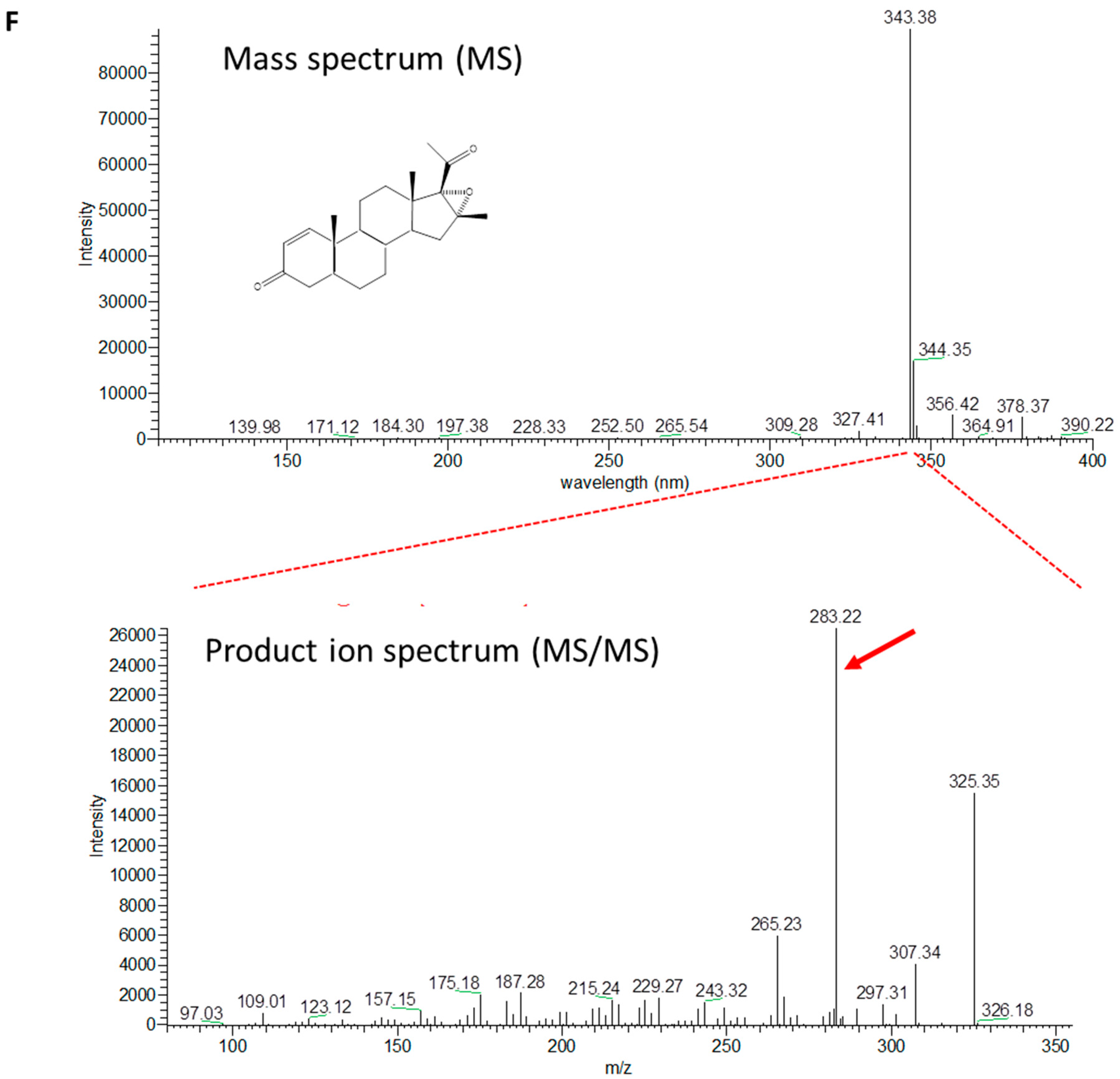
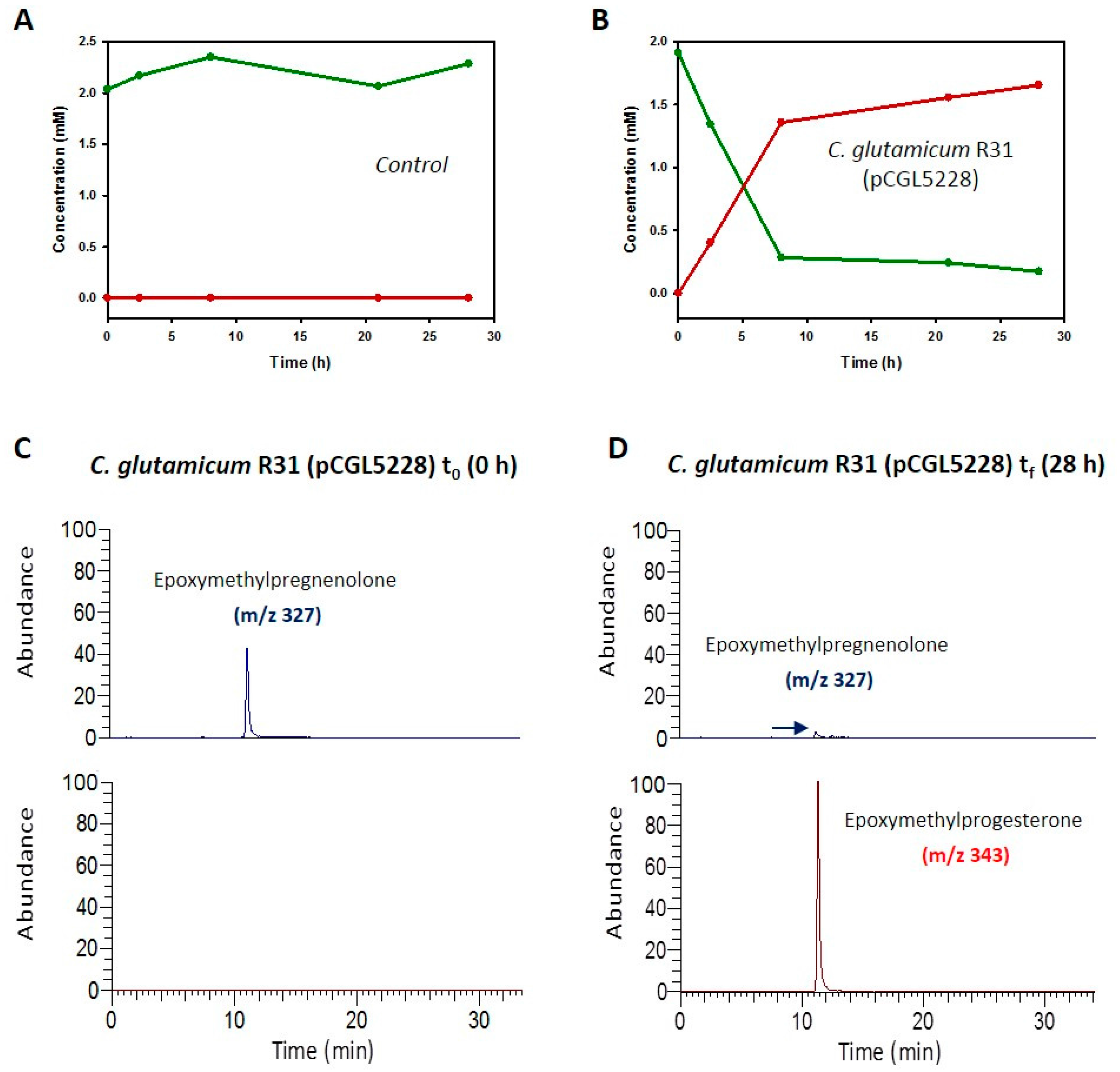
2.3. Production of 4-Pregnen-16α,17α-Epoxy-16β-Methyl-3,20-Dione by C. glutamicum (pCGL5228)

Taking advantage of our knowledge about the specificity of the steroid transport and since the 3β-HSD is active on C19, C21, and C27 steroids; we tested to determine whether the new biocatalyst was able to produce value-added compounds. The 4-pregnen-16α,17α-epoxy-16β-methyl-3,20-dione (epoxymethylprogesterone) (Figure 3) is used as an intermediate for the production of 16-methyl analogs of cortisone, hydrocortisone, megestrol, and other steroids of pharmaceutical interest. The production of epoxymethylprogesterone from 5-pregnen-16α,17α-epoxy-16β-methyl-3β-ol-20-one (epoxymethylpregnenolone) presents some difficulties to be oxidized by chemical procedures due to the presence of the epoxy ring. Figure 3 shows that C. glutamicum R31 (pCGL5228) is able to efficiently transport and transform epoxymethylpregnenolone into epoxymethylprogesterone, leading to a yield of almost 100% in 28 h.

3. Discussion

The 3-OH oxidation of ∆5-3-hydroxysteroids coupled to ∆5-∆4 isomerization, a key step in the transformation of sterols into many steroids of biological relevance (e.g., progesterone, testosterone, cortisol, aldosterone, and corticosterone), has been widely used for the quantification of cholesterol in clinical and food specimens [18,19], but there are only a few examples concerning its utilization in biocatalysis [20,21,22,23]. All these biocatalytic procedures have been carried out using ChOx from different sources, but none of them have been carried out with 3β-HSD enzymes. Here, we have explored the potential of the 3β-HSD enzyme from M. smegmatis expressed in a heterologous host as C. glutamicum, to achieve the biotransformation of sterols in their corresponding keto-derivatives.
Our analyses have demonstrated that the substrate specificity of the 3β-HSD from M. smegmatis (MSMEG_5228) is similar to that reported for the 3β-HSD of M. tuberculosis [24] but different from the 3β-HSD of Nocardia sp. Ch2-1 [25] since this enzyme was not active on DHEA [26]. These biochemical conclusions allowed us to use the 3β-HSD enzyme from M. smegmatis to constitute a proof of concept of the potential of Corynebacterium as a heterologous host for the industrial production of steroids.
We have been able to show that a recombinant strain of C. glutamicum carrying the 3β-HSD of M. smegmatis is able to catalyze the biotransformation of short chain (C19 and C21) steroids. However, we have observed that our recombinant Corynebacterium cannot transform cholesterol (C27) into cholestenone since it requires a specific cholesterol transport system [16,27]. Remarkably, among the substrates tested in this study, the production of epoxymethylprogesterone was very successful showing a biotransformation yield near to 100%.

4. Materials and Methods

4.1. Chemicals

Cholesterol, cholestenone, AD, DHEA, progesterone, and pregnenolone were purchased from Sigma-Aldrich (Merck KGaA, Darmstadt, Germany). 5-pregnen-16α,17α-epoxy-16β-methyl-3β-ol-20-one was provided by Crystal Pharma (Valladolid, Spain).

4.2. Bacterial Strains and Culture Conditions

The strains and plasmids used in this study are listed in Table 1. Escherichia coli and Rhodococcus erythropolis IGTS8 were cultured in LB medium at 37 °C and 30 °C, respectively, whereas Corynebacterium glutamicum R31 was cultured on Tryptic Soy Broth (TSB) at 30 °C. Antibiotics were used where indicated at the following concentrations: ampicillin (100 µg mL−1) for E. coli, chloramphenicol (Cm) (34 µg mL−1) for R. erythropolis IGTS8 and Cm (6 µg mL−1) for C. glutamicum R31. The oligonucleotides used in this work are listed in Table 2.

4.3. Expression of the MSMEG_5228 Protein in R. erythropolis IGTS8, E. coli BL21 and C. glutamicum R31

MSMEG_5228 gene was amplified by PCR from the total DNA of M. smegmatis mc2155 using primers listed in Table 2. This fragment was digested with NdeI and HindIII was cloned into pTipQC1 plasmid, delivering pTip5228 plasmid or digested with XhoI and HpaI and cloned into the pCGL0482 plasmid, delivering the construction named pCGL5228 (Table 1). The production of the 3β-HSD enzyme by BL2 1(DE3) (pET5228) (13) (Table 1) was induced by adding IPTG (0.2 mM) and analyzed by SDS-PAGE. Plasmid pTip5228 expressing the 3β-HSD activity was transformed into R. erythropolis IGTS8 electrocompetent cells and selected in LB agar plates containing Cm (34 µg mL−1). The production of the 3β-HSD enzyme with this construction was induced by adding thiostrepton (1 µg mL−1) and analyzed by SDS-PAGE. Plasmid pCGL5228 was further electroporated into C. glutamicum R31 competent cells and selected in TSA agar plates containing Cm (6 µg mL−1) to generate the recombinant C. glutamicum R31 (pCGL5228) strain.

4.4. Resting Cell Assay for Steroid Biotransformation Using C. glutamicum R31 (pCGL5228)

Recombinant C. glutamicum R31 (pCGL5228) cells were grown in 200 mL of TSB containing Cm (6 µg mL−1) at 30 °C and 250 rpm. For the biotransformation assay, C. glutamicum R31 (pCGL5228) cells were harvested by centrifugation, washed, and resuspended in 25 mM Hepes buffer (pH 8). The steroid was added at a final concentration of 2 mM from a 5 mM stock prepared in 10% (v/v) of Tyloxapol (Sigma, Merck KGaA, Darmstadt, Germany). Aliquots were taken at several times to analyze the biotransformation process during 28 h.

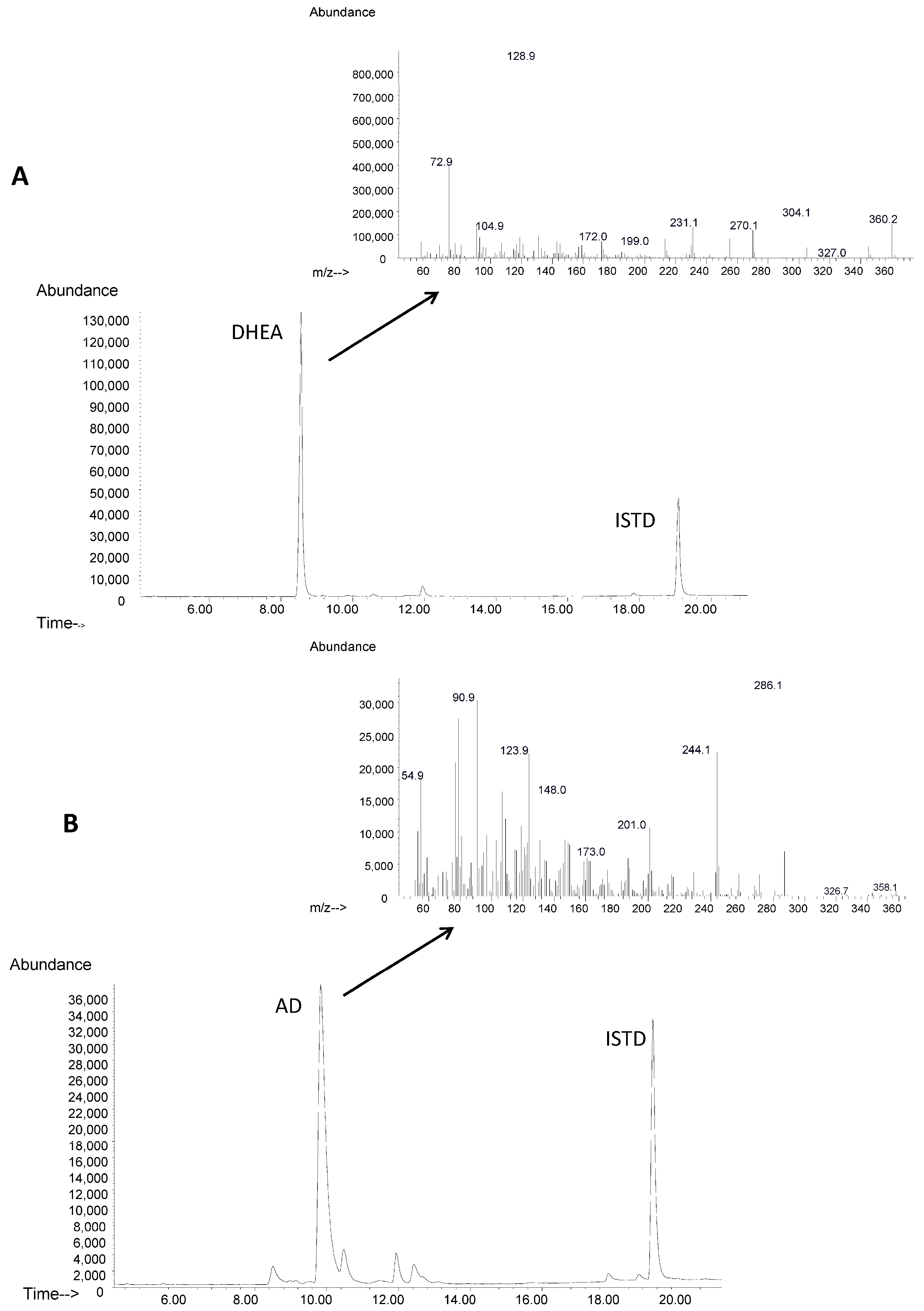
4.5. GC/MS Analysis: Identification and Quantification of DHEA and AD

To perform GC/MS analysis, culture aliquots were extracted twice at various extents of incubation with an equal volume of chloroform. Previous to its extraction, 700 mM of cholestenone dissolved in chloroform was added to the aliquots as internal standard (ISTD). The chloroform fraction was concentrated by evaporation and thetrimethylsilyl ether derivatives were formed by reaction with 50 μL of BSTFA and 50 μL of pyridine and heating at 60 °C for 45 min. Calibration standards were derivatized in the same way. The GC/MS analysis was carried out using an Agilent 7890A gas chromatograph coupled to an Agilent 5975C mass detector (Agilent Technologies, Palo Alto, CA, USA). Mass spectra were recorded in electron impact (EI) mode at 70 eV within the m/z range 50–550. The chromatograph was equipped with a 30 m × 0.25 mm i.d. capillary column HP-5MS (0.25 µm film thickness) (5% diphenyl 95% dimethylpolysiloxane from Agilent Technologies). Working conditions in the sample were as follows: split ratio (20:1), injector temperature, 240 °C; column temperature 240 °C for 3 min, then heated to 270 °C at 2.5 °C/min and heated again to 300 °C at 10 °C/min during 3 min. Total chromatographic time was 21 min. The mass spectra were recorded using full-scan mode (m/z 50–550) and SIM (selected ion monitoring) mode (m/z 360 (DHEA; characteristic ion 360.2 corresponding to trimethylsilyl ether derivatives); m/z 286 (AD)). EI mass spectra and retention data were used to assess the identity of compounds by comparing them with those of standards in the NIST Mass Spectral Database (NIST 2011) and commercial standards.

4.6. LC/MS Analysis: Identification and Quantification of Epoxymethylpregnenolone and Epoxymethylprogesterone

To perform LC/MS analysis, culture aliquots were extracted twice at various extents of incubation with an equal volume of chloroform. Previous to its extraction, 700 mM of cholestenone dissolved in chloroform was added to the aliquots as internal standard (ISTD). The chloroform fraction was concentrated by evaporation and then dissolved in acetonitrile, each sample (25 µL) was subjected to chromatographic analysis by LC/MS. Experiments were carried out using a DAD detector and a LXQ Ion Trap Mass Spectrometer, equipped with an atmospheric pressure chemical ionization source, and interfaced to a Surveyor Plus LC system (all from Thermo Electron, San Jose, CA, USA). Data were acquired with a Surveyor Autosampler and MS Pump and analyzed with the Xcalibur Software (from Thermo-Fisher Scientific, San Jose, CA, USA). All experiments were carried out with the following interface parameters: a capillary temperature of 350 °C, 60 °C for the gas temperature in the vaporizer, a capillary voltage of 41 V, an amplifier of 400 Vp, power source 6.00 µA and collision dissociation energy 15 eV. Chromatographic separation was performed on a Mediterranea Sea C18 column (4.6 mm × 150 mm, particle size 5 mm) (Teknokroma, Barcelona, Spain). The chromatography was performed using water containing 0.1% (v/v) of formic acid, acetonitrile containing 0.1% (v/v) of formic acid an isopropanol containing 0.1% (v/v) of formic acid as mobile phases A, B, and C, respectively (flow 1 mL min−1). Gradient was as follows: 45% A and 55% B for 5 min; reaching to 95% B and decreasing to 5% A in 10 min; hold 95% B, decreasing to 0% A and reaching to 5% C for 5 min; hold 0% A, decreasing to 70% B and reaching to 30% C for 15 min and return 45% A, 55% B, and 0% C in 5 min. MS analysis was performed in selected ion monitoring (SIM) mode by scanning all the daughter ions of the products in positive ionization mode. The quantification was performed from parent mass of compounds and the specificity was obtained by following the specific fragmentations of all compounds (m/z 327 (epoxymethylpregnenolone; characteristic ion 327 corresponding to dehydration after protonation of DHEA); m/z 343 (epoxymethylprogesterone)).

5. Conclusions

Our results demonstrate that the biotechnological production of epoxymethylprogesterone can be performed satisfactorily using C. glutamicum cells expressing the 3β-hydroxysteroid dehydrogenase/isomerase (MSMEG_5228) from M. smegmatis.
In addition, the results shown in this work allow us to highlight the biotechnological potential of Corynebacterium as a chassis for the steroid industry. This potential would be substantially increased by the heterologous expression of the Mce4 steroid-uptake system, which would allow for the use of natural sterols (cholesterol and phytosterols) as raw materials.

Supplementary Materials

The following are available online at www.mdpi.com/2073-4344/7/11/316/s1, Figure S1. Substrate specificity of the 3β-hydroxysteroid dehydrogenase/isomerase MSMEG_5228. The in vitro enzymatic assay was performed using protein extract from R. erythropolis (pTip5228). The specific activity for each substrate is shown. The substrates (150 μM) were solubilized in 3.33% ethanol and the enzymatic assay was carried out using buffer TAPS 100 mM pH 8.5 and 2.8 mM NAD+ as a cofactor.

Acknowledgments

The technical work of Ana Valencia is greatly appreciated. This work was supported by grants from the Ministry of Science and Innovation (BFU2009-11545-C03-03 and Ministry of Economy and Competitiveness (BIO2012-39695-C02-01; RTC-2014-2249-1). Grant RTC-2014-2249-1 is also supported by FEDER from the European Commission. J.G.-F. was supported by an FPI fellowship from the Spanish Ministry of Science and Innovation. We thank Xavier Meniche for providing plasmid pCGL0482.

Author Contributions

J.L.G. and B.G. conceived and designed the experiments; J.G.-F. performed the experiments; J.L.B. contributed reagents/materials/analysis tools; J.G.-F. and C.F.-S. analyzed the data; B.G. and J.L.G. wrote the paper.

Conflicts of Interest

The authors declare no conflicts of interest.

References

  1. Kalinowski, J.; Bathe, B.; Bartels, D.; Bischoff, N.; Bott, M.; Burkovski, A.; Dusch, N.; Eggeling, L.; Eikmanns, B.J.; Gaigalat, L.; et al. The complete Corynebacterium glutamicum ATCC 13032 genome sequence and its impact on the production of L-aspartate-derived amino acids and vitamins. J. Biotechnol. 2003, 104, 5–25. [Google Scholar] [CrossRef]
  2. Ikeda, M.; Nakagawa, S. The Corynebacterium glutamicum genome, features and impacts on biotechnological processes. Appl. Microbiol. Biotechnol. 2003, 62, 99–109. [Google Scholar] [CrossRef] [PubMed]
  3. De Lorenzo, V. Chassis organism from Corynebacterium glutamicum, the way towards biotechnological domestication of Corynebacteria. Biotechnol. J. 2015, 10, 244–245. [Google Scholar] [CrossRef] [PubMed]
  4. Unthan, S.; Baumgart, M.; Radek, A.; Herbst, M.; Siebert, D.; Brühl, N.; Bartsch, A.; Bott, M.; Wiechert, W.; Marin, K.; et al. Chassis organism from Corynebacterium glutamicum—A topdown approach to identify and delete irrelevant gene clusters. Biotechnol. J. 2014, 10, 290–301. [Google Scholar] [CrossRef] [PubMed]
  5. Becker, J.; Wittman, C. Bio-based production of chemicals, materials and fuels-Corynebacterium glutamicum as versatile cell factory. Curr. Opin. Biotechnol. 2012, 23, 631–640. [Google Scholar] [CrossRef] [PubMed]
  6. Charney, W.; Nobile, A.; Sutter, D.; Herzog, H.L.; Payne, C.C.; Tully, M.E.; Gentles, M.J.; Hershberg, E.B. Microbiological transformation of steroids—XI, The action of Corynebacterium simplex on non-corticoid steroid substrates. Tetrahedron 1962, 18, 591–596. [Google Scholar] [CrossRef]
  7. Zhang, X.; Wu, D.; Yanga, T.; Xu, M.; Rao, Z. Over-expression of Mycobacterium neoaurum 3-ketosteroid-Δ1-dehydrogenase in Corynebacterium crenatum for efficient bioconversion of 4-androstene-3,17-dione to androst-1,4-diene-3,17-dione. Elec. J. Biotechnol. 2016, 24, 84–90. [Google Scholar] [CrossRef]
  8. Donova, M.V.; Egorova, O.V. Microbial steroid transformations, current state and prospects. Appl. Microbiol. Biotechnol. 2012, 94, 1423–1447. [Google Scholar] [CrossRef] [PubMed]
  9. García, J.L.; Uhía, I.; Galán, B. Catabolism and biotechnological applications of cholesterol degrading bacteria. Microb. Biotechnol. 2012, 5, 679–699. [Google Scholar] [CrossRef] [PubMed]
  10. Galán, B.; Uhía, I.; García-Fernández, E.; Martínez, I.; Bahíllo, E.; de la Fuente, J.L.; Barredo, J.L.; Fernández-Cabezón, L.; García, J.L. Mycobacterium smegmatis is a suitable cell factory for the production of steroidic synthons. Microb. Biotechnol. 2017, 10, 138–150. [Google Scholar] [CrossRef] [PubMed]
  11. Donova, M.V. Steroid Bioconversions. Methods Mol. Biol. 2017, 1645, 1–13. [Google Scholar] [PubMed]
  12. Kreit, J. Microbial catabolism of sterols, focus on the enzymes that transform the sterol 3β-hydroxy-5-en into 3-keto-4-en. FEMS Microbiol. Lett. 2017, 364. [Google Scholar] [CrossRef] [PubMed]
  13. Uhía, I.; Galán, B.; Morales, V.; García, J.L. Initial step in the catabolism of cholesterol by Mycobacterium smegmatis mc2 155. Environ. Microbiol. 2011, 13, 943–959. [Google Scholar] [CrossRef] [PubMed]
  14. Molnár, I.; Hayashi, N.; Choi, K.-P.; Yamamoto, H.; Yamashita, M.; Murooka, Y. Bacterial cholesterol oxidases are able to act as flavoprotein-linked ketosteroid monooxygenases that catalyse the hydroxylation of cholesterol to 4-cholesten-6-ol-3-one. Mol. Microbiol. 1993, 7, 419–428. [Google Scholar] [CrossRef] [PubMed]
  15. Yamashita, M.; Toyama, M.; Ono, H.; Fujii, I.; Hirayama, N.; Murooka, Y. Separation of the two reactions, oxidation and isomerization, catalyzed by Streptomyces cholesterol oxidase. Protein Eng. 1998, 11, 1075–1081. [Google Scholar] [CrossRef] [PubMed]
  16. Mohn, W.W.; van der Geize, R.; Stewart, G.R.; Okamoto, S.; Liu, J.; Dijkhuizen, L.; Eltis, L.D. The actinobacterial mce4 locus encodes a steroid transporter. J. Biol. Chem. 2008, 283, 35368–35374. [Google Scholar] [CrossRef] [PubMed]
  17. García-Fernández, J.; Papavinasasundaram, K.; Galán, B.; Sassetti, C.M.; García, J.L. Unravelling the pleiotropic role of the MceG ATPase in Mycobacterium smegmatis. Environ. Microbiol. 2017, 19, 2564–2576. [Google Scholar] [CrossRef] [PubMed]
  18. Pollegioni, L.; Piubelli, L.; Molla, G. Cholesterol oxidase, biotechnological applications. FEBS J. 2009, 276, 6857–6870. [Google Scholar] [CrossRef] [PubMed]
  19. Kumari, L.; Kanwar, S.S. Cholesterol oxidase and its applications. Adv. Microbiol. 2012, 2, 49–65. [Google Scholar] [CrossRef]
  20. Alexander, D.L.; Fisher, J.F. A convenient synthesis of 7α-hydroxycholest-4-en-3-one by the hydroxypropyl-β-cyclodextrin-facilitated cholesterol oxidase oxidation of 3β,7α-cholest-5-ene-3,7-diol. Steroids 1995, 60, 290–294. [Google Scholar] [CrossRef]
  21. Labaree, D.; Hoyte, R.M.; Hochberg, R.B. A direct stereoselective synthesis of 7β-hydroxytestosterone. Steroids 1997, 62, 482–486. [Google Scholar] [CrossRef]
  22. Biellmann, J.F. Resolution of alcohols by cholesterol oxidase from Rhodococcus erythropolis, lack of enantiospecificity of the steroids. Chirality 2001, 13, 34–39. [Google Scholar] [CrossRef]
  23. Guo, L.-W.; Wilson, W.K.; Pang, J.; Shackleton, C.H.L. Chemical synthesis of 7- and 8-dehydro derivatives of pregnane-3,17α,20-triols, potential steroid metabolites in Smith-LemLi-Opitz syndrome. Steroids 2002, 68, 31–42. [Google Scholar] [CrossRef]
  24. Yang, X.; Dubnau, E.; Smith, I.; Sampson, N.S. Rv1106c from Mycobacterium tuberculosis is a 3beta-hydroxysteroid dehydrogenase. Biochemistry 2007, 46, 9058–9067. [Google Scholar] [CrossRef] [PubMed]
  25. Horinouchi, S.; Ishizuka, H.; Beppu, T. Cloning, nucleotide sequence, and transcriptional analysis of the NAD(P)-dependent cholesterol dehydrogenase gene from a Nocardia sp. and its hyperexpression in Streptomyces spp. Appl. Environ. Microbiol. 1991, 57, 1386–1393. [Google Scholar] [PubMed]
  26. Kishi, K.; Watazu, Y.; Katayama, Y.; Okabe, H. The characteristics and applications of recombinant cholesterol dehydrogenase. Biosci. Biotechnol. Biochem. 2000, 64, 1352–1358. [Google Scholar] [CrossRef] [PubMed]
  27. García-Fernández, J.; Papavinasasundaram, K.; Galán, B.; Sassetti, C.M.; García, J.L. Molecular and functional analysis of the MCE4 operon in Mycobacterium smegmatis. Environ. Microbiol. 2017, 19, 3689–3699. [Google Scholar] [CrossRef] [PubMed]
  28. Snapper, S.B.; Melton, R.E.; Mustafa, S.; Kieser, T.; Jacobs, W.R., Jr. Isolation and characterization of efficient plasmid transformation mutants of Mycobacterium smegmatis. Mol. Microbiol. 1990, 4, 1911–1919. [Google Scholar] [CrossRef] [PubMed]
  29. Kilbane, J.J. Desulfurization of coal, the microbial solution. Trends Biotechnol. 1989, 7, 97–101. [Google Scholar] [CrossRef]
  30. Santamaria, R.; Gil, J.A.; Martin, J.F. High-frequency transformation of Brevibacterium lactofermentum protoplasts by plasmid DNA. J. Bacteriol. 1985, 162, 463–467. [Google Scholar] [PubMed]
  31. Nakashima, N.; Tamura, T. Isolation and characterization of a rolling-circle-type plasmid from Rhodococcus erythropolis and application of the plasmid to multiple-recombinant-protein expression. Appl. Environ. Microbiol. 2004, 70, 5557–5568. [Google Scholar] [CrossRef] [PubMed]
  32. Peyret, J.L.; Bayan, N.; Joliff, G.; Gulik-Krzywicki, T.; Mathieu, L.; Schechter, E.; Leblon, G. Characterization of the cspB gene encoding PS2, an ordered surface-layer protein in Corynebacterium glutamicum. Mol. Microbiol. 1993, 9, 97–109. [Google Scholar] [CrossRef] [PubMed]
Figure 1. Expression of MSMEG_5228 gene from M. smegmatis in heterologous hosts. (A) SDS-gel electrophoresis of protein extracts. Lane 1 Molecular weight marker; Lanes 2 and 3, soluble and pellet fractions from E. coli (pET5228). (B) Lanes 1 and 6, Molecular weight markers; Lanes 2 and 3, soluble and pellet fractions from R. erythropolis (pTipQC1), respectively; Lanes 4 and 5, soluble and pellet fractions from R. erythropolis (pTip5228), respectively. (C) Lanes 1 and 3, soluble and pellet fractions from C. glutamicum R31 (pCGL5228), respectively. Lane 2, Molecular weight marker. (D) Reactions studied in this work that are carried out by the 3β-HSD mycobacterial enzyme.
Catalysts 07 00316 g001aCatalysts 07 00316 g001b
Figure 2. Transformation of DHEA into AD by C. glutamicum R31 (pCGL5228). Cells of C. glutamicum (pCGL5228) were incubated in a resting-cell medium for 28 h with 2 mM DHEA at 30 °C. (A) Analysis by GC-MS of the reaction products at 0 h and fragmentation pattern of DHEA. (B) Analysis by GC-MS of the reaction products after 28 h and fragmentation pattern of AD. ISTD: internal standard.
Catalysts 07 00316 g002
Figure 3. Production of epoxymethylprogesterone by C. glutamicum R31 (pCGL5228). Resting-cells assays of the control strain C. glutamicum R31 (pCGL0482) (A) or the producer strain C. glutamicum R31 (pCGL5228) (B). The data shown are representative of 3 independent experiments performed for 28 h with 2 mM epoxymethylpregnenolone at 30 °C. The concentrations of epoxymethylpregnenolone and epoxymethylprogesterone are shown in green and red colours, respectively. Analysis by LC-MS of products of the reaction performed by C. glutamicum R31 (pCGL5228) at 0 h (C) and after 28 h of incubation (D). Chromatogram (SIM) of characteristic ions of epoxymethylpregnenolone (m/z 327) (blue) and epoxymethylprogesterone (m/z 343) (red). Chemical structure, average of mass spectra (upper), and average MS/MS spectra (product ion mass spectra) (lower) of epoxymethylpregnenolone (E) and epoxymethylprogesterone (F).
Catalysts 07 00316 g003aCatalysts 07 00316 g003bCatalysts 07 00316 g003c
Table 1. Strains and plasmids used in this work.
StrainsGenotype and/or DescriptionSource or Reference
Escherichia coli DH10BF’, mcrA Δ(mrr hsdRMS-mcrBC) F80dlacDM15 ΔlacX74 deoR recA1 araD139 Δ(ara-leu)7697 galU galK λ rpsL endA1 nupGInvitrogen
Escherichia coli BL21 (DE3)F, ompT, hsdSB(rB-mB-), gal, dcm, λDE3 (harboring gene 1 of the RNA polymerase from the phage T7 under the PlacUV5 promoter)Invitrogen
Mycobacterium smegmatis mc2155ept-1, mc²6 mutant efficient for electroporation[28]
Rhodococcus erythropolis IGTS8Isolated for oil desulfurization using dsz genes[29]
Corynebacterium glutamicum R31MeLisR, AecR. Efficient for transformation[30]
Plasmids
pET29a(+)Cloning and expression vector, Kmr, oriColE1, T7 promoterNovagen
pET5228pET29 derivative containing MSMEG_5288 gene[13]
pTipQC1Bifunctional expression vector. PtipA, repAB (pRE2895), MCS type 1, CmR[31]
pCGL0482Bifunctional expression vector E. coli/C. glutamicum. PS1 promoter (modified from plasmid pCGL482), CmR[32] (Gift from X. Meniche)
pTip5228pTipQC1 harboring the MSMEG_5228 gene cloned in NdeI/HindIII, CmRThis study
pCGL5228pCGL0482 harboring the MSMEG_5228 gene cloned in XhoI/HpaI, CmRThis study
Table 2. Oligonucleotides used in this work.
Primer NameSequence 1 (5’-3’)Use 2
pTip5228 FCGCGCATATGGCTGACTCCACCACCGACAmplification of MSMEG_5228 for cloning in pTipQC1 (NdeI)
pTip5228 RCGCGAAGCTTCGTGCTGCCCTGAACTAGAmplification of MSMEG_5228 for cloning in pTipQC1 (HindIII)
pCGL5228 FCGCGCTCGAGATGGCTGACTCCACCACCGACCTCAmplification of MSMEG_5228 for cloning in pCGL0482 (XhoI)
pCGL5228 RCGCGGTTAACGCCCGTGCTGCCCTGAACAmplification of MSMEG_5228 for cloning in pCGL0482 (HpaI)
1 Restriction sites are underlined. 2 Restriction enzymes used for cloning are indicates in brackets.

