Article

Production of Hydroxylated Steroid Intermediates at 10-g Scale via the Original Sterol Modification Pathway in Mycolicibacterium neoaurum

1
School of Life Science and Biopharmaceuticals, Shenyang Pharmaceutical University, Shenyang 110016, China
2
Shenyang Botai Biopharmaceutial Ltd., Shenyang 110043, China
*
Authors to whom correspondence should be addressed.
Catalysts 2025, 15(5), 423; https://doi.org/10.3390/catal15050423
Submission received: 25 March 2025 / Revised: 20 April 2025 / Accepted: 22 April 2025 / Published: 25 April 2025

Abstract

:
The aerobic catabolism of steroids in bacteria is highly conserved, and the mechanism of steroid degradation in mycobacteria has been extensively studied. However, the branching modification pathways of steroids in mycobacteria remain a mystery, including the likely roles of cytochromes P450. In this study, we unraveled the CYP105S17 converting androst-4-ene-3,17-dione (AD) to 17β-hydroxy-4-androstene-3,16-dione (16-oxo-TS), which was subsequently reduced to 16α,17β-dihydroxy-androst-4-ene-3-one (16α-OH-TS) under reductive conditions in Mycolicibacterium neoaurum. By applying this modification pathway, the genetically modified strains overexpressing CYP105S17 were able to produce 16α-OH-TS at titers 13.0 g/L with a conversion rate of 91.9% (supplemented with 20 g/L phytosterols as the substrate) through a two-stage biotransformation process. This is the first instance of utilizing the native P450 of Mycobacterium to produce 16-hydroxylated steroid intermediates at the 10 g scale. This work provides invaluable perspectives and guidance to researchers seeking to understand the complexities of steroid metabolism in bacteria, and also highlights the great potential of Mycobacterium as a production platform for hydroxylated steroid intermediates or pharmaceuticals.

1. Introduction

Mycobacteria fall naturally and taxonomically into two main groups: slow- and fast-growers [1]. The latter group includes non-pathogenic species such as M. neoaurum, M. smegmatis, and M. fortuitum [1]. Genetically modified strains of these bacteria are frequently employed as cell factories to convert phytosterols into valuable steroid intermediates and drugs due to their environmentally friendly reaction condition [2,3,4]. Among the fast-growing mycobacteria, M. neoaurum has gained great attention as a model strain for steroid catabolic mechanism studies, owing to its robust steroid degradation capacity, small genome, and non-pathogenic model system [4]. In mycobacteria, steroids are aerobically mineralized through an intricate series of steps that involve modification of the steroid nucleus ring A, removal of the side chain, and cleavage of nucleus rings AB and CD (Figure 1) [5,6,7]. These degradation mechanisms are highly conserved, and some of the enzymes involved in these processes remain not fully understood. By inactivating kstD and kshA genes involved in steroid AB ring degradation, production strains for pharmaceutical precursors such as androsta-4-ene-3,17-dione (AD), androsta-1,4-diene-3,17-dione (ADD), and 9-hydroxyandrostra-4-ene-3,17-dione (9-OH-AD) were developed [8,9]. These precursors can be further modified enzymatically or chemically to various advanced steroid medicines.
Cytochromes P450 (CYPs or P450s) play a crucial role in the process of sterol degradation [10]. Specifically, CYP124, CYP125, and CYP142 are engaged in the initial oxidation of sterol alkyl side chains [11,12]. P450s belong to a family of heme monooxygenase enzymes that act with a conserved mechanism to selectively insert an oxygen atom from molecular dioxygen into a carbon–hydrogen bond of the substrate, forming an alcohol product [13,14]. In addition to their involvement in sterol degradation, microbial P450 enzymes mediating the hydroxylation of steroids serves as a crucial strategy for their structural modification and functional diversification [15]. For example, 16α-hydroxyl function plays a pivotal role in synthetic glucocorticoids like triamcinolone and dexamethasone [16,17,18,19]. Steroid hydroxylation research has predominantly focused on Escherichia coli whole-cell catalysis [20,21,22]. However, due to E. coli’s lack of a steroid transport system, absence of a native electron transport chain, and limited CYP450 substrate loading capacity, achieving steroid hydroxylation at 10 g scale remains challenging. In contrast, Mycobacterium possesses an inherent steroid transport system and a rich array of native electron transport chains, making it an ideal platform for CYP450-mediated steroid hydroxylation [23,24,25]. Furthermore, integrating steroid metabolic engineering with CYP450 hydroxylation may effectively address the challenges associated with low substrate loading capacity.
In our prior investigation, we found that the AD accumulation strain SL-01, obtained through knockout of the kstD and kshA genes from M. neoaurum DSM 44074, produced two primary impurities, 21-hydroxy-20-methylpregn-4-en-3-one (BA) and 3,24-dinor-chol-4-en-21-oic acid methyl ester (3-OPCM), during phytosterols conversion (Figure 1). Concurrently, an unidentified compound was noted, displaying intermittent occurrence across fermentation processes, resembling a transient occurrence within a limited fraction. These conspicuous by-products posed considerable impediments to the viable utilization of sterol-converting mycobacteria within industrial contexts. During the investigation of the sterol side chain β-oxidative dehydrogenase FadE26-FadE27 [26,27,28], the unknown compound significantly increased after knocking out fadE26-fadE27, which was purified and identified as 17β-hydroxy-4-androstene-3,16-dione (16-oxo-TS) by Mass spectrum and NMR spectroscopy (Figure S1). To our knowledge, this product has never been reported in mycobacteria before. We postulated that the production of 16-oxo-TS is related to P450s because this product features a ketone group at position 16 and a β-hydroxyl group at position 17, which is presumably produced through a series of isomerization following the introduction of a hydroxyl group at position 16 of the steroid by certain P450 monooxygenases. This discovery implies that disruption of steroid metabolic genes could trigger potential alternations in the pathways that are largely silent in wild strains. However, the alternative steroid metabolism pathways remain largely unexplored.
In this study, we found the novel sterol modification pathway, where CYP105S17 (David R, Nelson, University of Tennessee) (http://drnelson.uthsc.edu/CytochromeP450.html, accessed on 21 April 2025) catalyzed the conversion of androst-4-ene-3,17-dione (AD) into 17β-hydroxy-4-androstene-3,16-dione (16-oxo-TS), which was subsequently reduced to 16α,17β-dihydroxy-androst-4-ene-3-one (16α-OH-TS) under reductive conditions in M. neoaurum in vivo. By engineering this pathway, we achieved the production of 16α-OH-TS at the 10 g scale.

2. Results and Discussion

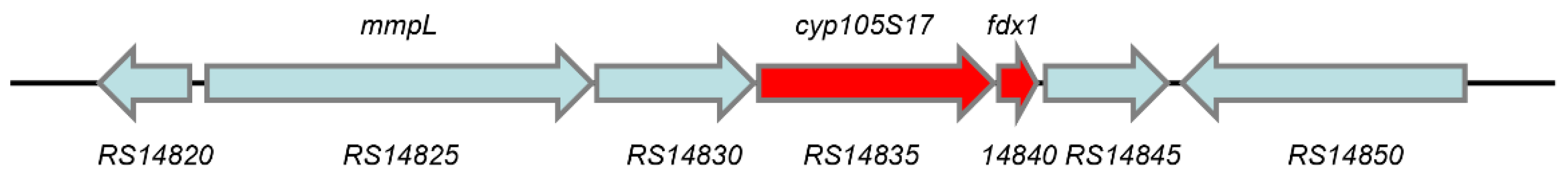
2.1. Screening of cyp450 Genes

During the examination of SL-01’s metabolism of phytosterols, we isolated a compound characterized as 16-oxo-TS from the resultant metabolites. We postulated that this compound could potentially arise from the hydroxylation catalyzed by a P450 enzyme. The protein blast annotation analysis of DSM 44074 genome (GenBank No. CCDR000000000) on NCBI (https://www.ncbi.nlm.nih.gov/, accessed on 21 April 2025) uncovered 32 P450s.
Bacterial P450s that are capable of hydroxylating the steroid at 16 position were reported as the CYP154 [29,30], CYP105 [31,32], and CYP109 families [33]. The CYP154 family predominantly targets progesterone for 16α-position hydroxylation, whereas the CYP105 and CYP109 families efficiently hydroxylate a variety of organic compounds, such as vitamin D3. In DSM 44074, only two P450s belonging to the CYP105 family were annotated, namely CYP105S17 (WP_030134122.1) and CYP105Q9 (WP_030137330.1), with the corresponding FDXs located in the same operon named FDX1 (WP_030134121.1) and FDX2 (WP_030137329.1), respectively. Three iron–sulfur protein reductases, registered in NCBI as WP_090559269.1, WP_030134859.1, and WP_030134758.1, were identified and designated as FDR1, FDR2, and FDR3, respectively. The antiSMASH analysis revealed that both cyp450 genes were not in the secondary metabolism gene clusters [34]. However, cyp105S17 was found to be co-localized with the MMPL family transporter on the same operon (Figure 2), suggesting its potential involvement in the synthesis or metabolism of specific lipids, and worth further investigation [35].
The CYP105 family comprises at least 17 subfamilies, predominantly identified in Streptomyces species [32,36]. Members of this family are generally categorized into two functional classes: those involved in the biosynthesis of bioactive compounds and those participating in the biotransformation and biodegradation of xenobiotics [37,38]. The broad substrate specificity and functional diversity observed among CYP105 enzymes suggest significant variability within the tertiary structural elements of their active sites. This structural diversity underpins their capacity to accommodate a wide range of substrates, positioning many CYP105 enzymes as promising biocatalysts for the synthesis of novel compounds [39]. Bacterial CYP105 enzymes have been extensively investigated for their catalytic potential in diverse applications, including steroid oxidation and the production of macrolide-derived metabolites [32,39]. For instance, CYP105A1 has been shown to metabolize sulfonylurea herbicides [40], while CYP105A2 catalyzes the hydroxylation of vitamin D3 to produce 25-hydroxyvitamin D3 [41]. CYP105D7 demonstrates the ability to hydroxylate steroid compounds at multiple positions [31]. Additionally, CYP105AB3 exhibits remarkable substrate promiscuity, catalyzing the hydroxylation of a wide range of molecules including fatty acids, steroids, and diverse aromatic compounds [39]. CYP105S17 is a member of the CYP105S subfamily and exhibits the closest phylogenetic relationship to the CYP105S1, CYP105C1, and CYP105AS1 [38,42,43], sharing 44.7%, 42.3%, and 44% sequence similarity, respectively. However, the functional roles of CYP105S1 and CYP105C1 remain uncharacterized, with CYP105C1 only known to be co-localized with a cholesterol oxidase gene within the same operon [42,44]. CYP105AS1 is capable of converting compactin into pravastatin [38]. Sequence alignment analysis indicated that CYP105S17 comprises 14 α-helices and 10 β-sheets. Notably, it possesses a conserved acid–alcohol residue pair (E242 and T243), whereas in CYP105C1, the corresponding glutamic acid is substituted by glycine. The threonine residue within this pair is highly conserved across cytochrome P450 enzymes and is critically involved in oxygen activation, a key step in CYP-mediated catalysis [45]. In addition, the ExxR sequence motif, which is important for maintaining the structural integrity of the enzyme, is conserved in CYP105S17 but absent in CYP105S1 due to the loss of the crucial arginine residue [36]. The conserved cysteine residue, which serves as the fifth ligand to the heme iron, is located at position 350 (Figure S3) [36]. Collectively, these features strongly suggest that CYP105S17 retains catalytic functionality, whereas CYP105C1 and CYP105S1 may be catalytically inactive due to the absence of key structural and functional elements. Furthermore, CYP105Q9 shares the highest sequence homology (73.2%) with the reported CYP105Q4, although the function of CYP105Q4 has yet to be elucidated [43]. Based on the aforementioned analyses, CYP105S17 and CYP105Q9 were chosen as the target genes for this study.

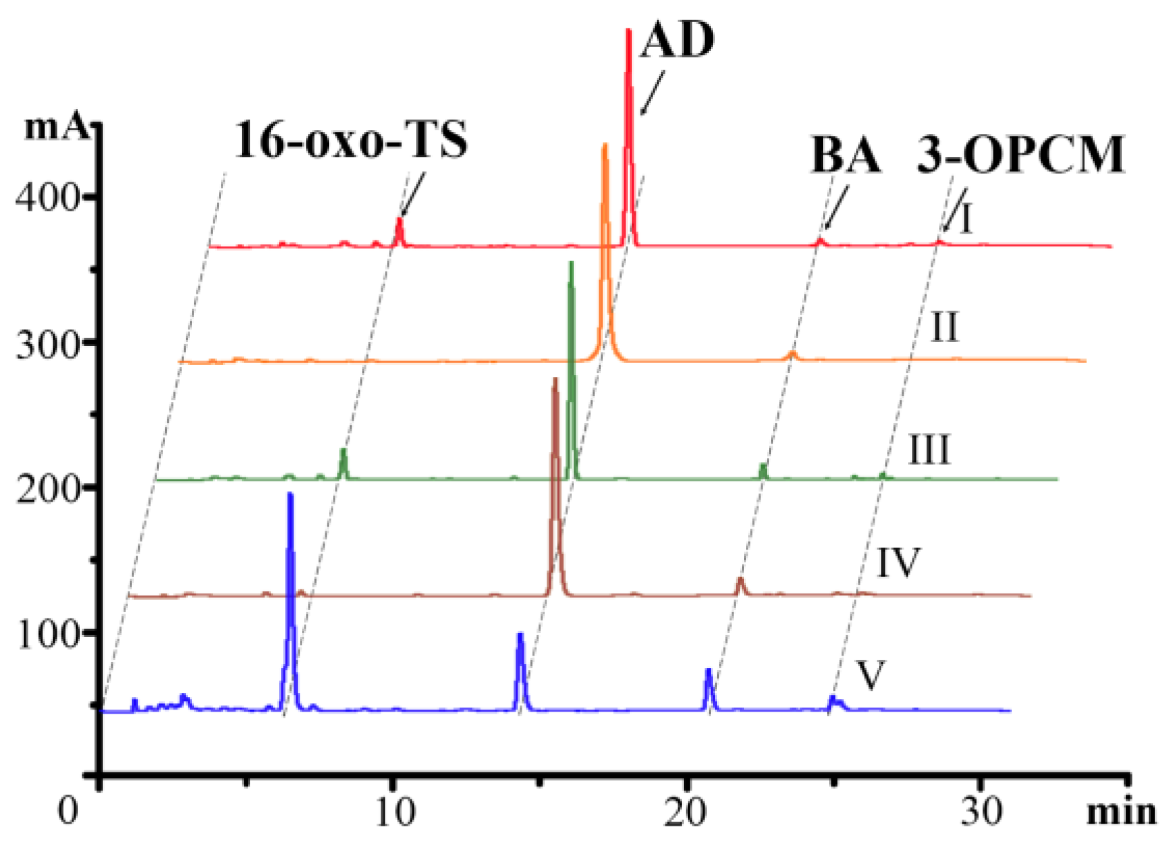
2.2. Analysis of Sterol Transformation Products of Mutant Strains

Five strains, namely SL-01, SL-01∆cyp105S17, SL-01∆cyp105Q9, SL-01 ∆cyp105S17cyp105S17, and SL-01∆cyp105S17cyp105S17-fdx1, were constructed (gene knockout results are shown in Figure S4) and subjected to fermentation with phytosterols as the substrate. The resulting samples were extracted and assayed by HPLC. The results indicated that in the absence of the cyp105S17 gene, the 16-oxo-TS was not found in sterol transformation. However, the 16-oxo-TS production was restored only after the corresponding fdx1 gene was co-expressed with cyp105S17 in the strain (Figure 3). This effect could be attributed to the polarity effect of the upstream cyp105S17 gene knockout. Furthermore, the amount of 16-oxo-TS increased in the complementation strain, which corresponded with an increased copy number of the P450 gene. Based on these results, it can be concluded that CYP105S17 plays a vital role in the formation of 16-oxo-TS, whereas no such association was observed for CYP105Q9.

2.3. P450 Substrate Determination

Since knockout of acyl-coenzyme A dehydrogenase gene fadE26-fadE27 resulted in an increase in 16-oxo-TS, we deleted fadE28-fadE29 and performed a combined knockdown of fadE26-fadE27 and fadE28-fadE29. The results showed that the yield of 16-oxo-TS in SL-01ΔfadE28-fadE29 sterol transformation remained the same, whereas SL-01∆fadE26-fadE27&fadE28-fadE29 did not produce any 16-oxo-TS. It is reasonable to speculate that 16-oxo-TS might originate from a downstream product of 3-OPC-CoA (Figure 4a). The degradation of sterol side chains after FadE28-FadE29 dehydrogenation requires two additional steps of hydration and aldol condensation for complete degradation. The combination deletion of fadE26-fadE27 and fadE28-fadE29 blocked the complete side chain degradation to AD through 3-OPDC-CoA and 17-HOPC-CoA [28] (Figure 1). Previously, we found that the transformation of sterols by SL-01Δltp2 led to the block of AD production (Figure 4a). Therefore, it was postulated that 16-oxo-TS may be derived from the complete sterol side chain degradation product AD. Thus, it was tentatively hypothesized that 16-oxo-TS likely results from the direct hydroxylation of AD.
Next, we performed fermentation transformation of AD using different SL-01 mutants. As shown in Figure 4b, SL-01 was able to convert AD to 16-oxo-TS in small amounts, while SL-01∆cyp105S17 was not, supporting the hypothesis that AD is the substrate of CYP105S17. When the transformation time reached 48 h, SL-01∆cyp105S17::cyp105S17-fdx1 generated two products. After isolation and purification, we found that the metabolite with a retention time of 4.8 min was 16α,17β-dihydroxyandrost-4-ene-3-one (16α-OH-TS) (Figure 4b and Figure S2), and the other metabolite with a retention time of 6.4 min was 16-oxo-TS. The generation of four transformation products of AD was observed at earlier transformation periods such as 24 h (Figure 4b). In addition to 16α-OH-TS and 16-oxo-TS, we also observed two metabolites with retention times of 6.2 min and 6.6 min with molecular weights of 302 and 304, respectively (Figure 4b). The metabolite with a molecular weight of 302 at a retention time of 6.2 min was isolated. But after purification and identification, it was still 16-oxo-TS. 16β-hydroxy-4-androstene-3,17-dione (16β-OH-AD) and 16-oxo-TS are a pair of isomers that can be interconverted into each other, with 16-oxo-TS being more stable. Under long-term room temperature or acidic conditions, 16β-OH-AD can be converted into 16-oxo-TS. This phenomenon has been observed during the conversion of AD in Aspergillus niger ATCC 9142 [46,47]. For the standard 16α-OH-AD, it has a retention time of 7.2 min (Figure 4b), which does not match the retention time of 6.2 min observed in this experiment. During the late stage of conversion, the pH of the culture medium was around 6, presenting an acidic environment conducive to the isomerization of 16β-OH-AD to16-oxo-TS. Unfortunately, we were unable to separate the metabolite with a molecular weight of 304 and a retention time of 6.6 min as it quickly disappeared in the culture medium. Its appearance in the liquid chromatography profile was sporadic and only observed in a few of the repeated conversions. However, these two metabolites eventually turned into 16-oxo-TS at 48 h (Figure 4b). It was also discovered that M. neoaurum hardly hydroxylated AD when the substrate concentration exceeded 0.5 g/L (P450 substrate inhibition) (Figure 4b). This phenomenon explains why SL-01ΔfadE26-fadE27 produced more 16-oxo-TS than M. neoaurum SL-01, because the knockout of fadE26-fadE27 weakens the metabolic process from phytosterols to AD, which in turn slows down the AD production, and facilitates the hydroxylation of AD by CYP105S17.
To further validate the CYP105S17 role, we constructed a series of E. coli expression strains for the gene expressions and biotransformation. We used E. coli BL21(DE3)-pRSFduet1 as the control and BL21(DE3)-pRSF-cyp105S17-fdx1-fdr1 as the experimental group for AD transformation. The results showed that BL21(DE3)-pRSF-cyp105S17-fdx1-fdr1 transformed AD with the same effect, producing the metabolite 16β-OH-AD and 16-oxo-TS (Figure S5). However, the former was all converted to 16-oxo-TS during the purification process, just like in mycobacteria. It was observed that all E. coli failed to produce the metabolite with a retention time of 6.6 min and molecular weight of 304, indicating that the metabolite is an intermediate of AD transformation in M. neoaurum.
In order to investigate the mechanism of product formation of CYP105S17, we employed the optimized AlphaFold protein model for molecular docking. The docking results revealed that the CYP105S17 model has a small substrate binding cavity that can accommodate AD (Figure S6a). The distance between C-16 of AD and the iron atom at the center of the porphyrin ring was 3.4 Å and the C16-H16-β-Fe angle was 144.8° (Figure S6b). These values fall within the range for hydroxylation, as the distance was less than 4.9 Å and the angle was between 135° and 180°, indicating that the 16β position can be hydroxylated [48,49]. Furthermore, CYP105S17 mainly forms two hydrogen bonds with the keto oxygen atoms at positions 3 and 17 of AD through the threonine and histidine at position 243 and 288, respectively (Figure S6b). CYP105S17 then stabilizes AD in the binding pocket through hydrophobic interactions of leucine at position 92, alanine at position 239, valine at position 286, phenylalanine at position 390, and isoleucine at position 391 (Figure S6b).
Structural prediction and molecular docking analyses of CYP105S17 with AD identified threonine at position 243 and histidine at position 288 as potentially critical residues for catalytic function. To validate their importance, site-saturation mutagenesis was performed at these two positions using pMV-cyp105S17-fdx1 as the template. The resulting mutant plasmids were transformed into M. neoaurum SL-01Δcyp105S17, and resting cell biotransformation assays were conducted with AD as the substrate. As summarized in Table S3, all amino acid substitutions at either position 243 or 288 completely abolished the enzymatic activity of CYP105S17 toward AD. These findings align with the docking predictions and highlight the essential role of hydrogen bonding in substrate recognition and catalytic turnover.

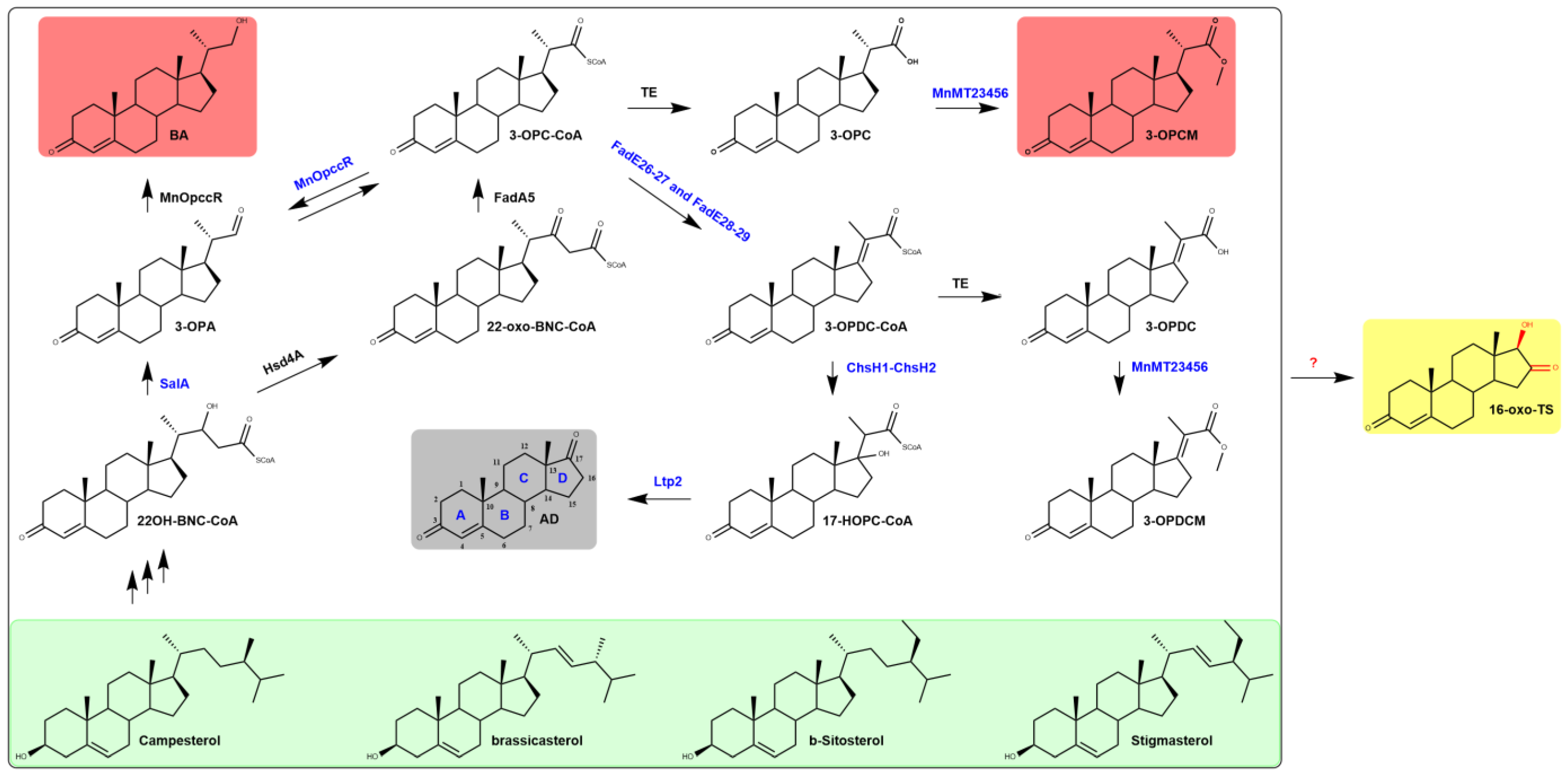
2.4. A Novel Steroid D-Ring Modification Pathway in M. neoaurum

Based on the results obtained from in vivo transformation experiments of AD M. neoaurum mutants, we postulated the mechanism of inter conversion of 16,17 oxidation products after sterol side chain degradation. The conversion of AD by CYP105S17 in SL-01 does not produce 16α-OH-AD (Figure 4b). 16β-OH-AD is quickly isomerized to 16-oxo-TS; the latter can be further reduced into 16α-OH-TS, rather than from 16α-OH-AD by 17β reduction (Figure 5). Initially, it was assumed that the structure of retention time 6.6 min metabolite (compound six) was 16β-OH-TS. However, the β-dehydrogenases present in Mycobacterium exhibit high stereospecificity in steroid compounds, mainly implicated in redox reactions at the 3- and 17-carbon positions of the steroid parent nucleus, and to some extent also at the 16-carbon position [50]. Nonetheless, the oxidation of the 17β-hydroxyl group will be impeded in the presence of the ketone group or the 16β-hydroxyl group at position 16 [50]. Conversely, the presence of the 17β-hydroxyl group will block the oxidation of the 16β-hydroxyl group [50]. If compound six is 16β-OH-TS, it will exist perpetually. However, during the conversion of AD by SL-01, compound six is only a transient intermediate and eventually becomes 16-oxo-TS, while 16β-OH-AD converts to 16-oxo-TS even during the purification process. Although the product of 16,17-diketo was not isolated in the experiment, a metabolite with a molecular weight of 300, compound four, was detected in one of the transformation experiments. Moreover, a small amount of 17α-hydroxy-androst-1,4-diene-3,16-dione has been isolated and identified during the purification of ADD impurities in our laboratory, which has the same steroid D ring structure as compound five. Therefore, it was postulated that the compound with a retention time of 6.6 min is compound six (16β,17α)-16,17-Dihydroxyandrost-4-en-3-one. This metabolite is known to be formed in mammalian liver microsomes during the metabolism of 16-dehydro derivatives of C18 and C19 steroids, via the intermediate 16α,17α-epoxy-4-androsten-3-one, under the action of microsomal epoxide hydratase [51,52,53,54]. Moreover, this compound can also be produced by Aspergillus niger ATCC 9142 during the transformation of testosterone [55]. It can be speculated that this compound may be a naturally occurring metabolite generated during the biological metabolism of certain steroid compounds. Numerous reductases are present in M. neoaurum, and some of them convert the 16-position ketone group of steroids to the α-hydroxyl group. Since these oxidoreductases are reversible, the 16α-hydroxyl group will eventually transform back into a ketone group. Interestingly, if a sufficient reductive environment is provided in the late stage of fermentation, 16-oxo-TS will continue to transform into 16α-OH-TS (Figure 6a), which is an inhibitor of aromatase and can also be utilized for the synthesis of glucocorticoids and estriol. As a result, the steroid D-ring modification pathway mediated by CYP105S17 was systematically elucidated (Figure 5).

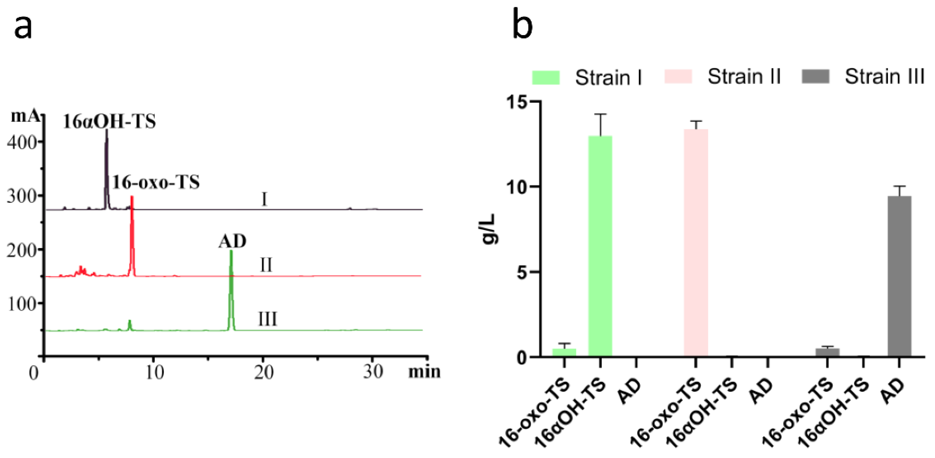
2.5. Construction and Evaluation of a High-Yield 16α-OH-TS Engineered Strain

Based on the steroid D-ring modification pathway, a combinatorial strategy was employed to achieve one-step production of 16α-OH-TS from phytosterols. To eliminate the impact of BA, the salA and opccR genes were knocked out, leading to the construction of engineered strain SL-01∆salA&opccR [56,57]. Upon transformation of 20 g/L phytosterols, SL-01∆salA&opccR yielded 9.5 g/L AD and 0.5 g/L 16-oxo-TS with no detectable 3-OPCM, achieving a sterol conversion efficiency of 75% (Figure 6a,b). Using a two-stage fermentation approach with engineered strain SL-01∆salA&opccRcyp105S17-fdx1 and 20 g/L sterols as the substrate, the production conversion rate of 16α-OH-TS reached 91.9%, with a titer of 13.0 g/L. This titer surpasses the current production yields attainable through E. coli catalysis and was achieved using unmodified CYP450 [29]. These results highlight the significant potential of this pathway and the considerable potential of integrating Mycobacterium steroid metabolic engineering with CYP450 expression for the efficient biosynthesis of hydroxylated steroid intermediates.
Engineered strains harboring CYP105S17 exhibit higher sterol conversion rates to 16-oxo-TS or 16α-OH-TS compared to AD-producing strains. This could be attributed to the intracellular localization and cytotoxicity of AD [58], while 16-oxo-TS or 16α-OH-TS can be excreted extracellularly for their stronger polarity. CYP105S17 serves as a detoxifier, which could be an inherent factor contributing to the production of 16-oxo-TS or 16α-OH-TS, similar to the role of carboxyl methyltransferase in converting 3-OPC to 3-OPCM [59]. The close phylogenetic relationship between the CYP105S subfamily and CYP105C1, which is co-localized with cholesterol oxidase within the same operon, suggests that steroid compounds are likely the physiological substrates of CYP105S17 enzymes. Given that cholesterol oxidase is indispensable for sterol degradation, it is plausible that at a certain stage of Mycolicibacterium evolution, the bacterium lacked the capacity for complete sterol catabolism. CYP105S17 may have been acquired through horizontal gene transfer to facilitate steroid detoxification. As Mycolicibacterium evolved to fully degrade sterols for enhanced energy acquisition, CYP105S17 was retained but gradually degenerated and became dissociated from the sterol degradation gene cluster.
The aforementioned findings unequivocally indicate that the metabolic fate of the compound in bacterial systems is not confined to a singular pathway. Under normal conditions, the predominant metabolic pathway governs the degradation of the compound, ensuring metabolic homeostasis. However, upon exposure to dynamic physicochemical contexts or genetic perturbations, bacteria exhibit ancillary routes for metabolism, thereby adapting to prevailing metabolic homeostasis and modulating diverse biological processes in nature. Remarkably, these transformed metabolites may possess profound industrial significance as intermediates or therapeutic agents. The ubiquity of such alternative metabolic pathways across microbial landscapes underscores their latent potential, awaiting comprehensive exploration and practical application. Future investigations should thus prioritize the systematic elucidation of these intricate metabolic networks, unveiling novel opportunities for drug discovery and biotechnological advancements.

3. Materials and Methods

3.1. Strains, Plasmids, Reagents, and Culture Conditions

The bacterial strains and plasmids used in this study are listed in Table S1. Oligonucleotides are listed in Table S2. All the mycobacterial strains were cultured in seed medium (soy peptone 5.0 g/L, yeast extract 0.5 g/L, glycerol 0.5% (v/v), glucose 1 g L−1, tween 80 0.4% (v/v), pH 7.0). The steroid biotransformation by Mycolicibacterium cells was executed in the fermentation medium, which contains soy peptone 10 g/L, NH4NO3 10 g/L, K2HPO4 5.0 g/L, ammonium ferric citrate 0.05 g/L, corn steep 10 g/L, sodium citrate 1.0 g/L, MgSO4·7H2O 0.2 g/L, glycerol 0.5% (v/v). The pH was adjusted to 7.0. Steroids (phytosterols and other steroidal compounds) were emulsified in tween 80 by grinding, and then transferred to the fermentation medium and autoclaved at 121 °C for 1 h. The final volume ratio of tween 80 cannot exceed 0.5%. The phytosterol mixture (β-sitosterol 47.5%, campesterol 26.4%, stigmasterol 17.7%, and brassicasterol 3.6%) was purchased from Davi Biochemistry (Huzhou, China). Cholesterol, AD, and TS were purchased from Shanghai Saen Chemical Technology Co. Ltd. (Shanghai, China). Other materials were purchased from DINGGUO CHANGSHENG, Beijing in China. Restriction enzymes, T4 DNA ligase, Q5 DNA polymerase, and other molecular biology experiment kits were purchased from Thermo Fisher Scientific (Waltham, MA, USA). Escherichia coli strain DH5α was here used for DNA transformation and plasmid replication. Kanamycin 50 mg l−1 was supplemented into LB medium (10 g/L tryptone, 10 g/L NaCl, and 5 g/L yeast extract) for the selection of E. coli transformants and kanamycin 50 mg/L and/or 80 g/L sucrose was supplemented into LB medium for the selection of mycobacterial transformants.

3.2. Gene Manipulation

Mutant strains of SL-01 were constructed by homologous recombination using the plasmid pK18mobSacB [60]. To delete the cyp genes, two ~1 Kb homologous fragments of the gene were amplified from the SL-01 genome with the primers U-f/r and D-f/r. Then, the two homologous fragments were digested with EcoRI/XbaI and XbaI/HindIII, respectively, while pK18mobSacB was digested with EcoRI and HindIII. The two DNA fragments and digested pK18mobSacB were ligated by T4 DNA ligase to create the gene knockout plasmids. The gene knockout plasmids were transformed into SL-01 by electroporation (12.5 kV/cm, 600 Ω, and 25 μF). The mutants were selected and confirmed by PCR with primer f/r, and then sequenced to confirm cyp genes had been knocked out. The knockout procedures of other genes were the same as above.
Plasmid pMV261 was used as overexpression and complementation vector. The cyp105S17 gene and the fdx1 gene were amplified for expression from SL-01 genomic DNA by PCR with the primers Mncyp105S17-f/r and Mncyp105S17-fdx1-f/r. The PCR fragments were inserted into the plasmid pMV261, which was digested by HindIII and EcoRI. The construct plasmids with the fdx1 and the cyp105S17 genes were transformed into SL-01, SL-01Δcyp105S17, and SL-01ΔopccR&salA via electroporation directly.
As for the construction of E. coli co-expression plasmids, vector pRSFduetI containing two T7 promoters was used. The cypS17-fdx1 gene and fdr genes were amplified by PCR. Then, the DNA fragments were inserted into pRSFduetI to construct two expression cassettes. Next, these recombinant plasmids were transformed into E. coli BL21(DE3).
Site-directed mutagenesis was performed using the 2× Platinum SuperFi PCR Master Mix from Geneview’s site-directed mutagenesis kit via a circular PCR reaction.
All recombinant plasmids were verified by Sanger sequencing.

3.3. Steroid Bioconversion by Mycobacterial Strains

M. neoaurum strains were cultured in 3 mL of nutrient broth medium at 32 °C for 48 h until reaching the desired optical density (OD600 = 0.8–1.0). A 0.1 mL aliquot of this culture was transferred to 5 mL of seed medium and cultivated for 48 h until the OD600 reached 2.5–3.0. Then, 5.0 mL of this culture was inoculated into 50 mL of fermentation medium containing phytosterols or steroid intermediates at a final concentration of 2 g/L-20 g/L and 0.1 g/L-1.0 g/L, respectively. In the two-stage fermentation process, 5 g/L of glucose was supplemented into the culture medium after 6 days of fermentation, and the shake flask was subsequently sealed with an airtight cap to facilitate anaerobic conditions for an additional 2 days of cultivation. The steroid bioconversion was sampled every 24 h.

3.4. Steroid Transformation by Resting E. coli Cell

Single colonies of recombinant bacteria were selected from fresh plates and cultured overnight in 5 mL LBK medium at 37 °C and 220 rpm. The cultures were then transferred to 50 mL TBK medium at 1% inoculum volume and incubated for 5 h. An amount of 5-Aminolevulinic acid (5-ALA) was added at a final concentration of 0.5 mM. After another 1 h incubation, isopropyl β-D-1-thiogalactopyranoside (IPTG) was added at a final concentration of 0.5 mM, and the cultures were induced at 25 °C for 24–36 h. The cells were harvested from the cultures by centrifugation at 5000 rpm for 10 min, washed with Resting Cell Transformation Buffer, and collected by centrifugation again. The weight of the wet cells was measured, and 10 mL of resting cell transformation buffer was added, along with 0.1 g/L of AD or TS (dissolved in methanol or dimethylformamide). The transformation reaction was carried out for 12 h to 48 h at 30 °C and 250 rpm.

3.5. Analysis of Fermentation Metabolites

Bioconversion samples were obtained from the transformation cultures. Aliquots of 1 mL were extracted with a triple volume of ethyl acetate by vortexing for one minute. The resulting ethyl acetate fraction (3 mL) was dried under a vacuum and subsequently dissolved in 500 μL of acetonitrile for HPLC assay.
For the quantification of steroid metabolites, HPLC (Shimadzu 20A, Kyoto, Japan) was performed using a C18-column (250 mm × 4.6 mm, 5 μm, 40 °C) with a gradient elution of acetonitrile (A) and water at a flow rate of 1.2 mL/min with UV absorption detection at 241 nm. The gradient elution program was as follows: 35% A at 0.01–14 min; 60% A at 14–20 min; 90% A at 20–25 min; 35% A at 25–26 min, and a re-equilibration time of 5 min for gradient elution. The injection volume of each sample was 10 μL. Results were calculated from three replicate extracts.
To identify the structures of steroid metabolites, the samples (10 μL) were subjected to LC-MS analysis using an Agilent 6125B instrument (Santa Clara, CA, USA). An Agilent XDB-C18 column (2.1 × 50 mm, 1.8 μm) was employed for chromatographic separation with a gradient elution of acetonitrile (A) and water (containing 0.1% formic acid) using the following program: 30% A at 0–15 min; 90% A at 15–30 min. The Agilent 6125B Accurate-Mass Q-TOF detector MS was operated in multiple reaction monitoring (MRM) modes, with a capillary voltage of 3000 V and full scanning from 150 m/z to 700 m/z. The ionization mode was EI (+) and electron energy was set at 70 eV. For isolation of the metabolites, the transformation mixture was extracted twice with ethyl acetate, and concentration under reduced pressure. The metabolites were purified by preparative HPLC, concentrated, and recrystallized to obtain a white powdery crystal, which was subjected to 1H NMR (600 MHz, CDCl3) and 13C NMR (150 MHz, CDCl3) analyses using a Bruker ascend series spectrometer for structural confirmation of the products.

3.6. Sequence Alignment and Molecular Docking Analysis

Amino acid sequences of CYP450s were aligned using ClustalW (https://www.genome.jp/tools-bin/clustalw, accessed on 21 April 2025). The model structure of the P450 protein was derived from AlphaFold Protein Structure Database (https://alphafold.ebi.ac.uk/entry/V5X884, accessed on 21 April 2025), followed by molecular dynamics optimization using the Protein Structure Prediction module of GalaxyWEB (http://galaxy.seoklab.org/, accessed on 21 April 2025). Molecular docking analysis was performed using Autodock vina1.2.2 [61], and the ligand–protein interactions were analyzed using Plip (plip-tool.biotec.tu-dresden.de/plip-web/plip, accessed on 21 April 2025). Pymol 2.3.3 was used for the visualization of protein structures.

4. Conclusions

In conclusion, we elucidated a naturally occurring steroid D-ring modification pathway in M. neoaurum catalyzed by CYP105S17. Leveraging this pathway, we systematically engineered a strain optimized for 16α-OH-TS production. Through a two-stage biotransformation process, the engineered strain achieved an 16α-OH-TS titer of 13.0 g/L with a target product yield of 91.9%. This titer surpasses the production levels currently achievable by Escherichia coli biotransformation for hydroxylated steroid production. This study offers valuable insights into the industrial biosynthesis of hydroxylated steroid intermediates in Mycolicibacterium and enhances our understanding of the intricate steroid metabolism within this genus.

5. Patents

The Chinese patent, entitled “A Cytochrome P450 Oxidase, Its Genetically Engineered Strain, and Applications”, has been filed under application number CN202311706077.6 and published under publication number CN117660378A.

Supplementary Materials

The following supporting information can be downloaded at: https://www.mdpi.com/article/10.3390/catal15050423/s1, Table S1. Strains and plasmids used in this study. Table S2. Main oligonucleotides used in this study. Table S3. Effect of site-directed mutation on CYP105X-1 relative activity. 0: No activity. Figure S1. 16-oxo-TS (in CDCl3). Figure S2. 16α-OH-TS (in CDCl3). Figure S3. Multiple sequence alignment with other homologous CYPs. CYP105AS1 from Amycolatopsis orientalis, CYPC1 from Streptomyces sp., CYP105S1 from S. tubercidicus, CYP105S17 from M. neoaurum. Secondary structural elements are shown above the aligned sequences. Figure S4. Colony PCR verification of cyp105S17, cyp105Q9, opccR and sal gene knockout mutants M1-M4: DNA Marker; 1,4,6,8: Control group; 2: Δcyp105S17; 3: Δcyp105Q9; 5: ΔopccR; 6: Δsal. Figure S5. The HPLC analysis of biotransformation products of AD by recombinant E. coli strains. (I) E. coli BL21(DE3)-pRSFduetI; (II) E. coli BL21(DE3)-pRSF-cyp105S17-fdx1-fdr1 for AD; (III) E. coli BL21(DE3)-pRSF-cyp105S17-fdx1-fdr2. Figure S6. CYP105S17 protein-substrate docking model. a. The surface of CYP105S17-AD docking model; b. Docking conformations of AD into the modeling structure of CYP105S17.

Author Contributions

L.Z. conceived this study. L.Z. designed the experiments. L.Z. and X.L. performed most of the experiments. L.Z. analyzed the data and drafted the manuscript. L.Z., X.S. and X.L. isolated, purified, and identified the steroid metabolites. Z.C. and S.C. were responsible for project administration, supervision, and the review and editing of the manuscript. All authors have read and agreed to the published version of the manuscript.

Funding

This research received no external funding.

Data Availability Statement

Data access is included in the manuscript and the Supplementary Information files.

Conflicts of Interest

Authors Lei Zou, Xue Li, Xue Sun, Shangfeng Chang and Zunxue Chang were employed by the company Shenyang Botai Biopharmaceutial Ltd. The remaining authors declare that the research was conducted in the absence of any commercial or financial relationships that could be construed as a potential conflict of interest.

References

  1. Parish, T.; Stoker, N.G. Mycobacteria. In Mycobacteria Protocols; Parish, T., Stoker, N.G., Eds.; Humana Press: Totowa, NJ, USA, 1998; pp. 1–13. [Google Scholar]
  2. Song, S.; He, J.; Gao, M.; Huang, Y.; Cheng, X.; Su, Z. Loop pathways are responsible for tuning the accumulation of C19- and C22-sterol intermediates in the mycobacterial phytosterol degradation pathway. Microb. Cell Factories 2023, 22, 19. [Google Scholar] [CrossRef] [PubMed]
  3. Yuan, C.; Song, S.; He, J.; Zhang, J.; Liu, X.; Pena Eliana, L.; Sun, J.; Shi, J.; Su, Z.; Zhang, B. Bioconversion of Phytosterols to 9-Hydroxy-3-Oxo-4,17-Pregadiene-20-Carboxylic Acid Methyl Ester by Enoyl-CoA Deficiency and Modifying Multiple Genes in Mycolicibacterium neoaurum. Appl. Environ. Microbiol. 2022, 88, e01303–e01322. [Google Scholar] [CrossRef]
  4. Zhao, A.; Zhang, X.; Li, Y.; Wang, Z.; Lv, Y.; Liu, J.; Alam, M.A.; Xiong, W.; Xu, J. Mycolicibacterium cell factory for the production of steroid-based drug intermediates. Biotechnol. Adv. 2021, 53, 107860. [Google Scholar] [CrossRef] [PubMed]
  5. Crowe Adam, M.; Casabon, I.; Brown Kirstin, L.; Liu, J.; Lian, J.; Rogalski Jason, C.; Hurst Timothy, E.; Snieckus, V.; Foster Leonard, J.; Eltis Lindsay, D. Catabolism of the Last Two Steroid Rings in Mycobacterium tuberculosis and Other Bacteria. mBio 2017, 8, e00321-17. [Google Scholar] [CrossRef] [PubMed]
  6. Crowe, A.M.; Krekhno, J.M.C.; Brown, K.L.; Kulkarni, J.A.; Yam, K.C.; Eltis, L.D. The unusual convergence of steroid catabolic pathways in Mycobacterium abscessus. Proc. Natl. Acad. Sci. USA 2022, 119, e2207505119. [Google Scholar] [CrossRef] [PubMed]
  7. Yao, K.; Wang, F.-Q.; Zhang, H.-C.; Wei, D.-Z. Identification and engineering of cholesterol oxidases involved in the initial step of sterols catabolism in Mycobacterium neoaurum. Metab. Eng. 2013, 15, 75–87. [Google Scholar] [CrossRef]
  8. Bragin, E.Y.; Shtratnikova, V.Y.; Schelkunov, M.I.; Dovbnya, D.V.; Donova, M.V. Genome-wide response on phytosterol in 9-hydroxyandrostenedione-producing strain of Mycobacterium sp. VKM Ac-1817D. BMC Biotechnol. 2019, 19, 39. [Google Scholar] [CrossRef]
  9. Wang, H.; Song, S.; Peng, F.; Yang, F.; Chen, T.; Li, X.; Cheng, X.; He, Y.; Huang, Y.; Su, Z. Whole-genome and enzymatic analyses of an androstenedione-producing Mycobacterium strain with residual phytosterol-degrading pathways. Microb. Cell Factories 2020, 19, 187. [Google Scholar] [CrossRef]
  10. Ouellet, H.; Guan, S.; Johnston, J.B.; Chow, E.D.; Kells, P.M.; Burlingame, A.L.; Cox, J.S.; Podust, L.M.; De Montellano, P.R.O. Mycobacterium tuberculosis CYP125A1, a steroid C27 monooxygenase that detoxifies intracellularly generated cholest-4-en-3-one. Mol. Microbiol. 2010, 77, 730–742. [Google Scholar] [CrossRef]
  11. Ghith, A.; Bruning, J.B.; Bell, S.G. The oxidation of cholesterol derivatives by the CYP124 and CYP142 enzymes from Mycobacterium marinum. J. Steroid Biochem. Mol. Biol. 2023, 231, 106317. [Google Scholar] [CrossRef]
  12. Ghith, A.; Bruning, J.B.; Bell, S.G. The catalytic activity and structure of the lipid metabolizing CYP124 cytochrome P450 enzyme from Mycobacterium marinum. Arch. Biochem. Biophys. 2023, 737, 109554. [Google Scholar] [CrossRef] [PubMed]
  13. McLean, K.J.; Belcher, J.; Driscoll, M.D.; Fernandez, C.C.; Le Van, D.; Bui, S.; Golovanova, M.; Munro, A.W. The Mycobacterium Tuberculosis Cytochromes P450: Physiology, Biochemistry & Molecular Intervention. Future Med. Chem. 2010, 2, 1339–1353. [Google Scholar] [CrossRef]
  14. Ortiz de Montellano, P.R. Substrate Oxidation by Cytochrome P450 Enzymes. In Cytochrome P450: Structure, Mechanism, and Biochemistry; Ortiz de Montellano, P.R., Ed.; Springer International Publishing: Cham, Switzerland, 2015; pp. 111–176. [Google Scholar]
  15. Zhang, X.; Peng, Y.; Zhao, J.; Li, Q.; Yu, X.; Acevedo-Rocha, C.G.; Li, A. Bacterial cytochrome P450-catalyzed regio- and stereoselective steroid hydroxylation enabled by directed evolution and rational design. Bioresour. Bioprocess. 2020, 7, 2. [Google Scholar] [CrossRef]
  16. Berrie, J.R.; Williams, R.A.D.; Smith, K.E. Microbial transformations of steroids-XI. Progesterone transformation by Streptomyces roseochromogenes–purification and characterisation of the 16α-hydroxylase system. J. Steroid Biochem. Mol. Biol. 1999, 71, 153–165. [Google Scholar] [CrossRef] [PubMed]
  17. Lobastova, T.G.; Gulevskaya, S.A.; Sukhodolskaya, G.V.; Donova, M.V. Dihydroxylation of dehydroepiandrosterone in positions 7α and 15α by mycelial fungi. Appl. Biochem. Microbiol. 2009, 45, 617–622. [Google Scholar] [CrossRef]
  18. Milecka-Tronina, N.; Kołek, T.; Świzdor, A.; Panek, A. Hydroxylation of DHEA and its analogues by Absidia coerulea AM93. Can an inducible microbial hydroxylase catalyze 7α- and 7β-hydroxylation of 5-ene and 5α-dihydro C19-steroids? Bioorg. Med. Chem. 2014, 22, 883–891. [Google Scholar] [CrossRef]
  19. Stanczyk, F.Z.; Archer, D.F. Gestodene: A review of its pharmacology, potency and tolerability in combined contraceptive preparations. Contraception 2014, 89, 242–252. [Google Scholar] [CrossRef]
  20. Chang, Y.; Liu, H.; Tian, W.; Chang, Z. Production of 14α-Hydroxy Progesterone Using a Steroidal Hydroxylase from Cochliobolus lunatus Expressed in Escherichia coli. Catalysts 2024, 14, 247. [Google Scholar] [CrossRef]
  21. Ke, X.; Dong, H.-D.; Zhao, X.-M.; Wang, X.-X.; Liu, Z.-Q.; Zheng, Y.-G. Functional Expression and Construction of a Self-Sufficient Cytochrome P450 Chimera for Efficient Steroidal C14α Hydroxylation in Escherichia coli. Biotechnol. Bioeng. 2025, 122, 724–735. [Google Scholar] [CrossRef]
  22. Zhang, Z.; Gao, C.; Zhao, J.; Peng, X.-G.; Li, Q.; Li, A. A Designed Chemoenzymatic Route for Efficient Synthesis of 6-Dehydronandrolone Acetate: A Key Precursor in the Synthesis of C7-Functionalized Steroidal Drugs. ACS Catal. 2023, 13, 13111–13116. [Google Scholar] [CrossRef]
  23. Epiktetov, D.O.; Karpov, M.V.; Donova, M.V. Identification of Electron Transfer in the System of Ferredoxins and Ferredoxin Reductases from Mycolicibacterium smegmatis. Microbiology 2024, 93, 149–153. [Google Scholar] [CrossRef]
  24. Rank, L.; Herring Laura, E.; Braunstein, M. Evidence for the Mycobacterial Mce4 Transporter Being a Multiprotein Complex. J. Bacteriol. 2021, 203, e00685-20. [Google Scholar] [CrossRef] [PubMed]
  25. Sui, L.; Chang, F.; Wang, Q.; Chang, Z.; Xia, H. Functional reconstitution of a steroidal hydroxylase from the fungus Thanatephorus cucumeris in Mycolicibacterium neoaurum for 15α-hydroxylation of progesterone. Biochem. Eng. J. 2023, 193, 108859. [Google Scholar] [CrossRef]
  26. Wipperman Matthew, F.; Yang, M.; Thomas Suzanne, T.; Sampson Nicole, S. Shrinking the FadE Proteome of Mycobacterium tuberculosis: Insights into Cholesterol Metabolism through Identification of an α2β2 Heterotetrameric Acyl Coenzyme A Dehydrogenase Family. J. Bacteriol. 2013, 195, 4331–4341. [Google Scholar] [CrossRef]
  27. Yang, M.; Lu, R.; Guja, K.E.; Wipperman, M.F.; St. Clair, J.R.; Bonds, A.C.; Garcia-Diaz, M.; Sampson, N.S. Unraveling Cholesterol Catabolism in Mycobacterium tuberculosis: ChsE4-ChsE5 α2β2 Acyl-CoA Dehydrogenase Initiates β-Oxidation of 3-Oxo-cholest-4-en-26-oyl CoA. ACS Infect. Dis. 2015, 1, 110–125. [Google Scholar] [CrossRef]
  28. Yuan, C.; Li, Y.; Han, S.; He, B.; Zhai, X.; Lin, W.; Shi, J.; Sun, J.; Zhang, B. Functional analysis of acyl-CoA dehydrogenases and their application to C22 steroid production. Appl. Microbiol. Biotechnol. 2023, 107, 3419–3428. [Google Scholar] [CrossRef]
  29. Bracco, P.; Janssen, D.B.; Schallmey, A. Selective steroid oxyfunctionalisation by CYP154C5, a bacterial cytochrome P450. Microb. Cell Factories 2013, 12, 95. [Google Scholar] [CrossRef]
  30. Fan, S.; Qin, M.; Wang, Q.; Jiang, Y.; Cong, Z. Semirationally Engineering an Efficient P450 Peroxygenase for Regio- and Enantioselective Hydroxylation of Steroids. ACS Catal. 2025, 15, 2977–2986. [Google Scholar] [CrossRef]
  31. Ma, B.; Wang, Q.; Ikeda, H.; Zhang, C.; Xu, L.-H. Hydroxylation of Steroids by a Microbial Substrate-Promiscuous P450 Cytochrome (CYP105D7): Key Arginine Residues for Rational Design. Appl. Environ. Microbiol. 2019, 85, e01530-19. [Google Scholar] [CrossRef]
  32. Moody, S.C.; Loveridge, E.J. CYP105—Diverse structures, functions and roles in an intriguing family of enzymes in Streptomyces. J. Appl. Microbiol. 2014, 117, 1549–1563. [Google Scholar] [CrossRef]
  33. Jóźwik, I.K.; Kiss, F.M.; Gricman, Ł.; Abdulmughni, A.; Brill, E.; Zapp, J.; Pleiss, J.; Bernhardt, R.; Thunnissen, A.-M.W.H. Structural basis of steroid binding and oxidation by the cytochrome P450 CYP109E1 from Bacillus megaterium. FEBS J. 2016, 283, 4128–4148. [Google Scholar] [CrossRef] [PubMed]
  34. Blin, K.; Medema, M.H.; Kottmann, R.; Lee, S.Y.; Weber, T. The antiSMASH database, a comprehensive database of microbial secondary metabolite biosynthetic gene clusters. Nucleic Acids Res. 2016, 45, D555–D559. [Google Scholar] [CrossRef] [PubMed]
  35. Melly, G.; Purdy, G.E. MmpL Proteins in Physiology and Pathogenesis of M. tuberculosis. Microorganisms 2019, 7, 70. [Google Scholar] [CrossRef] [PubMed]
  36. Kelly, S.L.; Kelly, D.E.; Jackson, C.J.; Warrilow, A.G.S.; Lamb, D.C. The Diversity and Importance of Microbial Cytochromes P450. In Cytochrome P450: Structure, Mechanism, and Biochemistry; Ortiz de Montellano, P.R., Ed.; Springer: Boston, MA, USA, 2005; pp. 585–617. [Google Scholar]
  37. Mnguni, F.C.; Padayachee, T.; Chen, W.; Gront, D.; Yu, J.-H.; Nelson, D.R.; Syed, K. More P450s Are Involved in Secondary Metabolite Biosynthesis in Streptomyces Compared to Bacillus, Cyanobacteria, and Mycobacterium. Int. J. Mol. Sci. 2020, 21, 4814. [Google Scholar] [CrossRef]
  38. McLean, K.J.; Hans, M.; Meijrink, B.; van Scheppingen, W.B.; Vollebregt, A.; Tee, K.L.; van der Laan, J.-M.; Leys, D.; Munro, A.W.; van den Berg, M.A. Single-step fermentative production of the cholesterol-lowering drug pravastatin via reprogramming of Penicillium chrysogenum. Proc. Natl. Acad. Sci. USA 2015, 112, 2847–2852. [Google Scholar] [CrossRef]
  39. Yasuda, K.; Sugimoto, H.; Hayashi, K.; Takita, T.; Yasukawa, K.; Ohta, M.; Kamakura, M.; Ikushiro, S.; Shiro, Y.; Sakaki, T. Protein engineering of CYP105s for their industrial uses. Biochim. Biophys. Acta (BBA)-Proteins Proteom. 2018, 1866, 23–31. [Google Scholar] [CrossRef]
  40. Omer, C.A.; Lenstra, R.; Litle, P.J.; Dean, C.; Tepperman, J.M.; Leto, K.J.; Romesser, J.A.; O’Keefe, D.P. Genes for two herbicide-inducible cytochromes P-450 from Streptomyces griseolus. J. Bacteriol. 1990, 172, 3335–3345. [Google Scholar] [CrossRef]
  41. Kawauchi, H.; Sasaki, J.; Adachi, T.; Hanada, K.; Beppu, T.; Horinouchi, S. Cloning and nucleotide sequence of a bacterial cytochrome P-450VD25 gene encoding vitamin D-3 25-hydroxylase. Biochim. Biophys. Acta (BBA)-Gene Struct. Expr. 1994, 1219, 179–183. [Google Scholar] [CrossRef]
  42. Horii, M.; Ishizaki, T.; Paik, S.Y.; Manome, T.; Murooka, Y. An operon containing the genes for cholesterol oxidase and a cytochrome P-450-like protein from a Streptomyces sp. J. Bacteriol. 1990, 172, 3644–3653. [Google Scholar] [CrossRef]
  43. Mohamed, H.; Child, S.A.; Doherty, D.Z.; Bruning, J.B.; Bell, S.G. Structural determination and characterisation of the CYP105Q4 cytochrome P450 enzyme from Mycobacterium marinum. Arch. Biochem. Biophys. 2024, 754, 109950. [Google Scholar] [CrossRef]
  44. Jungmann, V.; Molnár, I.; Hammer Philip, E.; Hill, D.S.; Zirkle, R.; Buckel Thomas, G.; Buckel, D.; Ligon James, M.; Pachlatko, J.P. Biocatalytic Conversion of Avermectin to 4″-Oxo-Avermectin: Characterization of Biocatalytically Active Bacterial Strains and of Cytochrome P450 Monooxygenase Enzymes and Their Genes. Appl. Environ. Microbiol. 2005, 71, 6968–6976. [Google Scholar] [CrossRef]
  45. Nagano, S.; Poulos, T.L. Crystallographic Study on the Dioxygen Complex of Wild-type and Mutant Cytochrome P450cam: IMPLICATIONS FOR THE DIOXYGEN ACTIVATION MECHANISM. J. Biol. Chem. 2005, 280, 31659–31663. [Google Scholar] [CrossRef]
  46. Kieslich, K.; Sebek, O.K. Microbial Transformations of Steroids. In Annual Reports on Fermentation Processes; Perlman, D., Ed.; Elsevier: Amsterdam, The Netherlands, 1979; Volume 3, pp. 275–304. [Google Scholar]
  47. Yamashita, H.; Shibata, K.; Yamakoshi, N.; Kurosawa, Y.; Mori, H. Microbial 16β-Hydroxylation of Steroids with Aspergillus niger. Agric. Biol. Chem. 1976, 40, 505–509. [Google Scholar] [CrossRef]
  48. Szklarz, G.D.; Halpert, J.R. Use of homology modeling in conjunction with site-directed mutagenesis for analysis of structure-function relationships of Mammalian cytochromes P450. Life Sci. 1997, 61, 2507–2520. [Google Scholar] [CrossRef]
  49. Lonsdale, R.; Harvey, J.N.; Mulholland, A.J. Inclusion of Dispersion Effects Significantly Improves Accuracy of Calculated Reaction Barriers for Cytochrome P450 Catalyzed Reactions. J. Phys. Chem. Lett. 2010, 1, 3232–3237. [Google Scholar] [CrossRef]
  50. Eppstein, S.H.; Meister, P.D.; Murray, H.C.; Peterson, D.H. Microbiological Transformations of Steroids and Their Applications to the Synthesis of Hormones. In Vitamins & Hormones; Harris, R.S., Marrian, G.F., Thimann, K.V., Eds.; Academic Press: Hoboken, NJ, USA, 1956; Volume 14, pp. 359–432. [Google Scholar]
  51. Gustafsson, J.-A. The formation of 16,17-dihydroxylated C19 steroids from 10-dehydro C19 steroids in liver microsomes from male and female rats. Biochim. Biophys. Acta (Bba)-Lipids Lipid Metab. 1973, 296, 179–188. [Google Scholar] [CrossRef]
  52. Rane, A.; Gustafsson, J.-Å. Formation of a 16,17-trans-glycolic metabolite from a 16-dehydro-androgen in human fetal liver microsomes. Clin. Pharmacol. Ther. 1973, 14, 833–839. [Google Scholar] [CrossRef]
  53. Siekmann, L.; Disse, B.; Breuer, H. Biosynthesis and metabolism of 16α,17-epoxy-C21-steroids in rat liver microsomes. J. Steroid Biochem. 1980, 13, 1181–1205. [Google Scholar] [CrossRef]
  54. Wood, A.; Swinney, D.; Thomas, P.; Ryan, D.; Hall, P.; Levin, W.; Garland, W. Mechanism of androstenedione formation from testosterone and epitestosterone catalyzed by purified cytochrome P-450b. J. Biol. Chem. 1988, 263, 17322–17332. [Google Scholar] [CrossRef]
  55. Peart, P.; Reynolds, W.; Reese, P. The facile bioconversion of testosterone by alginate-immobilised filamentous fungi. J. Mol. Catal. B Enzym. 2013, 95, 70–81. [Google Scholar] [CrossRef]
  56. Ke, X.; Cui, J.-H.; Ren, Q.-J.; Zheng, T.; Wang, X.-X.; Liu, Z.-Q.; Zheng, Y.-G. Rerouting phytosterol degradation pathway for directed androst-1,4-diene-3,17-dione microbial bioconversion. Appl. Microbiol. Biotechnol. 2024, 108, 186. [Google Scholar] [CrossRef] [PubMed]
  57. Wang, X.; Ke, X.; Zhao, X.; Ren, Q.; Cui, J.; Liu, Z.; Zheng, Y. Aldolase SalA dominates C24 steroidal side-chain-cleavage in the phytosterol degradation from Mycobacterium neoaurum. Process Biochem. 2023, 131, 217–225. [Google Scholar] [CrossRef]
  58. Su, L.; Xu, S.; Shen, Y.; Xia, M.; Ren, X.; Wang, L.; Shang, Z.; Wang, M. The Sterol Carrier Hydroxypropyl-β-Cyclodextrin Enhances the Metabolism of Phytosterols by Mycobacterium neoaurum. Appl. Environ. Microbiol. 2020, 86, e00441-20. [Google Scholar] [CrossRef] [PubMed]
  59. Lin, Z.; Hu, Z.; Zhou, L.; Liu, B.; Huang, X.; Deng, Z.; Qu, X. A large conserved family of small-molecule carboxyl methyltransferases identified from microorganisms. Proc. Natl. Acad. Sci. USA 2023, 120, e2301389120. [Google Scholar] [CrossRef]
  60. Pelicic, V.; Reyrat, J.-M.; Gicquel, B. Generation of unmarked directed mutations in mycobacteria, using sucrose counter-selectable suicide vectors. Mol. Microbiol. 1996, 20, 919–925. [Google Scholar] [CrossRef]
  61. Trott, O.; Olson, A.J. AutoDock Vina: Improving the speed and accuracy of docking with a new scoring function, efficient optimization, and multithreading. J. Comput. Chem. 2010, 31, 455–461. [Google Scholar] [CrossRef]
Figure 1. Schematic diagram of sterols side chain metabolism in M. neoaurum. Enzymes in blue: the indispensable enzymes. Enzymes in black: the redundancy enzymes.
Catalysts 15 00423 g001
Figure 2. Operonic organization of cyp105S17 gene.
Catalysts 15 00423 g002
Figure 3. Production of steroid metabolites in the M. neoaurum mutant strains. (I) SL-01; (II) SL-01∆cyp105S17; (III) SL-01∆cyp105Q9; (IV) SL-01∆cyp105S17cyp105S17; (V) SL-01∆cyp105S17cyp105S17-fdx1.
Catalysts 15 00423 g003
Figure 4. The substrate and product analysis of CYP105S17 in different strains. (a) Steroid metabolite analyses in the M. neoaurum mutant strains. (I) SL-01; (II) SL-01∆fadE26-fadE27; (III) SL-01∆fadE28-fadE29; (IV) SL-01∆fadE26-fadE27&fadE28-fadE29; (V) SL-01∆ltp2. (b) HPLC analysis of the biotransformation products of different concentration of AD by different M neoaurum mutant strains. Retention time = 4.8 min, 16α-OH-TS; retention time = 6.2 min, 16β-OH-AD; retention time = 6.4 min, 16-oxo-TS; retention time = 7.2 min,16α-OH-AD. (I) SL-01∆cyp105S17∷cyp105S17-fdx1 for 24 h with 0.2 g/L AD; (II) SL-01∆cyp105S17cyp105S17-fdx1 for 48 h with 0.2 g/L AD; (III) SL-01 with 0.2 g/L AD; (IV) SL-01∆cyp105S17 with 0.2 g/L AD; (V) SL-01∆cyp105S17cyp105S17-fdx1 with 0.5 g/L AD; (VI) 16α-OH-AD standards.
Catalysts 15 00423 g004
Figure 5. Biotransformation pathway of AD in M. neoaurum SL-01. Dotted arrows represent the speculative biochemical reactions. Solid line arrows represent the biochemical reactions proposed by this study.
Catalysts 15 00423 g005
Figure 6. Results of AD and 16α-OH-TS production strains phytosterols biotransformation. (a) The HPLC results of biotransformation products produced by recombinant engineered strains with phytosterols. (I) SL-01∆salA&opccRcyp105S17-fdx1 with oxygen deficit after 168 h; (II) SL-01∆salA&opccRcyp105S17-fdx1; (III) SL-01∆salA&opccR. (b) The yield of AD, 16αOH-TS, and 16-oxo-TS from phytosterols bioconversion by recombinant engineered strains. Error bars indicate SD (n = 3).
Catalysts 15 00423 g006
Disclaimer/Publisher’s Note: The statements, opinions and data contained in all publications are solely those of the individual author(s) and contributor(s) and not of MDPI and/or the editor(s). MDPI and/or the editor(s) disclaim responsibility for any injury to people or property resulting from any ideas, methods, instructions or products referred to in the content.

