Article

Influence of Preparation Method on Structure and Photocatalytic Performance of Bi2MoO6

1
School of Materials and Chemical Engineering, Functional Powder Materials Laboratory of Bengbu City, Anhui Provincial Engineering Laboratory of Silicon Based Materials, Engineering Technology Research Center of Silicon-Based Materials (Anhui), Bengbu University, Bengbu 233030, China
2
Triumph Science & Technology Co., Ltd., Bengbu 233030, China
*
Author to whom correspondence should be addressed.
Catalysts 2025, 15(3), 198; https://doi.org/10.3390/catal15030198
Submission received: 14 January 2025 / Revised: 12 February 2025 / Accepted: 19 February 2025 / Published: 20 February 2025
(This article belongs to the Section Photocatalysis)

Abstract

:
The emergence of bismuth molybdate (Bi2MoO6) as a promising visible-light catalyst has prompted researchers to increasingly focus on its investigation. To elucidate the impact of different preparation methods on the morphology and photocatalytic properties of Bi2MoO6, samples were synthesized via solvothermal, in situ conversion, solution combustion, precipitation, and sol-gel techniques. The physicochemical properties of Bi2MoO6 were characterized using X-ray diffraction (XRD), scanning electron microscopy (SEM), particle size analysis (PSA), fluorescence, and photocurrent measurements. The materials’ ability to degrade Rhodamine B (RhB) was evaluated. The results demonstrated that the crystallinity, morphology, bandgap width, and photogenerated carrier recombination of Bi2MoO6 varied significantly depending on the preparation method. Among these methods, the solvothermal route proved most effective, yielding Bi2MoO6 with the highest photocatalytic activity, achieving 97.5% RhB degradation within 25 min of light exposure. The low photogenerated carrier recombination rate was attributed to the large specific surface area and narrow bandgap (2.71 eV). This study provides valuable insights into preparing Bi2MoO6 with enhanced photocatalytic properties.

1. Introduction

Due to ongoing construction and the accelerated pace of modern life, environmental pollution is becoming increasingly severe. Therefore, it is imperative to implement measures to prevent and treat pollution without delay. Among various pollutants, organic dyes represent a significant challenge in pollution prevention and control, given their high toxicity, resistance to degradation, and complex structural composition [1,2,3]. Photocatalytic technology, which relies on the photogeneration of charges within a semiconductor material under sunlight, oxidizes and decomposes organic pollutants into small molecules or inorganic substances. This clean, efficient, and eco-friendly technology has emerged as a promising approach for pollution treatment, attracting wide research interest [4]. The most widely researched and applied photocatalytic materials include TiO2 [5,6], Bi2O3 [7], WO3 [8], and Bi2MoO6 [9,10].
As an important semiconductor material, Bi2MoO6 is extensively utilized in solar cells, gas sensors, ionic conductors, catalysts, and other applications due to its exceptional electrical and optical properties [11,12,13,14,15]. Notably, Bi2MoO6 is a direct bandgap semiconductor with a relatively narrow bandgap (2.6–2.8 eV), enabling it to respond to visible light and thus effectively catalyze the decomposition of dyes [16]. Bi2MoO6 is an Aurivillius-phase material, and its layered arrangement enhances photogenerated carrier separation efficiency [17]. Furthermore, the ohmic contact formed by Bi2MoO₆ and “Biene” with oxygen vacancies, due to the strong interaction of the built-in electric field, allows for nearly zero-resistance carrier transfer. This facilitates carrier separation and transfer, ultimately improving photocatalytic performance [18].
The photocatalytic performance of Bi2MoO6 is closely related to several factors, including its physical structure, particle morphology, grain size, and synthesis method [11]. Currently, the synthesis methods employed for Bi2MoO6 can be broadly classified into two main categories: solid- and liquid-phase methods. The solid-phase method requires a high energy input, which leads to poor crystallinity and harsh reaction conditions. Furthermore, controlling the morphology of the material solely by modifying the reaction conditions is challenging [19]. The most commonly employed approach is the liquid-phase method, which encompasses a number of techniques, including the hydrothermal [20], co-precipitation [21], microwave-assisted [22], and organic solvent thermal methods [23]. The hydrothermal method allows for the systematic regulation of reaction parameters and the effective control of the product’s physical phase, grain size, and morphology, although it requires a longer reaction time. The co-precipitation method is simple and suitable for industrial production. However, it is prone to creating regions of excessive local concentration, resulting in poor dispersion and a higher tendency towards agglomeration. The microwave method is distinctive, utilizing intramolecular heating for rapid warming, which not only ensures a more uniform heating profile but also reduces the reaction time. Bi2MoO6 prepared using different methods has shown distinct microstructures and grain sizes. Nevertheless, there is still a paucity of comprehensive studies examining how the preparation methods influence the microstructure and photocatalytic properties of Bi2MoO6.
In this study, Bi2MoO6 photocatalysts were prepared using five distinct methods: solvothermal [24], in situ transformation [25], solution combustion [26], precipitation [27], and sol-gel [28]. The objective was to produce samples with optimal photocatalytic efficacy. Various techniques, including X-ray diffraction (XRD), ultraviolet-visible (UV-vis) spectroscopy, scanning electron microscopy (SEM), and diffuse reflection spectroscopy, were employed to characterize the morphology, structure, and bandgap width of the samples. The degradation of rhodamine B (RhB) by samples prepared using different methods was compared, and the results are discussed. This study provides insights into the preparation of Bi2MoO6 with efficient photocatalytic performance and offers a potential solution for RhB degradation in practical applications.

2. Results and Discussion

2.1. Phase and Morphology

The crystal structure of Bi2MoO6 was analyzed by X-ray diffraction. Figure 1 shows the XRD patterns of Bi2MoO6 prepared using different methods. All peaks can be indexed to pure orthorhombic Bi2MoO6, in accordance with JCPDS No. 21-0102, with lattice parameters a = 5.506 Å, b = 16.226 Å, and c = 5.487 Å. The diffraction angles 2θ at approximately 10.9°, 28.3°, 32.5°, 46.7°, 55.4°, and 58.5° correspond to the (020), (131), (200), (202), (331), and (262) crystal planes, respectively. No additional impurity peaks were observed, indicating that the synthesized material had good purity and crystallinity [12,29]. The high intensity of the main Bi2MoO6 crystalline phase prepared by the precipitation method suggests a high degree of crystallinity, with the solvothermal method ranking second in terms of crystallinity. Overall, the diffraction peaks of the samples prepared by the solvothermal and precipitation methods were clearly shifted to smaller angles in the (131) crystal plane compared to those prepared by the solution combustion method. Typically, larger grain sizes correspond to diffraction peaks shifted to smaller angles. The grain sizes of Bi2MoO6 prepared by the precipitation, solvothermal, sol-gel, in situ conversion, and solution combustion methods are 15.6 nm, 10.8 nm, 10.3 nm, 9.9 nm, and 9.0 nm, respectively, as determined from the (131) crystal plane using Xie Le’s formula [30].
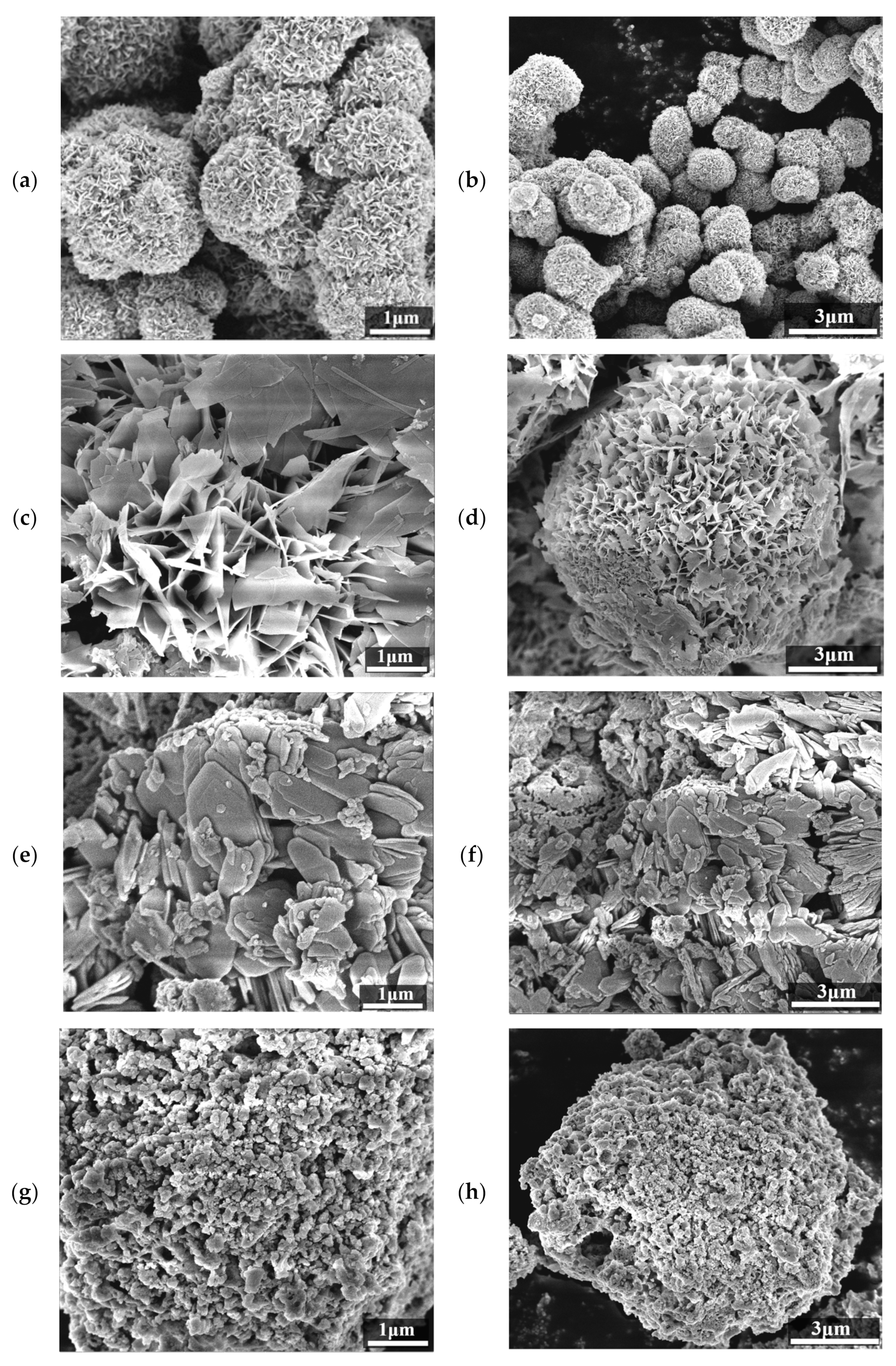
Figure 2 presents SEM images of the Bi2MoO6 samples. Bi2MoO6 prepared by the solvothermal method exhibited a flower-like, layered microspherical structure (Figure 2a,b) with an average sphere size of about 1.5 μm. These spheres were formed by the assembly of numerous Bi2MoO6 nanosheets with rough surfaces. Such rough surfaces increase the specific surface area, thereby enhancing the contact area between the catalyst and the dye molecules and improving the mobility of photogenerated electrons. Additionally, the rough surface led to significantly higher adsorption capacity, as observed in subsequent photocatalytic tests where Rhodamine B adsorbed readily onto Bi2MoO6. This factor indirectly influenced the photocatalytic efficiency.
Bi2MoO6 fabricated by the in situ conversion method (Figure 2c,d) resembled a ball composed of stacked smooth flakes, each approximately 450 nm long. The average size of these microspheres was about 10 μm. Although the Bi2MoO6 samples produced by the solvothermal and in situ conversion methods differed in size, their morphologies were somewhat similar. It is therefore hypothesized that their photocatalytic properties may follow similar trends, which will be examined through subsequent photocatalytic tests.
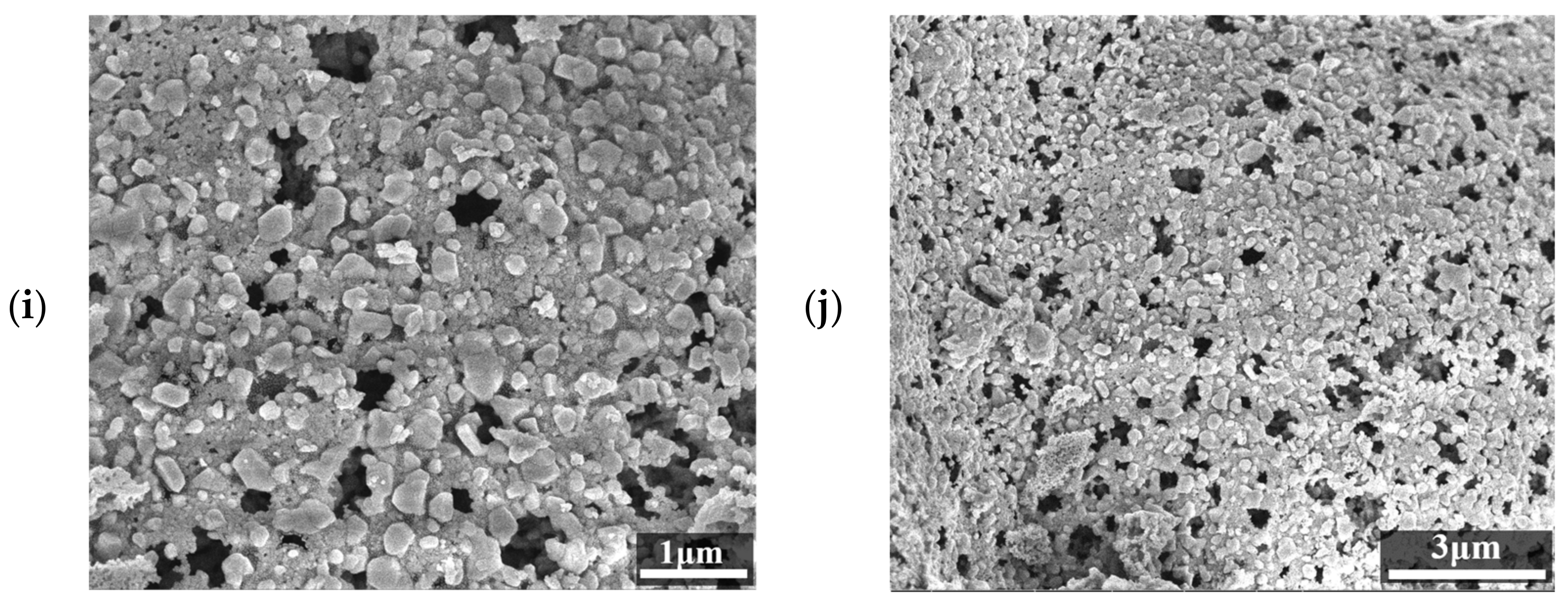
In contrast, Bi2MoO6 prepared using the solution combustion method (Figure 2e,f) consisted of disordered aggregates of irregular nanosheets with a high degree of agglomeration. This morphology suggests that the catalytic performance of Bi2MoO6 prepared by this method may differ significantly from that of the samples obtained by the solvothermal and in situ conversion methods. Under high-resolution SEM observations, the Bi2MoO6 samples synthesized by the precipitation and sol-gel methods consisted of numerous minute particles, approximately 200 nm in diameter, exhibiting a high degree of aggregation. The Bi2MoO6 samples prepared by the sol-gel method also displayed several smaller particles of varying sizes and irregular shapes on their surfaces, as well as an increased number of pores. Therefore, the SEM images confirm that the surface morphology and size of the Bi2MoO6 samples varied considerably among the five methods, highlighting the significant influence of experimental conditions and reaction concentrations on the final particle shape and size.
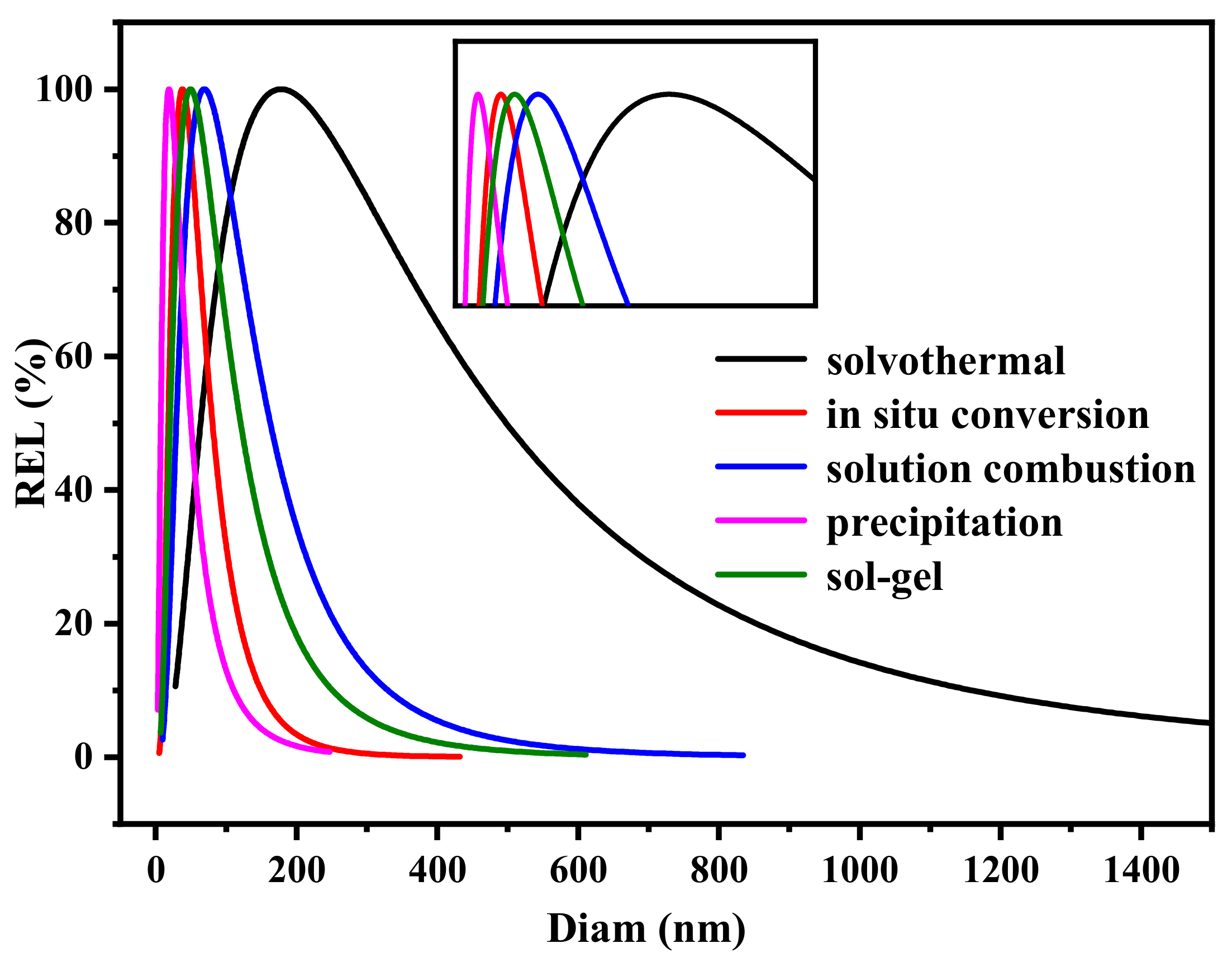
Figure 3 shows the particle size distribution of Bi2MoO6 prepared by the five different methods. The horizontal axis represents particle size, and the vertical axis indicates the percentage of particles of a given size relative to the total particle population. The primary size distribution of the Bi2MoO6 prepared using all five methods was less than 375 nm. The particle sizes, in descending order, were solvothermal > solution combustion > sol-gel > in situ transformation > precipitation. Compared with the other methods, the Bi2MoO6 particles produced by the solvothermal method exhibited the broadest size distribution range. In contrast, the other four methods yielded particle sizes that were not significantly different from one another and had relatively concentrated size distributions. The specific surface area and average pore size of the photocatalysts were determined using the Brunauer-Emmett-Teller (BET) method. It is noteworthy that the particle size distribution map in Figure 3 indicates that the size of Bi2MoO6 particles is considerably smaller than that observed in the scanning electron microscopy (SEM) images. For example, the size range of Bi2MoO6 particles prepared by the solvothermal method is less than 375 nm, whereas the particle sizes observed in the SEM images are approximately 1.5 μm. This discrepancy is believed to be primarily attributable to the agglomeration phenomenon that occurs in the sample during the preparation process. While smaller particles were formed during the initial stage of solvothermal preparation, these particles may have undergone agglomeration during the drying, transfer, and post-processing stages, resulting in the formation of larger particle agglomerates. The larger particle sizes observed in the SEM images are, in fact, the morphology of these agglomerates, whereas the size distribution plots reflect the size of the unaggregated smaller particles. Consequently, the range of smaller particle sizes depicted in the size distribution map accurately reflects the true size of the individual smaller particles in the sample. In contrast, the SEM image primarily demonstrates the overall morphology of the agglomerated particles, resulting in an apparent size difference between the two.
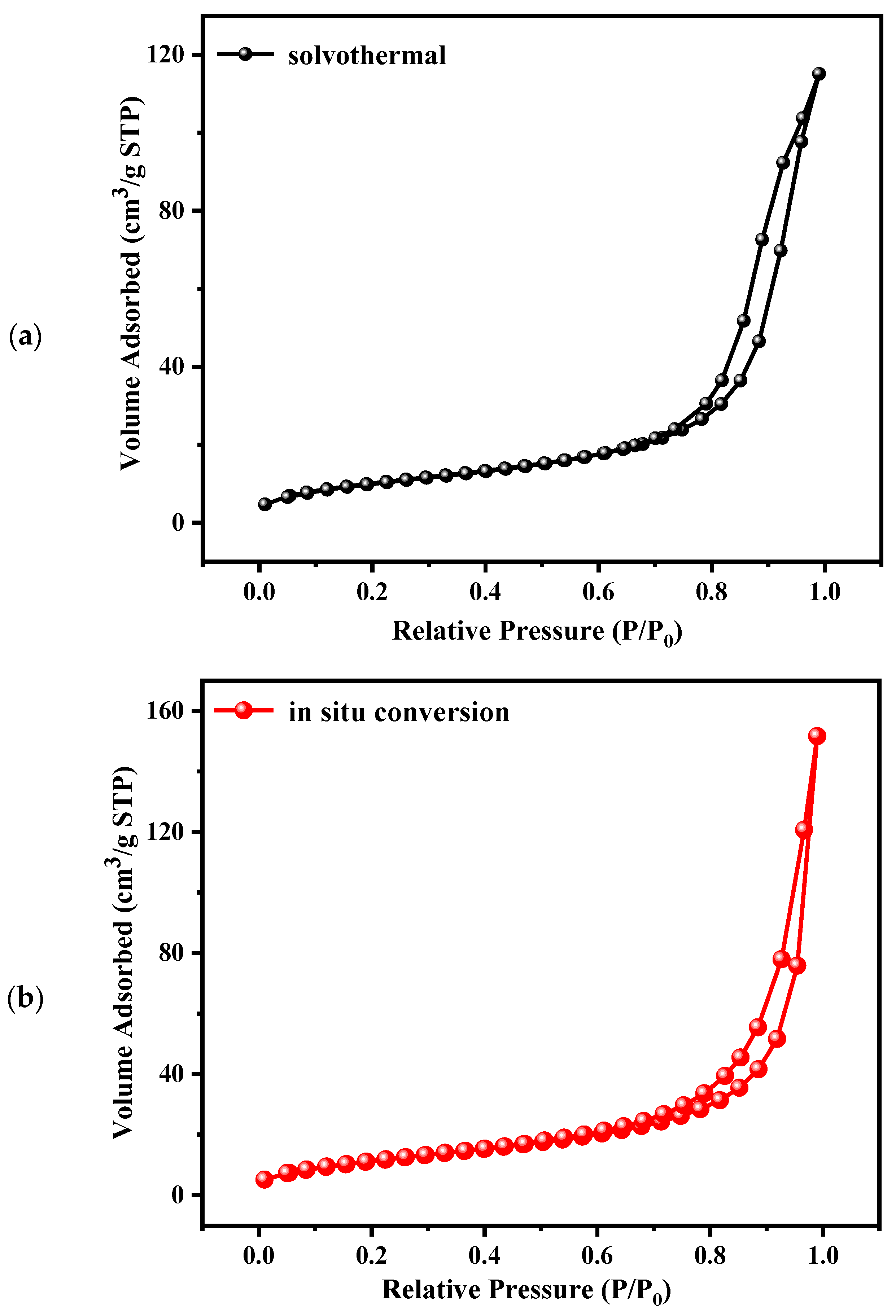
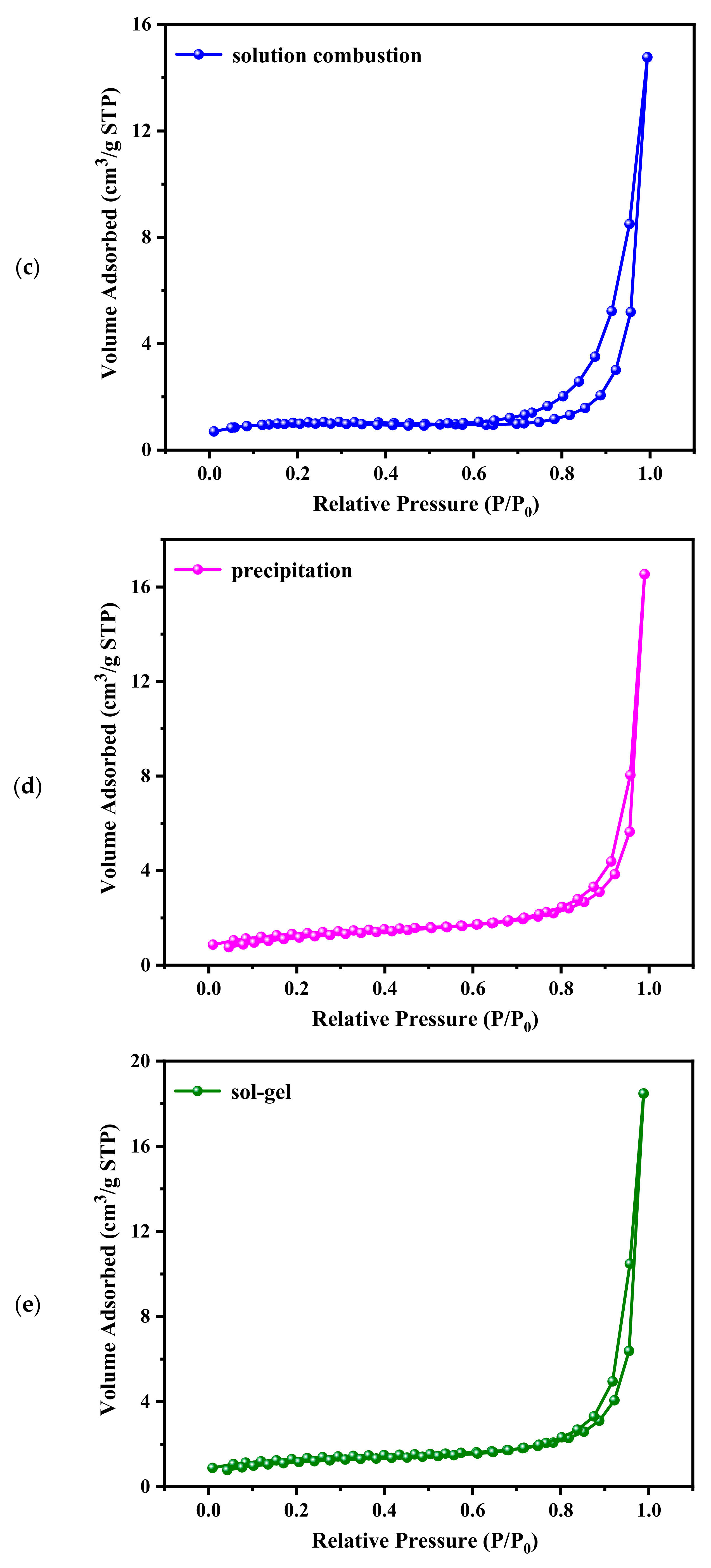
The nitrogen adsorption-desorption isotherms and pore size distribution curves of Bi2MoO6 prepared by the in situ conversion method are shown in Figure 4. The isotherm is type IV, and the hysteresis loop is type H3, indicating the presence of mesoporous structure in Bi2MoO6 flake particles [31,32]. The specific surface areas and average pore diameters of the samples are listed in Table 1. Generally, a decrease in the specific surface area of a photocatalyst leads to reduced photocatalytic activity due to fewer catalytically active sites. This trend was confirmed by subsequent photocatalytic activity characterization of Bi2MoO6.
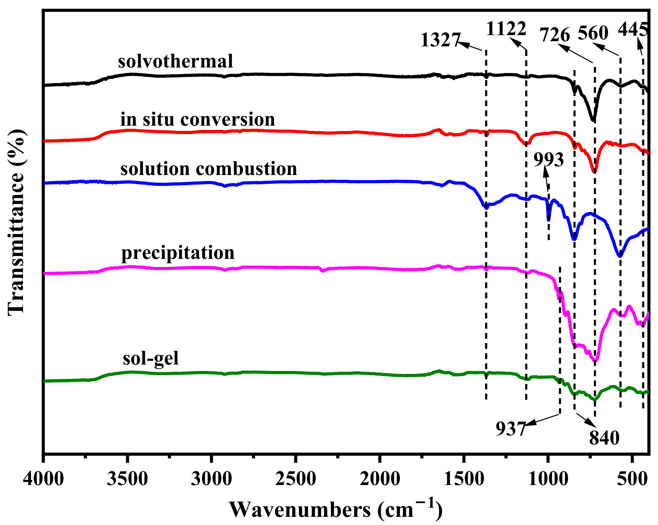
Figure 5 shows the FT-IR spectra of the prepared samples, revealing information about the surface chemical groups and atomic bonds. The characteristic vibrational absorption peaks of Bi2MoO6 appear predominantly between 400 and 1000 cm−1. The absorption peak at 445 cm−1 is attributed to the vibration of Bi–O. The bending and asymmetric stretching of Mo–O in the MoO6 octahedron produce peaks at 560 and 726 cm−1, associated with the equatorial oxygen atoms. The peak at 840 cm⁻1 corresponds to the symmetric stretching mode of the apical oxygen atom in MoO6. The Bi2MoO6 samples prepared by precipitation and the sol-gel methods exhibit an absorption peak at 937 cm−1, attributable to the symmetric stretching mode of O–Mo–O bonds. The peak at 993 cm−1 observed for Bi2MoO6 prepared using the solution combustion method may also originate from the Mo–O bond stretching mode in co-angular MoO6 octahedral. Peaks in the range of 1000–1700 cm−1 can be attributed to the vibrational stretching modes of C=O and C–O bonds present in CO2 impurities adsorbed on the catalyst surface. The broad band at 3200–3600 cm−1 and the peak at 1600 cm−1 indicate the presence of adsorbed water molecules, as evidenced by the -OH groups [33].

2.2. Spatial Charge Separation Capability

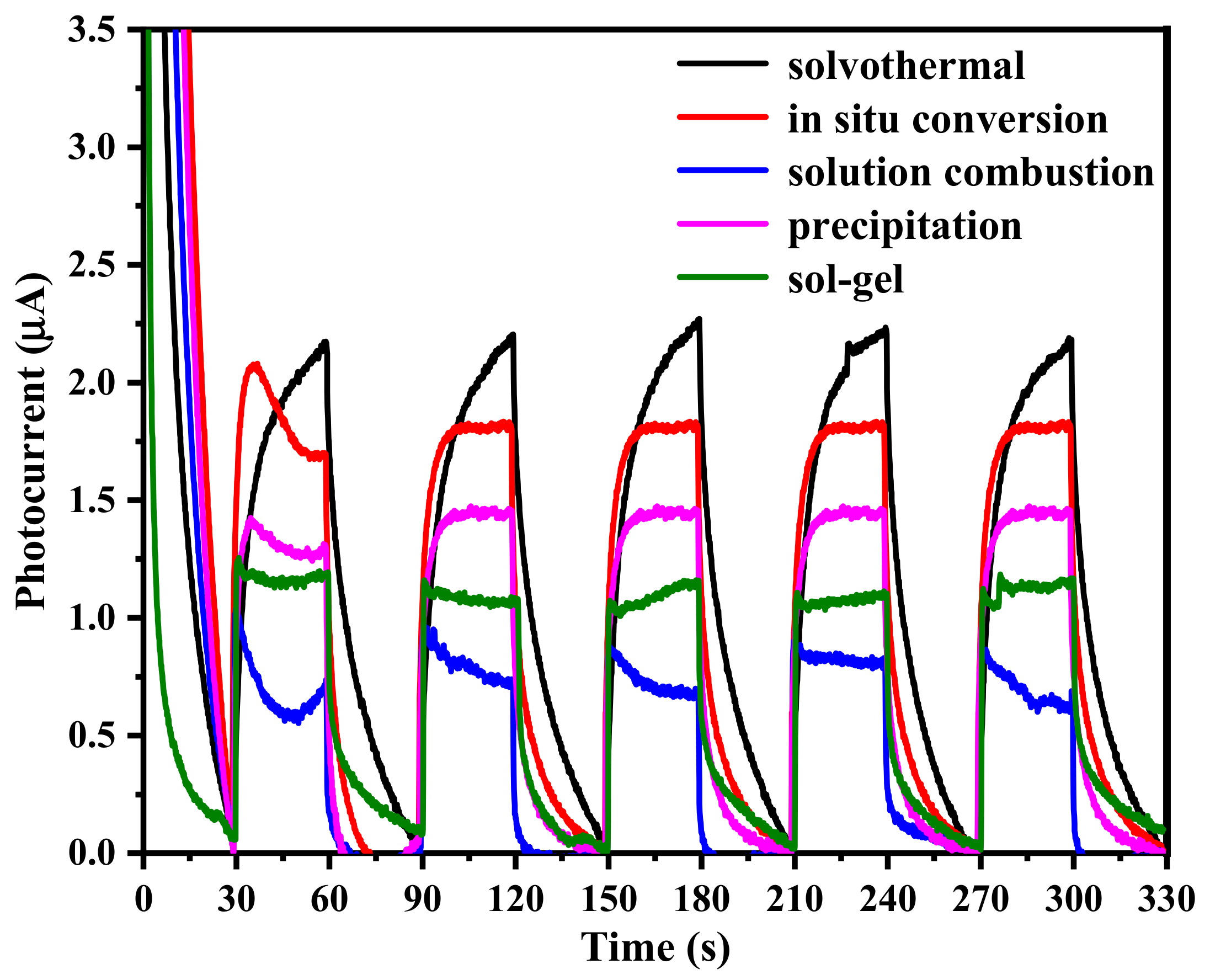
The interfacial charge separation and transfer efficiencies were investigated by analyzing the photocurrent-time response using an electrochemical workstation. As shown in Figure 6, the photocurrent rapidly increased upon illumination and decreased after the light was turned off, indicating that the samples exhibited excellent photosensitizing properties. The solvothermally prepared Bi2MoO6 displayed the highest photocurrent intensity, suggesting superior carrier transfer and separation efficiencies, which are advantageous for subsequent photocatalytic reactions.
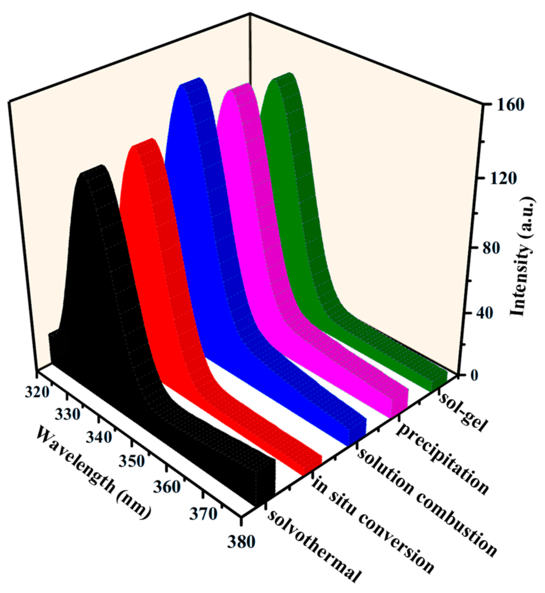
The complexation of photogenerated electrons and holes in the semiconductor samples was analyzed using photoluminescence spectroscopy. As shown in Figure 7, the excitation wavelength was 300 nm, and both the excitation and emission slit widths were 1.0 nm. The fluorescence spectra demonstrated that the Bi2MoO6 prepared by the solution combustion method exhibited a notably strong and broad fluorescence emission band, indicating a high degree of electron-hole pair recombination. The Bi2MoO6 prepared by the precipitation method followed in terms of recombination intensity. In contrast, the Bi2MoO6 prepared by the solvothermal method exhibited the weakest peak and lowest fluorescence intensity, suggesting the lowest rate of carrier recombination. A lower rate of electron-hole recombination is conducive to higher photocatalytic efficiency. Thus, the sequence of decreasing photoluminescence emission intensity aligns with the sequence of increasing photocatalytic performance. The superior photocatalytic performance of Bi2MoO6 prepared via the solvothermal method was confirmed by subsequent photocatalytic tests.

2.3. Optical Properties

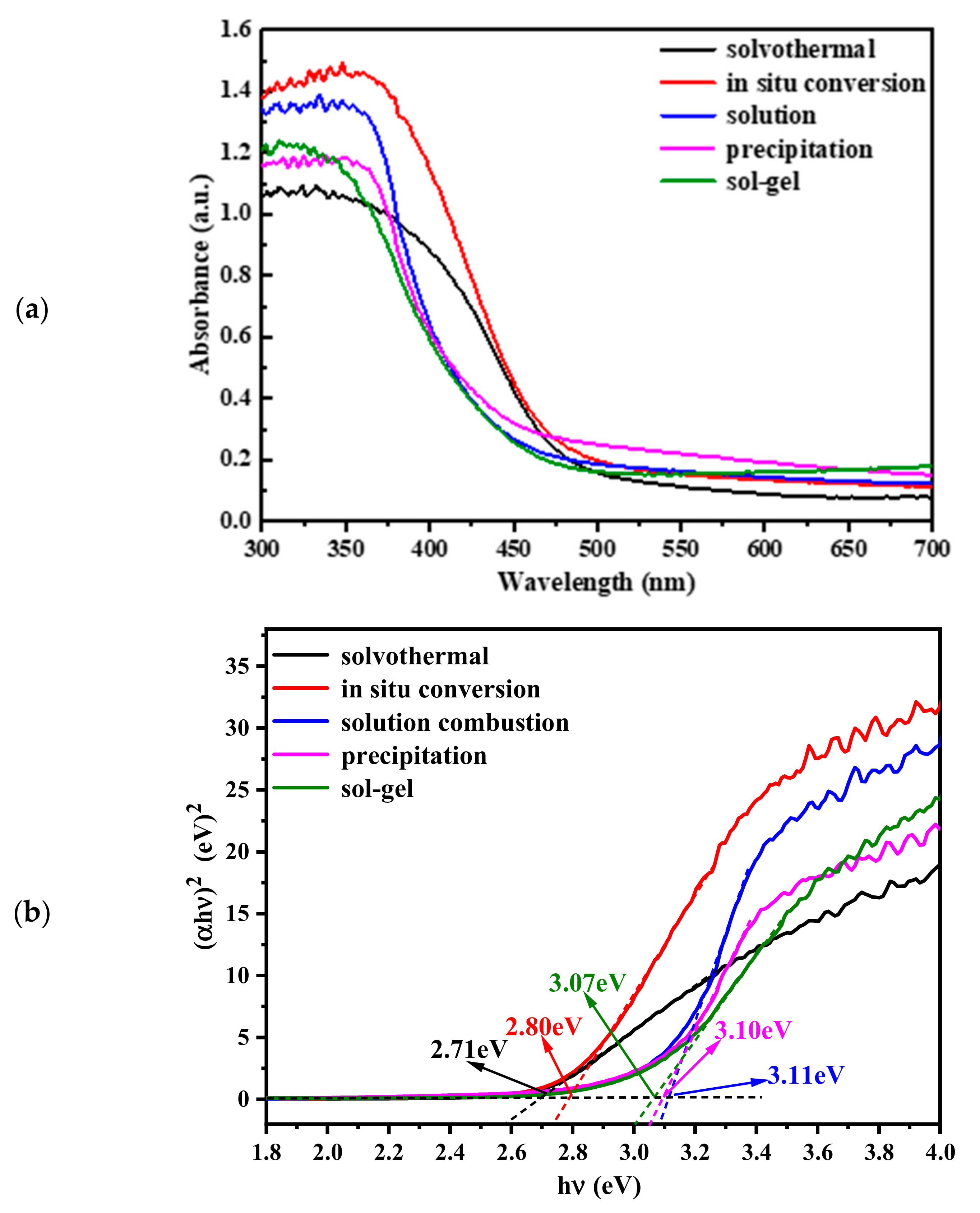
UV-visible diffuse reflectance spectroscopy was performed within the wavelength range of 300–700 nm to investigate the light absorption properties of the materials. As depicted in Figure 8, the five differently prepared Bi2MoO6 samples demonstrated significant absorption in the UV-visible range, with absorption edges at approximately 475 nm. The absorption edges of Bi2MoO6 prepared by the solvothermal and in situ conversion methods were red-shifted compared to those obtained by solution combustion, precipitation, and sol-gel methods, with the solvothermal method showing the most pronounced shift. The bandgap of Bi2MoO6 reflects its ability to absorb visible light. The bandgap energies were estimated using the Kubelka-Munk formula [34]:
α h ν = A ( h ν E g ) n / 2
where α is the absorption coefficient, h is Planck’s constant, ν is the frequency of the light, A is a constant, and Eg is the bandgap energy. Since Bi2MoO6 is a direct-bandgap semiconductor, n = 4. The calculated bandgaps for Bi2MoO6 prepared by solvothermal, in situ conversion, solution combustion, precipitation, and sol-gel methods were 2.71 eV, 2.80 eV, 3.11 eV, 3.07 eV, and 3.10 eV, respectively. A smaller bandgap energy extends absorption into the visible region, enhancing photocatalytic performance.

2.4. Photocatalytic Performances and Stability

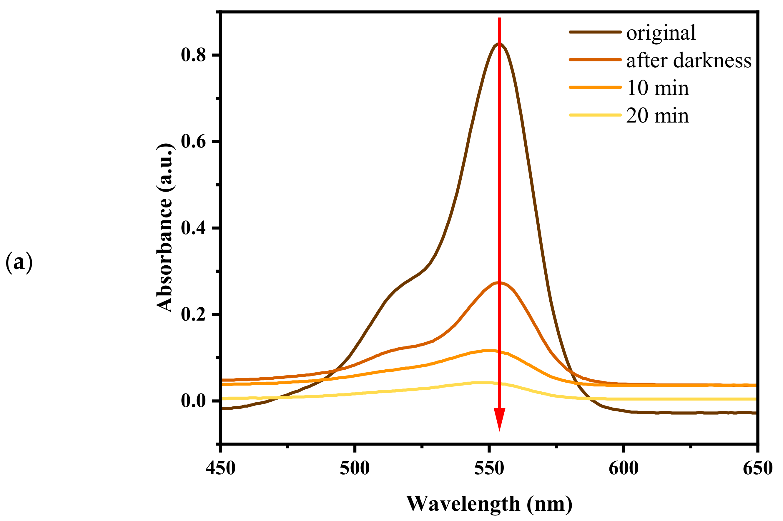
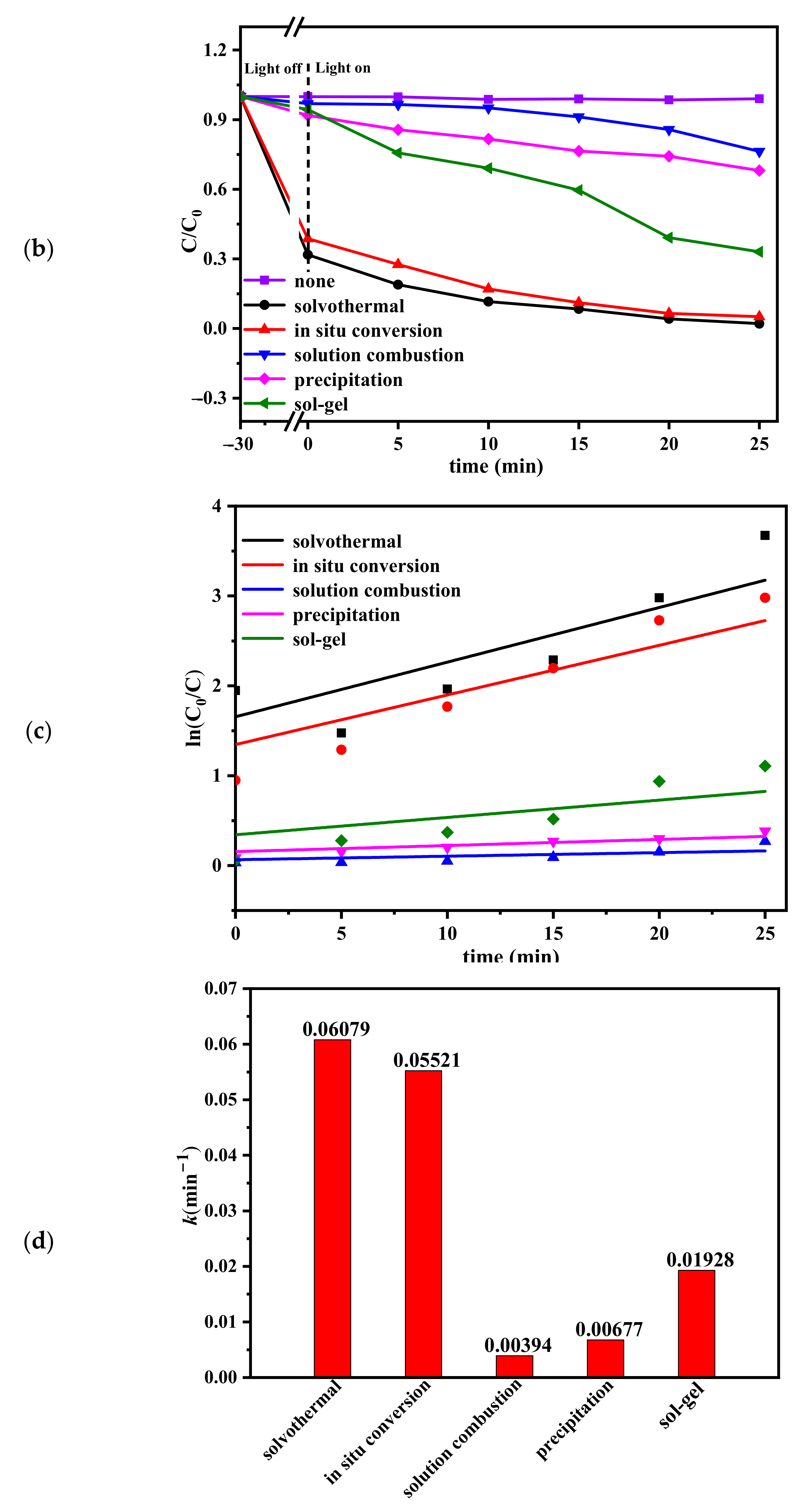
To investigate the photocatalytic activity of Bi2MoO6 prepared by the five different methods, Rhodamine B was selected as the target pollutant. Figure 9a illustrates the variation in the UV-Vis spectra of an RhB aqueous solution containing Bi2MoO6 over time with visible-light irradiation. The changes are evident in the UV-Vis absorption intensity. Under dark conditions, the absorption peak of the RhB solution remains unchanged, with only a decrease in intensity. However, under light irradiation, as the irradiation time increases, the strong absorption peak of RhB gradually diminishes, and notably, the peak position shifts significantly towards shorter wavelengths (blue shift). The absorption peak of the RhB solution remained unchanged with only a decrease in intensity, which may imply that adsorption alone has occurred, while the apparent shift in the peak position towards shorter wavelengths may be due to the degradation of RhB [35,36]. As illustrated in Figure 9b, Rhodamine B was not degraded under visible-light irradiation in the absence of a photocatalyst, nor in the presence of a photocatalyst without light irradiation. This indicates that the pollutant was stable at room temperature. The Bi2MoO6 samples prepared via the solvothermal and in situ transformation methods exhibited substantial RhB adsorption, whereas those prepared by the sol-gel, solution combustion, and precipitation methods showed minimal RhB adsorption. In the presence of visible light, all Bi2MoO6 samples exhibited notable RhB degradation efficiency, consistent with expectations. During the decomposition process, the solution initially appeared pink due to the dye, which gradually faded with increasing irradiation time. This suggests that RhB was first adsorbed onto the catalyst surface and subsequently decomposed. Bi2MoO6 prepared via the solvothermal and in situ conversion methods demonstrated superior RhB degradation rates of 97.5% and 94.9%, respectively, within 25 min.
Figure 9c shows the first-order kinetic curves of RhB degradation over Bi2MoO6 prepared using different methods. A linear relationship was observed between ln(C0/C) and the irradiation time (t), which can be expressed as lnC0/C = kt, where k is the apparent rate constant. Thus, the RhB photodegradation reaction followed first-order kinetics. A higher k value indicates better photocatalytic performance. As shown in Figure 9c,d, the Bi2MoO6 sample prepared via the solvothermal method exhibited the most pronounced photocatalytic activity under visible-light irradiation, with a rate constant k = 0.06079 min−1. The in situ conversion method ranked second, with k = 0.05520 min−1. In contrast, the Bi2MoO6 prepared by the solution combustion method displayed the lowest photocatalytic activity, with k = 0.00349 min−1.
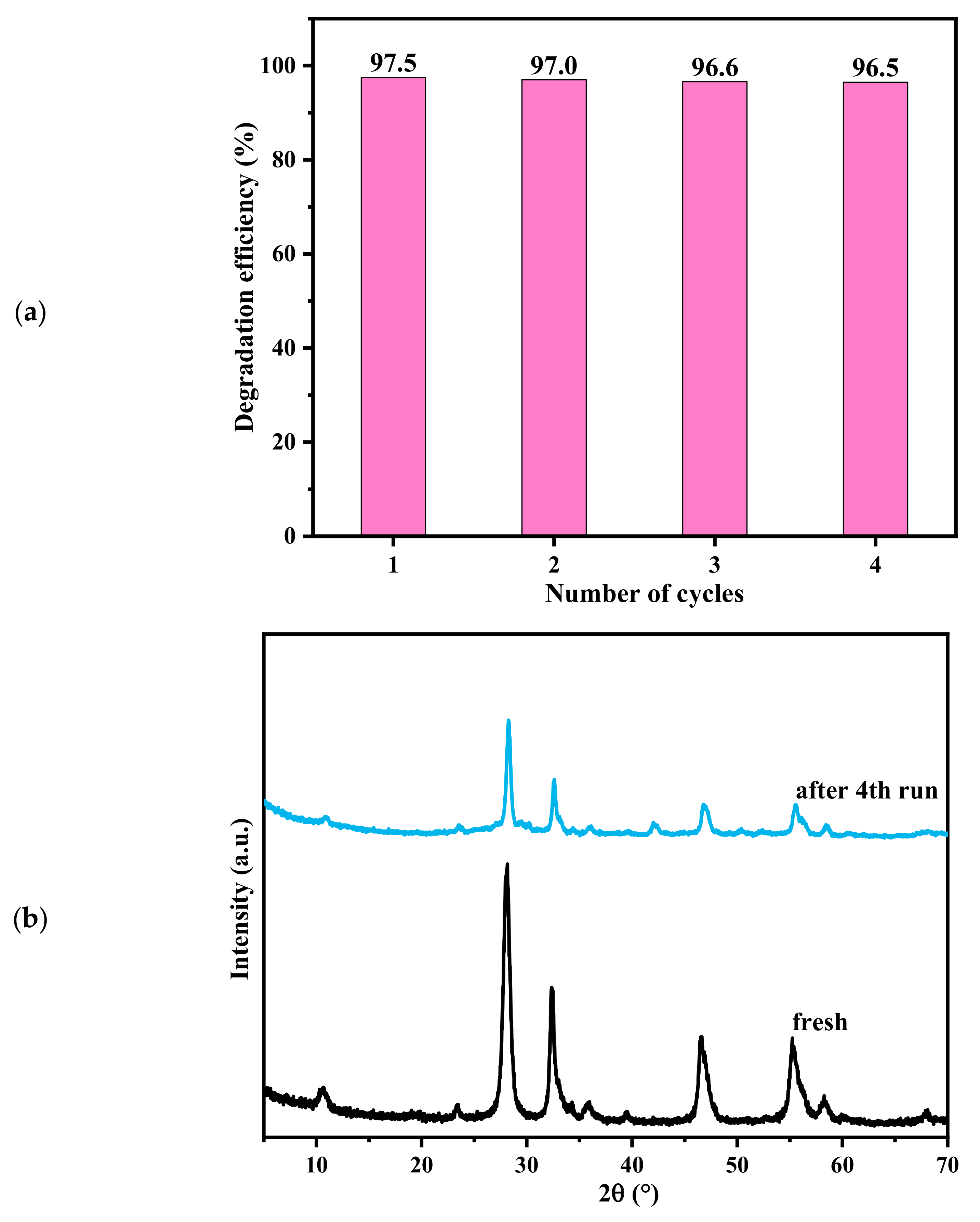
In the present study, Bi2MoO6 prepared by the solvothermal method demonstrated excellent photocatalytic properties. To evaluate its stability, reliability, and reproducibility, cycling experiments were performed. As shown in Figure 10a, the loss of photocatalytic performance was minimal after four cycles, indicating that Bi2MoO6 possessed good stability. It is important to highlight that no new phases were observed between the fresh and reused catalysts in Figure 10b. This suggests that bismuth molybdate demonstrates excellent stability throughout the photocatalytic reaction.

2.5. Photocatalytic Mechanism

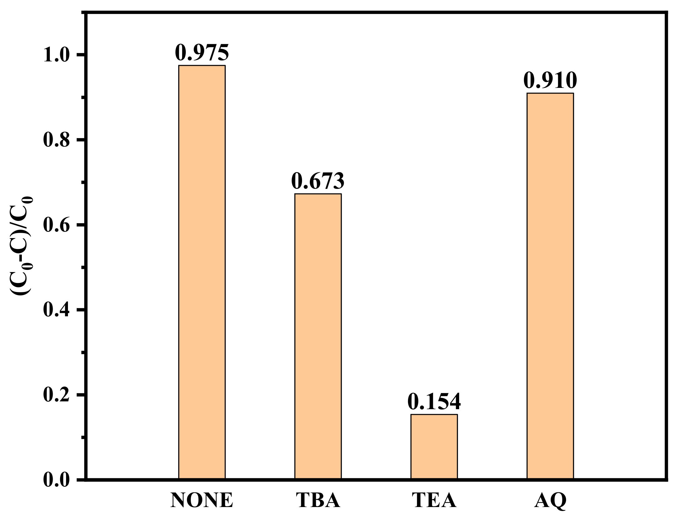
To gain further insight into the photodegradation mechanism of the Bi2MoO6 samples on organic compounds, the active species involved in the photocatalytic reaction were identified through trapping experiments. Tert-butanol (TBA), triethanolamine (TEA), and anthraquinone (AQ) were employed as scavengers of hydroxyl radicals (–OH), holes (h+), and superoxide radicals (–O2–), respectively. Radical scavengers (1 mmol/L) were introduced into Bi2MoO6 samples prepared via the solvothermal method for degradation experiments. As illustrated in Figure 11, the photocatalytic activity of Bi2MoO6 decreased slightly with the addition of TBA. However, the introduction of TEA led to a pronounced inhibition of the photocatalytic activity, whereas AQ had a comparatively minimal impact. Thus, holes (h+) and hydroxyl radicals (–OH) were identified as the primary active species in the degradation reaction of Bi2MoO6 on RhB.

3. Materials and Methods

3.1. Materials

Bismuth nitrate (Bi(NO3)3·5H2O, AR), ammonium molybdate ((NH4)6Mo7O24·4H2O, AR), and sodium molybdate (Na2MoO4·2H2O, AR) were obtained from Shanghai McLin Biochemical and Technology Co., Ltd. (Shanghai, China). Anhydrous ethanol (C2H5OH, AR) and ethylene glycol ((CH2OH)2, AR) were supplied by Sinopharm Chemical Reagent Co., Ltd. (Shanghai, China) Citric acid (CA, AR), concentrated ammonia solution (NH3·H2O, AR), and dilute nitric acid (HNO3, AR) were provided by various other chemical reagent companies in China.

3.2. Preparation of Bi2MoO6

Solvothermal method: A total of 1.69 g of Bi(NO3)3·5H2O and 0.42 g of Na2MoO4·2H2O were each dispersed in 10 mL of ethylene glycol. The mixtures were stirred vigorously until fully dissolved, ensuring that the components were well blended. The Na2MoO4 solution was then added dropwise to the Bi(NO3)3 solution under stirring. Once the two solutions were thoroughly mixed, 20 mL of ethanol was added dropwise, and the mixture was transferred to a PTFE-lined hydrothermal reactor. The reactor was placed in a constant-temperature blast drying oven at 160 °C for 10 h to ensure complete reaction. A yellowish precipitate formed, along with a layer of clear liquid above it. The precipitate was collected, washed three times with deionized water and ethanol, and dried in a constant-temperature blast drying oven at 60 °C for 24 h to yield Bi2MoO6 powder.
In situ conversion method: First, a BiOI microsphere network architecture was fabricated via a solvothermal route. For this purpose, 0.48 g of Bi(NO3)3·5H2O and 0.17 g of NaI were each dissolved in 20 mL of ethylene glycol to form separate Bi(NO3)3·5H2O and NaI solutions. These two solutions were mixed thoroughly and then transferred to a hydrothermal reactor, where they were reacted at 120 °C for 12 h. Subsequently, using the resulting BiOI powder as a template, 0.76 g of BiOI powder and 0.26 g of Na2MoO4·2H2O were dispersed in 20 mL of pure water. The mixture was stirred thoroughly, then poured into a 50 mL hydrothermal reactor and reacted at 120 °C for 8 h. Finally, the yellowish precipitate was collected, washed, and dried at 60 °C for 24 h to obtain the yellowish Bi2MoO6 powder.
Solution combustion method: The required amounts of Bi(NO3)3·5H2O (9.70 g) and (NH4)2MoO4 (1.96 g) were accurately weighed into a beaker. Next, 10 mL of anhydrous ethanol was added, and the contents were stirred for 10 min, ensuring thorough mixing. Stirring then continued for an additional 30 min. Using a pipette, an appropriate amount of the suspension was transferred into a combustion boat, which was placed into a tube furnace at 500 °C for 10 min to undergo a combustion reaction. After the reaction, the combustion boat was removed, and the product was collected to yield Bi2MoO6 powder.
Precipitation method: A total of 12.13 g of Bi(NO3)3·5H2O solid was weighed and dissolved ultrasonically in 10 mL of ethylene glycol (designated as Liquid A). Separately, 1.97 g of (NH4)2MoO4 and 0.098 g of polyvinylpyrrolidone (PVP) were dissolved ultrasonically in 20 mL of distilled water (designated as Liquid B). Liquid A was then added dropwise to Liquid B under vigorous stirring, resulting in the formation of a white precipitate. After the two solutions were combined, stirring continued for 10 min. The pH was adjusted to 5–7 by adding concentrated ammonia solution. The mixture was stirred for an additional three hours to ensure thorough mixing and precipitate formation. The resulting slurry was then subjected to suction filtration, and the precipitate was washed and dried at 80 °C. Finally, the dried precipitate was calcined at 400 °C for 2 h, and the roasted product was ground to obtain Bi2MoO6 powder.
Sol-gel method: First, 4.85 g of Bi(NO3)3·5H2O was dissolved in 50 mL of dilute nitric acid. Subsequently, 1.92 g of citric acid was added, and the mixture was heated and stirred until fully mixed, yielding Solution A. Another solution (Solution B) was prepared by dissolving 1.79 g of (NH4)2MoO4 in 50 mL of water and adding 3.84 g of citric acid, followed by heating and stirring until fully dissolved. Solution B was then slowly added to Solution A under stirring until a clear mixture was obtained. The pH was adjusted to 5–7 using concentrated ammonia, and the mixture was heated and stirred until it formed a gel. The gel, serving as a precursor for bismuth molybdate, was poured into a crucible and dried in an oven at 80 °C to obtain a dry coagulate. This dried product was removed, ground into a powder, placed in a crucible, and calcined at 400 °C for 5 h to obtain the Bi2MoO6 photocatalyst.

3.3. Sample Characterization

The samples were analyzed using an X-ray diffractometer (XRD, Rigaku, SmartLab SE, Tokyo, Japan) with a rotating copper target, Cu Kα radiation (λ = 0.154 nm), a scanning speed of 20°/min, and a step size of 0.02°. Scanning electron microscopy (SEM, Novanano 450, Hitachi, Tokyo, Japan) was used for morphological characterization at an operating voltage of 4.0 kV. A laser particle size analyzer (Nicomp380Z3000, PSS Particle Sizer, China Center of Excellence, Los Angeles, CA, USA) was employed to determine the particle size distribution of the samples. A Hitachi U3900 UV-Vis Multifunctional Spectrometer (Japan Scientific Instruments (Beijing) Co., Ltd., Beijing, China) was used for diffuse reflectance analysis of solid powder samples over a wavelength range of 300–700 nm. Furthermore, this instrument was employed to measure the photocatalytic degradation of rhodamine B, with a wavelength range of 450–650 nm and a scanning speed of 300 nm/min. For infrared absorption spectra, a small amount of the sample was mixed with KBr, ground, and pressed into pellets. The spectra were recorded using an IS10 instrument (Thermo Fisher Scientific Ltd., Waltham, MA, USA) over a range of 400–4000 cm⁻1. A fluorescence spectrometer (Cary Eclipse, Hi-Tech Scientific Naka Corporation, Naka-shi, Japan) was used to detect the separation of photogenerated electron–hole pairs after photoexcitation, using an excitation wavelength fixed at 300 nm and a scanning range of 300–600 nm.

3.4. Photoelectrochemical Measurements

The photocurrent of the materials was measured using a three-electrode CH760E (Shanghai Chenhua Instrument Co., Shanghai, China) electrochemical analyzer. A platinum wire served as the counter electrode, and an Ag/AgCl electrode served as the reference electrode. The electrolyte solution was 0.5 mol/L Na2SO4. To prepare the working electrode, 10 mg of the sample was added to 2 mL of water and sonicated to form a slurry, which was then dispersed onto pretreated indium tin oxide (ITO) conductive glass (1 × 2 cm2) and dried at 60 °C for 6 h. The light source was switched on and off every 30 s to measure the resulting photocurrent.

3.5. Photocatalytic Performance

Photodegradation experiments were conducted using rhodamine B (RhB, 10 mg/L) as the target pollutant. First, 50 mL of RhB solution was mixed with 0.2 g of Bi2MoO6, and the mixture was stirred in the dark for 30 min to achieve adsorption-desorption equilibrium on the catalyst surface. The suspension was then irradiated under a 500 W xenon lamp with continuous stirring. The temperature of the solution was monitored throughout the reaction. Samples were taken at 5 min intervals, centrifuged, and the supernatant was analyzed for absorbance using a U-3900 spectrophotometer. The RhB concentration was determined based on the measured absorbance.

4. Conclusions

This study presents the successful synthesis of Bi2MoO6 using five different methods. The crystallinity, morphology, bandgap width, and degree of photogenerated carrier complexation of Bi2MoO6 were significantly affected by the chosen preparation method, resulting in notable variations in photocatalytic performance. The solvothermal method produced a larger specific surface area, a greater number of surface-active sites, and enhanced photocatalytic performance of the resulting Bi2MoO6. Additionally, it exhibited a narrow bandgap and a low photogenerated carrier complexation rate, which contributed to its excellent photocatalytic performance. Although Bi2MoO6 prepared by the precipitation method showed favorable crystallization and small particle size, its large bandgap width and high photogenerated electron-hole complexation markedly reduced its photocatalytic performance. It can be seen that Bi2MoO6 with a small bandgap and a low photogenerated carrier complexation rate significantly improves photocatalytic performance. This study offers valuable insights into the preparation of Bi2MoO6 with highly efficient photocatalytic activity.

Author Contributions

Conceptualization, Q.W., W.L. and R.L.; methodology, Q.W.; validation, Q.W. and H.Z.; data acquisition, analysis, and interpretation, Q.W., J.G. and H.Z.; writing—original draft preparation, Q.W.; writing—review and editing, Q.W. and W.L.; funding acquisition, J.G. All authors have read and agreed to the published version of the manuscript.

Funding

This research was funded by the Excellent Innovative Scientific Research Team of Silicon-Based Materials (2022AH010101), Key Scientific Research Project of Bengbu University (2022ZR01zd) and Bengbu College Haikou Sanxin (Bengbu) New Energy Materials Co. (2023xqhz073).

Data Availability Statement

All data are available to download within the database.

Conflicts of Interest

Author Ruochen Li was employed by the company Triumph Science & Technology Co., Ltd., Bengbu, China. The remaining authors declare that the research was conducted in the absence of any commercial or financial relationships that could be construed as a potential conflict of interest.

References

  1. Qi, S.Y.; Wu, S.Q.; Zhang, K.Y.; Guan, L.; Hu, X.; Li, H.Y. Design, preparation and mechanism of Bi2MoO6-modified cobalt-doped CdS solid solution photocatalysts. J. Alloys Compd. 2024, 1003, 175565. [Google Scholar] [CrossRef]
  2. Sui, G.Z.; Li, J.L.; Du, L.J.; Zhuang, Y.; Zhang, Y.L.; Zou, Y.F.; Li, B. Preparation and characterization of g-C3N4/Ag-TiO2 ternary hollow sphere nano heterojunction catalyst with high visible light photocatalytic performance. J. Alloys Compd. 2020, 823, 153851. [Google Scholar] [CrossRef]
  3. Luo, C.W.; Liao, M.C.; Peng, D.Y.; Xiong, J.; Zeng, H.Y. Fe-doped MOF(Ti)/Bi2MoO6 heterostructure for boosting oxidation/reduction activities under visible light. Colloid Surf. A 2024, 19, 118565. [Google Scholar] [CrossRef]
  4. Dong, S.; Feng, J.L.; Fan, M.H.; Pi, Y.Q.; Hu, L.M.; Han, X.; Liu, M.L.; Sun, J.Y.; Sun, J.H. Recent developments in heterogeneous photocatalytic water treatment using visible light-responsive photocatalysts: A review. Rsc Adv. 2015, 5, 14610–14630. [Google Scholar] [CrossRef]
  5. Yang, S.; Lu, Q.P.; Wang, F.G.; Zhi, Y.H.; Chen, J.Y.; Wang, Y.H.; Zhang, H.; Yin, H.Q.; Sun, P.; Cao, W.B. S-scheme SnO/TiO2 heterojunction with high hole mobility for boosting photocatalytic degradation of gaseous benzene. Chem. Eng. J. 2023, 478, 147345. [Google Scholar] [CrossRef]
  6. Buddiga, L.R.; Gajula, G.R.; Sailaja, B.B.V.; Ch, M.L.V.P. Visible light photocatalytic exploit of P/Zr doped TiO2 nano particles for dye degradation of rose Bengal. Appl. Surf. Sci. 2023, 18, 100492. [Google Scholar] [CrossRef]
  7. Lu, P.; Peng, Y.Q.; Yang, Y.; Chen, S.Y.; Shang, J.; Yang, C.; Xu, M.M.; Bai, J.W.; Zhao, Z.L.; Hu, X.L. Visible-light-driven photocatalytic for degrading tetracycline wastewater by BiOI/Bi2O3 Z-scheme heterojunction. J. Environ. Chem. Eng. 2024, 12, 114395. [Google Scholar] [CrossRef]
  8. Xu, Y.X.; Chen, T.W. Development of nanostructured based ZnO@WO3 photocatalyst and its photocatalytic and electrochemical properties: Degradation of Rhodamine B. Int. J. Electrochem. Sci. 2023, 18, 100055. [Google Scholar] [CrossRef]
  9. Lin, Z.X.; Xu, J.X.; Gu, H.J.; Huang, J.Y.; Lin, J.H.; Shao, J.; Wang, D.D.; Li, H.J. A review on research progress in photocatalytic degradation of organic pollutants by Bi2MoO6. J. Environ. Chem. Eng. 2023, 11, 110911. [Google Scholar] [CrossRef]
  10. Hajiali, M.; Farhadian, M.; Tangestaninejad, S.; Khosravi, M. Synthesis and characterization of Bi2MoO6/MIL-101(Fe) as a novel composite with enhanced photocatalytic performance: Effect of water matrix and reaction mechanism. Adv. Power Technol. 2022, 33, 103546. [Google Scholar] [CrossRef]
  11. Ly, M.J.; Wang, C.M.; Rong, Y.Z.; Wei, J.W.; Yang, Y.K.; Liu, Y.Y.; Wei, G.X.; Zhang, Q.; Wang, C.; Xiu, J.S. Advances in modification of Bi2MoO6 and its photocatalysis: A review. J. Alloys Compd. 2024, 982, 173759. [Google Scholar] [CrossRef]
  12. Li, Z.X.; Qi, W.K.; Li, L.D.; Ma, Z.Y.; Lai, W.D.; Li, L.; Jin, X.S.; Zhang, Y.C.; Zhang, W.M. Preparation of carbon nanofibers supported Bi2MoO6 nanosheets as counter electrode materials on Titanium mesh substrate for dye-sensitized solar cells. Sol. Energy 2021, 214, 502–509. [Google Scholar] [CrossRef]
  13. Wu, K.D.; He, X.X.; Ly, A.; Lahem, D.; Debliquy, M.; Zhang, C. Highly sensitive and selective gas sensors based on 2D/3D Bi2MoO6 micro-nano composites for trimethylamine biomarker detection. Appl. Surf. Sci. 2023, 629, 157443. [Google Scholar] [CrossRef]
  14. Murugan, R. Investigation on ionic conductivity and Raman spectra of γ-Bi2MoO6. Phys. B 2004, 352, 227–232. [Google Scholar] [CrossRef]
  15. Ali, H.E.; Khairy, Y. Facile low temperature synthesis and characterization of bismuth molybdate (Bi2MoO6) nanostructures: An effect surfactant concentration. Optik 2019, 178, 90–96. [Google Scholar]
  16. Ashrafi, M.; Farhadian, M.; Nazar, A.R.S.; Hajiali, M.; Noorbaksh, A. The tetracycline degradation in a photocatalytic fuel cell microreactor using a ZnO nanorod/Bi2MoO6/ZIF-67 photocatalyst responsive to visible light. Energy Convers. Manag. 2024, 312, 118565. [Google Scholar] [CrossRef]
  17. Zhang, Y.; Zhu, Y.; Yu, J.; Yu, J.Q.; Yang, D.J.; Ng, T.W.; Wong, P.K.; Yu, J.C. Enhanced photocatalytic water disinfection propertiesof Bi2MoO6-RGO nanocomposites under visible light irradiation. Nanoscale 2013, 5, 6307–6310. [Google Scholar] [CrossRef]
  18. Yang, F.; Elnabawy, A.O.; Schimmenti, R.; Song, P.; Wang, J.W.; Peng, Z.Q.; Yao, S.; Deng, R.P.; Song, S.Y.; Lin, Y.; et al. Bismuthene for highly efficient carbon dioxide electroreduction reaction. Nat. Commun. 2020, 11, 1088. [Google Scholar] [CrossRef]
  19. Peng, Y.H.; Zhang, Y.; Tian, F.H.; Zhang, J.Q.; Yu, J.Q. Structure Tuning of Bi2MoO6 and Their Enhanced Visible Light Photocatalytic Performances. Crit. Rev. Solid State 2017, 42, 347–372. [Google Scholar] [CrossRef]
  20. Wang, M.; Han, J.; Guo, P.Y.; Sun, M.Z.; Zhang, Y.; Tong, Z.; You, M.Y.; Lv, C.M. Hydrothermal synthesis of B-doped Bi2MoO6 and its high photocatalytic performance for the degradation of Rhodamine B. J. Phys. Chem. Solids 2018, 113, 86–93. [Google Scholar] [CrossRef]
  21. Cruz, A.M.L.; Alfaro, S.O. Synthesis and characterization of γ-Bi2MoO6 prepared by co-precipitation: Photoassisted degradation of organic dyes under vis-irradiation. J. Mol. Catal. A-Chem. 2010, 320, 85–91. [Google Scholar] [CrossRef]
  22. Li, J.; Nie, X.; Meng, L.J.; Zhang, X.J.; Bai, L.M.; Chai, D.F.; Zhang, W.Z.; Zhang, Z.F.; Dong, G.H. Microwave-assisted hydrothermal synthesis of Ag/Bi2MoO6/ZnO heterojunction with nano Ag as electronic accelerator pump for high-efficienty photocatalytic degradation of levofloxacin. Appl. Surf. Sci. 2024, 678, 161143. [Google Scholar] [CrossRef]
  23. Wang, Y.J.; Wang, Q.Y.; Zhang, H.; Wu, Y.; Jia, Y.; Jin, R.C.; Gao, S.M. CTAB-assisted solvothermal construction of hierarchical Bi2MoO6/Bi5O7 Br with improved photocatalytic performances. Sep. Purif. Technol. 2020, 242, 11. [Google Scholar] [CrossRef]
  24. Liu, Z.; Tian, J.; Yu, C.L.; Fan, Q.Z.; Liu, X.Q. Solvothermal fabrication of Bi2MoO6 nanocrystals with tunable oxygen vacancies and excellent photocatalytic oxidation performance in quinoline production and antibiotics degradation. Chin. J. Catal. 2022, 43, 472–484. [Google Scholar] [CrossRef]
  25. Zhang, Q.; Wang, X.F.; Duan, F.; Chen, Q.M. Preparation and Photocatalytic Properties of Bi2MoO6 Hollow Microspheres. Chin. J. Inorg. Chem. 2015, 31, 2152–2158. [Google Scholar]
  26. Saha, D.; Madras, G. Solution combustion synthesis of γ(L)-Bi2MoO6 and photocatalytic activity under solar radiation. Mater. Res. Bull. 2011, 46, 1252–1256. [Google Scholar] [CrossRef]
  27. Hipólito, E.L.; Cruz, A.M.L.; Cuéllar, E.L. Synthesis, characterization, and photocatalytic properties of γ-Bi2MoO6 prepared by co-precipitation assisted with ultrasound irradiation. J. Taiwan Inst. Chem. E 2014, 45, 2749–2754. [Google Scholar] [CrossRef]
  28. Umapathy, V.; Manikandan, A.; Antony, S.A.; Ramu, P.; Neeraja, P. Structure, morphology and opto-magnetic properties of Bi2MoO6 nano-photocatalyst synthesized by sol-gel method. Trans. Nonferrous Met. Soc. China 2015, 25, 3271–3278. [Google Scholar] [CrossRef]
  29. Zhao, Z.W.; Zhang, W.D.; Sun, Y.Y.; Yu, J.Y.; Zhang, Y.X.; Wang, H.; Dong, F.; Wu, Z.B. Bi Cocatalyst/Bi2MoO6 microspheres nanohybrid with SPR-promoted visible-light photocatalysis. J. Phys. Chem. C 2016, 120, 11889–11898. [Google Scholar] [CrossRef]
  30. Zhang, M.Y.; Shao, C.L.; Mu, J.B.; Huang, X.M.; Zhang, Z.Y.; Guo, Z.C.; Zhang, P.; Liu, Y.C. Hierarchical heterostructures of Bi2MoO6 on carbon nanofibers: Controllable solvothermal fabrication and enhanced visible photocatalytic properties. J. Mater. Chem. 2012, 22, 577–584. [Google Scholar] [CrossRef]
  31. Li, J.L.; Liu, X.J.; Piao, X.Q.; Sun, Z.; Pan, L.K. Novel carbon sphere@Bi2MoO6 core-shell structure for efficient visible light photocatalysis. RSC Adv. 2015, 5, 16592–16597. [Google Scholar] [CrossRef]
  32. Li, Z.Q.; Chen, X.T.; Xue, Z.L. Bi2MoO6 microstructures: Controllable synthesis, growth mechanism, and visible-light-driven photocatalytic activities. CrystEngComm 2013, 15, 498–508. [Google Scholar] [CrossRef]
  33. Khazaee, Z.; Khavar, A.H.C.; Mahjou, A.R.; Motaeea, A.; Srivastavac, V.; Sillanpääc, M. Template-confined growth of X-Bi2MoO6 (X: F, Cl, Br, I) nanoplates with open surfaces for photocatalytic oxidation; experimental and DFT insights of the halogen doping. Sol. Energy 2020, 196, 567–581. [Google Scholar] [CrossRef]
  34. Hao, Z.Q.; Xu, L.L.; Wei, B.; Fan, L.L.; Liu, Y.; Zhang, M.y.; Gao, H. Nanosize α-Bi2O3 decorated Bi2MoO6 via an alkali etching process for enhanced photocatalytic performance. RSC Adv. 2015, 5, 12346–12353. [Google Scholar] [CrossRef]
  35. Miao, Y.C.; Pan, G.F.; Huo, Y.N.; Li, H.X. Aerosol-spraying preparation of Bi2MoO6: A visible photocatalyst inhollow microspheres with a porous outer shell and enhanced activity. Dyes Pigment. 2013, 99, 382–389. [Google Scholar] [CrossRef]
  36. Li, H.H.; Liu, C.Y.; Li, K.W.; Wang, H. Preparation, characterization and photocatalytic properties of nanoplate Bi2MoO6 catalysts. J. Mater. Sci. 2008, 43, 7026–7034. [Google Scholar] [CrossRef]
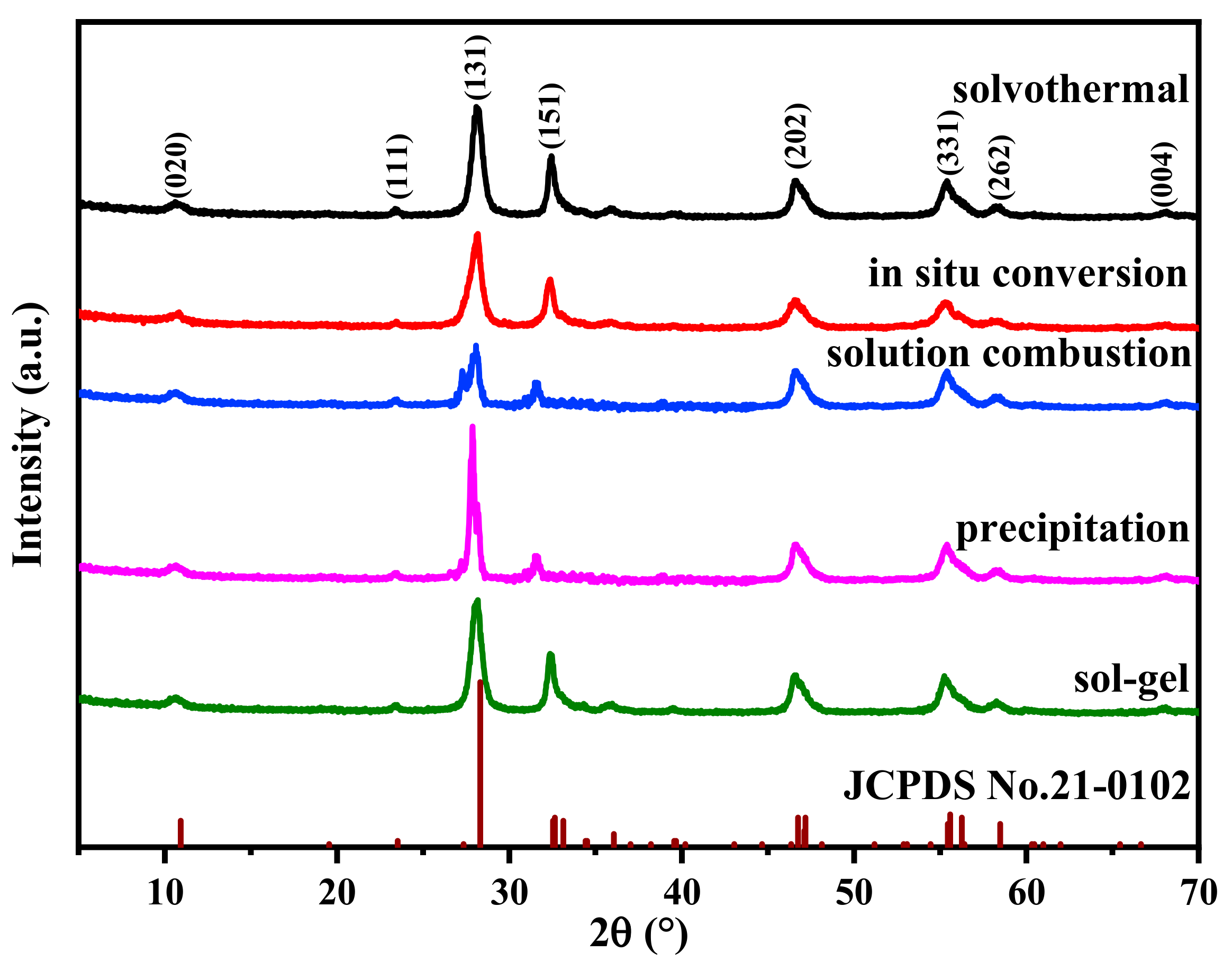
Figure 1. XRD patterns of Bi2MoO6 samples.
Catalysts 15 00198 g001
Figure 2. SEM image of Bi2MoO6 samples obtained by different preparation methods: (a,b) solvothermal; (c,d) in situ conversion; (e,f) solution combustion; (g,h) precipitation; (i,j) sol–gel.
Catalysts 15 00198 g002aCatalysts 15 00198 g002b
Figure 3. Particle size distribution of Bi2MoO6 prepared by different methods.
Catalysts 15 00198 g003
Figure 4. Nitrogen adsorption-desorption isotherms of Bi2MoO6 prepared by in situ transformation method: (a) solvothermal; (b) in situ conversion; (c) solution combustion; (d) precipitation; (e) sol-gel.
Catalysts 15 00198 g004aCatalysts 15 00198 g004b
Figure 5. FT-IR spectra of Bi2MoO6 prepared by different methods.
Catalysts 15 00198 g005
Figure 6. Photocurrent response of different samples.
Catalysts 15 00198 g006
Figure 7. Photoluminescence emission spectra of different Bi2MoO6.
Catalysts 15 00198 g007
Figure 8. (a) UV-Vis diffuse reflectance spectra of Bi2MoO6; (b) plots of the (αh)2 vs. the light energy for the Bi2MoO6 samples.
Catalysts 15 00198 g008
Figure 9. (a) UV-Vis spectral changes in RhB in aqueous Bi2MoO6 suspensions; (b) degradation rate of Rhodamine B under dark and visible light conditions; (c) plot of ln(C0/C) versus irradiation time; (d) photocatalytic kinetics constants for the as-prepared samples.
Catalysts 15 00198 g009aCatalysts 15 00198 g009b
Figure 10. (a) Cycling run in the photocatalytic reaction; (b) XRD patterns of the fresh and used Bi2MoO6 photocatalyst.
Catalysts 15 00198 g010
Figure 11. Influence of trapping agent on Bi2MoO6 prepared by solvothermal method.
Catalysts 15 00198 g011
Table 1. BET surface areas and average pore size of diverse samples.
SamplesBET Surface Area (m2/g)Average Pore Size (nm)
solvothermal38.510.0
in situ conversion43.07.1
solution combustion3.39.8
precipitation4.55.3
sol-gel4.46.4
Disclaimer/Publisher’s Note: The statements, opinions and data contained in all publications are solely those of the individual author(s) and contributor(s) and not of MDPI and/or the editor(s). MDPI and/or the editor(s) disclaim responsibility for any injury to people or property resulting from any ideas, methods, instructions or products referred to in the content.