Article

Genomic Identification of the Levansucrase Operon in Novel Bacillus velezensis HL25 in Sucrose Utilizing Pathway and Functional Characterization of Its Levansucrase

by
Hataikarn Lekakarn
1,
Jiruchaya Chaisuriyaphun
2,
Ruethaikan Junsuk
2,
Putanat Kornpitak
2,
Teeranart Komonmusik
1,
Wuttichai Mhuantong
3 and
Benjarat Bunterngsook
3,*
1
Department of Biotechnology, Faculty of Science and Technology, Thammasat University, Rangsit Campus, Pathum Thani 12120, Thailand
2
Science Classrooms in University-Affiliated School Project, Thammasat University-Suankularb Wittayalai Rangsit School Center, Khlong Luang, Pathum Thani 12120, Thailand
3
Enzyme Technology Research Team, Biorefinery Technology and Bioproduct Research Group, National Center for Genetic Engineering and Biotechnology, 113 Thailand Science Park, Khlong Luang, Pathum Thani 12120, Thailand
*
Author to whom correspondence should be addressed.
Catalysts 2025, 15(11), 1059; https://doi.org/10.3390/catal15111059
Submission received: 30 September 2025 / Revised: 31 October 2025 / Accepted: 4 November 2025 / Published: 6 November 2025
(This article belongs to the Section Biocatalysis)

Abstract

Levan and levan-type fructooligosaccharides (L-FOSs) are non-digestible fructans with prebiotic properties that promote gut microbiota growth. This study presents the first genomic analysis of a Bacillus velezensis HL25 strain with high fructan-producing efficiency, revealing genes involved in sucrose utilization and fructan biosynthesis. A putative levansucrase operon was identified in the HL25 genome, consisting of the sacB levansucrase gene classified in GH68 subfamily 1 and the following three GH32 genes: endo-levanase (lev), β-fructofuranosidase (ffase), and sucrose-6-phosphate hydrolase (scrB). Remarkably, sugars involved in levan biosynthesis are proposed to be transported through three distinct systems: a multiple-component ABC sugar transporter, a glucose/H+ symporter, and glucose- and fructose-specific phosphotransferase systems (PTS). Subsequently, recombinant HL25SacB levansucrase exhibited optimal activity at 40 °C and pH 5.0 in 50 mM sodium acetate buffer. The enzyme demonstrates high specificity in converting sucrose into a mixture of short-chain FOSs (DP 2–4) and levan, achieving a 62.5% conversion rate at 30 °C with 200 g/L sucrose over 24 h. These findings demonstrate the potential of this B. velezensis HL25 strain as an efficient whole-cell biocatalyst and highlight the applicability of the recombinant HL25SacB enzyme as a promising tool for sustainable production of FOSs and levan.

1. Introduction

Levan biopolymer and levan-type fructooligosaccharides (L-FOSs), as well as inulin and inulin-type FOSs, are non-digestible dietary fibers serving as prebiotics, promoting the growth of beneficial human gut microbiota such as Bacteroides and other beneficial gut bacteria. L-FOSs are short-chain oligomers composed of 2–10 fructose units linked by β-(2→6) glycosidic bonds, among which short-chain FOSs (comprising 3–5 fructose units) are particularly effective in promoting gut health [1]. In addition to their prebiotic function, FOSs serve as low-calorie sweeteners and are widely utilized in the food industry, pharmaceuticals, and animal feed due to their functional and health-promoting properties [2]. While levan is a fructan-type polysaccharide composed primarily of fructose units linked by β-(2→6) glycosidic bonds, with varying degrees of β-(2→1) branching depending on its biological origin. Due to its functional properties, levan is used in the food industry to enhance sweetness, improve texture, and act as a stabilizer. Additionally, levan has attracted interest for its potential applications in pharmaceuticals and biomedical fields owing to its biocompatibility, biodegradability, and reported health benefits, including prebiotic and antioxidant activities. Moreover, levan has potential applications in creating value-added products like short-chain L-FOSs by enzymatic hydrolysis [3]. Levan and FOSs are naturally found in some plants, such as timothy grass (Phleum pratense), and can also be synthesized by certain bacteria species through the transfructosylation action of levansucrase, for example, Priestia koreensis HL12, Gluconobacter japonicus LMG 1417, Limosilactobacillus reuteri JN101 (formerly known as Lactobacillus reuteri), Halomonas elongata 153B, Bacillus licheniformis NS032, Bacillus amyloliquefaciens, Leuconostoc citreum Strain BD1707, Leuconostoc mesenteroides MTCC10508, Zymomonas mobilis, Bacillus subtilis Natto, L. reuteri LTH5448, Halomonas smyrnensis AAD6T [4,5,6,7,8,9,10,11].
Enzymes involved in sucrose utilization and transfructosylation belong primarily to glycoside hydrolase families 32 (GH32) and 68 (GH68) [12,13]. Among them, levansucrase (SacB, EC 2.4.1.10), a GH68 enzyme, catalyzes both sucrose hydrolysis and transfructosylation reactions that produce β-2,6-linked levan polymers. Other GH68 members include inulosucrase (EC 2.4.1.9) and β-fructofuranosidase (EC 3.2.1.26). Most microbial SacB enzymes are extracellular and secreted via the Sec pathway [14]. Endo-levanase (LevB, EC 3.2.1.64 and 3.2.1.65), also referred to as β-2,6-fructanase, hydrolyzes internal β-2,6 linkages within the levan backbone, generating L-FOSs of varying lengths. According to amino acid classification, endo-levanase is categorized into glycoside hydrolase family 32 (GH32), as well as invertases (EC 3.2.1.26), endo-inulinases (EC 3.2.1.7), fructan β-2,6-fructosidase (EC 3.2.1.154), and various homologous enzymes [15,16]. For sugar transport, the systems reported for sucrose, glucose, and fructose include: (1) a multisugar ATP-binding cassette (ABC) transporter, which typically consists of substrate-binding proteins, membrane-spanning permeases, and ATPase components that use ATP hydrolysis to actively transport a range of sugars across the membrane; (2) a proton-coupled sugar symporter, which utilizes the proton motive force to drive the uptake of monosaccharides such as glucose and fructose, providing an energy-efficient mechanism under certain conditions; and (3) a phosphoenolpyruvate-dependent phosphotransferase system (PTS), which simultaneously transports and phosphorylates sugars, most notably glucose and sucrose, upon entry into the cell, linking transport directly to metabolism [17,18]. In Bacillus and related genera, genes associated with levan metabolism, including sacB and GH32 enzymes involved in sucrose hydrolysis and fructan modification, are often located within operons. However, the number, organization, and functional roles of these genes differ among species and strains, leading to variability in levan yield and properties. Despite extensive studies on levansucrases from Bacillus spp., information on how genomic variations influence enzyme specificity, catalytic efficiency, and product distribution remains limited. Therefore, addressing these knowledge gaps is essential for improving the enzymatic production of levan and fructooligosaccharides with desirable functional properties.
In this study, Bacillus velezensis HL25, which is a high levan-producing strain, was isolated from the rhizosphere of the sago palm (Cycas revoluta). Whole-genome sequencing led to the first identification of a putative levansucrase operon in B. velezensis, potentially involved in sucrose utilization and fructan biosynthesis. Given the central role of levansucrase in levan production, our primary focus was the experimental characterization of HL25SacB, the putative levansucrase encoded in this operon. The gene was functionally and biochemically analyzed, providing key insights into its catalytic role in sucrose conversion and levan synthesis.
In addition, in silico genomic analysis was performed to identify sugar transporters and associated enzymes within the operon and surrounding genomic regions. This allowed us to place HL25SacB in a broader metabolic context and predict the coordinated function of genes involved in sucrose uptake, hydrolysis, and fructan metabolism. Altogether, these findings offer new insights into the enzymatic behavior of HL25SacB and enhance our understanding of the genetic and metabolic framework underlying levan production in wild-type Bacillus strains.

2. Results and Discussion

2.1. Identification of Sucrose-Utilizing Bacteria and Genomic Analysis of Related Enzymes in Levansucrase Operon

The bacterial strain HL25 isolated from the rhizosphere of sago palm roots was screened on YPS agar supplemented with 100 g/L sucrose. After 3 days of incubation, the HL25 strain exhibited a mucoid colony morphology, indicating sucrose utilization and potential fructan production using sucrose as a sole carbon source. Regarding molecular identification, 16S rDNA sequence analysis indicated that the isolate HL25 is similar to B. velezensis CR-502 with 95.38% sequence similarity. To confirm species identification, taxonomic classification based on genome sequence analysis using Tetra Correlation Search (TCS) revealed that the strain HL25 is closely related to B. velezensis M27 with a Z-score of 0.99983, followed by B. subtilis B-1 (Z-score 0.99977), Bacillus sp. LK7 (Z-score 0.9997), and B. velezensis L-S60 (Z-score 0.99969). Therefore, the bacterial strain HL25 was classified as B. velezensis species.
Subsequently, genomic analysis revealed genes encoding enzymes associated with sucrose metabolism and fructan biosynthesis in B. velezensis HL25 genome, in which the nucleotide positions are listed in Table S1. Based on carbohydrate-active enzyme (CAZyme) prediction using dbCAN3, a putative levansucrase operon was identified in the genome of B. velezensis HL25. This operon comprises a levansucrase encoding gene (sacB) (ORF 1083) (nucleotide position 927,412–928,833) belonging to glycoside hydrolase family 68 (GH68) clustered with glycoside hydrolase family 32 (GH32): an endo-levanase encoding gene (lev) (ORF 1084) (nucleotide position 928,907–930,454) (Figure 1a). In addition, a β-fructofuranosidase encoding gene (ffase) (ORF 481) and a sucrose-6-phosphate hydrolase encoding gene (scrB) (ORF 830) belonging to GH32 were identified in the same scaffold.
Levansucrase (EC 2.4.1.10), also known as β-D-fructofuranosyl transferase, catalyzes the transfer of a fructosyl group from sucrose to acceptor molecules, forming β-2,6-linked fructooligosaccharides (FOSs) and levan polymers. This is a key enzyme to FOSs and levan biosynthesis, which are valuable as prebiotics in food and pharmaceutical industries. GH68 levansucrases are widely distributed across bacterial genera, including Acetobacter, Bacillus, Clostridium, Geobacillus, Lactobacillus, Leuconostoc, Pseudomonas, Zymomonas, Erwinia, Gluconobacter, Gluconacetobacter, Priestia, and Halomonas [8,10,19,20,21,22,23,24,25,26,27]. In contrast, endo-levanases (EC 3.2.1.65), enzymes that hydrolyze internal β-2,6-glycosidic linkages within levan polymers, play key roles in the degradation of levan, producing shorter fructooligosaccharides, and are functionally distinct from GH68 levansucrases involved in levan synthesis [28]. β-Fructofuranosidases (EC 3.2.1.26), commonly abbreviated as FFase, are enzymes that catalyze the hydrolysis of sucrose into its constituent monosaccharides comprising glucose and fructose. FFases specifically cleave the β-D-fructofuranosidic bond in sucrose and related fructosides. In addition to their hydrolytic activity, some FFases also exhibit transfructosylation activity, enabling the synthesis of FOSs [29,30]. Lastly, sucrose-6-phosphate hydrolases (ScrB) (EC 3.2.1.26) are involved in the hydrolysis of sucrose-6-phosphate into glucose-6-phosphate and fructose [31]. This reaction is a key step in the sucrose utilization pathway in many bacteria, particularly in the phosphoenolpyruvate-dependent phosphotransferase system (PTS), where sucrose is first phosphorylated upon uptake. The ScrB enzymes play critical roles in energy metabolism and carbon source utilization, contributing to bacterial growth on sucrose-containing media. In comparison with previous reports, levan synthesis and degradation genes in Bacillus subtilis are organized into two operons: the levansucrase operon (sacB and levB encoding endo-levanase) and the levanase operon containing exo-levanase (sacC) [3,32]. Both sacB and sacC are conserved in many levan-producing bacteria (e.g., Paenibacillus polymyxa, Cellulomonas cellulans, Z. mobilis, and B. subtilis) [33,34,35,36]. Basically, the sacC-encoded exo-levanase degrades levan into fructose. Notably, the genome of B. velezensis HL25 lacks a homolog of the exo-levanase gene (sacC), resulting in minimal fructose formation released from levan hydrolysis. This enables HL25 to convert sucrose primarily via levansucrase and endo-levanase, without exo-levanase-mediated degradation.
Besides functional genes related to fructan biosynthesis, the B. velezensis HL25 genome also contains gene clusters encoding sugar transporters implicated in sucrose utilization and levan biosynthesis (Figure 1b). These findings suggest that the B. velezensis HL25 strain has evolved three distinct and specialized sugar transport systems for sucrose, glucose, and fructose comprising the following: (1) a multisugar ABC transporter, (2) a proton-coupled sugar transporter, and (3) a phosphoenolpyruvate-dependent phosphotransferase system (PTS), reflecting a broader and more versatile carbohydrate uptake capability. Firstly, three multi-sugar ATP-binding cassette (ABC) transporters encoded by ORF 254, ORF 256, and ORF 624 were identified in the genome. ABC transporters are known to mediate the import of a wide range of sugars. Previous studies have shown that the multiple-sugar metabolism (Msm) ABC transporters in Escherichia coli, Streptococcus mutans, and Thermus thermophilus HB8 are capable of transporting sucrose, glucose, and fructose from the environment [18,37,38], suggesting a conserved functional role across diverse bacterial species. Secondly, five gene clusters (ORF 487, ORF 1000, ORF 1107, and ORF 1798) encoding proton-coupled sugar symporters, specifically glucose/H+ and fructose/H+ symporters, were identified. These transporters utilize the proton motive force to facilitate the active import of monosaccharides across the cell membrane, a mechanism that provides energetic advantages under nutrient-limited conditions. Lastly, six gene clusters (ORF 522, ORF 866, ORF 880, ORF 892, ORF 1152, and ORF 1348) were annotated as components of phosphoenolpyruvate (PEP)-dependent sugar phosphotransferase systems (PTS), specific for glucose and fructose transport. The PTS system is a multicomponent complex that couples sugar uptake with phosphorylation, allowing intracellular sugars to enter directly into metabolic pathways in their phosphorylated form. This mechanism not only conserves metabolic energy but also tightly regulates carbohydrate flux within the cell. Overall, the diversity and redundancy of these transport systems suggest that B. velezensis HL25 has evolved a broad and flexible capacity for sugar acquisition, potentially offering competitive advantages over other species within the genus in sugar-rich environments.
The capacity of B. velezensis HL25 to produce fructooligosaccharides (FOSs) and levan was subsequently evaluated. In the production of FOSs and levan by B. velezensis HL25 whole-cell biocatalysts using sucrose-rich media, a mixture of FOSs with varying degrees of polymerization of 3–4, representing 1-Kestose (GF2), 1,1-Kestotetraose (GF3), and high-molecular-weight levan polymers, was synthesized across sucrose concentrations of 100, 250, and 500 g/L. Furthermore, FOSs with degrees of polymerization greater than DP4 were also detected in minor quantities. The overall product yield increased progressively with both higher sucrose concentrations (100 and 200 g/L) and longer cultivation times (6, 24, and 48 h). However, the low FOSs yield observed at 500 g/L sucrose may be attributed to the inhibitory effect of high osmolarity on cell growth (Figure S1).

2.2. In Silico Analysis of Levansucrase from B. velezensis HL25

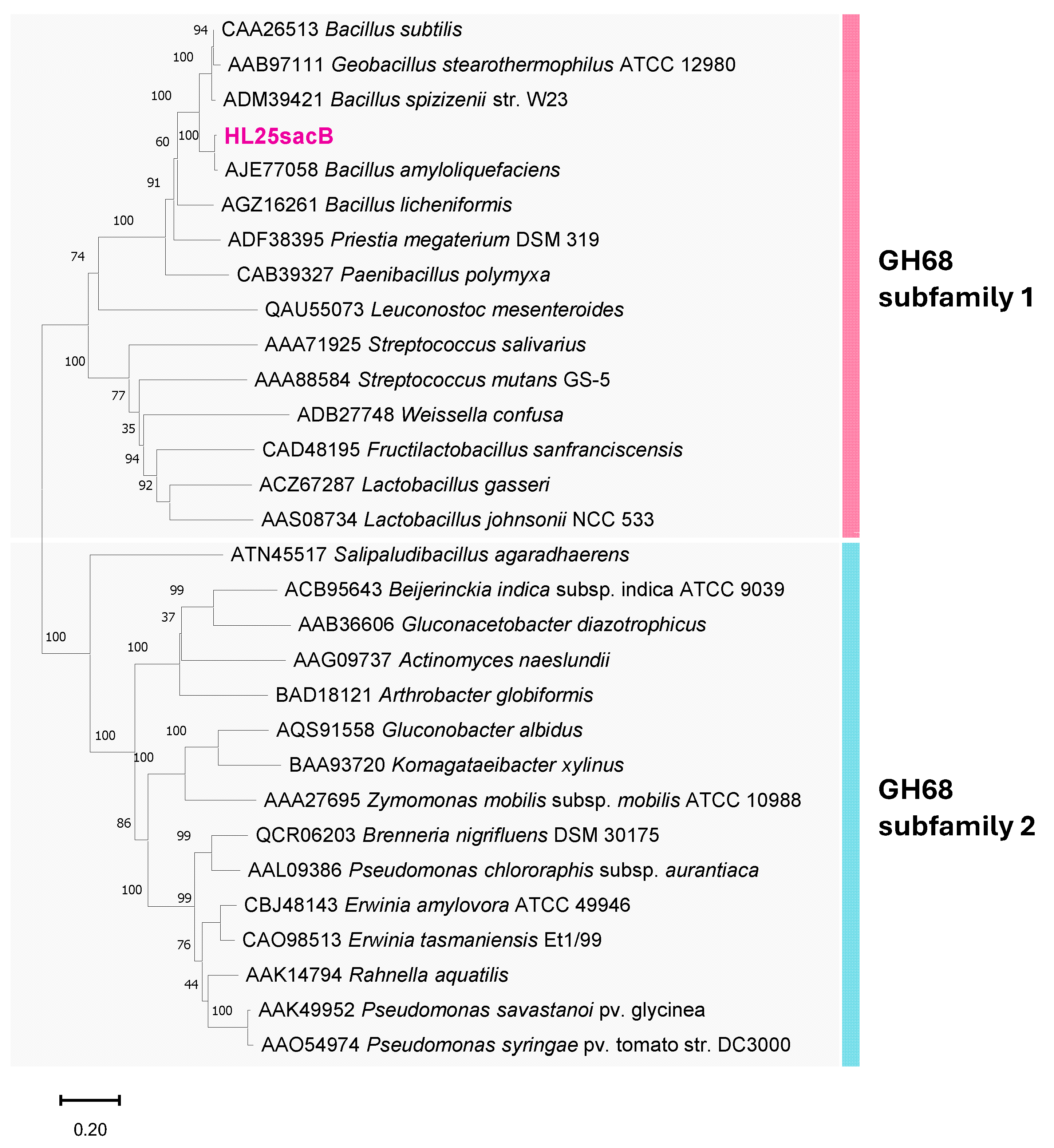
Focusing on levansucrase encoding gene, HL25SacB (GenBank accession number: PX097514) was isolated from B. velezensis HL25. The HL25SacB consists of 1419 bp, encoding 473 amino acids with an estimated molecular weight of 53 kDa. SignalP 6.0 analysis predicted a Sec-type signal peptide within the first 29 amino acids, with a cleavage site between 29 and 30 of HL25SacB. This Sec-type signal peptide corresponds to several Gram-positive levan producing bacteria such as B. subtilis, B. amyloliquefaciens, Streptococcus salivarius, Lactobacillus panis [39,40,41,42], indicating an extracellular secretion of levansucrase after cleavage. Based on sequence alignment, HL25SacB exhibited the highest similarity as glycoside hydrolase family 68 (GH68) protein from B. amyloliquefaciens with 99.58% similarity (GenBank accession number WP_132106502.1). Regarding protein evolutionary analysis comparing to the previously biochemically described GH68 shown in Figure 2, HL25SacB was classified as a levansucrase belonging to GH68 subfamily 1, clustering within the same phylogenetic clade as levansucrases from other Bacillus species. Notably, the classified GH68 subfamily 1 levansucrase also shows close evolutionary relationships with microbial inulosucrases from L. citreum, Lactobacillus gasseri, and L. reuteri [43].
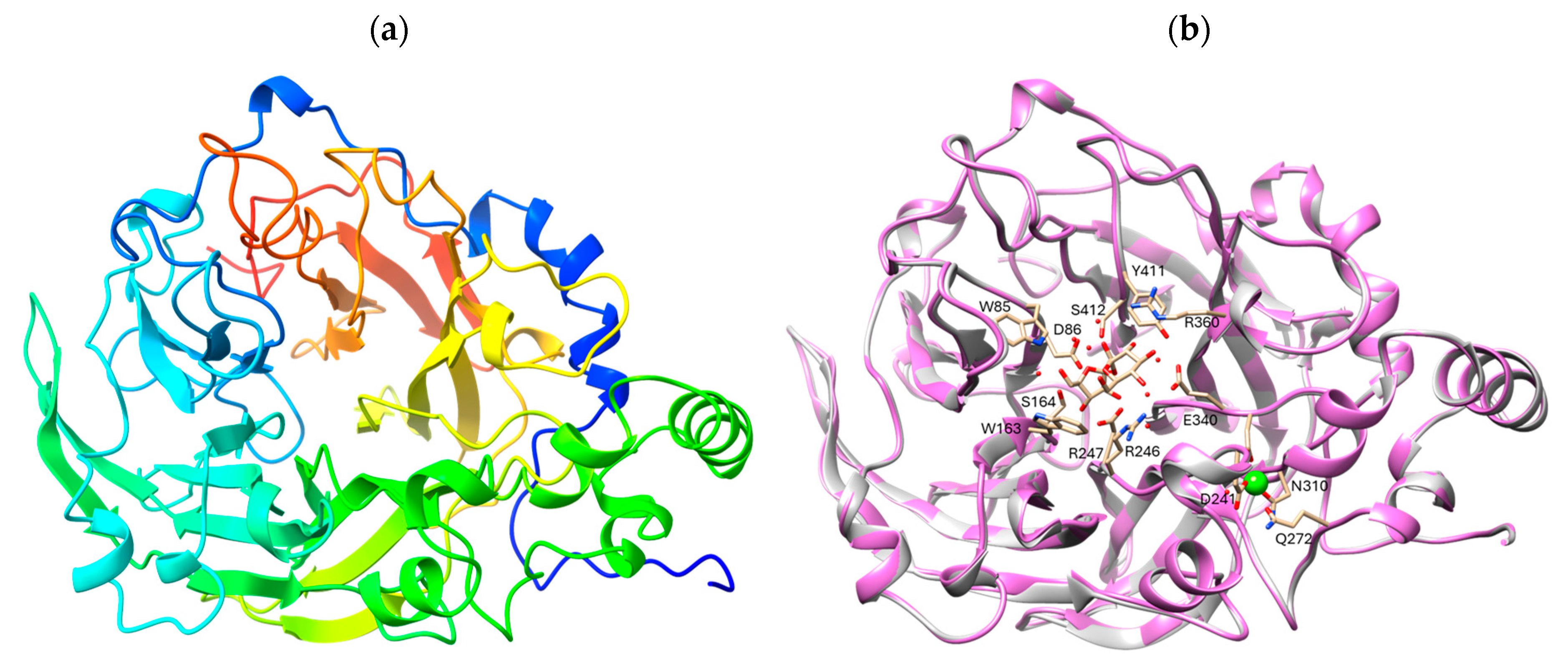
Secondary structure-based comparison of bacterial levansucrases revealed that HL25SacB shares an overall structural similarity with previously characterized levansucrases, including conserved sequence motifs (Figure S2). These highly conserved motifs, characteristic of glycoside hydrolase family 68 (GH68), likely contribute to the central framework of the β-propeller structure formed by HL25SacB. Identified motifs include 85WD86, 163WSGS166, 246RDP248, 339DEIER343, and 411YSH413, corresponding to those found in Priestia megaterium and P. koreensis [43,44].
The three-dimensional structure of HL25SacB was subsequently predicted using AlphaFold (Figure 3a,b). Structural assessment of the predicted model showed a high degree of similarity to the levansucrase from B. subtilis (PDB: 1pt2) with RMSD 0.158 angstroms. According to structural annotation, HL25SacB consists of a single domain exhibiting a five-bladed β-propeller architecture, in which each blade comprises four antiparallel β-strands forming the classical ‘W’-shaped topology. The active site is located at the base of the central cavity, with a funnel-like opening that provides access to a deep, negatively charged pocket on the protein surface, corresponding to the crystal structure of other microbial levansucrases [16]. The putative catalytic triad Asp86/Asp247/Glu340, located within the central active site, is predicted to function as the nucleophile, general acid-base catalyst, and transition-state stabilizer, respectively. These residues are conserved and functionally identical to those found in levansucrases from other species, including B. subtilis, P. megaterium, Gluconacetobacter diazotrophicus, Erwinia tasmaniensis, and Brenneria sp. [16,45,46,47,48].

2.3. Heterologous Expression and Purification

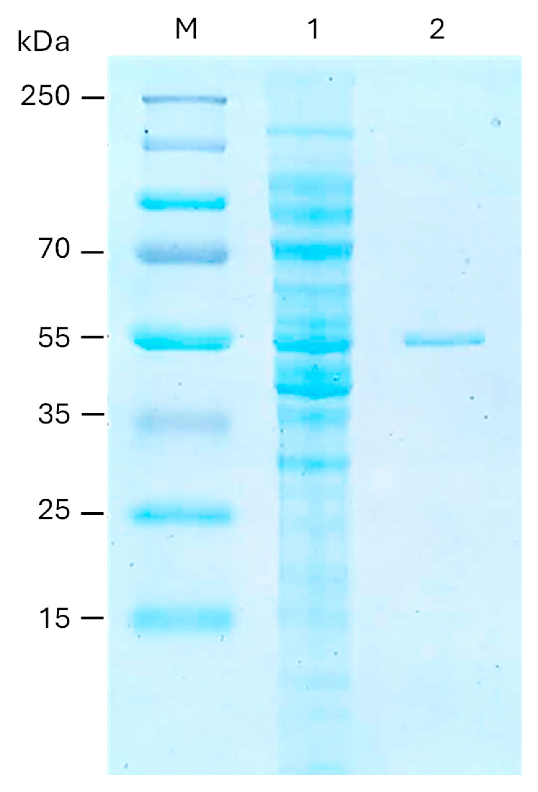
To characterize the function and biochemical properties of the levansucrase-encoding gene (ORF 1083) from B. velezensis HL25, the recombinant HL25SacB was produced heterologously in E. coli BL21(DE3). The full-length levansucrase encoding gene was expressed under the control of the T7 promoter and induced by 0.05 mM IPTG at 25 °C for 3 h. The expressed HL25SacB contained a 6xHistidine tag at the C-terminus and was subsequently purified to reach >95% homogeneity using a nickel ion affinity column (Figure 4).

2.4. Enzymatic Activity and Biochemical Characteristics of Recombinant HL25SacB

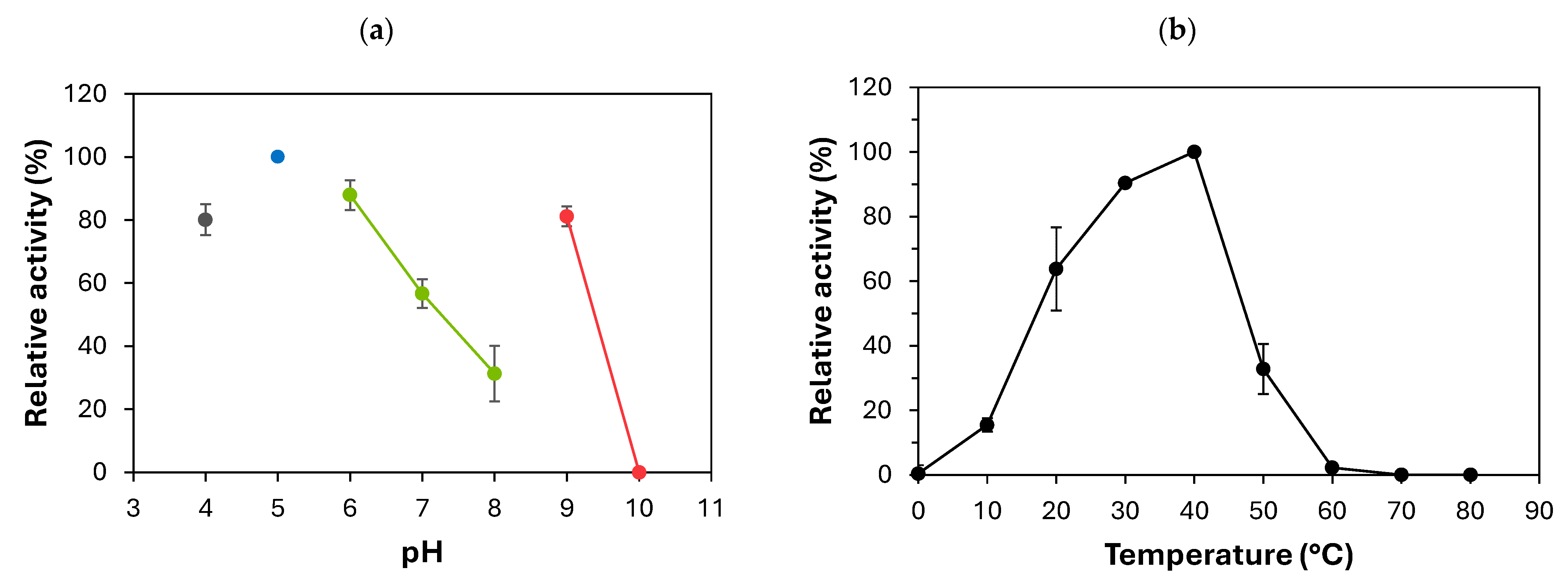
The levansucrase activity of recombinant HL25SacB was evaluated by measuring the amount of reducing sugars released from sucrose during the catalytic reaction, using the 3,5-dinitrosalicylic acid (DNS) assay. Regarding the effect of pH on enzyme activity presented in Figure 5a, the recombinant HL25SacB exhibited activity across a broad pH range in individual buffers, with the highest activity in 50 mM sodium acetate buffer at pH 5.0. Remarkably, over 80% of its maximum activity was retained at pH 4.0, 6.0, and 9.0, while complete loss of activity occurred at pH 10.0 in 50 mM glycine-NaOH buffer. The higher relative activity of HL25SacB observed at pH 5.0 not only indicates optimal catalytic conditions but also suggests a greater potential for levan synthesis, as the transfructosylation reaction responsible for polymer formation is favored under higher enzymatic activity. This broad pH tolerance is consistent with other microbial levansucrases, which typically show optimal activity under acidic conditions, generally in the range of pH 5.0–6.0 [16,43]. Almost all levansucrases from different sources are most stable and catalytically active under slightly acidic to neutral conditions, with reduced performance under both strongly acidic and basic environments. For instance, levansucrases from Gluconobacter oxydans exhibit peak activity at pH 4.0–5.0 but show significantly reduced activity outside this range [49]. Moreover, levansucrase from Bacillus siamensis displays clear pH sensitivity, maintaining optimal activity at pH 6.0. A significant decline in activity was observed at pH values above 7.0, indicating reduced stability or efficiency in more alkaline conditions. Additionally, acidic environments, particularly at pH 4.0, markedly inhibit activity of this levansucrase [50]. In contrast, the maximum activity was achieved in the alkaline region range (pH 8.0–9.0) for Vibrio natriegens levansucrase [49]. The levansucrase from B. amyloliquefaciens BH072, which shares 98.52% amino acid sequence identity with HL25SacB, exhibited maximal activity at pH 6.0 and less than 40% activity at pH 4.0 and 7.0. These biochemical differences from HL25SacB may result from seven amino acid substitutions, six of which are located near the substrate-binding pocket and ion-interaction regions [11]. Compared to these, HL25SacB demonstrates a superior broad pH range, especially under near neutral and mildly alkaline conditions, highlighting its potential for industrial applications requiring robust enzymatic performance across a broad pH spectrum.
The effect of temperature on levansucrase activity was also investigated. The results revealed that the recombinant HL25SacB exhibited increased activity at low temperatures ranging from 20 to 40 °C, with the highest activity at 40 °C (Figure 5b). The remaining enzyme activity was 90% and 64% relative to the maximum activity observed at 30 °C and 20 °C, respectively. At 50 °C and 10 °C, the activity decreased to 32% and 25%, respectively. Furthermore, the enzyme completely lost activity at elevated temperatures of 70 °C and 80 °C. The highest specific activity of the recombinant HL25SacB was detected at 40 °C in 50 mM sodium acetate buffer at pH 5.0. Levansucrase enzymes exhibit a range of optimal temperatures depending on their microbial sources. Reported optima include 30 °C for G. oxydans, L. mesenteroides MTCC10508, and Burkholderia graminis; 37 °C for B. siamensis; 40 °C for Aspergillus awamori EM66; 45 °C for V. natriegens; 45–50 °C for Novosphingobium aromaticivorans; and up to 65 °C for Pseudomonas orientalis [49,50,51]. Most levansucrases have optimal activity within 30–45 °C [51], and many, including those from B. subtilis ZW019, B. siamensis, and Bacillus sp. TH4-2 shows significant inhibition below 30 °C [16,20,50,52]. Only a few levansucrases retain high activity at low temperatures; examples include H. smyrnensis AAD6T and P. koreensis HL12 [27,43]. Notably, in this study, B. velezensis HL25 demonstrated cold-active levansucrase activity, maintaining over 60% activity even at 20 °C. This highlights its strong potential for low-temperature biotechnological applications, where energy efficiency and enzyme stability are critical. Kinetic analysis of the purified HL25SacB revealed a maximum velocity (Vmax) of 156 µmol/mg/min and a Michaelis–Menten constant (Km) of 22.97 mM for sucrose.

2.5. Biosynthesis of Fructooligosaccharides and Levan

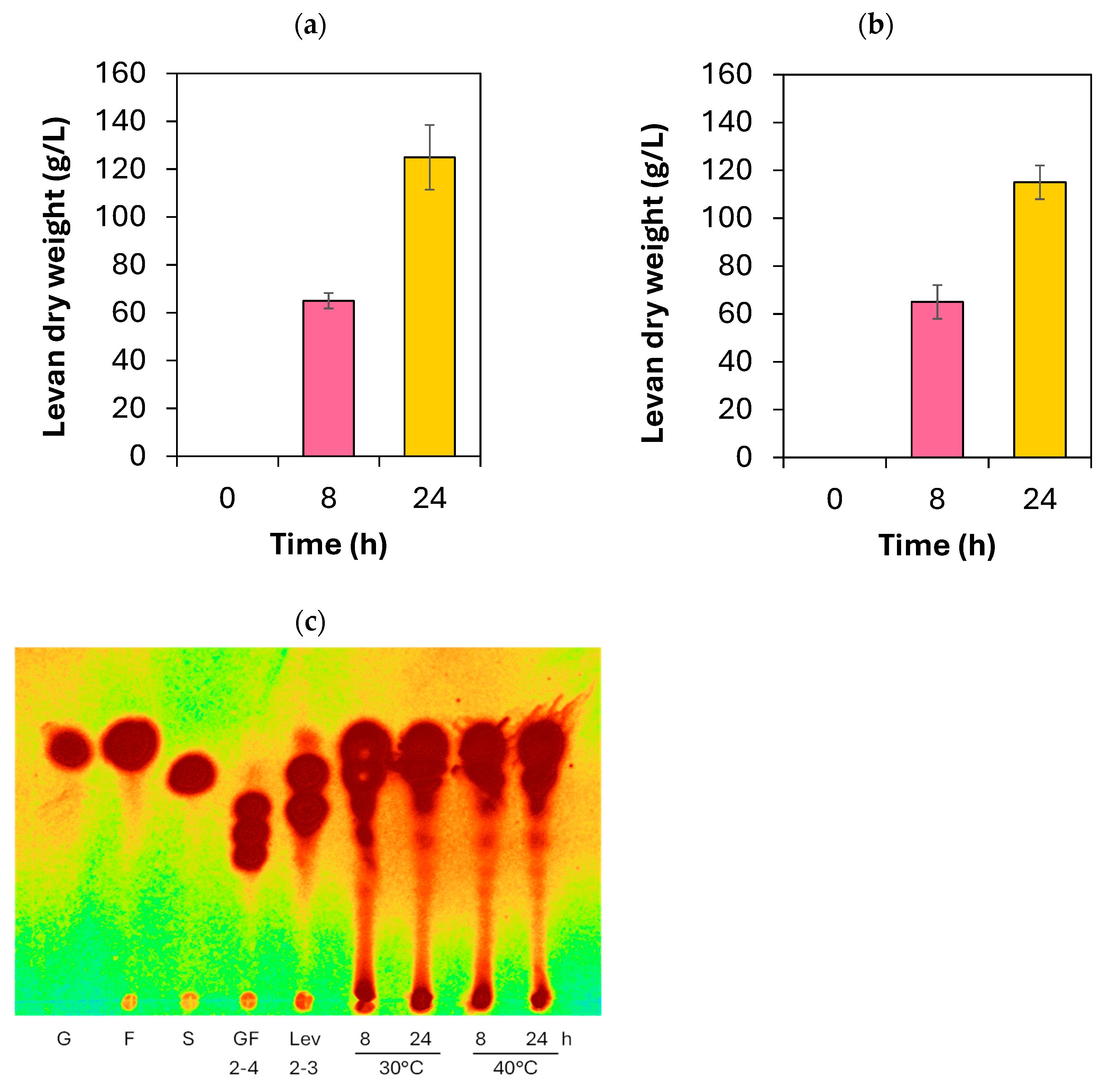
The fructooligosaccharides (FOSs) and levan synthesis potential of the recombinant HL25SacB enzyme were then evaluated using 200 g/L sucrose under optimized reaction conditions (30–40 °C, pH 5.0). The results revealed that the primary product released from sucrose catalyzed by HL25SacB was high-molecular-weight levan biopolymer (Figure 6a,b). In addition, a mixture of short-chain FOSs with degrees of polymerization (DP) ranging from 2 to 4 was also detected. The major FOSs components included levanbiose (DP2), 1-kestose (GF2), and 1,1-kestotetraose (GF3), while FOSs with DP > 4 were present in minor quantities (Figure 6c). The distribution of FOSs chain lengths was influenced by both enzyme concentration and incubation time, with longer incubation and higher enzyme levels promoting increased FOSs formation. Interestingly, levan production was significantly higher at 30 °C (125 ± 13.54 g/L levan, equivalent to 65.2% conversion) compared to 40 °C (115 ± 12.46 g/L levan, equivalent to 57.5% conversion) (Figure 6a,b). This may be attributed to the higher viscosity of sucrose at lower temperatures, which could reduce enzymatic hydrolysis activity and favor polymerization over degradation. These findings suggest that temperature plays a critical role in modulating the balance between FOSs synthesis and levan polymer formation. Levansucrases generally exhibit higher transfructosylation activity at lower temperatures, whereas hydrolytic activity becomes more prominent at elevated temperatures. For example, B. siamensis shows peak transfructosylation at 37 °C and hydrolytic activity at 50 °C, while L. reuteri LTH5448 displays a similar trend with optimal transfructosylation at 35 °C and hydrolysis at 45 °C. An exception is P. orientalis, which maintains high transfructosylation activity even at elevated temperatures [8,51]. Levan production by levansucrase depends on the microbial source, especially when using high initial sucrose concentrations (200–300 g/L). For example, recombinant levansucrase from Bacillus methylotrophicus SK 21.002 produced 100 g/L levan from 250 g/L sucrose (40% conversion) [53], and P. koreensis HL12 achieved 129 g/L (51.6%) from the same concentration [43]. Both V. natriegens and Bacillus indica subsp. indica converted 273 g/L sucrose into 85 g/L and 84 g/L levan, respectively, with yields around 31% [54]. B. subtilis also showed moderate efficiency (99.27 g/L from 300 g/L, 33.49%) [55]. In contrast, previous studies have demonstrated that levansucrases from Bacillus siamensis, Bacillus subtilis, and Erwinia amylovora exhibit markedly higher catalytic efficiency in fructooligosaccharides (FOSs) and levan biosynthesis compared to enzymes derived from other microorganisms (Table 1). The levansucrase from B. siamensis has been reported to produce 145.9 g/L of levan from a 200 g/L sucrose medium, corresponding to a 72.95% conversion yield, indicating substantial catalytic capability. Likewise, the enzyme from B. subtilis natto CCT7712 was shown to produce up to 366.01 g/L of FOSs and levan, with a 91.5% yield from 400 g/L sucrose, while the recombinant levansucrase from E. amylovora achieved 295 g/L of levan from 500 g/L sucrose, corresponding to a 59% yield [56,57]. In comparison, HL25SacB in the present study achieved a notably high conversion yield of 62.5% from 200 g/L sucrose, underscoring its outstanding catalytic efficiency and potential as a robust biocatalyst for industrial levan production.

3. Materials and Methods

3.1. Chemicals, Bacterial Strains and Plasmids

The bacterial strain B. velezensis HL25 was isolated from the rhizosphere of sago palm root (C. revoluta) collected from Trang Province, Thailand (GPS 7°31′21.0″ N 99°43′47.1″ E). Its sucrose utilizing capability was evaluated by cultivating YPS agar containing 10% (w/v) sucrose at 30 °C for 72 h [61]. Expression vector pET32a (Novagen, Darmstadt, Germany) was used for recombinant protein expression. The E. coli DH5α and E. coli BL21(DE3) (Novagen, Darmstadt, Germany) were used as bacterial hosts for cloning and recombinant protein production, respectively.

3.2. Bacterial Taxonomic Identification and Genome Sequence Analysis

Genomic DNA of B. velezensis HL25 was extracted using the GeneJET Genomic DNA Purification Kit (Thermo Fisher Scientific, Waltham, MA, USA) and quantified with a NanoDrop™ 2000 spectrophotometer (Thermo Fisher Scientific, Waltham, MA, USA). Bacterial taxonomic classification was conducted using 16S rRNA gene sequence analysis using Prok_BSF_8/20 (5′-AGAGTTTGATCCTGGCTCAG-3′) and Prok_REVB (5′-GGTTACCTTGTTACGACTT-3′) primers using PCR amplification, following previously described methods [61]. The genome sequencing of B. velezensis HL25 was performed through the Illumina 150 PE platform (Novogene) and subsequently annotated following the bioinformatics pipeline described in [10]. Taxonomic classification was performed based on genome sequence analysis against the entire genomes reference database GenomesDB using Tetra Correlation Search (TCS) [62]. All potential carbohydrate-active enzymes were predicted using dbCAN3, a web server for automated carbohydrate-active enzyme annotation (CAZymes) [63]. The carbohydrate-active enzyme candidates were subsequently verified by BLAST search against the NCBI database (http://www.ncbi.nlm.nih.gov/BLAST, accessed on 15 September 2025). The protein evolutionary analysis of HL25SacB against GH68_subfamily 1 and GH68_subfamily 2 sequence homologs was conducted using the MEGA11 program [64], using the Neighbor-Joining method [65] with bootstrap test (5000 replicates) [66]. The evolutionary distances were computed using the Kimura 2-parameter method [67]. For protein structure annotation, the conserved domain was analyzed by the NCBI Conserved Domains Database (CDD) [68]. The signal peptide involved in protein secretion pathway was analyzed by SignalP 6.0 [69]. Three-dimensional structure was predicted by homology modeling based on multiple sequence alignment with no gap, using AlphaFold in the ChimeraX 1.9 [70], and subsequently superimposed with levansucrase from B. subtillis (PDB: 1pt2) using Chimera [71].

3.3. Construction of pET32a-HL25SacB Recombinant Plasmid

The levansucrase encoding gene (sacB1083) (GenBank accession number: PX097514) was then amplified from B. velezensis HL25 genomic DNA using Phusion® High-Fidelity DNA Polymerase (Thermo Scientific, Waltham, MA, USA) with primers HL25SacB/NcoI/F (5′-GCCCATGGCAATGAACATCAAAAAATTTGCAAAA-3′) and HL25SacB/XhoI/R (5′-GCCTCGAGGTTGTTAACCGTAAGCTGTCCTTG-3′) incorporated with NcoI and XhoI restriction sites, respectively. The PCR reaction was carried out in Veriti™ Thermal Cycler (Thermo Scientific, Waltham, MA, USA). The thermal cycling conditions began at 95 °C for 5 min, followed by 25 cycles of denaturation at 95 °C for 30 s, annealing at 55 °C for 30 s, extension at 72 °C for 2 min, and final elongation at 72 °C for 10 min. The PCR product was then purified using the GeneJET Gel Extraction Kit (Thermo Scientific, Waltham, MA, USA), digested using NcoI and XhoI restriction enzymes, and ligated with the pET32a vector prior to digestion by the same restriction enzymes. The recombinant plasmid was transformed into E. coli DH5α using the heat-shock method, and its nucleotide sequence was confirmed by Sanger sequencing (Macrogen, Seoul, Republic of Korea). The recombinant plasmid pET32a-HL25SacB containing the HL25SacB gene with a 6xHistidine tag at the C-terminus was transformed into E. coli BL21(DE3) and spread on LB agar with 50 μg/mL ampicillin as a selectable marker.

3.4. Recombinant Protein Production and Purification

The recombinant colony was cultured in 5 mL LB medium containing 100 μg/mL ampicillin at 37 °C, 200 rpm for 18 h. The inoculum was then transferred into 800 mL of LB medium containing 100 μg/mL ampicillin with 1% inoculum and incubated at 37 °C for 3 h and subsequently induced by 0.05 mM IPTG with additional incubation at 25 °C, 200 rpm for 3 h. The cell was collected by centrifugation at 8000× g, 4 °C for 5 min and resuspended in 20 mM sodium phosphate buffer, pH 7.5. The cells were then disrupted using sonication (VCX 750 Ultrasonic Processors) on ice at 60% amplitude with a 9 s pulse on and a 9 s pulse off for a total of 10 cycles. The soluble protein fraction was harvested using centrifugation at 12,000× g, 4 °C for 30 min. The soluble protein was subsequently purified using a 5 mL HisTrap™ affinity column (GE Healthcare, Danderyd, Sweden) pre-equilibrated with binding buffer (20 mM sodium phosphate pH 7.4, 0.5 M NaCl, and 20 mM imidazole). The undesired proteins were removed by binding buffer. The recombinant SacB protein fused with 6xHis-tag was eluted using elution buffer containing 100 mM imidazole (20 mM sodium phosphate pH 7.4, 0.5 M NaCl, and 100 mM imidazole). Imidazole and salt were then removed using 30 kDa MWCO Amicon® Ultra-15 Centrifuge filter unit (Merck KGaA, Darmstadt, Germany). The purified protein was subsequently analyzed using 12% sodium dodecyl sulfate polyacrylamide gel electrophoresis, then stained with InstantBlue® Coomassie Protein Stain (Abcam, Singapore). The protein concentration was determined by measuring absorbance at 280 nm using a NanoDrop ND-100 spectrophotometer (Thermo Scientific, Waltham, MA, USA).

3.5. Enzyme Activity and Biochemical Property Characterization

The enzymatic activity of the recombinant HL25SacB levansucrase was evaluated using the 3,5-dinitrosalicylic acid (DNS) method [72], which quantifies the amount of reducing sugars released during catalysis. The reaction mixture consisted of 200 g/L sucrose, prepared in 50 mM sodium acetate buffer pH 5.0, and 0.8 µg (equivalent to 2.4 µg/mL) of enzyme. The mixture was then incubated at 40 °C for 30 min, followed by reaction termination at 100 °C for 10 min. The concentration of reducing sugars was determined by measuring absorbance at 540 nm using a spectrophotometer. All measurements were performed in triplicate to ensure reproducibility. One unit of levansucrase was defined as the amount of enzyme required to release 1 μmole of glucose per minute [25,60,73].
The effect of temperature on enzyme activity was assessed by a levansucrase activity assay conducted at temperatures ranging from 0 to 80 °C for 30 min in 50 mM sodium acetate buffer at pH 5.0. The effect of pH on enzyme activity was conducted at 40 °C for 30 min in 50 mM citrate buffer pH 4.0, 50 mM sodium acetate buffer pH 5.0, 50 mM sodium phosphate buffer pH 6.0–8.0, 50 mM glycine-NaOH buffer pH 9.0–10.0. The kinetic parameters of recombinant HL25SacB were determined using sucrose concentrations ranging from 1, 5, 10, 15, 20, 100, and 200 mg/mL in 50 mM sodium acetate buffer pH 5.0, incubated with 2 µg (equivalent to 5.9 µg/mL) of enzyme at 40 °C for 5–45 min. Kinetic parameters (Vmax and Km) were analyzed through the Michaelis–Menten equation using Prism software version 5.

3.6. Fructooligosaccharides and Levan Production

To evaluate the FOSs and levan synthesis activity of the recombinant HL25SacB, reactions were carried out using 200 g/L sucrose in 50 mM sodium acetate buffer pH 5.0 with 10 µg (equivalent to 20 µg/mL) of enzyme. Incubations were performed at 30 °C and 40 °C for 8 and 24 h, respectively. The short-chain FOSs profile liberated from enzyme catalysis was analyzed using thin layer chromatography using TLC Silica gel 60 F254 (Merck, Darmstadt, Germany) in 1-butanol:propan-2-ol:distilled water (50:30:20) as mobile phase. Spots were visualized using a developing solution containing 0.1% (w/v) orcinol in a 95:5 (v/v) mixture of ethanol and sulfuric acid, according to Lekakarn et al., 2025 [43]. Long-chain levan was precipitated by adding cold ethanol at a 3:1 (ethanol:reaction mixture) ratio [74], followed by incubation at 4 °C for 24 h. The precipitate was then collected by centrifugation at 12,000× g for 5 min and washed twice with 70% ethanol. The resulting pellet was dried in a desiccator, and the dry weight was subsequently measured.

4. Conclusions

This study is the first to report the organization of genes involved in fructan and levan-type fructooligosaccharides (L-FOSs) biosynthesis within the levansucrase operon, along with the associated sugar transport systems, in the identified novel B. velezensis HL25 strain. Moreover, functional and biochemical characterization of the recombinant HL25SacB levansucrase, classified into GH68 subfamily 1, provided insights into its role in the wild-type strain. Notably, HL25SacB demonstrated several industrially desirable features, including high conversion yield (62.5%), broad pH range (especially under near-neutral to mildly alkaline conditions), and cold-active functionality, maintaining over 60% activity even at 20 °C. These findings deepen our understanding of levan and L-FOSs biosynthesis mechanisms and highlight the biotechnological potential of HL25 and its levansucrase, HL25SacB, as a promising biocatalyst for developing robust, energy-efficient, and pH-tolerant bioprocesses using sucrose-rich feedstocks.

Supplementary Materials

The following supporting information can be downloaded at: https://www.mdpi.com/article/10.3390/catal15111059/s1, Figure S1: Product profile of fructooligosaccharides (FOSs) produced by the whole-cell biocatalyst B. velezensis HL25 cultivated in a sucrose-rich medium; Figure S2: Structure-based sequence alignment of levansucrase (HL25sacB) identified from B. velezensis HL25 and homologous sequences from previously biochemically characterized levansucrases classified into GH68_subfamily 1 and subfamily 2. Table S1: Nucleotide position of genes encoding enzymes associated with sucrose metabolism and fructan biosynthesis in B. velezensis HL25 genome.

Author Contributions

Conceptualization, H.L. and B.B.; methodology, H.L., J.C., R.J., P.K., T.K. and B.B.; software, W.M. and B.B.; validation, H.L. and B.B.; formal analysis, H.L. and B.B.; investigation, H.L. and B.B.; resources, H.L.; data curation, H.L. and B.B.; writing—original draft preparation, H.L., J.C., R.J., P.K., T.K. and B.B.; writing—review and editing, H.L., J.C., R.J., P.K., T.K., W.M. and B.B.; visualization, H.L. and B.B.; supervision, H.L. and B.B.; project administration, H.L.; funding acquisition, H.L. All authors have read and agreed to the published version of the manuscript.

Funding

This research project was supported by the Thailand Science Research and Innovation Fundamental Fund fiscal year 2025, Thammasat University.

Data Availability Statement

The raw data supporting the conclusions of this article will be made available by the authors on request.

Acknowledgments

The authors acknowledge the support provided by the Enzyme Technology Research Team, National Center for Genetic Engineering and Biotechnology, and Department of Biotechnology, Faculty of Science and Technology, Thammasat University. The authors also acknowledge the support from the Science Classrooms in University-Affiliated School Project (SCiUS), Thammasat University-Suankularb Wittayalai Rangsit School Center, and the 27th Young Scientist Competition (YSC 2025). The authors acknowledge support from S.M. Chemical Supplies Co., Ltd. (Bangkok, Thailand) for providing TLC Explorer for TLC visualization. The authors also acknowledge support from Kittipong Chaisuriyaphun.

Conflicts of Interest

The authors declare that they have no conflicts of interest with the contents of this article.

References

  1. Le Bourgot, C.; Rigaudier, F.; Juhel, C.; Herpin, F.; Meunier, C. Gastrointestinal tolerance of short-chain fructo-oligosaccharides from sugar beet: An observational, connected, dose-ranging study in healthy volunteers. Nutrients 2022, 14, 1461. [Google Scholar] [CrossRef] [PubMed]
  2. Dominguez, A.L.; Rodrigues, L.R.; Lima, N.M.; Teixeira, J.A. An overview of the recent developments on fructooligosaccharide production and applications. Food Bioprocess Technol. 2014, 7, 324–337. [Google Scholar] [CrossRef]
  3. Domżał-Kędzia, M.; Ostrowska, M.; Lewińska, A.; Łukaszewicz, M. Recent developments and applications of microbial levan, a versatile polysaccharide-based biopolymer. Molecules 2023, 28, 5407. [Google Scholar] [CrossRef]
  4. Bruni, G.O.; Qi, Y.; Terrell, E.; Dupre, R.A.; Mattison, C.P. Characterization of levan fructan produced by a Gluconobacter japonicus strain isolated from a sugarcane processing facility. Microorganisms 2024, 12, 107. [Google Scholar] [CrossRef]
  5. Erdal Altıntaş, Ö.; Toksoy Öner, E.; Çabuk, A.; Aytar Çelik, P. Biosynthesis of levan by Halomonas elongata 153B: Optimization for enhanced production and potential biological activities for pharmaceutical field. J. Polym. Environ. 2022, 31, 1440–1455. [Google Scholar] [CrossRef]
  6. Hövels, M.; Kosciow, K.; Kniewel, J.; Jakob, F.; Deppenmeier, U. High yield production of levan-type fructans by Gluconobacter japonicus LMG 1417. Int. J. Biol. Macromol. 2020, 164, 295–303. [Google Scholar] [CrossRef]
  7. Kim, K.H.; Chung, C.B.; Kim, Y.H.; Kim, K.S.; Han, C.S.; Kim, C.H. Cosmeceutical properties of levan produced by Zymomonas mobilis. J. Cosmet. Sci. 2005, 56, 395–406. [Google Scholar] [CrossRef]
  8. Ni, D.; Xu, W.; Bai, Y.; Zhang, W.; Zhang, T.; Mu, W. Biosynthesis of levan from sucrose using a thermostable levansucrase from Lactobacillus reuteri LTH5448. Int. J. Biol. Macromol. 2018, 113, 29–37. [Google Scholar] [CrossRef]
  9. Sarilmiser, H.K.; Ates, O.; Ozdemir, G.; Arga, K.Y.; Oner, E.T. Effective stimulating factors for microbial levan production by Halomonas smyrnensis AAD6T. J. Biosci. Bioeng. 2015, 119, 455–463. [Google Scholar] [CrossRef] [PubMed]
  10. Lekakarn, H.; Prongjit, D.; Mhuantong, W.; Trakarnpaiboon, S.; Bunterngsook, B. Exploring levansucrase operon regulating levan-type fructooligosaccharides (L-FOSs) production in Priestia koreensis HL12. J. Microbiol. Biotechnol. 2024, 34, 1959–1968. [Google Scholar] [CrossRef] [PubMed]
  11. Mu, D.; Zhou, Y.; Wu, X.; Montalban-Lopez, M.; Wang, L.; Li, X.; Zheng, Z. Secretion of Bacillus amyloliquefaciens levansucrase from Bacillus subtilis and its application in the enzymatic synthesis of levan. ACS Food Sci. Technol. 2021, 1, 249–259. [Google Scholar] [CrossRef]
  12. Xu, W.; Ni, D.; Zhang, W.; Guang, C.; Zhang, T.; Mu, W. Recent advances in levansucrase and inulosucrase: Evolution, characteristics, and application. Crit. Rev. Food. Sci. Nutr. 2019, 59, 3630–3647. [Google Scholar] [CrossRef]
  13. Tezgel, N.; Kırtel, O.; Van den Ende, W.; Toksoy Oner, E. Fructosyltransferase enzymes for microbial fructan production. In Microbial Enzymes: Roles and Applications in Industries; Arora, N.K., Mishra, J., Mishra, V., Eds.; Springer: Singapore, 2020; pp. 1–39. [Google Scholar]
  14. Seibel, J.; Moraru, R.; Götze, S.; Buchholz, K.; Na’amnieh, S.; Pawlowski, A.; Hecht, H.-J. Synthesis of sucrose analogues and the mechanism of action of Bacillus subtilis fructosyltransferase (levansucrase). Carbohydr. Res. 2006, 341, 2335–2349. [Google Scholar] [CrossRef]
  15. Zhang, W.; Xu, W.; Ni, D.; Dai, Q.; Guang, C.; Zhang, T.; Mu, W. An overview of levan-degrading enzyme from microbes. Appl. Microbiol. Biotechnol. 2019, 103, 7891–7902. [Google Scholar] [CrossRef] [PubMed]
  16. Xu, W.; Zhang, X.; Ni, D.; Zhang, W.; Guang, C.; Mu, W. A review of fructosyl-transferases from catalytic characteristics and structural features to reaction mechanisms and product specificity. Food Chem. 2024, 440, 138250. [Google Scholar] [CrossRef] [PubMed]
  17. Castrejón-Carrillo, S.; Morales-Moreno, L.A.; Rodríguez-Alegría, M.E.; Zavala-Padilla, G.T.; Bello-Pérez, L.A.; Moreno-Zaragoza, J.; López Munguía, A. Insights into the heterogeneity of levan polymers synthesized by levansucrase Bs-SacB from Bacillus subtilis 168. Carbohydr. Polym. 2024, 323, 121439. [Google Scholar] [CrossRef]
  18. Chandravanshi, M.; Sharma, A.; Dasgupta, P.; Mandal, S.K.; Kanaujia, S.P. Identification and characterization of ABC transporters for carbohydrate uptake in Thermus thermophilus HB8. Gene 2019, 696, 135–148. [Google Scholar] [CrossRef] [PubMed]
  19. Cheng, R.; Cheng, L.; Zhao, Y.; Wang, L.; Wang, S.; Zhang, J. Biosynthesis and prebiotic activity of a linear levan from a new Paenibacillus isolate. Appl. Microbiol. Biotechnol. 2021, 105, 769–787. [Google Scholar] [CrossRef]
  20. Wang, J.; Xu, X.; Zhao, F.; Yin, N.; Zhou, Z.; Han, Y. Biosynthesis and structural characterization of levan by a recombinant levansucrase from Bacillus subtilis ZW019. Waste Biomass Valorization 2022, 13, 4599–4609. [Google Scholar] [CrossRef]
  21. Jadaun, J.S.; Narnoliya, L.K.; Agarwal, N.; Singh, S.P. Catalytic biosynthesis of levan and short-chain fructooligosaccharides from sucrose-containing feedstocks by employing the levansucrase from Leuconostoc mesenteroides MTCC10508. Int. J. Biol. Macromol. 2019, 127, 486–495. [Google Scholar] [CrossRef]
  22. Polsinelli, I.; Caliandro, R.; Salomone-Stagni, M.; Demitri, N.; Rejzek, M.; Field, R.A.; Benini, S. Comparison of the Levansucrase from the epiphyte Erwinia tasmaniensis vs its homologue from the phytopathogen Erwinia amylovora. Int. J. Biol. Macromol. 2019, 127, 496–501. [Google Scholar] [CrossRef]
  23. Khandekar, S.; Srivastava, A.; Pletzer, D.; Stahl, A.; Ullrich, M.S. The conserved upstream region of lscB/C determines expression of different levansucrase genes in plant pathogen Pseudomonas syringae. BMC Microbiol. 2014, 14, 79. [Google Scholar] [CrossRef]
  24. Martinez-Fleites, C.; Ortiz-Lombardia, M.; Pons, T.; Tarbouriech, N.; Taylor, E.J.; Arrieta, J.G.; Hernandez, L.; Davies, G.J. Crystal structure of levansucrase from the Gram-negative bacterium Gluconacetobacter diazotrophicus. Biochem. J. 2005, 390, 19–27. [Google Scholar] [CrossRef]
  25. Gao, S.; Qi, X.; Hart, D.J.; Gao, H.; An, Y. Expression and characterization of levansucrase from Clostridium acetobutylicum. J. Agric. Food Chem. 2017, 65, 867–871. [Google Scholar] [CrossRef]
  26. Shaheen, S.; Aman, A.; Qader, S.A.; Siddiqui, N.N. Hyper-production of levansucrase from Zymomonas mobilis KIBGEIB14 using submerged fermentation technique. Pak. J. Pharm. Sci. 2017, 30, 2053–2059. [Google Scholar] [PubMed]
  27. Kirtel, O.; Menéndez, C.; Versluys, M.; Van den Ende, W.; Hernández, L.; Toksoy Öner, E. Levansucrase from Halomonas smyrnensis AAD6(T): First halophilic GH-J clan enzyme recombinantly expressed, purified, and characterized. Appl. Microbiol. Biotechnol. 2018, 102, 9207–9220. [Google Scholar] [CrossRef] [PubMed]
  28. Lekakarn, H.; Bunterngsook, B.; Jaikaew, P.; Kuantum, T.; Wansuksri, R.; Champreda, V. Functional characterization of recombinant endo-levanase (LevBk) from Bacillus koreensis HL12 on short-chain levan-type fructooligosaccharides production. Protein J. 2022, 41, 477–488. [Google Scholar] [CrossRef]
  29. Han, S.; Liu, H.; Li, S.; Zheng, Z.; Yan, Q.; Jiang, Z. High level production of a β-fructofuranosidase in Aspergillus niger for the preparation of prebiotic bread using in situ enzymatic conversion. Food Res. Int. 2025, 208, 116225. [Google Scholar] [CrossRef] [PubMed]
  30. Belmonte-Izquierdo, Y.; Salomé-Abarca, L.F.; González-Hernández, J.C.; López, M.G. Fructooligosaccharides (FOS) production by microorganisms with fructosyltransferase activity. Fermentation 2023, 9, 968. [Google Scholar] [CrossRef]
  31. de Lima, M.Z.T.; de Almeida, L.R.; Mera, A.M.; Bernardes, A.; Garcia, W.; Muniz, J.R.C. Crystal structure of a sucrose-6-phosphate hydrolase from Lactobacillus gasseri with potential applications in fructan production and the food industry. J. Agric. Food Chem. 2021, 69, 10223–10234. [Google Scholar] [CrossRef]
  32. Daguer, J.P.; Geissmann, T.; Petit-Glatron, M.F.; Chambert, R. Autogenous modulation of the Bacillus subtilis sacB-levB-yveA levansucrase operon by the levB transcript. Microbiology 2004, 150, 3669–3679. [Google Scholar] [CrossRef]
  33. Liyaskina, E.V.; Rakova, N.A.; Kitykina, A.A.; Rusyaeva, V.V.; Toukach, P.V.; Fomenkov, A.; Vainauskas, S.; Roberts, R.J.; Revin, V.V. Production and characterization of the exopolysaccharide from strain Paenibacillus polymyxa 2020. PLoS ONE 2021, 16, e0253482. [Google Scholar] [CrossRef] [PubMed]
  34. Senthilkumar, V.; Rajendhran, J.; Busby, S.J.; Gunasekaran, P. Characterization of multiple promoters and transcript stability in the sacB-sacC gene cluster in Zymomonas mobilis. Arch. Microbiol. 2009, 191, 529–541. [Google Scholar] [CrossRef] [PubMed]
  35. Tian, T.; Sun, B.; Shi, H.; Gao, T.; He, Y.; Li, Y.; Liu, Y.; Li, X.; Zhang, L.; Li, S.; et al. Sucrose triggers a novel signaling cascade promoting Bacillus subtilis rhizosphere colonization. ISME J. 2021, 15, 2723–2737. [Google Scholar] [CrossRef]
  36. Vu, T.H.N.; Quach, N.T.; Xuan, D.; Le, H.; Chi, L.; Lam, N.; Ngo, C.; Hà, H.; Chu, H.; Phi, Q.-T. A genomic perspective on the potential of termite-associated Cellulosimicrobium cellulans MP1 as producer of plant biomass-acting enzymes and exopolysaccharides. PeerJ 2021, 9, e11839. [Google Scholar] [CrossRef]
  37. Zeng, L.; Burne, R.A. Sucrose- and fructose-specific effects on the transcriptome of Streptococcus mutans, as determined by RNA sequencing. Appl. Environ. Microbiol. 2016, 82, 146–156. [Google Scholar] [CrossRef]
  38. Carreón-Rodríguez, O.E.; Gosset, G.; Escalante, A.; Bolívar, F. Glucose transport in Escherichia coli: From basics to transport engineering. Microorganisms 2023, 11, 1588. [Google Scholar] [CrossRef] [PubMed]
  39. Steinmetz, M.; Le Coq, D.; Aymerich, S.; Gonzy-Tréboul, G.; Gay, P. The DNA sequence of the gene for the secreted Bacillus subtilis enzyme levansucrase and its genetic control sites. Mol. Gen. Genet. 1985, 200, 220–228. [Google Scholar] [CrossRef]
  40. Waldherr, F.W.; Meissner, D.; Vogel, R.F. Genetic and functional characterization of Lactobacillus panis levansucrase. Arch. Microbiol. 2008, 190, 497–505. [Google Scholar] [CrossRef]
  41. Rathsam, C.; Jacques, N.A. Role of C-terminal domains in surface attachment of the fructosyltransferase of Streptococcus salivarius ATCC 25975. J. Bacteriol. 1998, 180, 6400–6403. [Google Scholar] [CrossRef]
  42. Borchert, T.V.; Nagarajan, V. Structure-function studies on the Bacillus amyloliquefaciens levansucrase signal peptide. In Genetics and Biotechnology of Bacilli; Zukowski, M.M., Ganesan, A.T., Hoch, J.A., Eds.; Academic Press: Cambridge, MA, USA, 1990; pp. 171–177. [Google Scholar]
  43. Lekakarn, H.; Phusiri, N.; Komonmusik, T.; Jaikaew, P.; Trakarnpaiboon, S.; Bunterngsook, B. Novel cold-active levansucrase (SacBPk) from Priestia koreensis HL12 for short-chain fructooligosaccharides and levan synthesis. Catalysts 2025, 15, 216. [Google Scholar] [CrossRef]
  44. Homann, A.; Biedendieck, R.; Götze, S.; Jahn, D.; Seibel, J. Insights into polymer versus oligosaccharide synthesis: Mutagenesis and mechanistic studies of a novel levansucrase from Bacillus megaterium. Biochem. J. 2007, 407, 189–198. [Google Scholar] [CrossRef] [PubMed]
  45. Strube, C.P.; Homann, A.; Gamer, M.; Jahn, D.; Seibel, J.; Heinz, D.W. Polysaccharide synthesis of the levansucrase SacB from Bacillus megaterium is controlled by distinct surface motifs. J. Biol. Chem. 2011, 286, 17593–17600. [Google Scholar] [CrossRef]
  46. Polsinelli, I.; Caliandro, R.; Demitri, N.; Benini, S. The structure of sucrose-soaked levansucrase crystals from Erwinia tasmaniensis reveals a binding pocket for levanbiose. Int. J. Mol. Sci. 2020, 21, 83. [Google Scholar] [CrossRef]
  47. Xu, W.; Ni, D.; Hou, X.; Pijning, T.; Guskov, A.; Rao, Y.; Mu, W. Crystal structure of levansucrase from the Gram-negative bacterium Brenneria provides insights into its product size specificity. J. Agric. Food Chem. 2022, 70, 5095–5105. [Google Scholar] [CrossRef]
  48. Raga-Carbajal, E.; Díaz-Vilchis, A.; Rojas-Trejo, S.P.; Rudiño-Piñera, E.; Olvera, C. The molecular basis of the nonprocessive elongation mechanism in levansucrases. J. Biol. Chem. 2021, 296, 100178. [Google Scholar] [CrossRef] [PubMed]
  49. Sahyoun, A.M.; Karboune, S. Unveiling four levansucrase enzymes: Insights into catalytic properties, kinetics, and end-product profiles. Process Biochem. 2024, 141, 39–49. [Google Scholar] [CrossRef]
  50. Phengnoi, P.; Thakham, N.; Rachphirom, T.; Teerakulkittipong, N.; Lirio, G.A.; Jangiam, W. Characterization of levansucrase produced by novel Bacillus siamensis and optimization of culture condition for levan biosynthesis. Heliyon 2022, 8, e12137. [Google Scholar] [CrossRef]
  51. Guang, C.; Zhang, X.; Ni, D.; Zhang, W.; Xu, W.; Mu, W. Identification of a thermostable levansucrase from Pseudomonas orientalis that allows unique product specificity at different temperatures. Polymers 2023, 15, 1435. [Google Scholar] [CrossRef]
  52. Ben Ammar, Y.; Matsubara, T.; Ito, K.; Iizuka, M.; Limpaseni, T.; Pongsawasdi, P.; Minamiura, N. Characterization of a thermostable levansucrase from Bacillus sp. TH4-2 capable of producing high molecular weight levan at high temperature. J. Biotechnol. 2002, 99, 111–119. [Google Scholar] [CrossRef]
  53. Zhang, T.; Li, R.; Qian, H.; Mu, W.; Miao, M.; Jiang, B. Biosynthesis of levan by levansucrase from Bacillus methylotrophicus SK 21.002. Carbohydr. Polym. 2014, 101, 975–981. [Google Scholar] [CrossRef]
  54. Hill, A.; Karboune, S.; Narwani, T.J.; de Brevern, A.G. Investigating the product profiles and structural relationships of new levansucrases with conventional and non-conventional substrates. Int. J. Mol. Sci. 2020, 21, 5402. [Google Scholar] [CrossRef] [PubMed]
  55. Hou, Y.; Huang, F.; Yang, H.; Cong, H.; Zhang, X.; Xie, X.; Yang, H.; Tong, Q.; Luo, N.; Zhu, P.; et al. Factors affecting the production and molecular weight of levan in enzymatic synthesis by recombinant Bacillus subtilis levansucrase SacB-T305A. Polym. Int. 2021, 70, 185–192. [Google Scholar] [CrossRef]
  56. Peng, J.; Xu, W.; Ni, D.; Zhang, W.; Zhang, T.; Guang, C.; Mu, W. Preparation of a novel water-soluble gel from Erwinia amylovora levan. Int. J. Biol. Macromol. 2019, 122, 469–478. [Google Scholar] [CrossRef]
  57. Magri, A.; Oliveira, M.R.; Baldo, C.; Tischer, C.A.; Sartori, D.; Mantovani, M.S.; Celligoi, M. Production of fructooligosaccharides by Bacillus subtilis natto CCT7712 and their antiproliferative potential. J. Appl. Microbiol. 2020, 128, 1414–1426. [Google Scholar] [CrossRef]
  58. Lu, L.; Fu, F.; Zhao, R.; Jin, L.; He, C.; Xu, L.; Xiao, M. A recombinant levansucrase from Bacillus licheniformis 8-37-0-1 catalyzes versatile transfructosylation reactions. Process Biochem. 2014, 49, 1503–1510. [Google Scholar] [CrossRef]
  59. Liu, Q.; Yu, S.; Zhang, T.; Jiang, B.; Mu, W. Efficient biosynthesis of levan from sucrose by a novel levansucrase from Brenneria goodwinii. Carbohydr. Polym. 2017, 157, 1732–1740. [Google Scholar] [CrossRef]
  60. Xu, W.; Ni, D.; Yu, S.; Zhang, T.; Mu, W. Insights into hydrolysis versus transfructosylation: Mutagenesis studies of a novel levansucrase from Brenneria sp. EniD312. Int. J. Biol. Macromol. 2018, 116, 335–345. [Google Scholar] [CrossRef]
  61. Lekakarn, H.; Bunterngsook, B.; Pajongpakdeekul, N.; Prongjit, D.; Champreda, V. A novel low temperature active maltooligosaccharides-forming amylase from Bacillus koreensis HL12 as biocatalyst for maltooligosaccharide production. 3 Biotech 2022, 12, 134. [Google Scholar] [CrossRef] [PubMed]
  62. Richter, M.; Rosselló-Móra, R.; Oliver Glöckner, F.; Peplies, J. JSpeciesWS: A web server for prokaryotic species circumscription based on pairwise genome comparison. Bioinformatics 2015, 32, 929–931. [Google Scholar] [CrossRef] [PubMed]
  63. Zheng, J.; Ge, Q.; Yan, Y.; Zhang, X.; Huang, L.; Yin, Y. dbCAN3: Automated carbohydrate-active enzyme and substrate annotation. Nucleic Acids Res. 2023, 51, W115–W121. [Google Scholar] [CrossRef] [PubMed]
  64. Tamura, K.; Stecher, G.; Kumar, S. MEGA11: Molecular evolutionary genetics analysis version 11. Mol. Biol. Evol. 2021, 38, 3022–3027. [Google Scholar] [CrossRef] [PubMed]
  65. Saitou, N.; Nei, M. The neighbor-joining method: A new method for reconstructing phylogenetic trees. Mol. Biol. Evol. 1987, 4, 406–425. [Google Scholar] [CrossRef] [PubMed]
  66. Sanderson, M.J. Confidence limits on phylogenies: The bootstrap revisited. Cladistics 1989, 5, 113–129. [Google Scholar] [CrossRef]
  67. Kimura, M. A simple method for estimating evolutionary rates of base substitutions through comparative studies of nucleotide sequences. J. Mol. Evol. 1980, 16, 111–120. [Google Scholar] [CrossRef]
  68. Marchler-Bauer, A.; Derbyshire, M.K.; Gonzales, N.R.; Lu, S.; Chitsaz, F.; Geer, L.Y.; Geer, R.C.; He, J.; Gwadz, M.; Hurwitz, D.I.; et al. CDD: NCBI’s conserved domain database. Nucleic Acids Res. 2014, 43, D222–D226. [Google Scholar] [CrossRef]
  69. Teufel, F.; Almagro Armenteros, J.J.; Johansen, A.R.; Gislason, M.H.; Pihl, S.I.; Tsirigos, K.D.; Winther, O.; Brunak, S.; von Heijne, G.; Nielsen, H. SignalP 6.0 predicts all five types of signal peptides using protein language models. Nat. Biotechnol. 2022, 40, 1023–1025. [Google Scholar] [CrossRef]
  70. Meng, E.C.; Pettersen, E.F.; Couch, G.S.; Huang, C.C.; Ferrin, T.E. Tools for integrated sequence-structure analysis with UCSF Chimera. BMC Bioinform. 2006, 7, 339. [Google Scholar] [CrossRef]
  71. Pettersen, E.F.; Goddard, T.D.; Huang, C.C.; Couch, G.S.; Greenblatt, D.M.; Meng, E.C.; Ferrin, T.E. UCSF Chimera—A visualization system for exploratory research and analysis. J. Comput. Chem. 2004, 25, 1605–1612. [Google Scholar] [CrossRef]
  72. Miller, G.L. Use of dinitrosalicylic acid reagent for determination of reducing sugar. Anal. Chem. 1959, 31, 426–428. [Google Scholar] [CrossRef]
  73. Belghith, K.S.; Dahech, I.; Belghith, H.; Mejdoub, H. Microbial production of levansucrase for synthesis of fructooligosaccharides and levan. Int. J. Biol. Macromol. 2012, 50, 451–458. [Google Scholar] [CrossRef] [PubMed]
  74. Chidambaram, J.; Veerapandian, B.; Sarwareddy, K.K.; Mani, K.P.; Shanmugam, S.R.; Venkatachalam, P. Studies on solvent precipitation of levan synthesized using Bacillus subtilis MTCC 441. Heliyon 2019, 5, e02414. [Google Scholar] [CrossRef] [PubMed]
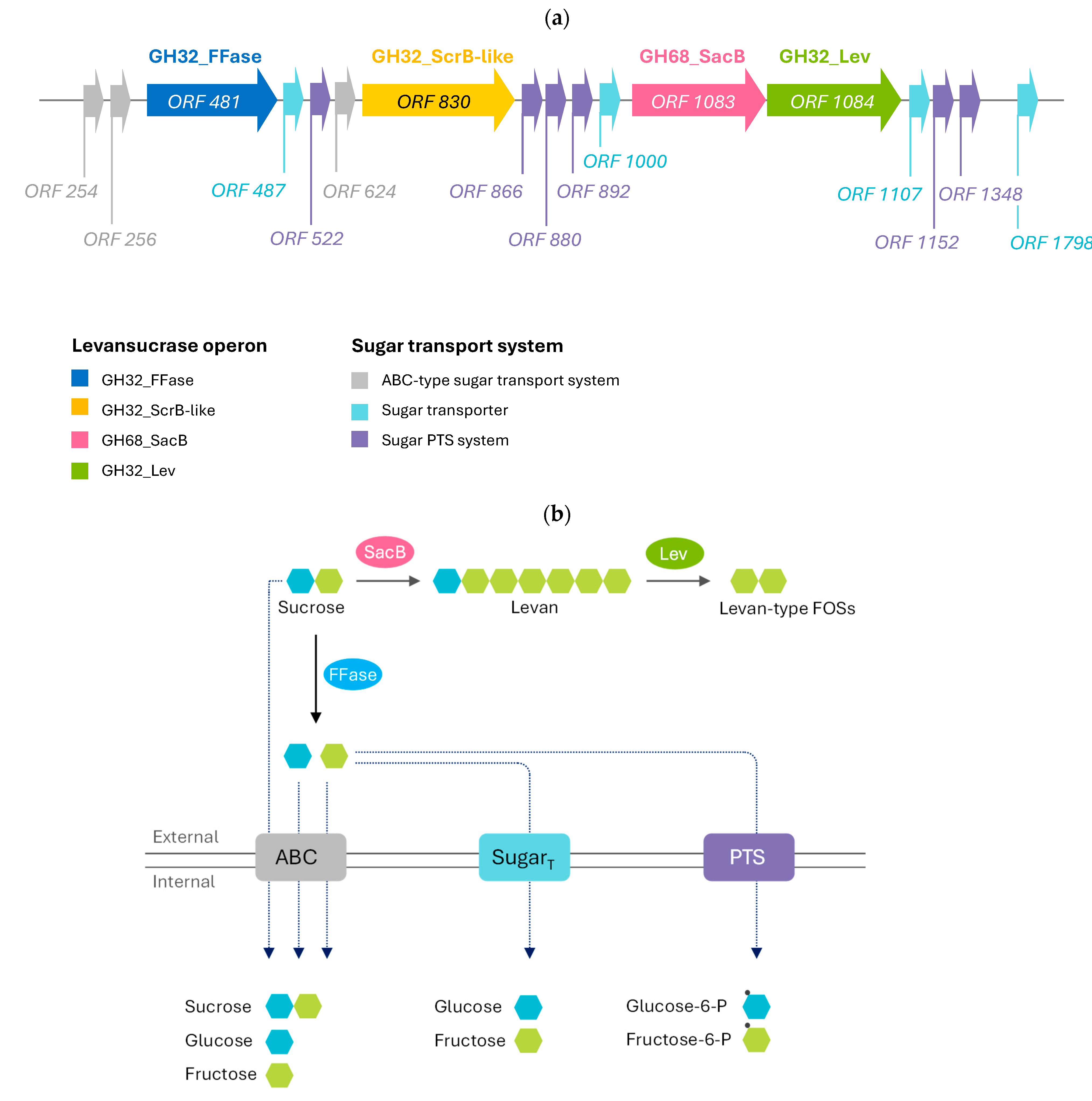
Figure 1. Functional annotation and gene organization of the levansucrase operon identified in the B. velezensis HL25 genome. (a) Gene organization of the levansucrase operon and sugar transport systems; (b) proposed mechanism for levan-type fructooligosaccharides (L-FOSs) biosynthesis and sugar transport pathways.
Catalysts 15 01059 g001
Figure 2. Evolutionary analysis of the levansucrase-encoding gene (HL25SacB) identified in the B. velezensis HL25 genome, along with homologous sequences classified into GH68_subfamily 1 and 2.
Catalysts 15 01059 g002
Figure 3. Predicted protein structure of the levansucrase from the B. velezensis HL25 (HL25SacB). (a) Cartoon representation of HL25sacB comprising GH68 catalytic domain (b) Structural comparison between HL25SacB (pink) and the levansucrase from B. subtilis (1pt2) (gray), highlighting the proposed key amino acid residues involving in sucrose binding site of HL25SacB. The green sphere represents calcium ion.
Catalysts 15 01059 g003
Figure 4. SDS-PAGE analysis of heterologous expression of HL25SacB-encoded levansucrase from B. velezensis HL25. Lane M: PageRuler™ Unstained Protein Ladder (Thermo Scientific, Waltham, MA, USA); Lane 1: crude protein extract from E. coli BL21(DE3) harboring pET32a/HL25sacB plasmid; Lane 2: purified HL25SacB protein.
Catalysts 15 01059 g004
Figure 5. Effect of pH and temperature on the levansucrase activity of recombinant HL25SacB. (a) The effect of pH on HL25SacB activity. Hydrolytic activity was measured using 200 g/L sucrose as the substrate at 40 °C for 10 min in different buffer systems: 50 mM sodium citrate buffer pH 4.0 (grey), 50 mM sodium acetate buffer pH 5.0 (blue), 50 mM sodium phosphate buffer pH 6.0–8.0 (green), and 50 mM glycine-NaOH buffer pH 9.0–10.0 (red). (b) Optimal temperature profile of HL25SacB activity. Reactions were performed in 50 mM sodium phosphate buffer at pH 6.0 and incubated on ice (0 °C) and at various temperatures ranging from 10 °C to 80 °C for 10 min. All assays were conducted in three replicates.
Catalysts 15 01059 g005
Figure 6. The product profile analysis of FOSs and levan synthesis by recombinant HL25SacB from B. velezensis HL25. (a,b) Levan was produced from 200 g/L sucrose in 50 mM sodium acetate buffer at pH 5.0 for 24 h at 30 °C and 40 °C, respectively. (c) The qualitative product profile before ethanol precipitation was analyzed by the TLC method. The sugar standards are glucose (Glc), fructose (Fru), sucrose (Suc), levanbiose (Lev2), levantriose (Lev3), 1-kestose (GF2), 1,1-kestotetraose (GF3), and 1,1,1-kestopentaose (GF4).
Catalysts 15 01059 g006
Table 1. List of reported biochemical characterization of the levansucrases.
OrganismOptimal pHOptimal Temp
(°C)
Enzyme Activity
(U/mg Protein)
Km (mM)VmaxSucrose Concentration (g/L)Yield of
Levan (g/L)
Reference
Bacillus velezensis HL2554028.6822.97156 µmol/mg/min200125This study
Bacillus siamensis53715.95N.D.N.D.200145.9[50]
Bacillus amyloliquefaciens7.03593.40.710.49 μmol/mL/min20078.34[11]
Bacillus amyloliquefaciens63782N.D.N.D.500200[50]
Bacillus subtilis5.240N.D.25.63N.D.10030.6[20]
Bacillus licheniformis6.545N.D.152.790.03 mM/min2747.1[58]
Brenneria goodwinii635N.D.N.D.N.D.500185[59]
Priestia koreensis HL12635–40167.4646.1528.54 mole/mg/min500129[43]
Brenneria sp. EniD3126.545N.D.N.D.N.D.25085[60]
Erwinia amylovora6.535N.D.N.D.N.D.500295[56]
N.D. indicates not determined.
Disclaimer/Publisher’s Note: The statements, opinions and data contained in all publications are solely those of the individual author(s) and contributor(s) and not of MDPI and/or the editor(s). MDPI and/or the editor(s) disclaim responsibility for any injury to people or property resulting from any ideas, methods, instructions or products referred to in the content.