Article

Enhancement of Photocatalytic and Anticancer Properties in Y2O3 Nanocomposites Embedded in Reduced Graphene Oxide and Carbon Nanotubes

by
ZabnAllah M. Alaizeri
*,
Syed Mansoor Ali
and
Hisham A. Alhadlaq
*
Department of Physics and Astronomy, College of Science, King Saud University, Riyadh 11451, Saudi Arabia
*
Authors to whom correspondence should be addressed.
Catalysts 2025, 15(10), 960; https://doi.org/10.3390/catal15100960
Submission received: 30 July 2025 / Revised: 18 September 2025 / Accepted: 24 September 2025 / Published: 6 October 2025

Abstract

Due to their excellent physicochemical properties, the nanoparticles (NPs) have been utilized in various potential applications, including environmental remediation, energy storage, and nanomedicine. In this work, the ultrasonic and manual stirring approaches were used to integrate yttrium oxide (Y2O3) nanoparticles (NPs) into reduced graphene oxide (RGO) and carbon nanotubes (CNTs) to enhance their photocatalytic and anticancer properties. Pure Y2O3NPs, Y2O3/RGO NCs, and Y2O3/CNTs NCs were characterized using different analytical techniques, such as XRD, SEM, EDX with Elemental Mapping, FTIR, UV-Vis, PL, and DLS to investigate their improved structural, surface morphological, chemical bonding, optical, and surface charge properties. XRD data confirmed the successful integration of Y2O3into RGO and CNTs, with minor changes in crystallite sizes. SEM images with EDX analysis revealed that Y2O3NPs were uniformly distributed on RGO and CNTs, reducing aggregation. Chemical bonding and interactions between Y2O3and carbon materials were investigated using Fourier Transform Infrared (FTIR) analysis. UV and PL results suggest that the optical studies showed a shift in absorption peaks upon integration with RGO and CNTs. This indicates enhanced light absorption and modifications to the band gap between (3.79–4.40 eV) for the obtained samples. In the photocatalytic experiment, the degradation efficiency of bromophenol blue (BPB) dye for Y2O3RGO NCs was up to 87.3%, outperforming pure Y2O3NPs (45.83%) and Y2O3/CNTs NCs (66.78%) after 120 min of UV irradiation. Additionally, the MTT assay demonstrated that Y2O3/RGO NCs exhibited the highest anticancer activity against MG-63 bone cancer cells with an IC50 value of 45.7 µg/mL compared to Y2O3CNTs NCs and pure Y2O3NPs. This work highlights that Y2O3/RGO NCs could be used in significant applications, including environmental remediation and in vivo cancer therapy studies.

1. Introduction

Carbon materials combined with metal oxide NPs have been explored for potential applications, including enhanced photocatalytic and anticancer properties [1,2,3]. These hybrid materials have exhibited enhanced photocatalytic activity for pollutant removal, addressing issues like rapid photocarrier recombination and high band gap energy in metal oxides [4]. The integration of carbon nanomaterials with metal oxides improves light absorption and organic pollutant adsorption [5]. Among these metal oxides, the yttrium oxide (Y2O3) nanoparticles (NPs) have attracted significant interest due to their excellent stability and photocatalytic properties, which make them potentially applicable in environmental remediation and the biomedical field [6,7]. However, the incorporation of carbon materials such as GO, RGO, and different types of CNTs with metal oxide forms hybrid nanocomposites (NCs) that exhibit good photocatalytic and therapeutic performance [8,9,10]. Recent studies have combined various metal oxides with carbon materials, such as SnO2/RGO NCs [11], ZnO/CNTs NCs [12], TiO2/GO NCs [12], and ZrO2/MWCNT NCs [13], to improve photocatalytic and biological properties compared to their pure metal oxide.
Several researchers are excited about yttrium oxide nanoparticles (Y2O3 NPs) and their hybrids with other nanomaterials, which are attracting interest as potential applications in catalytic, medical imaging, and targeted therapy [14]. For example, Emad et al. [15] investigated the cytotoxicity of Y2O3NPs toward MDA-MB-231 cells, with an IC50 value of 74.4 µg/mL. They found high biocompatibility in normal retina REP1 and human dermal fibroblast HDF cell lines. Another study, R. Govindasamy et al. [15], shows that the photocatalytic degradation, antibacterial, and anticancer properties of Y2O3NPs have been enhanced using a green process compared with chemical synthesis. R. Vatset al., [16] used the sol–gel chemical process to prepare cubic Y2O3 nanocrystals, which displayed good capacity for methylene blue degradation. Moreover, CeO2/Y2O3 NCs prepared via the hydrothermal route demonstrated photocatalytic degradation and antibacterial activity [17]. Similarly, F. El-Sayed et al. [18] used chemical synthesis of CuO/Y2O3 NCs, which exhibit high degradation of MB under UV and visible irradiation. ZnO/Y2O3 NCs have been prepared using the co-precipitation process and achieved high degradation efficiency (96%) compared to ZnO NPs (86%) in 120 min, as well as antibacterial activity [11]. Additionally, Mo/ZnO/RGO NCs synthesized using an eco-friendly approach with date palm fruit extract showed higher anticancer activity in colon and breast cancer cells compared to pure ZnO NPs [19].
The development of metal oxide/carbon materials as NCs, like graphene oxide (GO), reduced graphene oxide (RGO), and carbon nanotubes (CNTs), has been explored for photocatalytic and anticancer applications owing to their high surface area [20,21]. Our study [22] reported that In2O3 embedded on RGO NCs has higher photocatalytic and cytotoxicity activities compared to pure In2O3 NPs. On the other hand, F. Zhang et al. [23] suggested that Y-TiO2/CNTs NCs exhibit excellent photocatalytic degradation of MB dye compared to individual samples, due to generating more OH groups that can be adsorbed on the surface. Furthermore, K. Bhuvaneswari et al. [24] showed that the integration of MWCNT into MnO2N NPs enhances photocatalytic and anticancer performance. S. S. Wagh et al. [25] investigated that synthesized Ag-doped ZnO/CNTs NCs exhibit rapid dye degradation, as well as antifungal and antibacterial activity, compared to the single samples. Particularly, T. Saravanan et al. [26] synthesized Y2O3/RGO NCs using a low-temperature solution process and displayed enhanced photocatalytic performance compared to pure Y2O3 NPs.
The present work was designed to optimize the photocatalytic and biological properties of Y2O3 NPs by integrating RGO and CNTs through ultrasonication and manual stirring processes. XRD, SEM, EDX, FTIR, UV-Vis, PL, and DLS techniques were employed to investigate the improved physicochemical properties of the prepared NCs. The photocatalytic activity of the prepared samples was evaluated by measuring the degradation of bromophenol blue (BPB) dye under UV light for 120 min. Moreover, an MTT assay on MG-63 bone cancer cells for 24 h was used to evaluate the anticancer properties of the synthesized samples at various concentrations.

2. Results and Discussions

2.1. X-Ray Diffraction (XRD)

The crystal structure and phase of the obtained RGO, CNTs, pure Y2O3 NPs, Y2O3/RGO NCs, and Y2O3/CNTs NCs were investigated through XRD analysis, as shown in Figure 1(i–v). As depicted in Figure 1(i), the XRD pattern of reduced graphene oxide (RGO) sheet shows the broad peak at 2θ = 25°, matching the (002) plane [27]. On the other hand, carbon nanotubes (CNTs) (Figure 1(ii)) display a sharp peak at 2θ = 26.2°, corresponding to the (002) plane, as reported in a previous study [28]. In Figure 1(iii), the XRD pattern of pure Y2O3 NPs reveals distinct peaks at 2θ = 29.1°, 33.6°, 48.4°, and 57.6°, which are indexed to the (111), (200), (220), and (311) planes, respectively. These peaks confirm the cubic crystal structure of pure Y2O3 NPs with high crystallinity, consistent with previous studies. It can be observed in Figure 1(iv,v) that both CNT and RGO peaks exist in the XRD patterns of Y2O3/RGO NCs and Y2O3 Y2O3/CNTs NCs. These peaks indicate that pure Y2O3 Y2O3 Y2O3 NPs are successfully anchored onto the RGO sheet and CNTs, as agreed with SEM images (Figure 2b,c). Figure 1b shows the high resolution of the XRD pattern of the prepared samples for (220) and (311) planes. It can be observed that there is a very slight shift in the peak positions of the samples. The Debye-Scherrer formula was applied to estimate the average crystallite size of the prepared samples using the (111) peak, as shown in Equation (1).
D n m = k λ β C O S θ
where D is the crystallite size, λ is the X-ray wavelength, β is the full width at half maximum (FWHM) of the diffraction peak, and θ is the Bragg angle. Furthermore, the crystallite sizes (Table 1) of pure Y2O3 NPs, Y2O3/RGO NCs, and Y2O3/CNTs NCs are 10.12 nm, 9.10 nm, and 11.11 nm, respectively. These slight decreases in crystallite size in Y2O3/RGO NCs compared with pure Y2O3 NPs confirmed the interaction between Y2O3 NPs and CNTs. Moreover, the dislocation density (δ), macrostrain (ε), and stacking fault (γs) of all the samples were calculated as shown in Table 1. These values suggest that the addition of RGO and CNTs can influence the crystal growth of pure Y2O3 NPs during preparation. Additionally, XRD patterns show that the presence of RGO and CNTs in the XRD pattern confirms the successful integration of Y2O3 NPs with RGO and CNTs, as reported in earlier studies [29,30].

2.2. SEM Characterization

The surface morphology and distribution of NPs and NCs were further investigated through scanning electron microscopy (SEM) techniques, as illustrated in Figure 2a–c. As seen in Figure 2a, the SEM image of pure Y2O3 NPs shows spherical particles with a smooth surface and uniform distribution, albeit with high agglomerations [31]. These properties suggest a high degree of crystallinity of pure Y2O3 NPs, which plays a role in improving their properties when combined with carbon materials. As observed in Figure 2b, the SEM image of Y2O3/RGO NCs confirms that the Y2O3 NPs are successfully loaded on the surface of the RGO sheet with homogeneous distribution and less aggregation. This integration indicates that the RGO sheet can enhance the morphology and structure properties of metal oxide NPs (Y2O3 NPs), similar to previous studies [32,33]. Correspondingly, the SEM image of Y2O3/CNTs NCs (Figure 2c) exhibits that the Y2O3 NPs were anchored and distributed with less heavy aggregation onto the CNT, which has a tubular structure. These results indicate that the Y2O3/RGO NCs and Y2O3/CNTs NCs were successfully fabricated due to the strong interaction of the Y2O3 NP with the RGO sheet and CNTs. SEM images show the successful preparation of Y2O3-based nanocomposites with RGO and CNTs.

2.3. EDX and Elemental Mapping Analysis

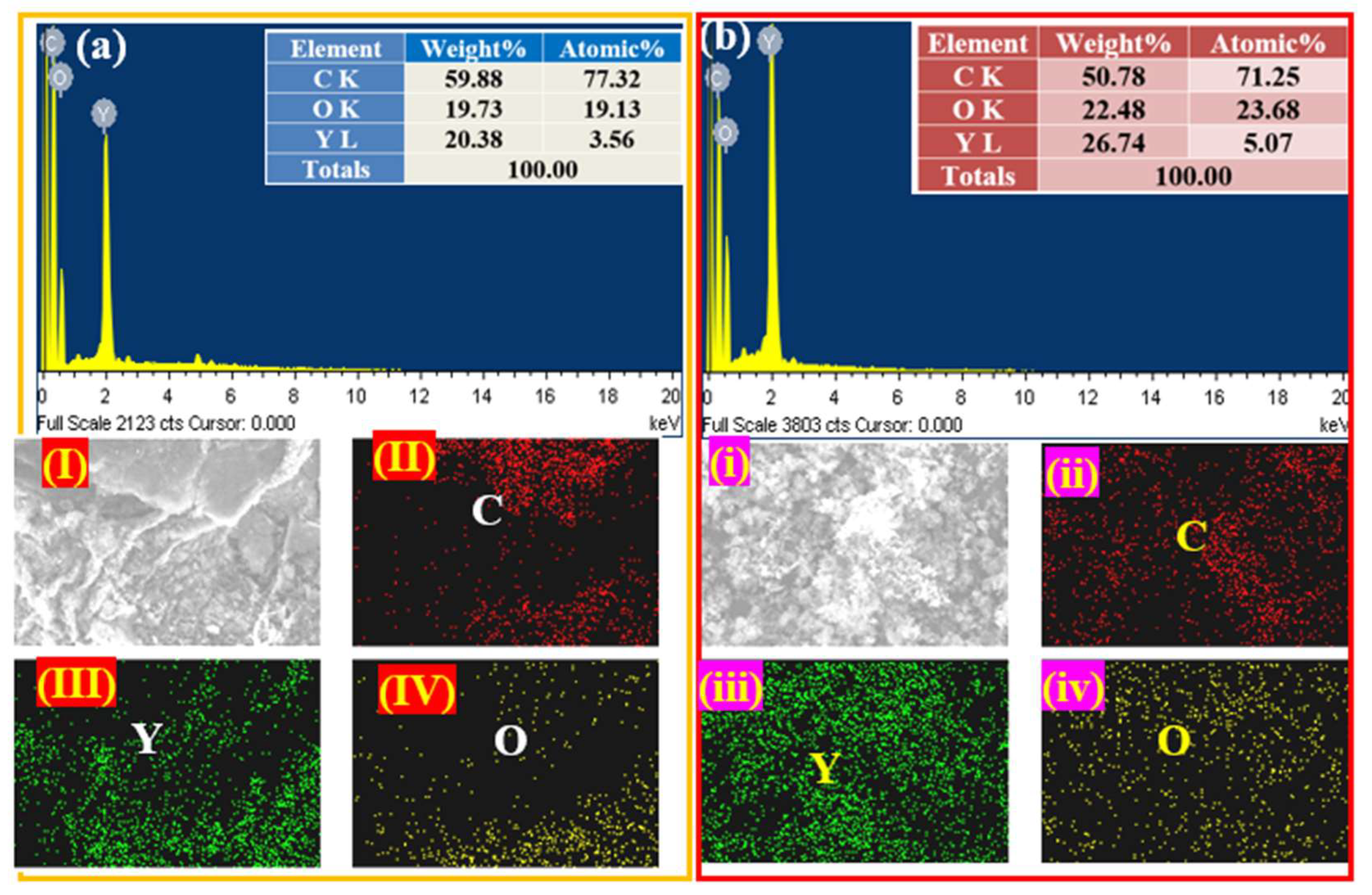
The EDX with mapping analysis was used in the present work to confirm the presence of the compositions and their distribution in synthesized Y2O3/RGO NCs and Y2O3/CNTs NCs, as demonstrated in Figure 3a,b. It can be observed in Figure 3a that the EDX spectrum of Y2O3/RGO NCs shows weight percentages of 59.88%, 19.73%, and 20.38% for Carbon (C), Oxygen (O), and Yttrium (Y), respectively. These percentages indicate that the Y2O3 NPs are fruitfully integrated onto RGO sheets. Moreover, the mapping images in Figure 3 (I–IV) display the distribution of these elements (C, O, and Y) within the obtained Y2O3/RGO NCs. As observed, the EDX data (Figure 3a) were further matched with elemental mapping in Figure 3(II–IV). Equally, Figure 3b shows the EDX spectrum of Y2O3/CNTs NCs, with percentages of 50.78%, 22.48%, and 26.74% for C, O, and Y, respectively. Furthermore, the mapping images (i–iv) also confirm the uniform distribution of these elements in Y2O3/CNTs NCs. As shown in EDX and mapping analysis, the carbon percentage and its distribution in Y2O3/RGO NCs (Figure 3a) were increased compared to Y2O3/CNTs NCs (Figure 3b). As shown in the results, the EDX spectrum and element mapping confirm the successful preparation of Y2O3/RGO NCs and Y2O3/CNTs NCs, highlighting their potential for various applications, such as environmental remediation and cancer therapy. The presented results agreed with XRD data (Figure 1) and were similar to previous studies [34,35].

2.4. X-Ray Photoelectron Spectroscopy (XPS)

Figure 4 provides the comprehensive information about the surface chemical composition and bonding states of the prepared Y2O3/RGO NCs. The survey scan (Figure 4a) confirms the presence of chemical composition in Y2O3/RGO NCs (yttrium (Y), oxygen (O), and carbon (C)) with binding energies corresponding to their respective core levels. Moreover, Figure 4b shows the XPS high-resolution of Y 3d spectra, which exhibit peaks at 159.6 eV (Y 3d5/2) and 157.7 eV (Y 3d3/2), while the peaks at 161.1 eV and 161.8 eV were assigned for 3d5/2 and Y 3d3/2 respectively. These peaks confirmed that the Y2O3 NPs were successfully prepared, as expected from earlier investigations [36,37]. In addition, the O 1s spectra (Figure 4b) reveal peaks at 530.3 eV, 531.7 eV, and 528.4 eV, attributed to Y–O bonds, hydroxyl groups, and oxygenated groups on the graphene surface, respectively. These functionalities are crucial for the interaction between Y2O3 and the RGO sheet, as reported in previous studies [38,39]. Additionally, the C 1s spectrum reveals peaks at 284.4 eV, 285.2 eV, and 288.1 eV, corresponding to C–C, C–O, and C=O bonds, respectively. These peaks confirm the presence of oxygenated functionalities on the RGO surface, which improve the properties of NCs [40]. XPS results showed that the loading of Y2O3 NPs onto the RGO surface played a role in enhancing the properties of NCs, as agreed with consistent studies [41,42].

2.5. Fourier Transform Infrared (FTIR) Spectroscopy

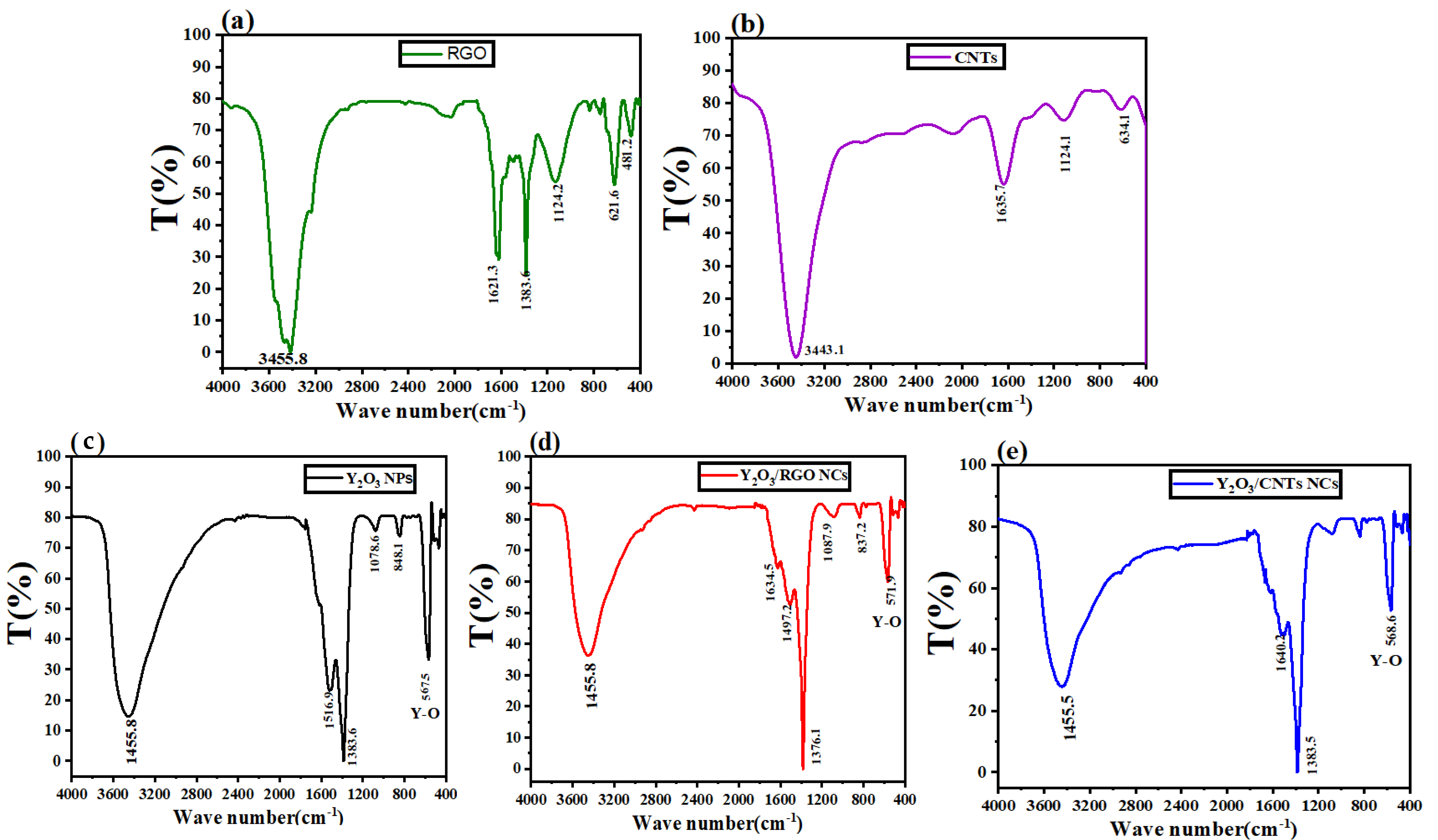
Figure 5a–e shows the FTIR spectrum of RGO, CNTs, pure Y2O3 NPs, Y2O3/RGO NCs, and Y2O3/CNTs NCs, respectively. FTIR spectrum (Figure 5a) of the RGO sheet reveals strong absorption peaks at 508.4 cm−1, 1124.2 cm−1, and 1383.6 cm−1 for C–C stretching and another band at 1620.4 cm−1 and ranges between 3405.1 and 1501.9 cm−1, which are associated with C–O and O–H stretching, respectively. These bands indicate that the RGO sheet has been successfully prepared due to the presence of oxygenated functional groups, as reported in a previous study [43]. Also, the FTIR spectrum of CNTs (Figure 5b) demonstrates the broad peaks at 1124.1 cm−1 and 1640.4 cm−1 for C–C stretching, with 3445.12 cm−1 for O–H stretching [44]. As observed in the FTIR spectrum for pure Y2O3 NPs (Figure 5c), the prominent peak at 567.5 cm−1 is assigned due to the Y–O stretching vibration, and another bands at 1383.6 cm−1 and 1516.9 cm−1 for C–C stretching with an O–H bending vibration at 3452.2 cm−1, as agreed with earlier studies [45,46]. For Y2O3/RGO NPs, the FTIR spectrum (Figure 5d) exhibits similar peaks at 571.9 cm−1 of Y2O3 NPs with a slight difference in intensity and shifting due to the RGO sheet addition, which includes the C=C and C–O stretching bands at 1376.1 cm−1 and 1634.5 cm−1, respectively [47]. This confirmed the integration of Y2O3 into the RGO sheets, as supported in XRD (Figure 1a) and SEM (Figure 2b) results. Similarly, the Y2O3/CNTs NCs spectrum shows the CNT-specific peaks at 1640.2 cm−1 and the broad O–H stretching band at 3445.5 cm−1, while also showing the Y–O stretch at 568.6 cm−1. This phenomenon indicates successful synthesis of the NCs. FTIR analysis shows that the Y2O3 NPs interact with surface functional groups on RGO and CNTs to prepare novel NCs with excellent physicochemical properties. These results suggest that these NCs have potential applications such as catalysis, energy storage, and cancer therapy.

2.6. Optical Properties Analysis

Figure 6a,b exhibit the optical properties, including absorption with the band gap energy and emission peaks, for prepared pure Y2O3 NPs, Y2O3/RGO NCs, and Y2O3/CNTs NCs. The UV-Vis absorbance spectra (Figure 6a) show that all the synthesized samples reveal a similar trend with their notable variations. These variations indicate the influence of surface modification on the optical properties of samples. Nevertheless, the absorbance peak of pure Y2O3 NPs (Figure 6a) was associated with around 200–300 nm, which is characteristic of the Y2O3 NPs, as agreed in a previous study [48]. On the other hand, the absorbance peak of pure Y2O3 NPs is shifting to a higher wavelength upon the integration of RGO and CNTs. In comparison between CNT and RGO, the shift in the absorption edge of Y2O3 NPs with RGO was more suitable than that of the individual samples alone. This shift suggests that RGO plays a role in influencing the charge carrier dynamics and improving light absorption capabilities. The inset in Figure 6a displays the calculated band gap energies (Eg) from the Tauc plot, with values of 3.79 eV, 4.40 eV, and 3.91 eV for Y2O3 NPs, Y2O3 RGO NCs, and Y2O3/CNTs NCs, respectively [49]. These values reveal that the band gap energy of Y2O3 NPs was slightly increased compared to pure Y2O3 NPs and Y2O3 CNTs NCs, which can be attributed to the modification effects from RGO sheets. Figure 5b depicts the PL spectrum of the synthesized samples. Moreover, the PL emission peak of Y2O3 NPs exhibits around 335.66 nm and 378.89 nm with a slight shift for Y2O3 CNTs NCs and Y2O3 RGO NCs. Notably, the shift in the PL emission peak was observed for Y2O3 RGO NCs and Y2O3 CNTs, further confirming the influence of RGO and CNTs on the optical properties. Furthermore, the PL intensity of Y2O3 RGO NCs is relatively lower compared to the pure Y2O3 NPs and Y2O3 CNTs, which can be attributed to the electron transfer and charge separation effects facilitated by CNTs, reducing recombination rates and enhancing the photocatalytic efficiency. The larger shift in the PL spectrum of Y2O3 CNTs NCs compared to Y2O3 RGO NCs is likely due to the stronger electron transfer and charge separation facilitated by the CNTs’ conductive properties, which influence the optical behaviour more significantly than RGO. Similarly, Shashikumara et al. [50] showed the tuning of electronic structures in Y2O3 NPs by incorporating RGO and Cr3+, resulting in more efficient light absorption and charge carrier dynamics. UV-Vis and PL results demonstrate the successful integration of RGO and CNTs into Y2O3 NPs, leading to an enhancement in band gap energy and changes in the photoluminescent behaviour. This study showed that the Y2O3/RGO NCs could be promising in potential applications such as environmental remediation and the therapeutic field.

2.7. DLS Analysis

DLS analysis provided information on the behavior of the samples, including agglomeration, distribution, and surface charge in aqueous media. Figure 7a,b depict the zeta potential and the electrophoretic mobility distribution for prepared pure Y2O3 NPs, Y2O3/RGO NCs, and Y2O3/CNTs NCs. However, the zeta potential analysis (Figure 6a) demonstrates the surface charge properties of three synthesized samples. Pure Y2O3 NPs (Record 361) exhibit a broader distribution with a negative zeta potential of −19.6 ± 5.79 mV, as shown in Table 1, which agrees with an earlier study [51]. This value indicates that pure Y2O3 NPs may be stable in aqueous media. In contrast, the negative zeta potential (−24.5 ± 5.16 mV) of Y2O3 RGO NCs (Record 364) displayed a relatively narrow distribution. This reveals an increase in stability compared to the pure NPs. The Y2O3/CNTs NCs (Record 365) show the negative Zeta potential (−22.1 ± 4.53 mV), which indicates that the integration of CNTs significantly affects the surface charge properties, leading to reduced stability in aqueous media.
In Figure 7b, the electrophoretic mobility distribution confirmed the values of the zeta potential of the three obtained samples. It can be observed in Table 1 that the negative mobility values of pure Y2O3 NPs, Y2O3/RGO NCs, and Y2O3/CNTs NCs were 1.54 ± 0.68, 1.92 ± 1.02, and −1.75 ± 0.97 µmcm/Vs, respectively, as reported in similar nanoparticles [52]. These values were in good agreement with the positive zeta potential observed (Figure 6a and Table 2). Our DLS results indicate that the Y2O3 RGO NCs have improved dispersion stability compared to pure Y2O3 NPs and Y2O3/CNTs NCs because RGO sheets can generate hydroxyl groups in solution. This study is making them promising for potential applications requiring stable nanomaterials, such as catalytic and cancer therapy.

2.8. Photocatalytic Activity

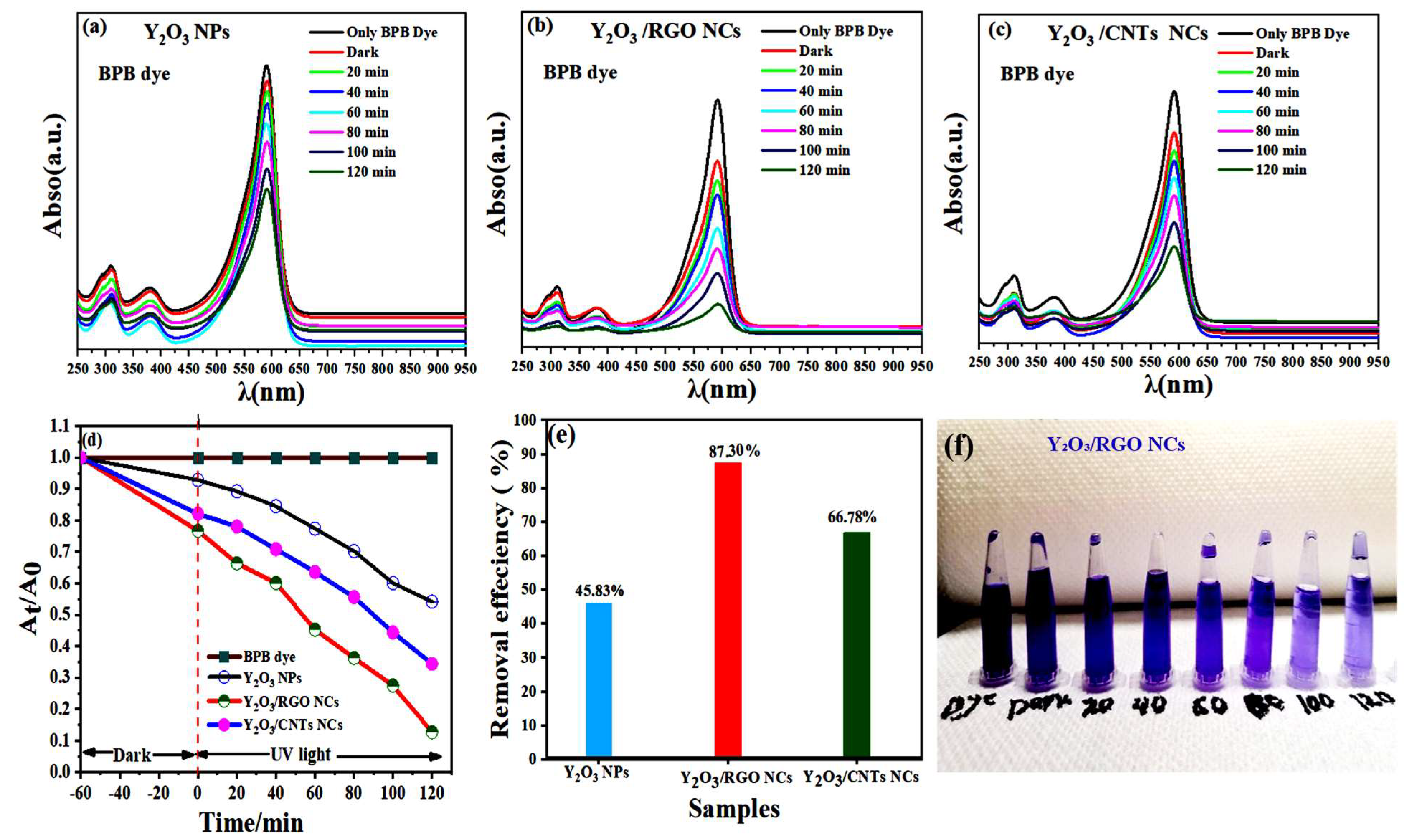
The photocatalytic performance of the obtained pure Y2O3 NPs, Y2O3 RGO NCs, and Y2O3/CNTs NCs was evaluated by the degradation of BPB dye under UV irradiation, as presented in Figure 8a–f. It can be observed in Figure 8a–c that the absorption peaks of the BPB dye solution at 590 nm, using all the prepared samples, decrease gradually with a clear shift in the spectra as the exposure time increases. Specifically, the absorption peaks of the BPB dye solution using Y2O3/RGO NCs were significantly reduced compared to both pure Y2O3 NPs and Y2O3/CNTs NCs. This indicates that the addition of RGO significantly enhances degradation, allowing the BPB dye solution to be degraded when exposed to UV light due to the high surface area of RGO compared with CNTs. To confirm that, Figure 8d shows the degradation profiles. Moreover, the Y2O3/RGO NCs exhibited the highest rate of degradation, resulting in a significant reduction in dye concentration. On the other hand, pure Y2O3 NPs and Y2O3/CNTs NCs demonstrate slower degradation of the dye. This is supported by Figure 8e, which shows the removal efficiencies of Y2O3/RGO (87.30%), Y2O3/CNTs NCs (66.78%), and Y2O3 NPs (45.83%) after 120 min. The visual evidence in Figure 8f confirms these results. We observed that the colour of the BPB dye solution was fading significantly faster in the presence of Y2O3/RGO NCs. These results suggest that Y2O3/RGO NCs have superior photocatalytic activity attributed to the synergistic effect of RGO, which enhances charge carrier separation and increases the surface area for dye adsorption. Table 3 shows the comparison of the photocatalytic degradation efficiency of Y2O3-based NCs with reported catalysts. We observed that the Ag/Zn/CNTs NCs have been achieved, contributing to their high efficiency in the degradation of BPB dye in the visible light zone. Overall, this study highlights that RGO and CNTs in metal oxide nanoparticles (NPs) can enhance photocatalytic efficiency for wastewater treatment applications.

2.8.1. Stability

The stability results of the Y2O3 NPs, Y2O3/RGO NCs, and Y2O3/CNTs NCs show significant differences across four runs, as demonstrated in Figure 9. Y2O3 NPs show the lowest stability, with performance percentages (D%) declining from 45.8% in Run #1 to 44.9% in Run #4, indicating a substantial loss of stability over repeated cycles. In contrast, Y2O3/RGO NCs keep a higher and more consistent stability, ranging from 65.5% to 65% across the runs, likely due to the stabilizing effects of reduced graphene oxide. Nevertheless, the Y2O3/CNTs NCs reveal the highest overall stability, starting at 87.3% in Run #1 and only dropping slightly to 86.6% by Run #4, highlighting the significant role of carbon nanotubes in reinforcing the composite and preventing degradation. These results agreed with previous studies showing that CNTs and RGO improve the mechanical and structural integrity of nanocomposites, leading to enhanced long-term performance [58,59]. Pure Y2O3 NPs, lacking a stabilizing matrix, are more prone to performance degradation, as seen in the results, which supports prior findings about the challenges of using unmodified nanoparticles in cyclic applications [60]. Overall, incorporating CNTs or RGO into Y2O3 significantly develops its stability, making it a more reliable material for applications requiring repeated cycles.

2.8.2. Reaction Mechanism

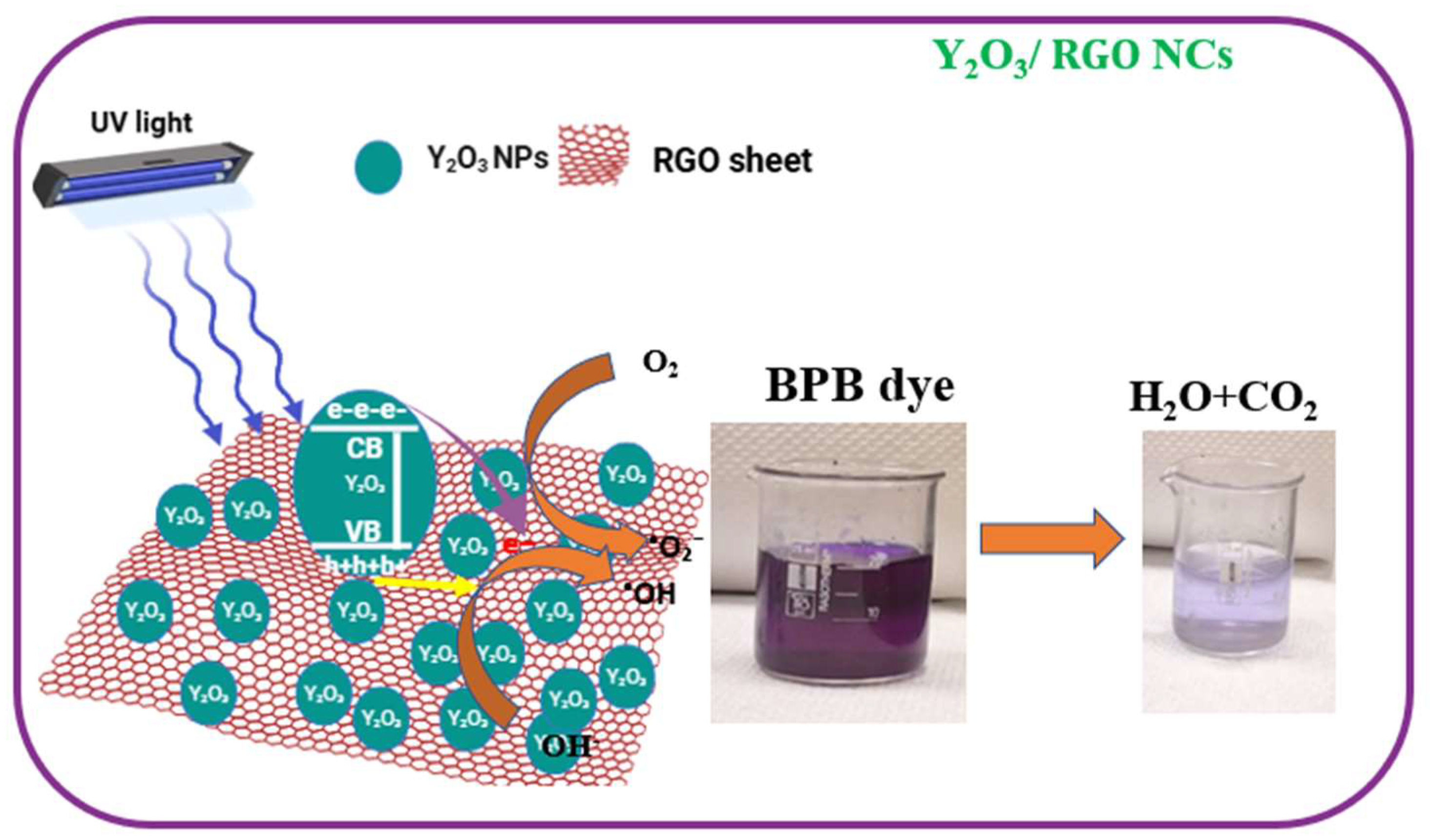
Figure 10 describes the reaction mechanism of degradation for BPB dye using the prepared catalysts under UV light. Initially, the Y2O3 NPs were exposed to UV irradiation. Then, the electrons (e) were excited to the conduction band (CB) of Y2O3 NPs, while the holes (h+) remain in the valence band (VB), as shown in Equation (2). These generated electrons were transferred to the reduced graphene oxide (RGO) sheet, as demonstrated in Equation (3). This phenomenon may play a role in enhancing charge separation and preventing recombination between electrons(e) and holes (h+). The photogenerated electrons on the surface of RGO and CB in Y2O3 NPs react with oxygen molecules (O2) present in BPB dye solution to produce superoxide anions (O2), as illustrated in Equation (4). In parallel, the holes in the VB of Y2O3 were reacted with water (H2O) to generate hydroxyl radicals (OH•), as shown in Equation (5). Subsequently, produced superoxide anions (O2) and hydroxyl radicals (OH•) could react with molecules (O2) present in BPB dye (lighter color) to generate water (H2O) and carbon dioxide (CO2)(smaller color), as revealed in Equation (6). This visual change in beakers (Figure 8) confirms the successful photocatalytic degradation of BPB dye by the Y2O3 RGO NCs under UV light for 120 min. These reactions are described in the following equations.
Y2O3 RGO NCs + hv ⟶ Y2O3 (e + h+)/CNTs NCs
Y2O3 (e) ⟶ e (RGO)
e (RGO) + O2 ⟶ O2
h+ + OH ⟶ •OH
(O2,•OH) + BPB dye ⟶ Mineralization (H2O + CO2)

2.9. Anticancer Study

The Y2O3 nanostructure has been attracting potential applications such as catalytic and biomedical fields, as demonstrated by many researchers [44,51,61]. By MTT assay, the anticancer activity of Y2O3 NPs, Y2O3/RGO NCs, and Y2O3/CNTs NCs was performed against human bone cancer cell lines (MG-63 cells) for 24 h. As shown in Figure 11a–c, the viability percentage of MG-63 cells vs. logarithmic concentration of obtained samples is presented in Figure 11a–c. Following a 24 h treatment period, the effects of each sample on cell viability were measured at different concentrations of 0, 1, 5, 10, 25, 50, and 100 µg/mL. Figure 11a shows that the pure Y2O3 NPs exhibit an IC50 value of 158.12 µg/mL, as indicated by the logarithmic curve fitting (Log IC50 = 2.19, R2 = 0.9671). This high IC50 value shows that pure Y2O3 NPs induce low cytotoxicity (high cell viability). The Y2O3 RGO NCs (Figure 11b) illustrate enhanced cytotoxicity activity, with an IC50 value of 45.7 µg/mL (Log IC50 = 1.66, R2 = 0.9975). However, the lower IC50 value indicates that these NCs exhibit stronger cytotoxic effects than pure Y2O3 NPs due to the strong interaction between Y2O3 NPs and the RGO sheet. Similarly, the Y2O3 3/CNTs NCs (Figure 11c) exhibit the IC50 value of 73.45 µg/mL (Log IC50 = 1.86, R2 = 0.9764), indicating moderate cytotoxicity. Although this value is lower than that of Y2O3 RGO NCs, it still shows a significant reduction in cell viability compared to pure Y2O3 3 NPs. Table 4 summarizes the IC50 values of the prepared samples against the bone (MG-63) cancer cell line after 24 h of treatment. It can be observed in these data that the reduction in IC50 value indicates that the integration of RGO into Y2O3 NPs induced higher cytotoxicity compared with CNTs. These results suggest that these NCs show promise for anticancer applications due to their enhanced bioactivity.

3. Methodology

3.1. Materials and Chemicals Used

The yttrium oxide nanoparticles (Y2O3NPs) (nanopowder, <50 nanoparticle size) were MTT reagent [3-(4,5-dimethylthiazol-2-yl)-2,5-diphenyltetrazolium bromide, dimethyl sulfoxide (DMSO), Bromophenol Blue (3′,3′′,5′,5′′-Tetrabromophenolsulfonephthalein)(BPB) dye were purchased from Sigma-Aldrich (St. Louis, MO, USA). Dulbecco’s Modified Eagle’s Medium (DMEM) and fetal bovine serum (FBS) were obtained from Invitrogen (Carlsbad, CA, USA) for use in cell culture experiments. Ethanol and deionized water were further utilized as solvents throughout the experiment. To ensure accuracy in our methodologies, all supplies were obtained from Sigma-Aldrich, unless otherwise stated.

3.2. Preparation of Reduced Graphene Oxide (RGO) and Carbon Nanotubes (CNTs)

The reduced graphene oxide (RGO) was prepared as reported in our previous study [62]. The chemical vapor deposition (CVD) method was used to synthesize carbon nanotubes (CNTs). Firstly, the acetylene (C2H2) gas was passed over a catalyst (typically iron, Fe) supported on a silica substrate. Next, this reaction was performed at 700 °C. After that, the produced CNTs were collected as a precipitate. Then, the collected CNTs were washed with hydrochloric acid (HCl) to eliminate metal catalysts and other impurities. The precipitated CNTs were also dried at 50 °C under vacuum for 24 h.

3.3. Preparation of Y2O3/RGO NCs, and Y2O3/CNTs NCs and Characterization

In the present work, the Y2O3/RGO NCs were successfully synthesized using the ultrasonic technique (an ultrasonic,100W, 42 kHz, Vernon Hills, IL, USA) and manual stirring procedures. Initially, 1 g of Y2O3 NPs was dissolved in 50 mL of a solution of distilled water/ethanol (ratio of 50:50) under ultrasonication at 60 kW for 1 h as the first solution. Next, 5% of prepared reduced graphene oxide (RGO) was also dispersed in 50 mL of a solution of distilled water/ethanol (ratio of 50:50) under ultrasonication at 60 kW for 1 h, as the second solution. After 1 h of ultrasonication, the two solutions were mixed in a 150 mL beaker and subjected to ultrasonication for an additional 3 h. Then, the mixture solution was dried under vacuum at 60 °C for 12 h. The obtained precipitate was manually stirred through grinding for 15 min to obtain Y2O3/RGO NCs as nanopowder, as described in Scheme 1a. Following the same protocols above, Y2O3/CNTs NCs were prepared by incorporating 5% CNTs in place of RGO, as illustrated in Scheme 1b. In this study, the different analytical tools such as X-ray diffraction (XRD)(PanAnalytic X’Pert Pro, Malvern Instruments, Malvern, UK) with Cu-Kα radiation (=0.15405 nm, at 45 kV and 40 mA), scanning electron microscopy (SEM), energy-dispersive X-ray (EDX), Fourier transform infrared (FTIR) spectroscopy, ultraviolet-visible (UV-Vis) absorption, photoluminescence (PL) spectroscopy, and dynamic light scattering (DLS) were carefully used to examine the physicochemical properties, as described in our previous studies [63,64,65].

3.4. Photocatalytic Degradation Experiment

The degradation of BPB dye under UV irradiation was used to evaluate the photocatalytic performance of the obtained catalysts (pure Y2O3 NPs, Y2O3/RGO NCs, and Y2O3 CNTs NCs). The UV light source in this experiment was a 400 W Xenon lamp (CEL-HXF300, Beijing, China) with a wavelength of less than 420 nm. A 50 mL solution of BPB dye at an initial concentration of 10 mg/L (10 ppm) was dissolved in distilled water. Then, 0.025 g of each catalyst type was added to the BPB dye solution. After that, the dye solution with the catalyst was stirred for 1 h in the dark to ensure adsorption equilibrium between the dye and the catalyst surface. Next, this solution was irradiated with UV light (λ = 365 nm) in a photoreactor at a constant distance from the UV source to ensure uniform light intensity. At regular intervals (20, 40, 60, 80, 100, and 120 min), 2 mL of the suspended solution was collected and centrifuged to separate the catalyst from the dye solution. The absorbance values of the BPB dye solution were recorded by UV-Vis spectroscopy at a wavelength of 590 nm to determine the removal efficiency using the following Equation (7).
Removal efficiency (%) = [1 − At/A0] × 100
where A0 denotes the initial absorbance of BPB, and At is the final absorbance of the dye after a specified reaction time (min). Additionally, the stability of prepared samples was evaluated for four cycles.

3.5. MTT Experiment with Statistical Analysis

In the present study, the bone cancer cell lines (MG-63) were used to investigate the cytotoxicity of prepared NPs and NCs using the MTT assay, following the same protocol as in our previous studies [28] with some improvements. The five tested concentrations (0, 1, 5, 10, 25, 50, and 100 µg/mL) were applied to MG-63 cancer cells for 24 h. After treatment, cell viability percentage was measured using a microreader at a wavelength of 630 nm. Moreover, the half-maximal inhibitory concentration (IC50) for MG-63 cancer cells was calculated to assess the cytotoxic activity of the prepared samples against bone (MG-63) cancer cell lines for 24 h. The biological data were analyzed by applying one-way ANOVA and Dunnett’s multiple comparison tests. The value p < 0.05 was assigned as statistical significance.

4. Conclusion

This study aimed to synthesize and characterize Y2O3-based RGO and carbon nanotubes (CNTs) nanocomposites (NCs) to enhance their photocatalytic and anticancer properties using simple ultrasonic and stirring processes. The successful preparation of Y2O3 NPs onto RGO and CNTs was confirmed through various analytical techniques, including X-ray diffraction (XRD), scanning electron microscopy (SEM), energy-dispersive X-ray (EDX), Fourier-transform infrared (FTIR), UV-Vis, Photoluminescence (PL), and Dynamic Light Scattering (DLS). XRD results indicate that the RGO sheet and CNTs have a strong interaction with Y2O3 NPs. This affects the crystallinity sizes, which range between 9.10 nm and 11.11 nm, for the prepared samples. SEM images and EXD with mapping confirmed the uniform dispersion of Y2O3 on RGO and CNTs, as well as the chemical composition (Y, C, O), which reduces particle aggregation and enhances surface area. UV-vis spectra showed that the band gap energies of Y2OPs, Y2O3/RGO NCs, and Y2O3/CNTs NCs were 3.79 eV, 4.40 eV, and 3.91 eV, respectively. The reduced PL intensity confirmed improved optical properties of Y2O3/RGO NCs compared with individual samples, owing to reduced recombination rate3, resulting in better light absorption and increased photocatalytic efficiency. DLS analysis showed that the zeta potential of Y2O3 NPs, Y2O3/RGO NCs, and Y2O3/CNTs NCs were found to be −19.6 ± 3.79 mV, −24.5 ± 5.16 mV, and −22.1 ± 4.53 mV, respectively. For applications, the Y2O3/RGO NCs have exhibited the highest degradation efficiency of BPB dye (87.3%) compared to pure Y2O3 NPs (45.83%) and Y2O3/CNTs NCs (66.78%). For nanomedicine, the MTT assay showed that Y2O3/RGO NCs (IC50 = 45.7 µg/mL) exhibit a higher cytotoxic effect than Y2O3 CNTs NCs (IC50 = 73.45 µg/mL) and pure Y2O3 NPs (IC50 = 158.12 µg/mL). These results highlight the dual functionality of Y2O3 RGO NCs for dye degradation and cancer therapy applications. This study recommended further studying the possibilities of in vivo models, advanced environmental remediation, and targeted drug delivery.

Author Contributions

Z.M.A. conceptualized the study, and the investigations and methods were carried out by Z.M.A., H.A.A. and S.M.A. The original draft was prepared by Z.M.A. and S.M.A., and Review and editing were conducted by Z.M.A., H.A.A. and S.M.A. All authors have read and agreed to the published version of the manuscript.

Funding

The authors would like to thank Ongoing Research Funding Program, (ORFFT-2025-069-1), King Saud University, Riyadh, Saudi Arabia for financial support.

Data Availability Statement

The raw data supporting the conclusions of this article will be made available by the authors on request.

Conflicts of Interest

The authors declare no conflict of interest.

References

  1. Dabhi, S.D.; Gahlot, S.; Mirza, S.; Pandya, M.; Rawal, R.; Kulshrestha, V.; Jha, P.K. Study of Anticancer Properties of Graphene Oxide–Metal Oxide Composite. Macromol. Symp. 2024, 413, 2300086. [Google Scholar] [CrossRef]
  2. Loh, T.A.J.; Hu, Y.; Pham, K.-C.; Tan, Z.; Chua, D.H.C. Multifunctional Metal Oxides and 2D Materials Utilizing Carbon Nanotubes as a Base Template for Clean Energy and Other Applications. In Proceedings of the 2016 IEEE 16th International Conference on Nanotechnology (IEEE-NANO), Sendai, Japan, 22–25 August 2016; pp. 899–900. [Google Scholar] [CrossRef]
  3. Bijesh, P.; Selvaraj, V.; Andal, V. A Review on Synthesis and Applications of Nano Metal Oxide/Porous Carbon Composite. Mater. Today Proc. 2021, 55, 212–219. [Google Scholar] [CrossRef]
  4. Ansari, A.S.; Azzahra, G.; Nugroho, F.G.; Mujtaba, M.M.; Ahmed, A.T.A. Oxides and Metal Oxide/Carbon Hybrid Materials for Efficient Photocatalytic Organic Pollutant Removal. Catalysts 2025, 15, 134. [Google Scholar] [CrossRef]
  5. Silva, C.; Pastrana Martinez, L.; Morales-Torres, S. When Carbon Meets Light: Synergistic Effect between Carbon Nanomaterials and Metal Oxide Semiconductors for Photocatalytic Applications. Boletín Grupo Español Carbón 2016, 40, 28–35. [Google Scholar]
  6. Parangusan, K.; Subramanium, V.; Sundarabharathi, L.; Kannan, K.; Radhika, D. Influence of PH on Structural, Morphological, Optical, Photocatalytic, and Antibacterial Properties of Yttrium Oxide Nanoparticles via Co-Precipitation Method. Res. Sq. 2021. [Google Scholar] [CrossRef]
  7. Myvizhi, G.; Krishna, S.K. Structural and Optical Studies of Hydrothermal Synthesis of Yttrium Oxide Nanoparticles: In Vitro Antioxidant Activity. Asian J. Chem. 2022, 34, 651–656. [Google Scholar] [CrossRef]
  8. Lagos, K.J.; Buzzá, H.H.; Bagnato, V.S.; Romero, M.P. Carbon-Based Materials in Photodynamic and Photothermal Therapies Applied to Tumor Destruction. Int. J. Mol. Sci. 2022, 23, 22. [Google Scholar] [CrossRef]
  9. Soren, S.; Chakroborty, S.; Pal, K. Enhanced in Tunning of Photochemical and Electrochemical Responses of Inorganic Metal Oxide Nanoparticles via RGO Frameworks (MO/RGO): A Comprehensive Review. Mater. Sci. Eng. B 2022, 278, 115632. [Google Scholar] [CrossRef]
  10. Rasheed, T.; Ahmad, N.; Nawaz, S.; Sher, F. Photocatalytic and Adsorptive Remediation of Hazardous Environmental Pollutants by Hybrid Nanocomposites. Case Stud. Chem. Environ. Eng. 2020, 2, 100037. [Google Scholar] [CrossRef]
  11. Gang, R.; Xu, L.; Xia, Y.; Zhang, L.; Wang, S.; Li, R. Facile One-Step Production of 2D/2D ZnO/RGO Nanocomposites under Microwave Irradiation for Photocatalytic Removal of Tetracycline. ACS Omega 2021, 6, 3831–3839. [Google Scholar] [CrossRef]
  12. Zhu, H.; Pan, Y.; Wang, Y.; Xiang, Y.; Han, R.; Huang, R. Photocatalytic Activity and Antibacterial Properties of ZnO/CNTs Composites. J. Nano Res. 2024, 82, 55–75. [Google Scholar] [CrossRef]
  13. Gangadhar, A.; Ramesh, A.M.; Krishnegowda, J.; Shivanna, S. Photo-Catalytic Dye Degradation of Methylene Blue by Using ZrO2/MWCNT Nanocomposites. Water Pract. Technol. 2021, 16, 1265–1276. [Google Scholar] [CrossRef]
  14. Mohamed, H.R.; Hemdan, S.H.A.; El-Sherif, A.A. Y2O3 NPs Induce Selective Cytotoxicity, Genomic Instability, Oxidative Stress and ROS Mediated Mitochondrial Apoptosis in Human Epidermoid Skin A-431 Cancer Cells. Sci. Rep. 2025, 15, 1543. [Google Scholar] [CrossRef]
  15. Mohamed, M.; Sedky, A.; Alshammari, A.S.; Khan, Z.R.; Bouzidi, M.; Gandouzi, M. Structural, Morphological, Optical, Photocatalytic Activity Investigations of Bi Doped ZnO Nanoparticles. Opt. Mater. 2023, 136, 113347. [Google Scholar] [CrossRef]
  16. Vats, R.; Bhukkal, C.; Goswami, B.; Rani, N.; Ahlawat, R. Structural and Dye Degradation Study of Cubic Nanocrystalline Yttria. AIP Conf. Proc. 2021, 2352, 040035. [Google Scholar] [CrossRef]
  17. Magdalane, C.M.; Magdalane, C.M.; Kaviyarasu, K.; Kaviyarasu, K.; Vijaya, J.J.; Siddhardha, B.; Jeyaraj, B. Facile Synthesis of Heterostructured Cerium Oxide/Yttrium Oxide Nanocomposite in UV Light Induced Photocatalytic Degradation and Catalytic Reduction: Synergistic Effect of Antimicrobial Studies. J. Photochem. Photobiol. B 2017, 173, 23–34. [Google Scholar] [CrossRef]
  18. El-Sayed, F.; Ganesh, V.; Hussien, M.S.A.; AlAbdulaal, T.; Zahran, H.Y.; Yahia, I.S.; Abdel-wahab, M.S.; Shakir, M.; Bitla, Y. Facile Synthesis of Y2O3/CuO Nanocomposites for Photodegradation of Dyes/Mixed Dyes under UV and Visible Light Irradiation. J. Mater. Res. Technol. 2022, 19, 4867–4880. [Google Scholar] [CrossRef]
  19. Ahamed, M.; Akhtar, M.J.; Khan, M.A.M.; Alhadlaq, H.A. Enhanced Anticancer Performance of Eco-Friendly-Prepared Mo-ZnO/RGO Nanocomposites: Role of Oxidative Stress and Apoptosis. ACS Omega 2022, 7, 7103–7115. [Google Scholar] [CrossRef]
  20. El-Shafai, N.M.; Ramadan, M.S.; Amin, M.A.; El-Mehasseb, I.M. Graphene Oxide/Cellulose Derivative Nanohybrid Membrane with Yttrium Oxide: Upgrading the Optical and Electrochemical Properties for Removing Organic Pollutants and Supercapacitors Implementations. J. Energy Storage 2021, 44, 103344. [Google Scholar] [CrossRef]
  21. Zhang, Y.; Yuan, S.; Zhao, Y.; Wang, H.; He, C. Synthesis of Novel Yttrium-Doped Graphene Oxide Nanocomposite for Dye Removal. J. Mater. Chem. 2014, 2, 7897–7903. [Google Scholar] [CrossRef]
  22. Alaizeri, Z.A.M.; Alhadlaq, H.A.; Aldawood, S.; Akhtar, M.J.; Aziz, A.A.; Ahamed, M. Photocatalytic Degradation of Methylene Blue and Anticancer Response of In2O3/RGO Nanocomposites Prepared by a Microwave-Assisted Hydrothermal Synthesis Process. Molecules 2023, 28, 5153. [Google Scholar] [CrossRef]
  23. Zhang, F.; Chen, M.; Zhang, K.; Oh, W.-C. Visible Light Photoelectrocatalytic Properties of Novel Yttrium Treated Carbon Nanotube/Titania Composite Electrodes. Bull. Korean Chem. Soc. 2010, 31, 133–139. [Google Scholar] [CrossRef]
  24. Bhuvaneswari, K.; Sreeja, B.S.; Radha, S.; Saranya, J.; Palanisamy, G.; Srinivasan, M.; Pazhanivel, T. Facile Assembly of Effective Carbon Quantum Dots and Multiwall Carbon Nanotubes Supported MnO2 Hybrid Nanoparticles for Enhanced Photocatalytic and Anticancer Activity. Inorg. Chem. Commun. 2022, 148, 110250. [Google Scholar] [CrossRef]
  25. Wagh, S.S.; Chougale, A.S.; Survase, A.A.; Patil, R.S.; Naik, N.; Naushad, M.; Pathan, H.M. Rapid Photocatalytic Dye Degradation, Enhanced Antibacterial and Antifungal Activities of Silver Stacked Zinc Oxide Garnished on Carbon Nanotubes. Sci. Rep. 2024, 14, 14045. [Google Scholar] [CrossRef]
  26. Phophayu, S.; Pimpang, P.; Wongrerkdee, S.; Sujinnapram, S.; Wongrerkdee, S. Modified Graphene Quantum Dots-Zinc Oxide Nanocomposites for Photocatalytic Degradation of Organic Dyes and Commercial Herbicide. J. Reinf. Plast. Compos. 2020, 39, 81–94. [Google Scholar] [CrossRef]
  27. Alaizeri, Z.M.; Alhadlaq, H.A.; Aldawood, S.; Akhtar, M.J.; Ahamed, M. One-Pot Synthesis of SnO2-RGO Nanocomposite for Enhanced Photocatalytic and Anticancer Activity. Polymers 2022, 14, 2036. [Google Scholar] [CrossRef]
  28. Alaizeri, Z.A.M.; Alahmari, A.S.; Alhadlaq, H.A.; Aziz, I.M.; Mohammed, O.B.; Ali, S.M. Enhancement of Physicochemical Properties and Biological Impact of Fe2O3/ZnO/ZrO2 Nanocomposites in Cancer Applications. J. Cryst. Growth 2025, 667, 128250. [Google Scholar] [CrossRef]
  29. Alaizeri, Z.A.M.; Alhadlaq, H.A.; Aldawood, S.; Ahamed, M. Chemical Synthesis, Characterization, and Anticancer Potential of CuO/ZrO2/TiO2/RGO Nanocomposites against Human Breast (MCF-7) Cancer Cells. RSC Adv. 2024, 14, 37697–37708. [Google Scholar] [CrossRef] [PubMed]
  30. Alaizeri, Z.A.M.; Alhadlaq, H.A.; Aldawood, S.; Javed Akhtar, M.; Ahamed, M. One-Step Preparation, Characterization, and Anticancer Potential of ZnFe2O4/RGO Nanocomposites. Saudi Pharm. J. 2023, 31, 101735. [Google Scholar] [CrossRef] [PubMed]
  31. Amir Faiz, M.S.; Che Azurahanim, C.A.; Raba’ah, S.A.; Ruzniza, M.Z. Low Cost and Green Approach in the Reduction of Graphene Oxide (GO) Using Palm Oil Leaves Extract for Potential in Industrial Applications. Results Phys. 2020, 16, 102954. [Google Scholar] [CrossRef]
  32. Ngoma, M.M.; Mathaba, M.; Moothi, K. Effect of Carbon Nanotubes Loading and Pressure on the Performance of a Polyethersulfone (PES)/Carbon Nanotubes (CNT) Membrane. Sci. Rep. 2021, 11, 23805. [Google Scholar] [CrossRef] [PubMed]
  33. Mandal, G.; Choudhary, R.B. RGO–Y2O3 Intercalated PANI Matrix (PANI–RGO–Y2O3) Based Polymeric Nanohybrid Material as Electron Transport Layer for OLED Application. Res. Chem. Intermed. 2019, 45, 3755–3775. [Google Scholar] [CrossRef]
  34. Kumar, S.; Tripathi, H.; Bhardwaj, S.; Garbiec, D.; Gupta, A.; Batra, U.; Sharma, J.D. Structural and Opto-Electrical Properties of Y2O3 Nanopowders Synthesized by Co-Precipitation Method. J. Mol. Struct. 2024, 1302, 137463. [Google Scholar] [CrossRef]
  35. Porosnicu, I.; Butnaru, C.M.; Tiseanu, I.; Stancu, E.; Munteanu, C.V.A.; Bita, B.I.; Duliu, O.G.; Sima, F. Y2O3 Nanoparticles and X-Ray Radiation-Induced Effects in Melanoma Cells. Molecules 2021, 26, 3403. [Google Scholar] [CrossRef]
  36. Zhou, S.; Deng, Z.; Wu, Z.; Xie, M.; Tian, Y.; Wu, Y.; Liu, J.; Li, G.; He, Q. Ta2O5/RGO Nanocomposite Modified Electrodes for Detection of Tryptophan through Electrochemical Route. Nanomaterials 2019, 9, 811. [Google Scholar] [CrossRef]
  37. Tamang, S.; Rai, S.; Bhujel, R.; Bhattacharyya, N.K.; Swain, B.P.; Biswas, J. A Concise Review on GO, RGO and Metal Oxide/RGO Composites: Fabrication and Their Supercapacitor and Catalytic Applications. J. Alloys Compd. 2023, 947, 169588. [Google Scholar] [CrossRef]
  38. Alaizeri, Z.A.M.; Alhadlaq, H.A.; Aldawood, S.; Akhtar, M.J.; Ahamed, M. Bi2O3-Doped WO3 Nanoparticles Decorated on RGO Sheets: Simple Synthesis, Characterization, Photocatalytic Performance, and Selective Cytotoxicity toward Human Cancer Cells. ACS Omega 2023, 8, 25020–25033. [Google Scholar] [CrossRef] [PubMed]
  39. Pirsaheb, M.; Hossaini, H.; Fatahi, N.; Jafari, Z.; Jafari, F.; Jafari Motlagh, R. Photocatalytic Removal of Organophosphorus Pesticide by the WO3-Fe3O4/RGO Photocatalyst under Visible Light. Environ. Sci. Pollut. Res. Int. 2024, 31, 2555–2568. [Google Scholar] [CrossRef]
  40. Song, J.B.; Kim, J.T.; Oh, S.G.; Yun, J.Y. Contamination Particles and Plasma Etching Behavior of Atmospheric Plasma Sprayed Y2O3 and YF3 Coatings under NF3 Plasma. Coatings 2019, 9, 102. [Google Scholar] [CrossRef]
  41. Yao, Q.-L.; He, M.; Kong, Y.-R.; Gui, T.; Lu, Z.-H. Y2O3-Functionalized Graphene-Immobilized Ni–Pt Nanoparticles for Enhanced Hydrous Hydrazine and Hydrazine Borane Dehydrogenation. Rare Met. 2023, 42, 3410–3419. [Google Scholar] [CrossRef]
  42. Mariscal-Becerra, L.; Vázquez-Arreguín, R.; Balderas, U.; Carmona-Téllez, S.; Murrieta Sánchez, H.; Falcony, C. Luminescent Characteristics of Layered Yttrium Oxide Nano-Phosphors Doped with Europium. J. Appl. Phys. 2017, 121, 125111. [Google Scholar] [CrossRef]
  43. Saeed, M.; Marwani, H.M.; Shalauddin, M.; Alfaifi, S.Y.; Akhter, S.; Alzahrani, K.A.; Jefrey Basirun, W.; Rahman, M.M. Sensitive Detection of Unsafe Nitrite Chemical Based on GO@Fe2O3/Y2O3 Nanocomposite by Electrochemical Approach for Environmental Assessment. J. Ind. Eng. Chem. 2025, 144, 552–564. [Google Scholar] [CrossRef]
  44. Saravanan, T.; Anandan, P.; Azhagurajan, M.; Arivanandhan, M.; Pazhanivel, K.; Hayakawa, Y.; Jayavel, R. Synthesis and Characterization of Y2O3 -Reduced Graphene Oxide Nanocomposites for Photocatalytic Applications. Mater. Res. Express 2016, 3, 075502. [Google Scholar] [CrossRef]
  45. Zhidkov, I.S.; Kukharenko, A.I.; Maksimov, R.N.; Finkelstein, L.D.; Cholakh, S.O.; Osipov, V.V.; Kurmaev, E.Z. Optical Transparency and Local Electronic Structure of Yb-Doped Y2O3 Ceramics with Tetravalent Additives. Symmetry 2019, 11, 243. [Google Scholar] [CrossRef]
  46. Kandasamy, K.; Kumpati, P. In Vitro Antioxidant and Antibacterial Potential of Biosynthesized Yttrium Oxide Nanoparticles Using Floral Extract of Illicium Verum. J. Appl. Biol. Biotechnol. 2023, 11, 112–118. [Google Scholar] [CrossRef]
  47. Johra, F.T.; Jung, W.-G. Hydrothermally Reduced Graphene Oxide as a Supercapacitor. Appl. Surf. Sci. 2015, 357, 1911–1914. [Google Scholar] [CrossRef]
  48. Shaddad, M.N.; Alanazi, A.A.; Aldawsari, A.M.; Alotaibi, M.A.; Aladeemy, S.A.; Abdulaziz, F.; Aljohani, Y.S. Multifunctional Nitrogen Doped Carbon Nanotube-Encapsulated Defect-Rich Cobalt-Iron Oxide Catalyst for Efficient Water Electrolysis and and Dye Removal from Water. J. Ind. Eng. Chem. 2025. [Google Scholar] [CrossRef]
  49. Basavegowda, N.; Mishra, K.; Thombal, R.S.; Kaliraj, K.; Lee, Y.R. Sonochemical Green Synthesis of Yttrium Oxide (Y2O3) Nanoparticles as a Novel Heterogeneous Catalyst for the Construction of Biologically Interesting 1,3-Thiazolidin-4-Ones. Catal. Lett. 2017, 147, 2630–2639. [Google Scholar] [CrossRef]
  50. Zhang, L.; Li, Z.; Zhen, F.; Wang, L.; Zhang, Q.; Sun, R.; Selim, F.A.; Wong, C.; Chen, H. High Sinterability Nano-Y2O3 Powders Prepared via Decomposition of Hydroxyl-Carbonate Precursors for Transparent Ceramics. J. Mater. Sci. 2017, 52, 8556–8567. [Google Scholar] [CrossRef]
  51. Shashikumara, J.K.; Kalaburgi, B.; Swamy, B.E.K.; Nagabhushana, H.; Sharma, S.C.; Lalitha, P. Effect of RGO-Y2O3 and RGO-Y2O3:Cr3+ Nanocomposite Sensor for Dopamine. Sci. Rep. 2021, 11, 9372. [Google Scholar] [CrossRef]
  52. Iqbal, T.; Khan, M.A.R.; Tahir, H.M. Green Synthesis of Yttrium Oxide (Y2O3) Nanoparticles by Agathosma Betulina Leaf Extract: Structural, Optical and Antimicrobial Properties. Int. J. Mod. Phys. B 2024, 38, 2450278. [Google Scholar] [CrossRef]
  53. Marzouk, M.A.; Fayad, A.M. Optical Band Gap and Structural Study on GeO2- and Y2O3-Doped Barium Aluminoborate Glasses. Appl. Phys. A Mater. Sci. Process 2016, 122, 931. [Google Scholar] [CrossRef]
  54. Johnson, A.; Welsher, K.D. Single-Nanoparticle Electrophoretic Mobility Determination and Trapping Using Active-Feedback 3D Tracking. bioRxiv 2024. [Google Scholar] [CrossRef]
  55. Narayan, H.; Alemu, H.; Macheli, L.; Thakurdesai, M.; Rao, T.K.G. Synthesis and Characterization of Y3+-Doped TiO2 Nanocomposites for Photocatalytic Applications. Nanotechnology 2009, 20, 255601. [Google Scholar] [CrossRef] [PubMed]
  56. Nachimuthu, S.; Thangamani, C.; Thiyagarajulu, N.; Thangaraj, K.; Paramasivam, D.; Thangavel, S.; Kannan, K.; Parvathiraja, C.; Vibala, B.V.; Velmurugan, P.; et al. Facile Synthesis of ZnO-Y2O3 Nanocomposite for Photocatalytic and Biological Applications. Catal. Commun. 2023, 184, 106786. [Google Scholar] [CrossRef]
  57. Ali Baig, A.B.; Rathinam, V.; Palaninathan, J. Photodegradation Activity of Yttrium-Doped SnO2 Nanoparticles against Methylene Blue Dye and Antibacterial Effects. Appl. Water Sci. 2020, 10, 76. [Google Scholar] [CrossRef]
  58. Yadav, A.A.; Hunge, Y.M.; Kang, S.W.; Fujishima, A.; Terashima, C. Enhanced Photocatalytic Degradation Activity Using the V2O5/RGO Composite. Nanomaterials 2023, 13, 338. [Google Scholar] [CrossRef]
  59. Aragaw, B.A.; Dagnaw, A. Copper/Reduced Graphene Oxide Nanocomposite for High Performance Photocatalytic Methylene Blue Dye Degradation. Ethiop. J. Sci. Technol. 2020, 12, 125–137. [Google Scholar] [CrossRef]
  60. Ahmad, M.; Ahmad, I.; Ahmed, E.; Akhtar, M.S.; Khalid, N.R. Facile and Inexpensive Synthesis of Ag Doped ZnO/CNTs Composite: Study on the Efficient Photocatalytic Activity and Photocatalytic Mechanism. J. Mol. Liq. 2020, 311, 113326. [Google Scholar] [CrossRef]
  61. Alomayrah, N.; Ikram, M.; Zulfiqar, S.; Alomairy, S.; Al-Buriahi, M.S.; Shakir, I.; Warsi, M.F.; Cochran, E.W. Fabrication of a Highly Efficient CuO/ZnCo2O4/CNTs Ternary Composite for Photocatalytic Degradation of Hazardous Pollutants. RSC Adv. 2024, 14, 24874–24897. [Google Scholar] [CrossRef]
  62. Govindasamy, R.; Govindarasu, M.; Alharthi, S.S.; Mani, P.; Bernaurdshaw, N.; Gomathi, T.; Ansari, M.A.; Alomary, M.N.; Atwah, B.; Malik, M.S.; et al. Sustainable Green Synthesis of Yttrium Oxide (Y2O3) Nanoparticles Using Lantana Camara Leaf Extracts: Physicochemical Characterization, Photocatalytic Degradation, Antibacterial, and Anticancer Potency. Nanomaterials 2022, 12, 2393. [Google Scholar] [CrossRef] [PubMed]
  63. Akhtar, M.J.; Ahamed, M.; Alrokayan, S.A.; Ramamoorthy, M.M.; Alaizeri, Z.A.M. High Surface Reactivity and Biocompatibility of Y2O3 NPs in Human MCF-7 Epithelial and HT-1080 Fibro Blast Cells. Molecules 2020, 25, 1137. [Google Scholar] [CrossRef] [PubMed]
  64. Gao, C.; Jin, Y.; Jia, G.; Suo, X.; Liu, H.; Liu, D.; Yang, X.; Ge, K.; Liang, X.; Wang, S.; et al. Y2O3 Nanoparticles Caused Bone Tissue Damage by Breaking the Intracellular Phosphate Balance in Bone Marrow Stromal Cells. ACS Nano 2018, 13, 313–323. [Google Scholar] [CrossRef] [PubMed]
  65. Rajakumar, G.; Mao, L.; Bao, T.; Wen, W.; Wang, S.; Gomathi, T.; Gnanasundaram, N.; Rebezov, M.; Shariati, M.A.; Chung, I.-M.; et al. Yttrium Oxide Nanoparticle Synthesis: An Overview of Methods of Preparation and Biomedical Applications. Appl. Sci. 2021, 11, 2172. [Google Scholar] [CrossRef]
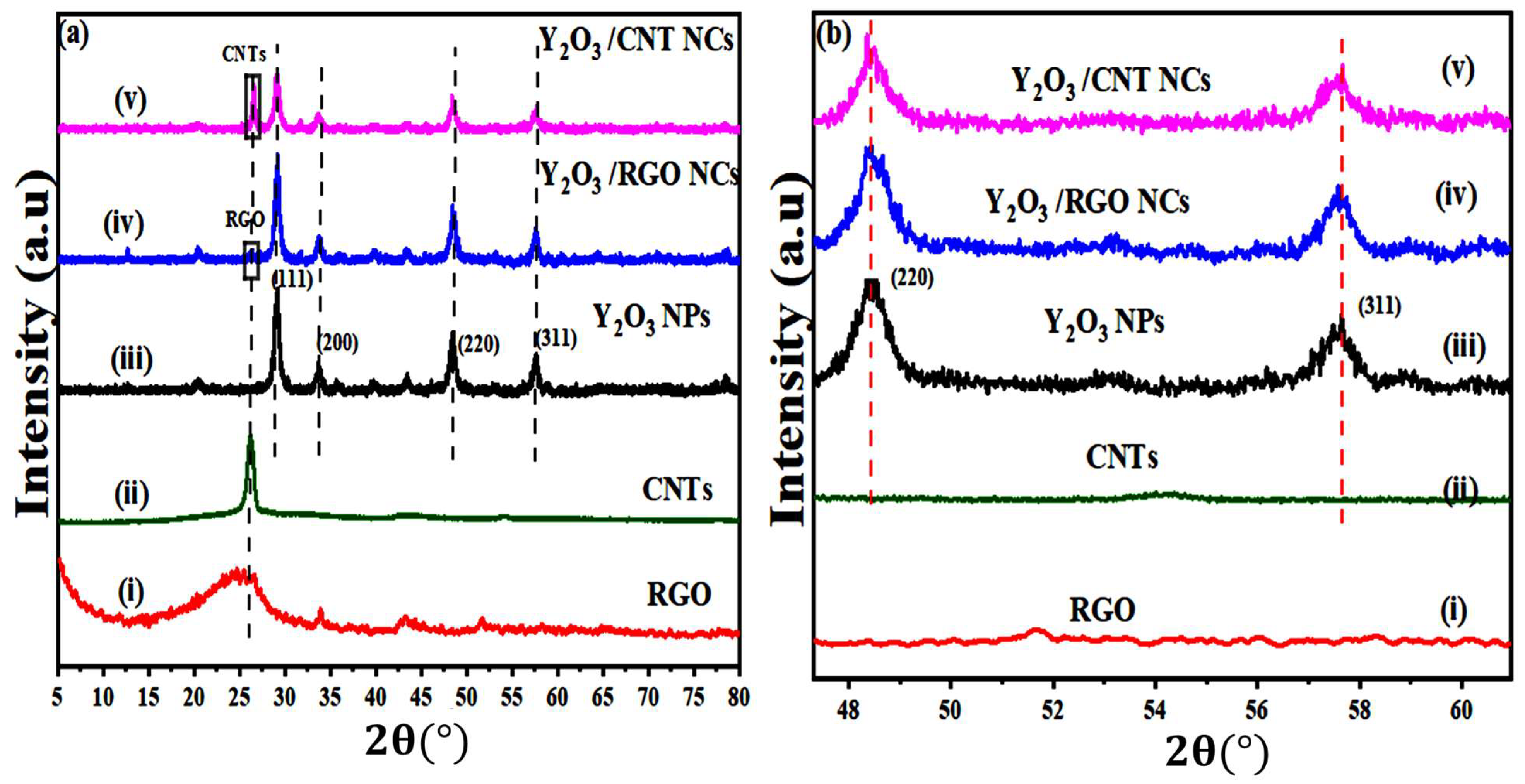
Figure 1. (a) X-ray diffraction pattern and (b) magnification of XRD spectra of (220) and (311) planes for (i) RGO, (ii) CNTs, (iii) pure Y2O3 Y2O3 NPs, (iv) Y2O3 Y2O3/RGO NCs, and (v) Y2O3 Y2O3/CNTs NCs.
Catalysts 15 00960 g001
Figure 2. SEM images for (a) pure Y2O3 NPs, (b) Y2O3/RGO NCs, and (c) Y2O3/CNTs NCs.
Catalysts 15 00960 g002
Figure 3. (a) EDX mapping analysis for Y2O3/RGO NCs: (I) electron image, (II) Carbon (c), (III) Yttrium (Y), and (IV) Oxygen (O). (b) EDX with mapping analysis FOR Y2O3/CNTs NCs: (i) Electron image, (ii) Carbon (c), (iii) Yttrium (Y), and (iv) Oxygen (O).
Catalysts 15 00960 g003
Figure 4. XPS spectra of Y2O3/RGO NCs: (a) Survey spectra of Y2O3/RGO NCs, (b) High resolution of XPS for Y 3d, (c) High resolution of XPS for O 1s, and (d) High resolution of XPS for C 1s.
Catalysts 15 00960 g004
Figure 5. FTIR spectrum of (a) RGO, (b) CNTs, (c) pure Y2O3 NPs, (d) Y2O3/RGO NCs, and (e) Y2O3/CNTs NCs.
Catalysts 15 00960 g005
Figure 6. (a) UV-Vis spectrum of the absorption with the band gap energy and (b) PL spectrum for pure Y2O3 NPs, Y2O3/RGO NCs, and Y2O3/CNTs NCs.
Catalysts 15 00960 g006
Figure 7. (a) Zeta potential analysis and (b) Electrophoretic Mobility Distribution of (red) pure Y2O3 NPs, (green) Y2O3/RGO NCs, and (blue) Y2O3/CNTs NCs.
Catalysts 15 00960 g007
Figure 8. Photocatalytic degradation of BPB dye using pure Y2O3 NPs, Y2O3/RGO NCs, and Y2O3/CNTs NCs: (ac) UV-Vis absorption spectrum of BPB dye solution in prepared samples under UV light for 120 min, (d) degradation analysis of BPB dye over time under UV light and dark conditions for synthesized samples, (e) Removal efficiency of BPB dye through produced samples, and (f) degradation of BPB dye visualized in test tubes using Y2O3/RGO NCs after 120 min.
Catalysts 15 00960 g008
Figure 9. Stability of degradation of Y2O3 NPs, Y2O3/RGO NCs, and Y2O3/CNTs NCs.
Catalysts 15 00960 g009
Figure 10. Possible reaction mechanism of degradation for BPB dye for Y2O3/RGO NCs.
Catalysts 15 00960 g010
Figure 11. Cell viability (%) versus logarithm of the concentration and IC50 values obtained for (a) pure Y2O3 NPs, (b) Y2O3/RGO NCs, and (c) Y2O3/CNTs NCs.
Catalysts 15 00960 g011
Scheme 1. The Steps for the preparation of (a) Y2O3/RGO NCs and (b) Y2O3/CNTs NCs.
Catalysts 15 00960 sch001
Table 1. Structural parameters of synthesized samples: crystallite size (D), dislocation density (δ), macrostrain (ε), and stacking fault ( γ s ) of prepared samples.
SamplesD (nm)δ (Lines/m2) × 10−3ε × 10−3 γ s (J/m2)
Pure Y2O3 NPs10.12 ± 1.83.21 ± 0.69.74 ± 0.24.52 ± 1.6
Y2O3/RGO NCs9.10 ± 1.34.12 ± 0.312.10 ± 0.77.01 ± 1.1
Y2O3/CNTs NCs11.11 ± 2.31.27 ± 0.18.30 ± 0.52.16 ± 0.8
Table 2. Zeta potential and electrophoretic mobility values in aqueous media of synthesized samples.
SamplesZeta Potential (mV ± SD)Electrophonic Mobility (µmcm/Vs ± SD)
Pure Y2O3 NPs−19.6 ± 3.79 −1.54 ± 0.68
Y2O3/RGO NCs−24.5 ± 5.16 −1.92 ± 1.02
Y2O3/CNTs NCs−22.1 ± 4.53 −1.75 ± 0.97
Table 3. Comparison of the photocatalytic activity of reported catalysts with recent work.
Sample TypeAverage Size (nm)Time (min)Light SourceD (%)Ref.
Y2O3 RGO NCs9.10120UV light87.83This work
Y2O3/TiO2 NCs73.0180UV light86.00[53]
ZnO/Y2O3 NCs15.3120UV light96.00[51]
Y/SnO2 NPs27.1180Visible light92.34[46]
V2O5/RGO NCs.----100Visible light63.00[54]
Cu/GO NCs12.250Visible light94.00[55]
Ag/Zn/CNTs NCs37.0120Visible light100.00[56]
CuO/ZnCo2O4/CNTs NCs9.890Sunlight87.00[57]
Table 4. Cytotoxic activity of prepared samples against bone (MG-63) cancer cell lines for 24 h.
SampleIn Vitro Cytotoxicity (IC50) Values for 24 h
IC50Log IC50R2
Y2O3 NPs158.122.190.9671
Y2O3/RGO NCs45.701.660.9975
Y2O3/CNTs NCs73.451.860.9764
Disclaimer/Publisher’s Note: The statements, opinions and data contained in all publications are solely those of the individual author(s) and contributor(s) and not of MDPI and/or the editor(s). MDPI and/or the editor(s) disclaim responsibility for any injury to people or property resulting from any ideas, methods, instructions or products referred to in the content.