Article

Multienzyme Synthesis of Glycyrrhetic Acid 3-O-mono-β-d-glucuronide by Coupling UGT73F15 to UDP-Glucuronic Acid Regeneration Module

1
National Engineering Laboratory for Industrial Enzymes, Tianjin Institute of Industrial Biotechnology, Chinese Academy of Sciences, Tianjin 300308, China
2
National Technology Innovation Center of Synthetic Biology, Tianjin 300308, China
*
Author to whom correspondence should be addressed.
Catalysts 2023, 13(1), 104; https://doi.org/10.3390/catal13010104
Submission received: 9 November 2022 / Revised: 12 December 2022 / Accepted: 14 December 2022 / Published: 3 January 2023
(This article belongs to the Section Biocatalysis)

Abstract

:
Glycyrrhetic acid 3-O-mono-β-d-glucuronide (GAMG), a rare and innovative compound in licorice, exhibits high-potency sweetness and improved physiological activities. However, low amounts of GAMG from plants cannot meet the demands of growing markets. In this study, an efficient one-pot multienzyme cascade reaction for GAMG biosynthesis was constructed using a coupled catalysis of glycosyltransferase and uridine 5′-diphosphate (UDP) glucuronic acid (GlcA) regeneration system. The Glycyrrhiza uralensis glycosyltransferase UGT73F15 was expressed in Escherichia coli BL21 (DE3). The optimal reaction conditions of UGT73F15 were found to be pH 7.5 and 35 °C. The catalytic efficiency (kcat/Km) for glycyrrhetic acid (GA) was 2.14 min−1 mM−1 when using UDP-GlcA as sugar donor. To regenerate costly UDP-GlcA, the one-pot multienzyme cascade reaction including UGT73F15, sucrose synthase, UDP-glucose dehydrogenase, and lactate dehydrogenase was adopted to synthesize GAMG from GA on the basis of the UDP-GlcA regeneration system. By optimizing the cascade reaction conditions, the GAMG production successfully achieved 226.38 mg/L. Our study developed an economical and efficient one-pot multienzyme cascade method for facile synthesis of GAMG and other bioactive glucuronosides.

1. Introduction

Due to the health problems caused by excessive consumption of sugar, increasing attention and demand exist for new alternative natural sweeteners with high-intensity, non-caloric, safe, and taste-modifying properties. Natural sweeteners, such as licorice extract, mogrosides from Siraitia grosvenorri, and steviol glycosides from Stevia rebaudiana Bertoni, were found to be 100–400 times sweeter than sucrose and showed low calories [1,2,3]. Glycyrrhizinate (GL) is the main bioactive constituent of licorice, which is 160 times sweeter than sucrose and possesses valuable bioactivities, including anticancer and antiviral activities [3,4,5]. However, GL has low absorption in the bloodstream [6]. The ionic metabolic equilibrium may be disturbed due to high-level intake of GL, causing adverse effects in humans [7,8]. Compared with GL, glycyrrhetic acid 3-O-mono-β-d-glucuronide (GAMG) showed stronger physiological activities, increased bioavailability, and better taste, and it was found to be 941 times sweeter than sucrose [9]. In recent years, due to its improved biological activities, GAMG has been developed as an innovative functional sweetener and a promising drug for the treatment of cancer and inflammation [10].
GAMG is an oleanane-type triterpenoid saponin that contains one molecule glycyrrhetic acid (GA) as aglycone and one glucuronic acid (GlcA) attached to the C-3 hydroxy group of GA (Figure 1). Biological strategies were developed to increase GAMG yields to effectively improve its industrial application in the food, pharmaceutical, and cosmetics industries. At present, the major approach for GAMG production is biotransformation of GL catalyzed by the enzyme β-glucuronidase (GUS, EC.3.2.1.31), which is utilized to hydrolyze a distal glucuronyl unit of GL in vivo/vitro [11]. By using this strategy, the yield of GAMG reached 2.62 g/L [12]. This method is convenient and efficient, but it relies on the substrate specificity of GUS and consumes extracted GL from plants as the substrate. Alternatively, GA has been de novo synthesized in Saccharomyces cerevisiae by introducing the entire heterogeneous biosynthetic pathway of the compound, which is a potential and ideal platform to eliminate the dependence on plant extracts [13,14]. Therefore, glycosylation of GA is the key process in the biosynthesis of GAMG.
Uridine diphosphate glycosyltransferases (UGTs) are the key factor to the glycosylation of GA in the biosynthesis pathway of GAMG. Several reports could be found on UGTs related to the catalysis of GA glycosylation (Table 1). Most of UGTs, including glycosyltransferases UGT73C11, UGT73F24, and Yjic, could transfer the glucose (Glc) moiety to the OH group at C3 and/or C30 of GA [15,16,17]. GmSGT2 from Glycine max transferred a galactose to the sugar moiety of monosaccharide derivatives of GA [18]. Two cellulose synthase superfamily-derived glycosyltransferases were reported to catalyze 3-O-glucuronosylation of triterpenoid aglycones [19]. UGT73F15 (GuGT14) from Glycyrrhiza uralensis is one of the few UGTs that could transfer GlcA to the OH group at the 3-position of GA and produce GAMG [20]. By introducing UGT73F15 to the engineered GAMG-producing yeast strains, the production of GAMG was determined to be 92.00 μg/L in shake flasks [21]. UGT1A1 from mammals was expressed in S. cerevisiae to construct microbial cell factory containing the complete biosynthesis pathway of GL. However, the productions of GL and GAMG were only 5.98 ± 0.47 and 2.31 ± 0.21 mg/L, respectively [22]. Therefore, effective UGTs and synthesis platform are urgently needed to biosynthesize high valued GAMG.
In vitro enzymatic biosystems have emerged as an important manufacturing platform. In the glucuronosylation of GA in vitro, the costly UDP-GlcA is required, which restricts the synthesis of GAMG and the practical application of UGTs. Therefore, a useful and efficient in situ regeneration of UDP-GlcA system platform is needed to minimize the process costs [23]. In the present study, the key enzyme UGT73F15 was first characterized in vitro by using GA and UDP-GlcA as sugar acceptor and donor, respectively. Then, a one-pot in vitro multienzyme cascade catalysis system for producing the sweetener GAMG based on the UDP-GlcA regeneration pathway was constructed. In the system, UDP was converted into UDP-Glc by sucrose synthase with the use of sucrose as substrate. UDP-GlcA was generated from UDP-Glc by UDP-Glc dehydrogenase. The GlcA residue was then transferred from UDP-GlcA to GA by UGT73F15, producing GAMG and the byproduct UDP, which could enter the first step. During the process, nicotinamide adenine dinucleotide (NAD+) could be regenerated by lactate dehydrogenase (LDH). The one-pot reaction conditions were also optimized to synthesize GAMG efficiently.
Table 1. Summary of enzymes showing glycosylation activity toward GA and its glycosides.
NoEnzymesSubstrates 1Sugar-DonorRef.
1UGT73C11GAUDPG[15]
2UGT73C33GAUDPG[16]
3UGT73F24GAUDPG[16]
4Bs-YjicGA/GLUDPG[17]
5GmSGT2GAMG/GLMGUDP-Gal[18]
6UGT109A3GAUDPG[24]
7UGT73F17GLUDPG, UDP-Xyl, UDP-Gal, and UDP-Ara[25]
8GuUGATGAUDPGlcA[26]
9GuUGTGAUDPG[27]
10UGT73P12GAMGUDPG/ UDPGlcA[28]
11GuGT14GAUDPG/ UDPGlcA[20]
12GuGT33GAUDPG[20]
13GuCslGAUDPGlcA[19]
14CslsGAUDPGlcA[19]
15UGT1A1GA/GAMGUDPGlcA[22]
1 GA—glycyrrhetinic acid, GL—glycyrrhizin, GAMG—glycyrrhetinic acid-3-O-monoglucuronide, GLMG—glycyrrhetinic acid-3-O-monoglucose.

2. Results

2.1. Expression and Characterization of UGT73F15

The glycosyltransferase UGT73F15 from licorice catalyzed the glycosylation of the OH group at the 3- and 30-positions of GA [20]. The codon-optimized gene was cloned into plasmid pET32a and overexpressed in E. coli BL21 (DE3). The recombinant enzyme of UGT73F15 was purified by Ni-NTA Sefinose column and analyzed by SDS-PAGE. The molecular mass of the purified recombinant enzyme was found to be approximately 74.5 kDa with TrxA tag (Figure S1).
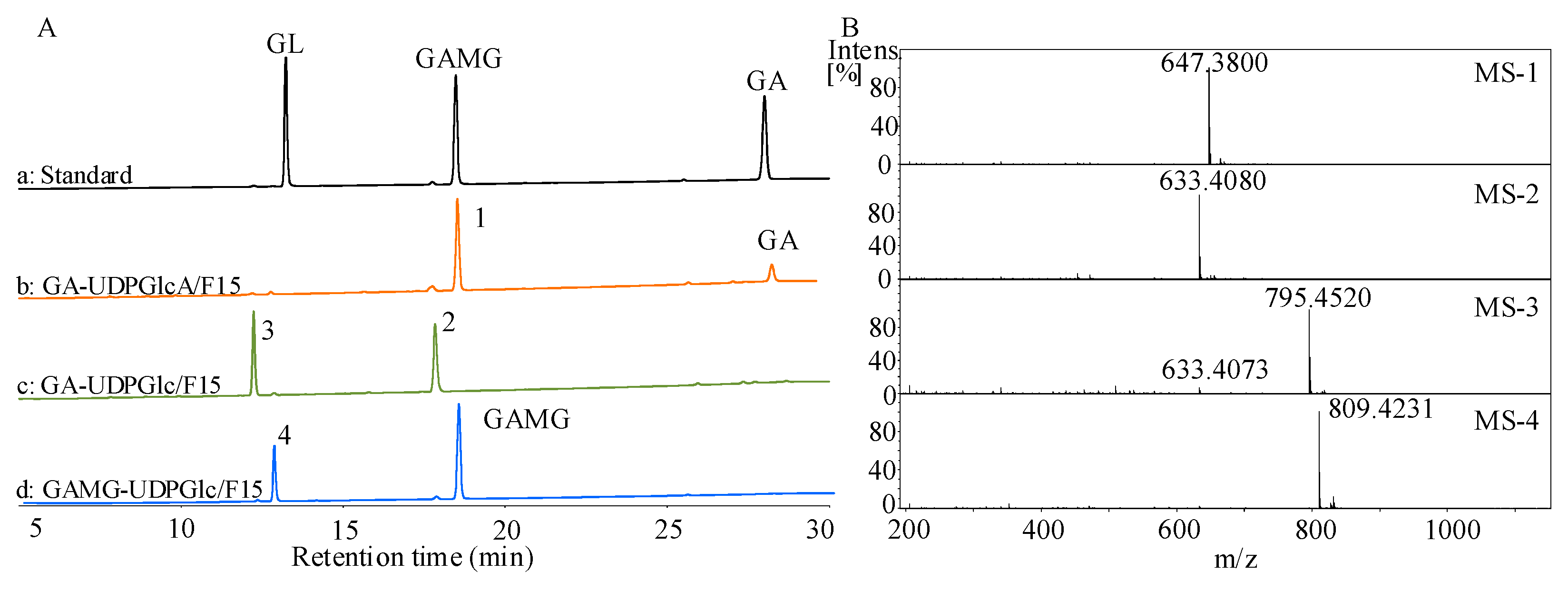
First, GA was utilized as the sugar receptor, while UDP-Glc or UDP-GlcA was tested as the sugar donor. As shown in Figure 2, UGT73F15 transferred one GlcA residue from UDP-GlcA to GA and produced a single product (1, GAMG), of which the molecular ion [M + H]+ was at m/z 647.38. The result indicated that UGT73F15 could be a suitable enzyme for the biosynthesis of GAMG. When UDP-Glc was utilized as sugar donor, two products (2 and 3) were detected in the GA glycosylation. Mass analysis of product 2 ([M + H] + m/z 633.41) and product 3 ([M + H] + m/z 795.45, [M − Glc + H]+ m/z 633.41) and their retention time showed that these products were GA-3-O-β-d-glucopyranoside (GA-Glc) and GA-3-O-β-d-glucopyranosyl-30-O-β-d-glucopyranoside, respectively, as confirmed in our recent study [17]. UGT73F15 was also demonstrated to glycosylate GAMG with UDP-Glc as a sugar donor by its accurate molecular ion [M + H]+ at m/z 809.43 (product 4).

2.2. Enzymatic Properties of UGT73F15

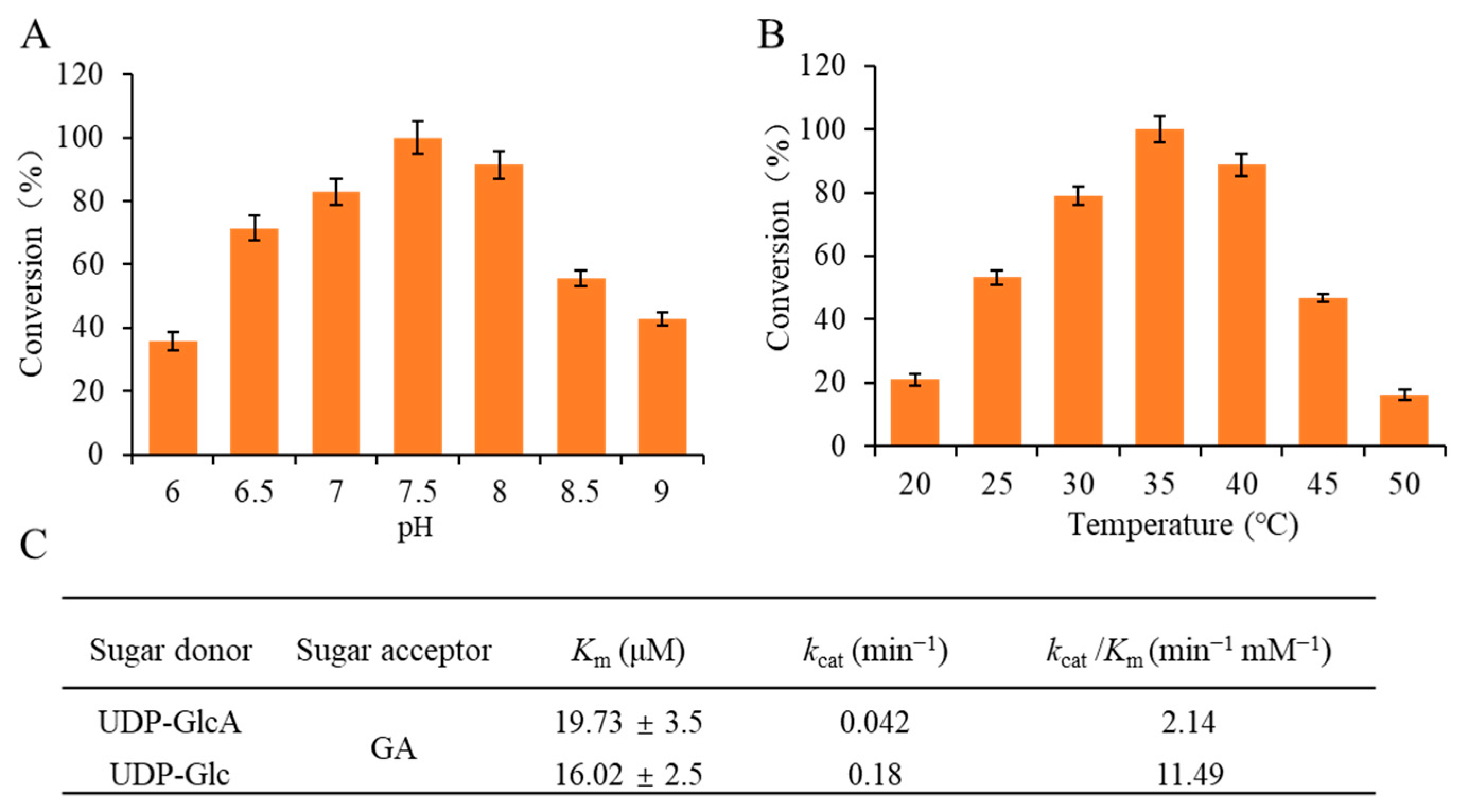
The pH and temperature profiles of UGT73F15 were investigated after the recombinant UGT73F15 was purified. As shown in Figure 3A, the optimal pH of UGT73F15 for glycosylation of GA was 7.5. The enzyme retained more than 60% of its activity detected at pH 7.5 when the pH ranged from 6.5 to 8. The optimal reaction temperature for UGT73F15 was 35 °C (Figure 3B). Under optimal temperature and pH conditions, the kinetic parameters of UGT73F15 in the glycosylation of GA were determined using UDP-GlcA and UDP-Glc as sugar donors. The Km values of UGT73F15 towards GA, using UDP-GlcA and UDP-Glc as sugar donor, were found to be 19.00 and 16.02 μM, respectively (Figure 3C). The kcat/Km values in the glucuronidation of GA was 2.14 min−1 mM−1, which was five times lower than that in the glycosylation of GA (11.49 min−1 mM−1). Compared with the kinetic parameters of B. vulgaris UGT73C11 [15] and Bacillus UGT109A3 [24], the catalytic efficiency of UGT73F15 against GA using UDP-Glc as sugar donor was slightly lower. Considering the enzymatic substrate specificity, it is of particular interest to engineer UGT73F15 to obtain an effective biocatalyst and synthesize a specific GA glucoside (GAMG).
The structure of UGT73F15 was predicted, and the docking of the enzyme with UDP-Glc/UDP-GlcA was studied to further elucidate the recognition of UDP-sugar. A homology model of UGT73F15 was constructed using glycosyltransferase PaGT3 from Phytolacca americana (PDB ID: 6lzx; identity: 47.1%) as the template by SWISS-MODEL. As shown in Figure 4A, the UGT73F15 structure displayed the conserved GT-B fold, consisting of a N-terminal domain and a C-terminal domain [29]. The reported UGT structures revealed a catalytic dyad (the highly conserved His-Asp) near the acceptor binding pocket, which was critical for catalytic reactions [29,30]. The corresponding residues were identified as His53 and Asp160 in UGT73F15 (Figure 4). UDP-GlcA and UDP-Glc were docked into the homology model of UGT73F15 with AutoDock, respectively, and interactions between UDP-sugar and the enzyme were analyzed by PyMOL. Five residues, including A371, S396, G393, N392, and W391 were involved in the recognition of the UDP moiety of UDP-sugar through the formation of hydrogen bonds. Residue E412 formed hydrogen bonds with the sugar moiety (Figure 4B,C). The plant secondary product glycosyltransferase (PSPG) motif in the C-terminal domain is highly conserved, and it interacts with the sugar donor. Therefore, PSPG-box sequence alignment was performed between UGT73F15 and other UGTs which used UDP-GlcA as a sugar donor, and the sequence identity was 79.5% (Figure 4D and Figure S3).

2.3. Construction of Multienzyme System for GAMG Synthesis

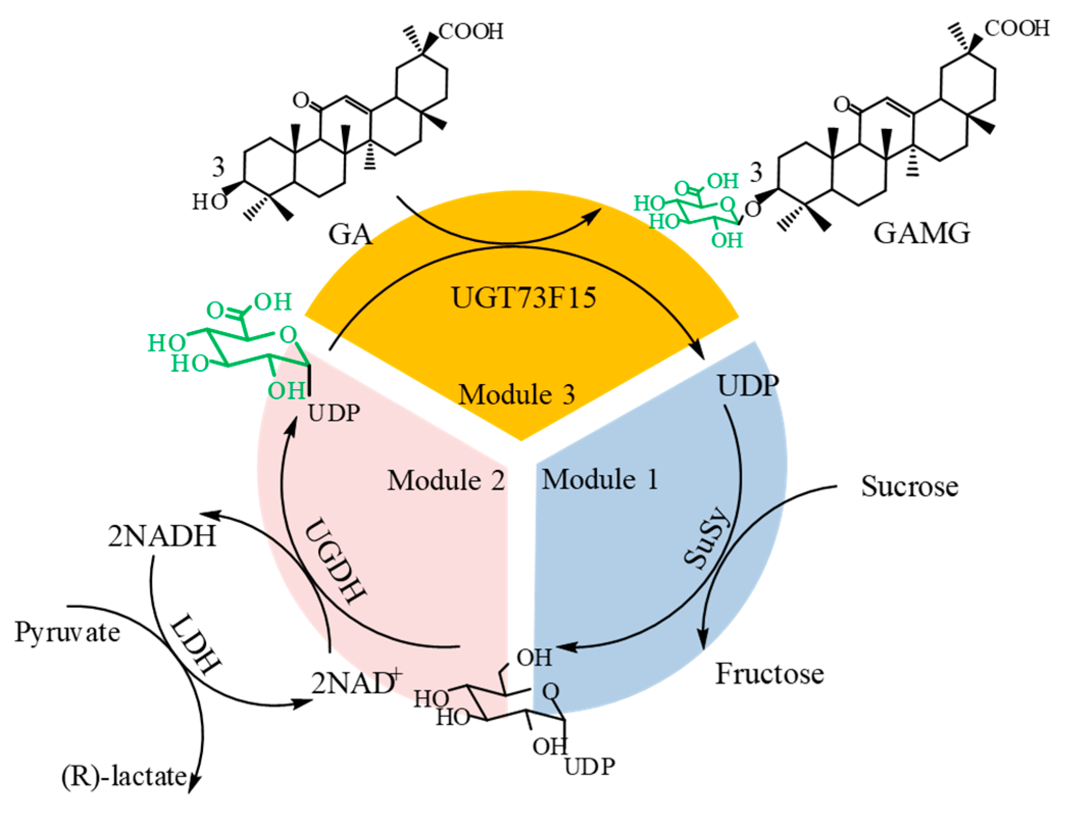
UDP-GlcA was the vital and high-cost sugar donor in the biosynthesis of high-intensity sweetener GAMG. To provide the sugar donor in the in vitro biosynthesis of GAMG, enzyme reaction system including in situ regeneration of UDP-GlcA and cofactor NAD+ was constructed by the combination of three enzymes (Figure 5). In the first module, UDP-Glc was synthesized from cheap sucrose and UDP using sucrose synthase (SuSy) as an efficient biocatalyst. In the second module, UGDH subsequently converted UDP-Glc into UDP-GlcA, in which NAD+ was consumed and converted into NADH. LDH from E. coli was integrated for in situ regeneration of NAD+ [23]. In the third module, glucuronyltransferase transferred GlcA from UDP-GlcA into GA and the byproduct UDP, which could enter the first module. Therefore, UDP-GlcA recycling could be constructed in a one-pot reaction.
The genes AcSusy from A. caldus, UGDH, and LDH from E. coli were cloned and inserted into the expression plasmid pET28 or pET32. All enzymes were expressed in E. coli BL21 (Figure S1). The SDS-PAGE results of the enzymes showed that the molecular masses of AcSusy (91.2 kDa), UGDH (60.5 kDa), LDH (53.4 kDa), and UGT73F15 (74.5 kDa) were in agreement with the sizes predicted by their amino acid sequences. The supernatants containing the soluble protein acted as crude enzyme after the harvested cells were sonicated and centrifugated. The activity of the enzymes was detected using respective substrates. As shown in Table 2, the crude enzymes for AcSusy, UGDH, LDH, and UGT73F15 at 35 °C were 130, 26, 370, and 4.3 mU/mg, respectively. Among them, UGT73F15 showed the lowest activity, indicating that the glucuronidation module could be the rate-limiting step in the GAMG biosynthesis pathway. The multienzyme system of GAMG was conducted by mixing 150 mU/mL AcSusy, LDH, UGT73F15, and 300 mU/mL UGDH with 1 mM UDP, 500 mM sucrose, 1 mM NAD+, 5 mM DTT, 0.2 mM GA, and 5 mM pyruvate. The platform successfully produced 0.085 mM of GAMG, demonstrating the feasibility of the biosynthetic pathway.

2.4. Optimizing the Parameters of the Enzyme Module System

For further improvement of the GAMG conversion, the key parameters of the enzyme module system on GAMG production were investigated to obtain the optimal reaction conditions for the in vitro cascade system, including pH, temperature, and concentrations of sucrose, UDP, GA, and NAD+. The influence of different temperatures on GAMG production was investigated at 25 °C–45 °C. As shown in Figure 6A, the highest yield of GAMG was obtained at 35 °C, which was chosen as the optimal temperature for the cascade reactions. The influence of pH (5.5–7.5) on GAMG biosynthesis system was also determined. As shown in Figure 6B, the highest conversion rate of GAMG was achieved at pH 6.5.
The effects of different substrate concentrations on the GAMG conversion rate were examined. The conversion decreased to 48.8% when the concentration of GA increased from 0.1 mM to 0.4 mM (Figure 6C). With the increase in the amount of sucrose and UDP, the glycosylation rate of GA increased and the highest title was reached at 750 mM sucrose and 1 mM UDP (Figure 6D,E). The conversion rate did not obviously change when the concentration of NAD+ was less than 1.0 mM, whereas higher concentrations inhibited the glycosylation activity, which was influenced by the decline in pH (Figure 6F). In summary, the parameters 0.4 mM GA, 750 mM sucrose, 1 mM UDP, 0.5 mM NAD+, pH 6.5, and 35 °C temperature were selected in the cascade reaction for GAMG-synthesis.

2.5. Biosynthesis of GAMG by One-Pot Cascade Reaction

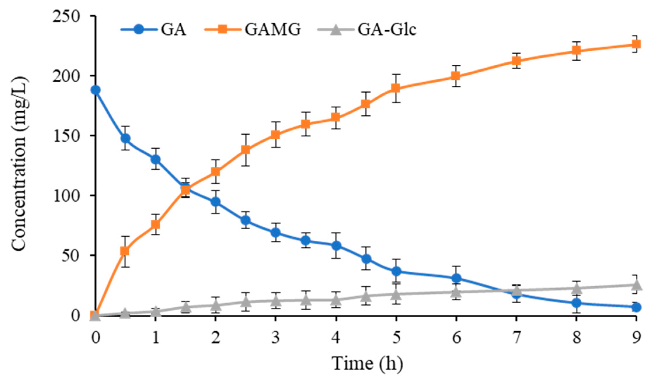
The one-pot cascade reaction for GAMG biosynthesis was executed under the optimized reaction conditions (Figure 7). The final concentrations of GAMG and GA-Glc reached 226.38 and 25.8 mg/L, respectively, and the molar conversion of GA was about 96.1%, after 9 h of incubation. To the best of our knowledge, this work was the first to report triterpene glucuronosylation using a one-pot cascade reaction. The byproduct GA-Glc was produced due to the promiscuousness of UGT73F15 towards different sugar donors (UDP-Glc) and resulted in the consumption of the substrate. Therefore, the enzyme specificity for sugar donor (UDP-GlcA) selection would be improved in the future on the basis of protein engineering, thus reducing the formation of byproduct. The UGT73F15 activity, which was lower than that of other enzymes in the one-pot cascade reaction, was another main rate-limiting factor, and the activity of the enzyme should be enhanced to improve the yield of GAMG.

3. Discussion

GAMG is an innovative high-potency sweetener. The compound presents better taste than untreated licorice extract and remarkable pharmacological activities, attracting considerable attention in the food and pharmaceutical industries. In the enzymatic synthesis of GAMG, the expensive sugar donor UDP-GlcA was required as a glycosyl donor for GA glycosylation. Therefore, a one-pot multienzyme system was constructed to generate UDP-GlcA by regeneration of UDP with cheap sucrose to complete the UDP/UDP-GlcA cycle [23]. Accordingly, an economical and efficient method for the production of GAMG was developed using sucrose synthetase, UDP-Glc dehydrogenase, lactate dehydrogenase, and glycosyltransferase.
At present, GAMG is mainly obtained via plant extraction, chemical synthesis, and biotransformation. The GAMG content in the roots of wild licorice is extremely low, thus reducing the efficiency of GAMG extraction (<0.1 mg/g) [31]. Moreover, overharvesting of wild licorice leads to environmental damage and even desertification [32]. Compared with chemical methods which involve strong and harsh conditions and inevitable environmental pollution, the biosynthesis of GAMG possesses milder reaction conditions and higher yields [33]. As one of the major and efficient approaches, hydrolysis of GL was utilized for GAMG production, which was catalyzed by enzyme GUSs or microbial strains harboring GUSs. However, there are still some limitations such as the burden of heavy reliance on plant extracts and environmental damage concerns [12,21]. Recently, heterologous biosynthesis of GAMG was reported in engineered yeast strains, resulting in a small amount of GAMG (2.31 mg/L). Therefore, it is necessary to develop an efficient and economical method for the production of high valued triterpenoid glycosides.
In situ regeneration of UDP-GlcA by coupling UGT with the UDP-GlcA regeneration module could effectively reduce the reaction cost. In the one-pot cascade reaction, sucrose synthase, UDP-Glc dehydrogenase, and lactate dehydrogenase were coupled to effectively generate the UDP-GlcA; UGT transferred the GlcA motif from UDP-GlcA to GA. After optimization, the titer of target product GAMG reached 226.38 mg/L, which is 98-fold higher than that of the engineered yeast strain mentioned above [21]. Moreover, multienzyme synthesis avoided the inhibitory effect of secondary metabolites on the growth of engineered strains, inefficient transport of substrates/products across membranes, and complex metabolism regulation.
Glucuronidation catalyzed by UGTs is the key step in GAMG biosynthesis, increasing the sweetness and bioavailability of the molecule. However, few UGTs were reported to show 3-O-glucuronosylation of GA. Two cellulose synthase superfamily-derived glycosyltransferases showed the needed activity. Both enzymes were predicted transmembrane proteins and only characterized in vivo [30]. UGT1A1 exhibited the catalytic potential to produce GL by catalyzing the native substrate GA with GAMG as a mid-product [22]. In this study, UGT73F15 showed strong regioselectivity and stereospecificity with GA. When UDP-GlcA was utilized as sugar donor, UGT73F15 could only glycosylate GA at the C-3 hydroxyl and produce GAMG. Moreover, the enzyme was confirmed to produce di-glycosides 3 and 4 when UDP-Glc and GA (or GAMG) were utilized as substrates, indicating the catalytic promiscuity towards both the sugar donors and aglycon acceptors.
Plant UGTs harbor a PSPG-box (highly conserved consensus sequence with 44 amino acids) near the C-terminus of the enzymes, which was proposed to interact with sugar donors [33]. It was reported that recognition of UDP-glucose can be partially correlated with the last amino acid within the PSPG-box, in which glutamine (Q) is necessary for glucosyl transfer [34]. The relatively conserved motif can influence the sugar donor specificity of UGTs for natural products glycosylation. Both the sugar donor specificity and the activity of enzymes can be operated through sequence/structure-based protein engineering around that motif or substrate binding pocket [35]. UGT73F15 was predicted to depict a GT-B structural fold and has a remarkable PSPG-box (W370-Q413). It was identified that UDP-Glc and UDP-GlcA interacted with the same the amino acid residues (A371, S396, G393, N392, W391, and E412) around the binding pocket of UGT73F15. Among those sites, E412, which formed hydrogen bonds with the sugar moiety, would be selected for mutagenesis to enhance the sugar donor specificity of UGT73F15 towards UDP-GlcA.
In conclusion, the recombinant glycosyltransferase UGT73F15 from G. uralensis exhibited promising application in the biosynthesis of GAMG. Furthermore, enzymatic biosynthesis of GAMG by coupling UGT73F15 to the UDP-glucuronic acid regeneration module showed the great potential versatility of this system in synthesizing diverse and bioactive glucuronides and laid a solid foundation for promoting the application of triterpenoid saponins in various fields.

4. Materials and Methods

4.1. Materials

UDP, UDP-Glc, and UDP-GlcA were purchased from Sigma–Aldrich (Shanghai, China). GA, GAMG, and GL were purchased from Chengdu Biopurify Phytochemicals. All other chemicals were of analytical grade.

4.2. Heterologous Expression and Purification of Enzymes

The genes of AcSusy-L637M-T640V (GenBank ID: KP284426.1), UDP-glucose dehydrogenase (UGDH, GenBank ID: ACT43781), LDH (GenBank ID: AVI55897.1), and UGT73F15 (GenBank ID: QDM38902.1) were synthesized by Genscript (Nanjing) Co., Ltd. and inserted into the expression plasmid pET28 (AcSusy) or pET32 (UGDH, LDH and UGT73F15). The recombinant vectors with the above genes were then transformed into E. coli BL21 (DE3) for expression.
Positive single colonies containing the abovementioned genes were inoculated into LB medium (5 mL, 100 μg/mL ampicillin) and grown at 37 °C and 200 rpm overnight. Then, 1% (v/v) inoculum was transferred to 200 mL LB liquid culture medium with ampicillin (100 μg/mL) and cultured at 37 °C and 200 rpm until the OD600 reached 0.6−0.8. isopropyl-β-d-thiogalactopyranoside was added to the medium at a final concentration of 0.2 mM at 16 °C and 200 rpm for 20 h for further incubation. The cells were harvested by centrifugation at 5000 g for 10 min and resuspended in 2 mL of 50 mM Tris-HCl (pH 7.0) after being washed twice with 10 mL buffer. The harvested broth was sonicated and centrifugated at 17,000 g for 60 min at 4 °C to yield the supernatant as crude enzyme containing the soluble protein. The enzymes were purified utilizing the 6× His-tag in the proteins by nickel-nitrilotriacetic acid (Ni-NTA) affinity chromatography. The enzyme purity was assessed by SDS-PAGE, and the protein concentration was determined using a NanoDrop 2000 (Thermo Scientific, Waltham, MA, USA).

4.3. Enzyme Activity Assays

The glycosylation of UGT was assayed in a 300 μL reaction mixture containing 1 mM UDP-Glc or UDP-GlcA, 0.2 mM GA or GAMG (dissolved in DMSO), 50 mM Tris-HCl (pH 7.0), 10 mM MgCl2, and 100 μL of UGT. The reaction was incubated at 35 °C for 3 h and terminated by adding an equal volume of methanol. After centrifugation was performed, the reactants were filtered through a 0.22 μm filter and analyzed by HPLC or HPLC-MS, as shown in Section 4.7
The catalytic activity of AcSuSy was examined in a 300 μL volume containing 1 mM UDP, 750 mM sucrose, 50 mM Tris-HCl (pH 7.0), and 10 μL AcSuSy for 10 min at 35 °C. The sample was analyzed by measuring the amount of fructose via bicinchoninic acid method [36].
The activity of UGDH was analyzed at 35 °C in 200 μL reaction assay buffer consisting of 5 mM UDP-Glc, 5 mM NAD+, 50 mM Tris-HCl buffer (pH 7.0), 5 mM DTT, and 10 μL UGDH. The reaction was monitored at 340 nm with a UV spectrophotometer for 30 min [37].
LDH activity was assayed by detecting the decrease in absorbance at 340 nm as NADH was oxidized to NAD+ as catalyzed by LDH. The LDH assay mixture solution (200 μL) was as follows: 5 mM NADH, 5 mM sodium pyruvate, 50 mM Tris-HCl buffer (pH 7.0), and 10 μL LDH [38].

4.4. Kinetic Parameters of UGTs

Kinetic analysis of UGT73F15 towards GA was conducted in the following reaction (300 μL): 1 mM UDP-Glc or UDP-GlcA, 1 mM MgCl2, 50 mM Tris-HCl (pH 7.5), GA (varying from 0.01 mM to 0.08 mM), and 0.2 mg of the purified enzyme. The reaction was incubated at 35 °C for 2 h and then stopped by adding 300 μL of methanol. The kinetic parameters were calculated by nonlinear regression of the Michaelis−Menten equation on GraphPad Prism 5.

4.5. Homology Modeling and Molecular Docking

The sequence of UGT73F15 was used to align and search suitable models for homology modeling by the SWISS-MODEL server (http://swissmodel.expasy.org/, accessed on 1 August 2021). The structure of PaGT3 from Phytolacca americana (PDB ID: 6lzx, 47.1% sequence identity with that of UGT73F15) was utilized as a template for model building. The molecule of UDP-GlcA was docked into the structure of UGT73F15 by using AutoDock 4.0. PyMOL was also used to visualize the models and prepare the figures. Sequence alignments were created using Clustal Omega and ESPript [30].

4.6. Cascade Reaction Optimization

For glycosylation of GA in the multienzyme system using sucrose as sugar donor, a reaction mixture containing 1 mM UDP, 500 mM sucrose, 1 mM NAD+, 5 mM DTT, 0.2 mM GA, 5 mM pyruvate, 50 mM Tris-HCl (pH 7.0), 150 mU/mL AcSuSy, LDH, UGT, and 300 mU/mL UGDH was constructed at 35 °C for 5 h. The effects of different concentrations of substrates, including sucrose (250−1000 mM), UDP (0.1−1.5 mM), NAD+ (0.1−1.5 mM), and GA (0.1−0.6 mM), and different pH values from 5.5 to 7.5 (50 mM acetic acid−sodium acetate buffer for pH 5.5−6.0, 50 mM Tris-HCl buffer for pH 6.5−7.5) and temperatures (25 °C−45 °C) on the conversion rate of GA were evaluated.

4.7. Biotransformation of GA by Cascade Reaction

The reaction mixture (10 mL) included 1 mM UDP, 750 mM sucrose, 0.5 mM NAD+, 5 mM DTT, 0.4 mM GA, 5 mM pyruvate, 50 mM Tris-HCl (pH 6.5), 150 mU/mL AcSuSy, LDH, and UGT, and 300 mU/mL UGDH. The reaction was performed at 35 °C and 150 rpm. The sample (150 μL) was taken and stopped by adding an equal volume of methanol at certain times and analyzed by Agilent 1260 HPLC system.

4.8. Analytical Methods

GA, GAMG, and their glycosides were analyzed by HPLC or HPLC-electrospray ionization (ESI)-MS (Agilent 1260 HPLC system coupled to a Bruker micrOTOF II mass spectrometer) using a reversed-phase column (Ultimate C18 column, 4.6 mm × 250 mm, 5 μm particle, Welch, Shanghai, China). The ESI methods were as described before (Li et al., 2020). The operating conditions were 1 mL/min and a wavelength of 250 nm. The gradient program was as follows: distilled water with 0.1% formic acid (solvent A) and acetonitrile with 0.1% formic acid (solvent B) using a gradient program of 25–85% B in 0–25 min, followed by 85% B for 5 min.

Supplementary Materials

The following supporting information can be downloaded at: https://www.mdpi.com/article/10.3390/catal13010104/s1, Figure S1: SDS-PAGE of recombinant enzymes utilized in this study; Figure S2: Determination of kinetic parameters for recombinant UGT73F15 on GA; Figure S3: Multiple alignment of UGT73F15 with other UGTs identified with UDP-GlcA activity.

Author Contributions

Conceptualization, methodology, and writing—original draft preparation, J.L.; validation, T.C. and X.Z.; formal analysis, J.Y. and Y.Z.; writing—review and editing, Y.M. and Y.S. All authors have read and agreed to the published version of the manuscript.

Funding

This work was supported by the National Natural Science Foundation of China (No. 32101885), the Tianjin Synthetic Biotechnology Innovation Capacity Improvement Project (No. TSBICIP-CXRC-023), the National Key Research and Development Program of China (2019YFA0905100), as well as the China Postdoctoral Science Foundation (No. 2021M693349).

Data Availability Statement

Data is contained within the article or Supplementary Material.

Conflicts of Interest

The authors declare no conflict of interest.

References

  1. Matsumoto, K.; Kasai, R.; Ohtani, K.; Tanaka, O. Minor cucurbitane-glycosides from fruits of Siraitia grosvenori (Cucurbitaceae). Himalayan Chem. Pharm. Bull. 1990, 38, 2030–2032. [Google Scholar] [CrossRef] [Green Version]
  2. Ceunen, S.; Geuns, J.M.C. Steviol glycosides: Chemical diversity, metabolism, and function. J. Nat. Prod. 2013, 76, 1201–1228. [Google Scholar] [CrossRef]
  3. Kitagawa, I. Licorice root. A natural sweetener and an important ingredient in Chinese medicine. Pure Appl. Chem. 2002, 74, 1189–1198. [Google Scholar] [CrossRef] [Green Version]
  4. Roohbakhsh, A.; Iranshahy, M.; Iranshahi, M. Glycyrrhetinic acid and its derivatives: Anti-cancer and cancer chemopreventive properties, mechanisms of action and structure-cytotoxic activity relationship. Curr. Med. Chem. 2016, 23, 498–517. [Google Scholar] [CrossRef] [PubMed]
  5. Dastagir, G.; Rizvi, M.A. Review—Glycyrrhiza glabra L. (liquorice). Pak. J. Pharm. Sci. 2016, 29, 1727–1733. [Google Scholar] [PubMed]
  6. Akao, T. Difference in the metabolism of glycyrrhizin, glycyrrhetic acid and glycyrrhetic acid monoglucuronide by human intestinal flora. Biol. Pharm. Bull. 2000, 23, 1418–1423. [Google Scholar] [CrossRef] [PubMed] [Green Version]
  7. Homma, M. Education program of kampo-medicine for undergraduates in preparation for clinical setting. Yakugaku Zasshi J. Pharm. Soc. Jpn. 2016, 136, 417–422. [Google Scholar] [CrossRef] [Green Version]
  8. Wang, C.; Guo, X.X.; Wang, X.Y.; Qi, F.; Feng, S.J.; Li, C.; Zhou, X.H. Isolation and characterization of three fungi with the potential of transforming glycyrrhizin. World J. Microbiol. Biotechnol. 2013, 29, 781–788. [Google Scholar] [CrossRef]
  9. Mizutani, K.; Kuramoto, T.; Tamura, Y.; Ohtake, N.; Doi, S.; Nakaura, M.; Tanaka, O. Sweetness of glycyrrhetic acid 3-O-β-d-monoglucuronide and the related glycosides. Biosci. Biotechnol. Biochem. 1994, 58, 554–555. [Google Scholar] [CrossRef]
  10. Edwards, C.H.; Rossi, M.; Corpe, C.P.; Butterworth, P.J.; Ellis, P.R. The role of sugars and sweeteners in food, diet and health: Alternatives for the future. Trends Food Sci. Technol. 2016, 56, 158–166. [Google Scholar] [CrossRef]
  11. Feng, S.J.; Li, C.; Xu, X.L.; Wang, X.Y. Screening strains for directed biosynthesis of β-d-mono-glucuronide-glycyrrhizin and kinetics of enzyme production. J. Mol. Catal. B Enzym. 2006, 43, 63–67. [Google Scholar] [CrossRef]
  12. Kaleem, I.; Shen, H.; Lv, B.; Wei, B.; Rasool, A.; Li, C. Efficient biosynthesis of glycyrrhetic acid 3-O-mono-β-d-glucuronide (GAMG) in water-miscible ionic liquid by immobilized whole cells of Penicillium purpurogenum Li-3 in alginate gel. Chem. Eng. Sci. 2014, 106, 136–143. [Google Scholar] [CrossRef]
  13. Wang, C.; Su, X.; Sun, M.; Zhang, M.; Wu, J.; Xing, J.; Wang, Y.; Xue, J.; Liu, X.; Sun, W.; et al. Efficient production of glycyrrhetinic acid in metabolically engineered Saccharomyces cerevisiae via an integrated strategy. Microb. Cell Fact. 2019, 18, 1–15. [Google Scholar] [CrossRef] [Green Version]
  14. Zhu, M.; Wang, C.; Sun, W.; Zhou, A.; Wang, Y.; Zhang, G.; Li, C. Boosting 11-oxo-β-amyrin and glycyrrhetinic acid synthesis in Saccharomyces cerevisiae via pairing novel oxidation and reduction system from legume plants. Metab. Eng. 2018, 45, 43–50. [Google Scholar] [CrossRef]
  15. Liu, X.; Zhang, L.; Feng, X.; Lv, B.; Li, C. Biosynthesis of glycyrrhetinic acid-3-O-monoglucose using glycosyltransferase UGT73C11 from Barbarea vulgaris. Ind. Eng. Chem. Res. 2017, 56, 14949–14958. [Google Scholar] [CrossRef]
  16. Zhang, L.; Ren, S.; Liu, X.; Liu, X.; Guo, F.; Sun, W.; Li, C. Mining of UDP-glucosyltrfansferases in licorice for controllable glycosylation of pentacyclic triterpenoids. Biotechnol. Bioeng. 2020, 117, 3651–3663. [Google Scholar] [CrossRef] [PubMed]
  17. Dai, L.; Li, J.; Yang, J.; Men, Y.; Zeng, Y.; Cai, Y.; Sun, Y. Enzymatic synthesis of novel glycyrrhizic acid glucosides using a promiscuous Bacillus glycosyltransferase. Catalysts 2018, 8, 615. [Google Scholar] [CrossRef] [Green Version]
  18. Gao, Y.; Zhang, L.; Feng, X.; Liu, X.; Guo, F.; Lv, B.; Li, C. Galactosylation of monosaccharide derivatives of glycyrrhetinic acid by UDP-glycosyltransferase GmSGT2 from Glycine max. J. Agric. Food Chem. 2020, 68, 8580–8588. [Google Scholar] [CrossRef]
  19. Jozwiak, A.; Sonawane, P.D.; Panda, S.; Garagounis, C.; Papadopoulou, K.K.; Abebie, B.; Aharoni, A. Plant terpenoid metabolism co-opts a component of the cell wall biosynthesis machinery. Nat. Chem. Biol. 2020, 16, 740–748. [Google Scholar] [CrossRef]
  20. Chen, K.; Hu, Z.M.; Song, W.; Wang, Z.L.; He, J.B.; Shi, X.M.; Ye, M. Diversity of O-glycosyltransferases contributes to the biosynthesis of flavonoid and triterpenoid glycosides in Glycyrrhiza uralensis. ACS Synth. Biol. 2019, 8, 1858–1866. [Google Scholar] [CrossRef]
  21. Huang, Y.; Jiang, D.; Ren, G.; Yin, Y.; Sun, Y.; Liu, T.; Liu, C. De Novo Production of Glycyrrhetic Acid 3-O-mono-β-d-glucuronide in Saccharomyces cerevisiae. Front. Bioeng. Biotech. 2021, 9, 709120. [Google Scholar] [CrossRef] [PubMed]
  22. Xu, K.; Zhao, Y.J.; Ahmad, N.; Wang, J.N.; Lv, B.; Wang, Y.; Li, C. O-glycosyltransferases from Homo sapiens contributes to the biosynthesis of glycyrrhetic acid 3-O-mono-β-d-glucuronide and glycyrrhizin in Saccharomyces cerevisiae. Synth. Syst. Biotechnol. 2021, 6, 173–179. [Google Scholar] [CrossRef] [PubMed]
  23. Engels, L.; Henze, M.; Hummel, W.; Elling, L. Enzyme Module Systems for the Synthesis of Uridine 5′-Diphospho-α-d-glucuronic Acid and Non-Sulfated Human Natural Killer Cell-1 (HNK-1) Epitope. Adv. Synth. Catal. 2015, 357, 1751–1762. [Google Scholar] [CrossRef]
  24. Ali, M.Y.; Chang, Q.; Yan, Q.; Qian, Z.; Guo, X.; Thow, K.; Wu, J.; Zhang, Y.; Feng, Y. Highly Efficient Biosynthesis of Glycyrrhetinic Acid Glucosides by Coupling of Microbial Glycosyltransferase to Plant Sucrose Synthase. Front. Bioeng. Biotechnol. 2021, 9, 645079. [Google Scholar] [CrossRef]
  25. He, J.; Chen, K.; Hu, Z.M.; Li, K.; Song, W.; Yu, L.Y.; Ye, M. UGT73F17, a new glycosyltransferase from Glycyrrhiza uralensis, catalyzes the regiospecific glycosylation of pentacyclic triterpenoids. Chem. Commun. 2018, 54, 8594–8597. [Google Scholar] [CrossRef]
  26. Xu, G.J.; Cai, W.; Gao, W.; Liu, C.S. A novel glucuronosyltransferase has an unprecedented ability to catalyse continuous two-step glucuronosylation of glycyrrhetinic acid to yield glycyrrhizin. New Phytol. 2016, 212, 123–135. [Google Scholar] [CrossRef] [Green Version]
  27. Huang, Y.; Li, D.; Wang, J.; Cai, Y.; Dai, Z.; Jiang, D.; Liu, C. GuUGT, a glycosyltransferase from Glycyrrhiza uralensis, exhibits glycyrrhetinic acid 3-and 30-O-glycosylation. R. Soc. Open Sci. 2019, 6, 191121. [Google Scholar] [CrossRef] [Green Version]
  28. Nomura, Y.; Seki, H.; Suzuki, T.; Ohyama, K.; Mizutani, M.; Kaku, T.; Muranaka, T. Functional specialization of UDP-glycosyltransferase 73P12 in licorice to produce a sweet triterpenoid saponin, glycyrrhizin. Plant J. 2019, 99, 1127–1143. [Google Scholar] [CrossRef] [Green Version]
  29. Li, J.; Yang, J.; Mu, S.; Shang, N.; Liu, C.; Zhu, Y.; Ma, Y. Efficient O-glycosylation of triterpenes enabled by protein engineering of plant glycosyltransferase UGT74AC1. ACS Catal. 2020, 10, 3629–3639. [Google Scholar] [CrossRef]
  30. Huang, W.; He, Y.; Jiang, R.; Deng, Z.; Long, F. Functional and Structural Dissection of a Plant Steroid 3-O-Glycosyltransferase Facilitated the Engineering Enhancement of Sugar Donor Promiscuity. ACS Catal. 2022, 12, 2927–2937. [Google Scholar] [CrossRef]
  31. Song, W.; Qiao, X.; Chen, K.; Wang, Y.; Ji, S.; Feng, J.; Ye, M. Biosynthesis-based quantitative analysis of 151 secondary metabolites of licorice to differentiate medicinal Glycyrrhiza species and their hybrids. Anal. Chem. 2017, 89, 3146–3153. [Google Scholar] [CrossRef] [PubMed]
  32. Szakiel, A.; Pączkowski, C.; Henry, M. Influence of environmental biotic factors on the content of saponins in plants. Phytochem. Rev. 2011, 10, 493–502. [Google Scholar] [CrossRef]
  33. Guo, L.; Katiyo, W.; Lu, L.; Zhang, X.; Wang, M.; Yan, J.; Zhao, W. Glycyrrhetic Acid 3-O-mono-β-d-glucuronide (GAMG): An Innovative High-Potency Sweetener with Improved Biological Activities. Compr. Rev. Food Sci. Food Saf. 2018, 17, 905–919. [Google Scholar] [CrossRef] [PubMed] [Green Version]
  34. Caputi, L.; Malnoy, M.; Goremykin, V.; Nikiforova, S.; Martens, S. A genome-wide phylogenetic reconstruction of family 1 UDP-glycosyltransferases revealed the expansion of the family during the adaptation of plants to life on land. Plant J. 2012, 69, 1030–1042. [Google Scholar] [CrossRef]
  35. Rahimi, S.; Kim, J.; Mijakovic, I.; Jung, K.H.; Choi, G.; Kim, S.C.; Kim, Y.J. Triterpenoid-biosynthetic UDP-glycosyltransferases from plants. Biotechnol. Adv. 2019, 37, 107394. [Google Scholar] [CrossRef] [PubMed]
  36. Dai, L.; Liu, C.; Li, J.; Dong, C.; Yang, J.; Dai, Z.; Sun, Y. One-pot synthesis of ginsenoside Rh2 and bioactive unnatural ginsenoside by coupling promiscuous glycosyltransferase from Bacillus subtilis 168 to sucrose synthase. J. Agric. Food Chem. 2018, 66, 2830–2837. [Google Scholar] [CrossRef]
  37. Chu, X.; Han, J.; Guo, D.; Fu, Z.; Liu, W.; Tao, Y. Characterization of UDP-glucose dehydrogenase from Pasteurella multocida CVCC 408 and its application in hyaluronic acid biosynthesis. Enzyme Microb. Technol. 2016, 85, 64–70. [Google Scholar] [CrossRef]
  38. Yang, G.; Tian, J.; Li, J. Fermentation of 1,3-propanediol by a lactate deficient mutant of Klebsiella oxytoca under microaerobic conditions. Appl. Microbiol. Biotechnol. 2007, 73, 1017–1024. [Google Scholar] [CrossRef]
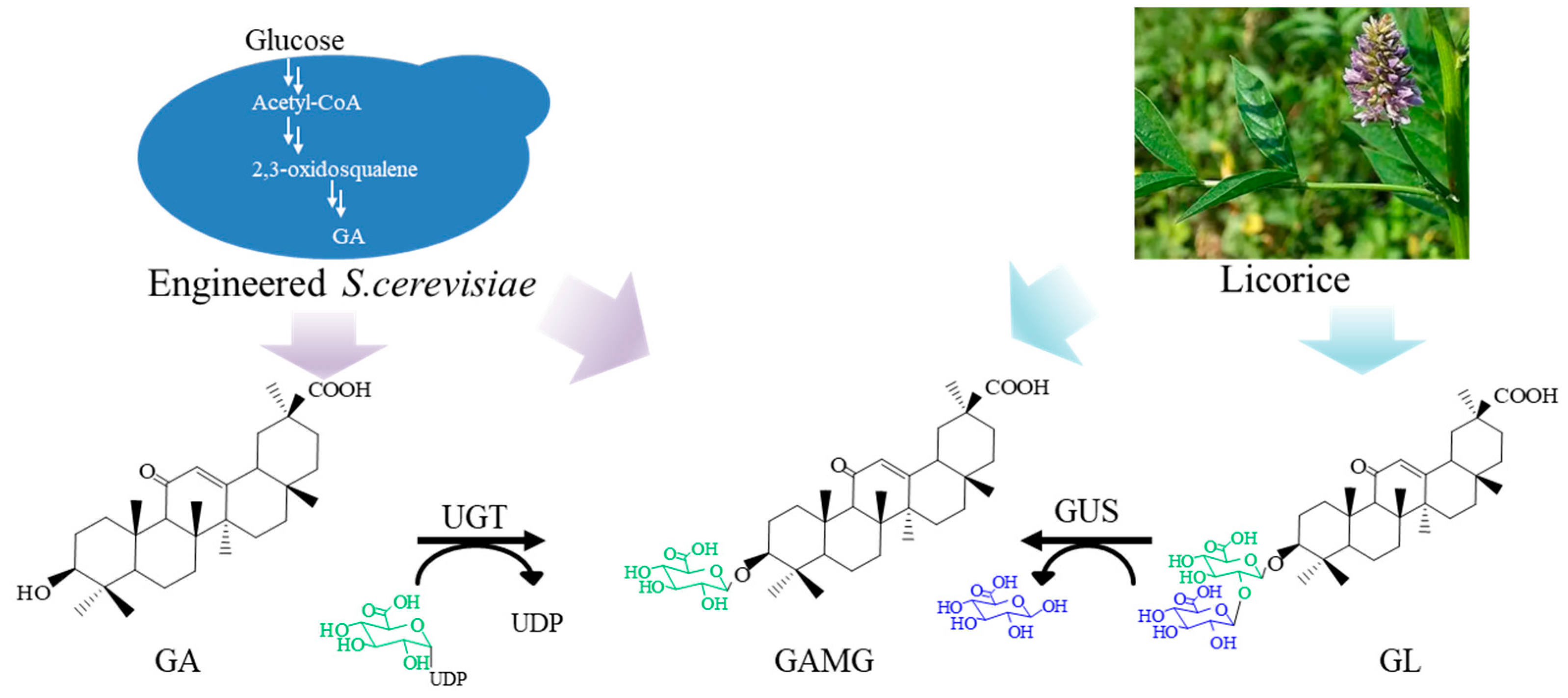
Figure 1. Biosynthesis of glycyrrhetic acid 3-O-mono-β-d-glucuronide (GAMG) from glycyrrhetic acid (GA) catalyzed by glycosyltransferase (UGT) or glycyrrhizin (GL) catalyzed by β-glucuronidase (GUS).
Catalysts 13 00104 g001
Figure 2. HPLC-MS analysis of the glycosylated products of GA and GAMG. (A) HPLC chromatograms of GAMG standards (a), UGT73F15-catalyzed reaction using GA and UDP-GlcA as substrates (b), UGT73F15-catalyzed reaction using GA and UDP-Glc as substrates (c), and UGT73F15-catalyzed reaction using GAMG and UDP-Glc as substrates (d). (B) MS spectra for products 1–4.
Catalysts 13 00104 g002
Figure 3. The effects of pH (A) and temperature (B) on the UGT73F15-catalyzed glycosylation of GA. (C) Kinetic parameters of UGT73F15 toward GA using UDP-GlcA or UDP-Glc as sugar donor. Error bars represent the standard deviation from three repeats.
Catalysts 13 00104 g003
Figure 4. Sugar donor (UDP-GlcA) recognition of UGT73F15. (A) Homology model of UGT73F15 using glycosyltransferase PaGT3 from Phytolacca americana as template. (B) Interaction analysis between UDP-Glc and UGT73F15 in the docking model. Hydrogen bonds are represented by yellow dashed lines. Residues involved in the interaction with UDP-Glc are shown in blue sticks. Catalytic residues are shown in pink sticks. UDP-Glc is shown in yellow sticks. (C) Interaction analysis between UDP-GlcA and UGT73F15 in the docking model. UDP-GlcA is shown in purple/blue sticks. (D) PSPG-box sequence alignment of UGT73F15 and other UGTs identified with UDP-GlcA activity.
Catalysts 13 00104 g004
Figure 5. Schematic representation of GAMG synthesis using UGT73F15 as catalyst, based on in situ regeneration of UDP-GlcA by combination of sucrose synthase (SuSy), UDP-Glc-dehydrogenase (UGDH), and lactate dehydrogenase (LDH).
Catalysts 13 00104 g005
Figure 6. Effects of key parameters on the glycosylation of GA enzyme module system. Effects of temperature (A) and pH (B) on the cascade reaction. Effects of the concentration of GA (C), sucrose (D), UDP (E), and NAD+ (F) on the cascade reaction. Error bars represent the standard deviation from three repeats.
Catalysts 13 00104 g006
Figure 7. Time course of glycosylation of GA into GAMG and GA-Glc in the enzyme module system. The cascade reaction contained 1 mM UDP, 750 mM sucrose, 0.5 mM NAD+, 5 mM DTT, 0.4 mM GA, 5 mM pyruvate, 50 mM Tris-HCl (pH 6.5), 150 mU/mL AcSuSy, LDH, UGT, and 300 mU/mL UGDH and was performed at 35 °C and 150 rpm for 9 h.
Catalysts 13 00104 g007
Table 2. Properties of enzymes utilized in this study.
Enzyme SourceGenBankMW (kDa)Activity (mU/mg) 1
AcSusyA. caldusKP284426.191.2130
UGDHE. coliACT4378160.526
LDHE. coliAVI55897.153.4370
UGT73F15LicoriceQDM38902.174.54.3
1 The activity was detected using the crude enzymes from cell lysates.
Disclaimer/Publisher’s Note: The statements, opinions and data contained in all publications are solely those of the individual author(s) and contributor(s) and not of MDPI and/or the editor(s). MDPI and/or the editor(s) disclaim responsibility for any injury to people or property resulting from any ideas, methods, instructions or products referred to in the content.