Article

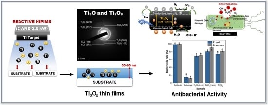
A Study on the Characteristic and Antibacterial Activity of Ti3Ox Thin Films

1
Department of Tropical Agriculture and International Cooperation, National Pingtung University of Science & Technology, Pingtung 91201, Taiwan
2
Department of Agricultural Product Technology, Faculty of Agricultural Technology, Universitas Brawijaya, Malang 65145, Indonesia
3
Department of Materials Engineering, National Pingtung University of Science & Technology, Pingtung 91201, Taiwan
4
Kaohsiung Veterans General Hospital, Kaohsiung City 813414, Taiwan
5
Institute of Materials Science and Engineering, National Central University, Zhongli 32001, Taiwan
6
Department of Biological Science and Technology, National Pingtung University of Science & Technology, Pingtung 91201, Taiwan
*
Author to whom correspondence should be addressed.
Catalysts 2021, 11(11), 1416; https://doi.org/10.3390/catal11111416
Submission received: 30 October 2021 / Revised: 18 November 2021 / Accepted: 21 November 2021 / Published: 22 November 2021
(This article belongs to the Special Issue Photocatalytic Nanomaterials for Abatement of Microorganisms)

Abstract

:
A pure Ti target in Ar/O2 gas mixture was used to synthesize Ti3Ox thin film on a glass substrate by Reactive High-Power Impulse Magnetron Sputtering (HiPIMS) under different sputtering power (2 and 2.5 kW). The influence of HiPIMS parameters on thin films’ structural, morphological, chemical composition, optical and photocatalytic, and antibacterial properties was investigated. In this study, Ti3Ox thin films can be synthesized using the HiPIMS method without the post-annealing process. Two co-existence phases (hexagonal Ti3O and base-centered monoclinic Ti3O5 phases) existed on the Ti3Ox films. It is found that the peak intensity of (006) Ti3O hexagonal slightly increased as the sputtering power increased from 2 to 2.5 kW. The Ti3Ox thin-film bandgap values were 3.36 and 3.50 eV for 2 and 2.5 kW, respectively. The Ti3Ox films deposited at 2.5 kW showed good photocatalytic activity under UV light irradiation, with a higher methylene blue dye degradation rate than TiO2 thin films. The antibacterial study on Ti3Ox thin films exhibited a high inhibition percentage against E. coli and S. aureus. This study demonstrates that Ti3Ox thin films can promote high photocatalytic and antibacterial activity.

Graphical Abstract

1. Introduction

Titanium sub-oxides (TSO) have drawn much attention as they possess interesting properties for metal oxide thin films which have a variety of crystalline structures; unique physical properties; and a wide range of electrical, optical, and electrochemical properties such as electro-optical devices [1], self-cleaning [2], solar cell [3], and photocatalyst [4]. Magnéli-phase titanium oxides, the TinO2n−1, (3 ≤ n ≤ 10) exhibit specific oxygen vacancies organization that garnered substantial interest owing to their excellent electric conductivity, high corrosion resistance, and optical properties [5,6]. According to Xu et al. [7], Magnéli-phase on TSO is a mixed-valence molecule composed of two Ti3+ (3d1 electronic configuration) and (n—2)Ti4+ (3d0) ions. The existence of both Ti3+ and Ti4+ ions in the crystal opens up several possible cation configurations, resulting in a variety of charge-ordered states.
Titanium sub-oxides can be grown by a series of methods, including low-energy ion bombardment [8], pulsed layer deposition [9,10], a chemical vapor deposition (CVD) method [11], sol–gel combined with the energy-efficient vacuum-carbothermic (SF-VC) process [6], and a sputtering method [12,13,14]. Among all the deposition techniques, reactive HiPIMS has the capability to grow titanium sub-oxides thin films with alterable oxidation states such as TiO, Ti2O3, Ti3O5, and TiO2 crystals with minor oxygen concentration on TiO2 thin film [15,16,17]. Numerous studies have highlighted the favorable impact of reactive HiPIMS on thin-film performance, including denser microstructures, higher hardness, lower surface roughness, high degree of crystallinity, and extended phase stabilities or enhanced surface coverage of complex-shaped substrate [18,19,20]. The reactive sputtering method is typically characterized by the target’s composition, influencing the sputtering yield and secondary electron emission coefficient that vary between Ti metal and TiO2 films [21]. Moreover, besides the elemental coefficients, deposition condition parameters, such as sputtering power, will influence thin films’ structure, composition, and photocatalytic properties [22,23,24,25].
The properties of single-crystalline TSO such as Ti2O, Ti2O3, Ti3O5, Ti4O7, TiO2, etc., have been studied extensively by a few research groups [26,27,28]. However, it was reported that multiphase composition (Ti20O39, TiO2, Ti3O, Ti17O33, Ti5O9, and Ti2O) on TiO2 thin films could promote better photocatalytic activity than single-phase titanium [29]. Mixed phase particles of 30% anatase, 25% Ti4O7, and 20% Ti5O9 also demonstrated the highest photocatalytic H2 evolution activity [30]. Instead of photocatalytic activity, TiO2-containing Magnéli phases such as Ti4O7, Ti3O5, and Ti2O3 was reported can enhance the photocatalytic activity [30,31] and antibacterial properties of Hydroxyapatite (Hap) against E. coli bacteria [32,33]. The thin-film characteristic has considerable importance, especially for the photocatalytic and antibacterial performance of TSO.
In this study, we have successfully developed multiphase composition on Ti3Ox thin films prepared using reactive-HiPIMS without the post-annealing process. Ti3Ox thin films were composed of both Ti3O and Ti3O5 phases. Ti3O5, as one of the Magnéli-phase titanium oxides, has been explored in previous studies as an attractive material for its properties of structural transformation. Ti3O5 is shown to have excellent photo-reversibility, photocatalysis, oxygen sensitivity, and low resistance temperature coefficient compared to TiO2 thin film [34,35,36,37]. Nevertheless, limited published work refers to the in-cooperation of Ti3O and Ti3O5 phase properties on thin-film photocatalytic and antibacterial activity. In order to observe the fundamental relations between the mixture phase composition (Ti3O and Ti3O5) on the structural, morphology, optical, photocatalytic, and antibacterial properties of Ti3Ox films, different deposition parameters (sputtering power 2 and 2.5 kW at a constant oxygen flow) were applied in the thin film’s preparation. The described synthesis demonstrates that titanium sub-oxides (Ti3Ox) thin films having Ti3O and Ti3O5 can be successfully synthesized using reactive-HiPIMS methods. This study explores the multi-phase TSO structure, microstructure, chemical composition, optical, photocatalytic properties, and antibacterial activity on Ti3Ox thin films.

2. Results and Discussion

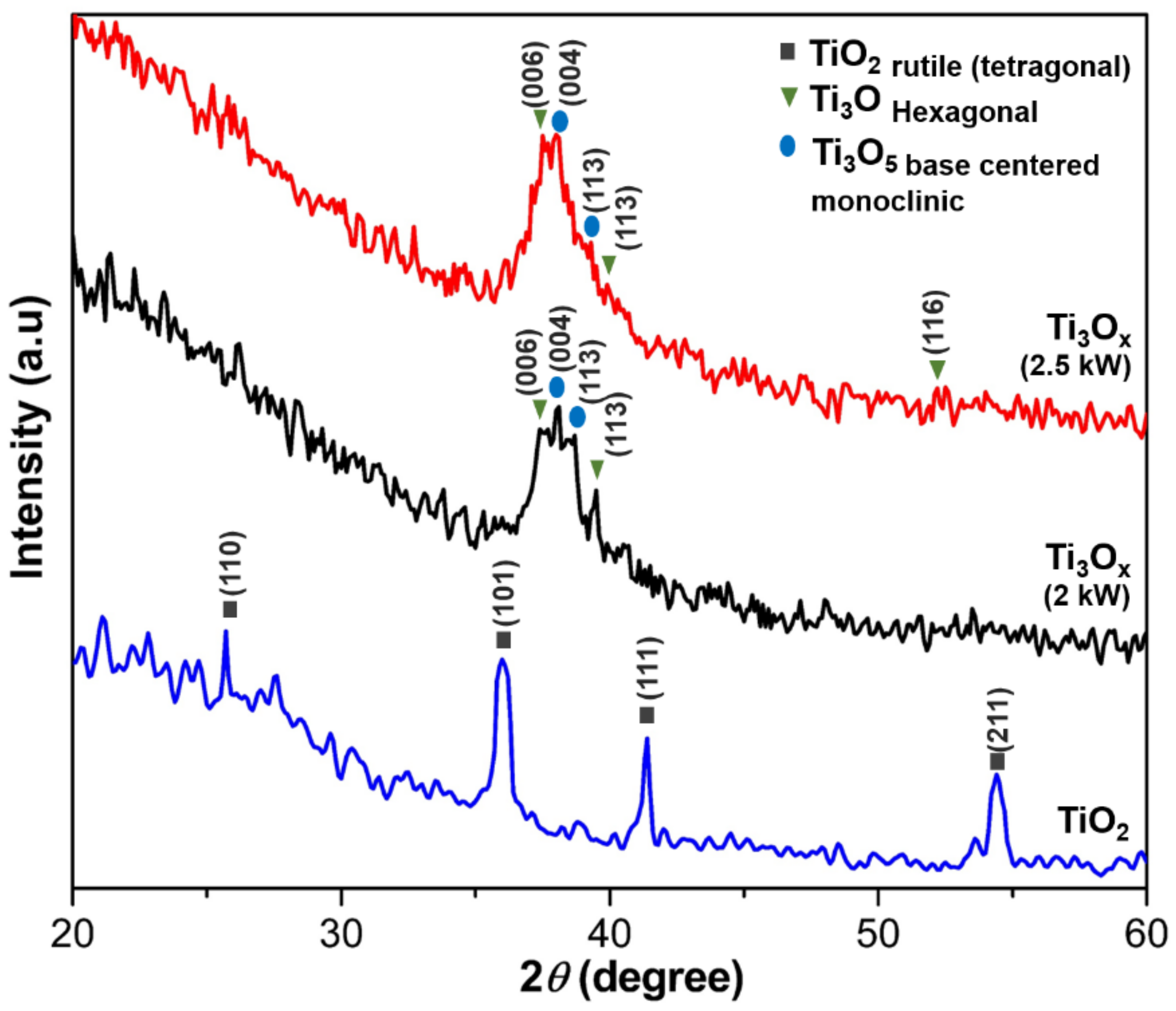
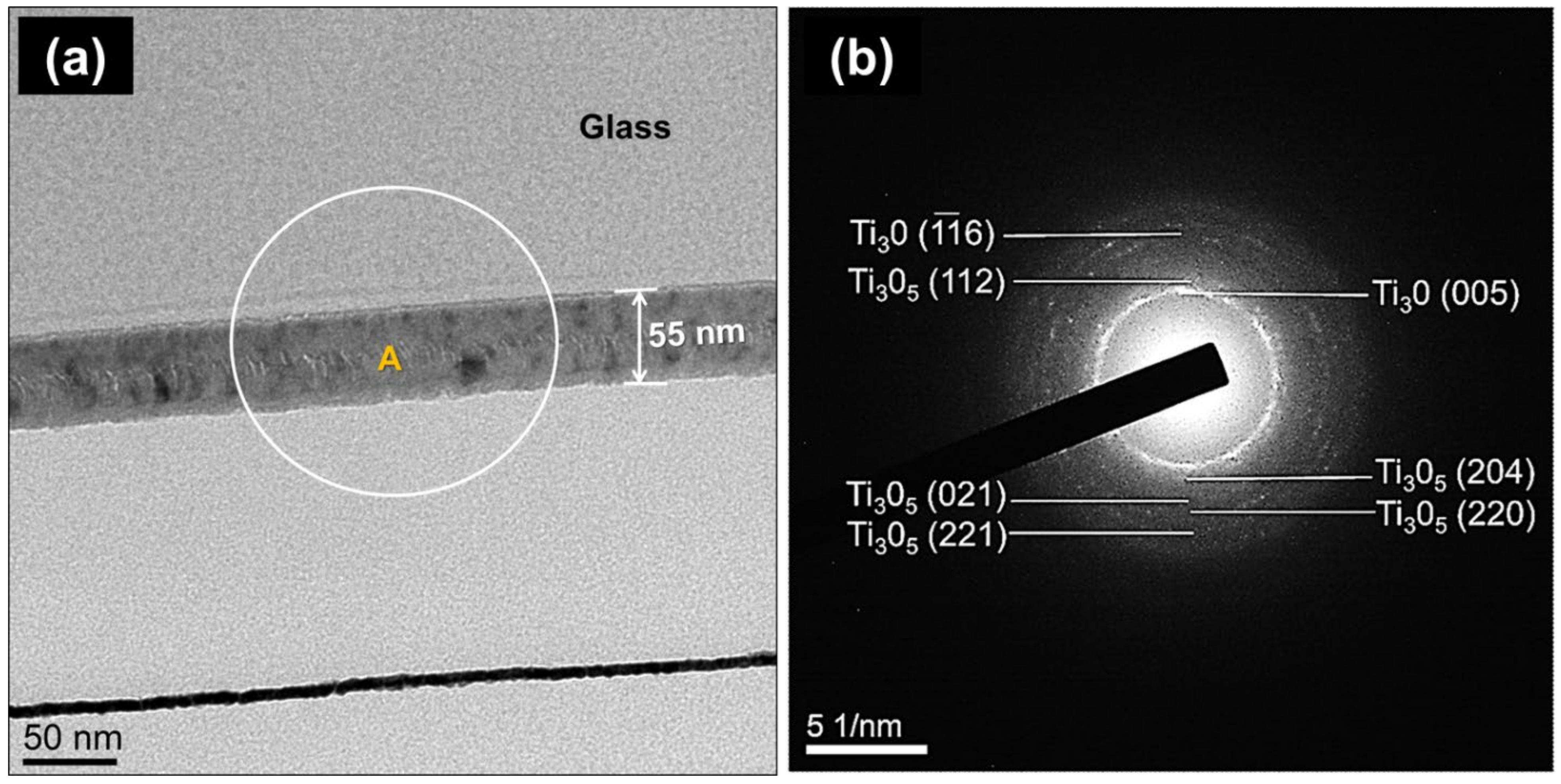
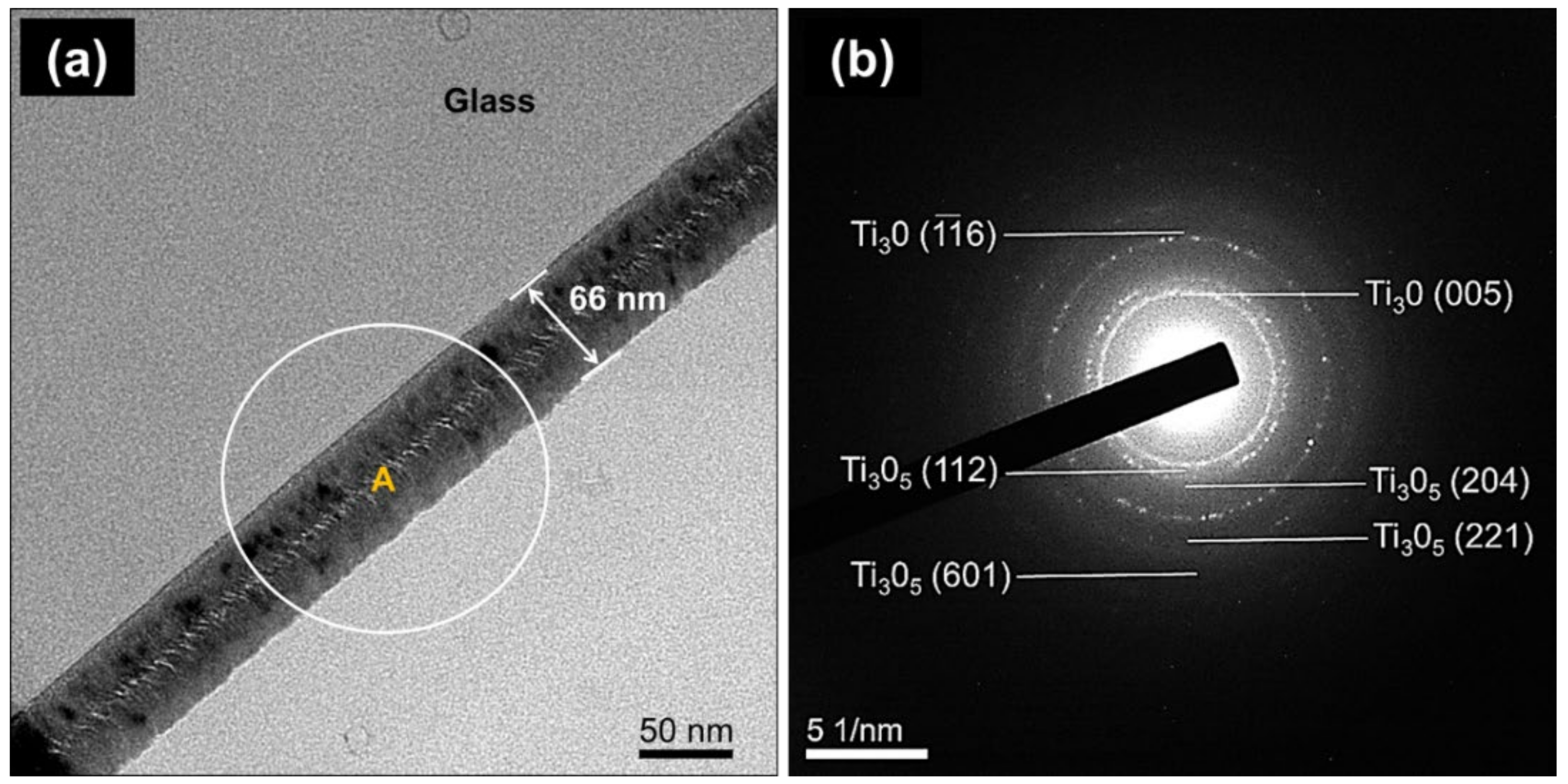
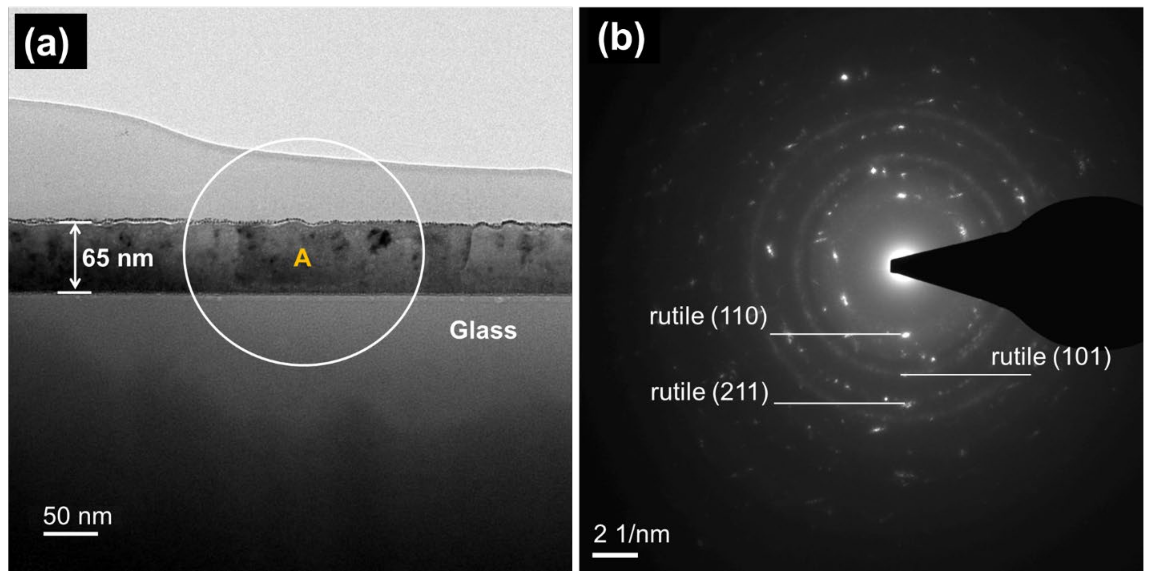
2.1. Phase Structure and Microstructure of Ti3Ox Films

The crystal phase of the Ti3Ox thin films on the glass substrate was characterized by X-ray diffraction (XRD) analysis. The TiO2 thin films were used to compare with Ti3Ox thin films samples. Figure 1 shows the XRD patterns of the Ti3Ox thin films deposited on a glass substrate without post-annealing process as a function of the sputtering power. The presence of the peaks at 2θ = 37.78°, 39.76°, and 52.30° correspond to (006), (113), and (116) plane of hexagonal Ti3O phase (JCPDS card: 76-1644), respectively. Furthermore, (004) and (113) planes resemble the monoclinic Ti3O5 phase (JCPDS card: 72-0519). Nupriyonok et al. [38] reported that Ti3O and Ti3O5 were found through the thermal vacuum treatment of Ti films. It is well known that the two essential variables affecting the crystallization of the film during the sputtering process are the thermal energy generated by the substrate heating and the energy of sputtered particles impinging on the substrate surface [25]. However, in this study, the substrates were not intentionally thermal annealing. The crystallization formed in Ti3Ox film is observed due to the impinging of energetic sputtered particles, which are under high sputtering power. However, when Ti3Ox films were annealed at 550 °C for 3 h, it was found that the Ti3O/Ti3O5 phases were transferred to the rutile TiO2 phase (JCPDS card: 21-1272).
It was observed that the peak intensity of (006) Ti3O hexagonal slightly increased as the sputtering power increased from 2 to 2.5 kW. The results indicate that Ti3Ox films can be prepared using HiPIMS under sputtering power at >2 kW and Ar/O2 ratio at 200/5 (O2 ratio: 2.5%). It was reported that Ti3O5 is one of the most promising materials for its properties of phase transition photo reversibility, photocatalysis, and oxygen sensitivity [36]. Further study of the microstructure using FE-TEM will be performed to prove this supposition.
The influence of sputtering power on the microstructure of Ti3Ox as-deposited films was confirmed using High-resolution TEM (HR-TEM). Figure 2a shows the bright cross-sectional field of Ti3Ox thin film, which is prepared at 2 kW sputtering power. The thickness of the film is approximately 55 nm. The selected zone (inset A) for electron diffraction analysis was used to identify the crystalline phase with a circle area around 200 nm. According to the electron diffraction pattern analysis, the crystalline planes belong to the Ti3O and Ti3O5 phases, as shown in Figure 2b. This observation is consistent with the XRD results, as explained in Figure 1.
Figure 3 exposes the HR-TEM micrographs and selected area diffraction (SAD) pattern of the sample with sputtering power of 2.5 kW. When the sputtering power was enhanced to 2.5 kW, it is obvious that the intensities of the diffraction rings of Ti3OX deposited at 2.5 kW were stronger than those 2 kW ones. These results implied that enhancing sputtering to 2.5 kW can promote better crystallinity of Ti3Ox thin films, as shown in Figure 3a. This finding is also in agreement with XRD analysis (Figure 1) that showed peak intensity of (006) Ti3O and (004) Ti3O5 slightly increased as the sputtering power increased from 2 to 2.5 kW. According to SAD pattern analysis in Figure 3b, the crystalline planes have referred to hexagonal Ti3O and base-centered monoclinic Ti3O5 phases. It found that the increasing sputtering power up to 2.5 kW, drives to the Ti3O and Ti3O5 crystalline intensity increased. Moreover, the intensities of the diffraction rings of (116) Ti3O plane at 2.5 kW were stronger than at 2 kW. This result is agreed with the XRD analysis (Figure 1) that showed the (116) Ti3O peak only observed on Ti3Ox thin film deposited at 2.5 kW.
When Ti3Ox films were annealed at 550 °C for 3 h, the Ti3O/Ti3O5 phases were directly transferred to the rutile TiO2 phase, as shown in Figure 4b. It shows that the films have good crystallinity. According to SAD pattern analysis, the crystalline planes (110), (211), and (101) belong to the rutile phase in the thin films. It has been reported that TiO2 films can be prepared using sputtered titanium film by thermal oxidation [4,39,40]. The single-phase rutile TiO2 can be obtained at 550 °C annealing, which agrees with the literature [28].
The influence of sputtering power on film chemical state, Ti2p, and O1s core level spectra of Ti3Ox films were detected using X-ray photoelectron spectroscopy analysis (Figure 5). Photoelectron peaks for Ti and O were clearly recorded for all samples. XPS Ti0 signals are 453.57 and 459.73 eV and Ti3+ signals are 457.1 and 463.1 eV, as shown on Figure 5a. The binding energy differences (ΔEb) were found at 6.16 and 5.87 eV for Ti0 and Ti3+, respectively, which is in agreement with the literature [16,41]. After subtracting a Shirley background, the XPS spectra were fitted to the titanium oxide component and an asymmetric line-shape function to fit the metal component. Both Ti0 (~453 eV) and Ti3+ (457.1 eV) chemical states in Ti3Ox films have referred to Ti3O and Ti3O5 phases. In addition, the binding energy curves observed at ~458 and ~463 eV correspond to Ti4+ that indicated TiO2 phases. The shifted peak positions on TiO2 films, shown in Figure 5a, indicate that the thermal annealing shifted the binding energy to higher values, effected by surface oxidation, and this effect ascribed to the O-Ti bonding [42].
Moreover, the increasing peak intensity of Ti0 chemical state correlated with high metallic titanium when the sputtering power is increased from 2 to 2.5 kW. Godfroid et al. published that the films deposited at the high sputtering power attain the metallic state more rapidly [16].
Figure 5b shows an asymmetric high binding energy curve for O1s lines with an intense peak at 529 eV, which is in good agreement with a previous report [43]. The co-existence of Ti3O and Ti3O5 phases in Ti3Ox films was observed using HiPIMS at 2 kW and 2.5 kW with a low oxygen ratio (2.5%), indicating the Ti3Ox films can be prepared at higher sputtering power and lower oxygen concentration. It is found that lower oxygen concentration plays a role in the co-existence of Ti3O and Ti3O5 crystal phases. However, the O1s peak intensity was decreased significantly when the sputtering power increased from 2.0 kW to 2.5 kW.
The chemical composition of Ti3Ox films at different sputtering power was analyzed based on XPS data. The concentrations of titanium and oxygen are listed in Table 1. Ti to O proportion at different sputtering powers is 0.95 and 1.20 for 2 kV and 2.5 kV, respectively. The results indicate that the oxygen concentration in Ti3Ox films was decreased with increasing sputtering power up to 2.5 kW. It means that the Ti3O phase increased at 2.5 kV in the films. This result is consistent with the XRD and TEM analysis, as exhibited in Figure 1 and Figure 3. Increasing the sputtering power up to 2.5 kW leads to shifting the binding energy to lower energy due to lower oxidation state changes in the Ti-O element. Furthermore, the atomic concentration of Ti and O in the TiO2 films was 29.94 and 70.06%, respectively. Ti to O proportion is 0.42. More increased oxygen existence in TiO2 films was expected after thermal annealing [44].

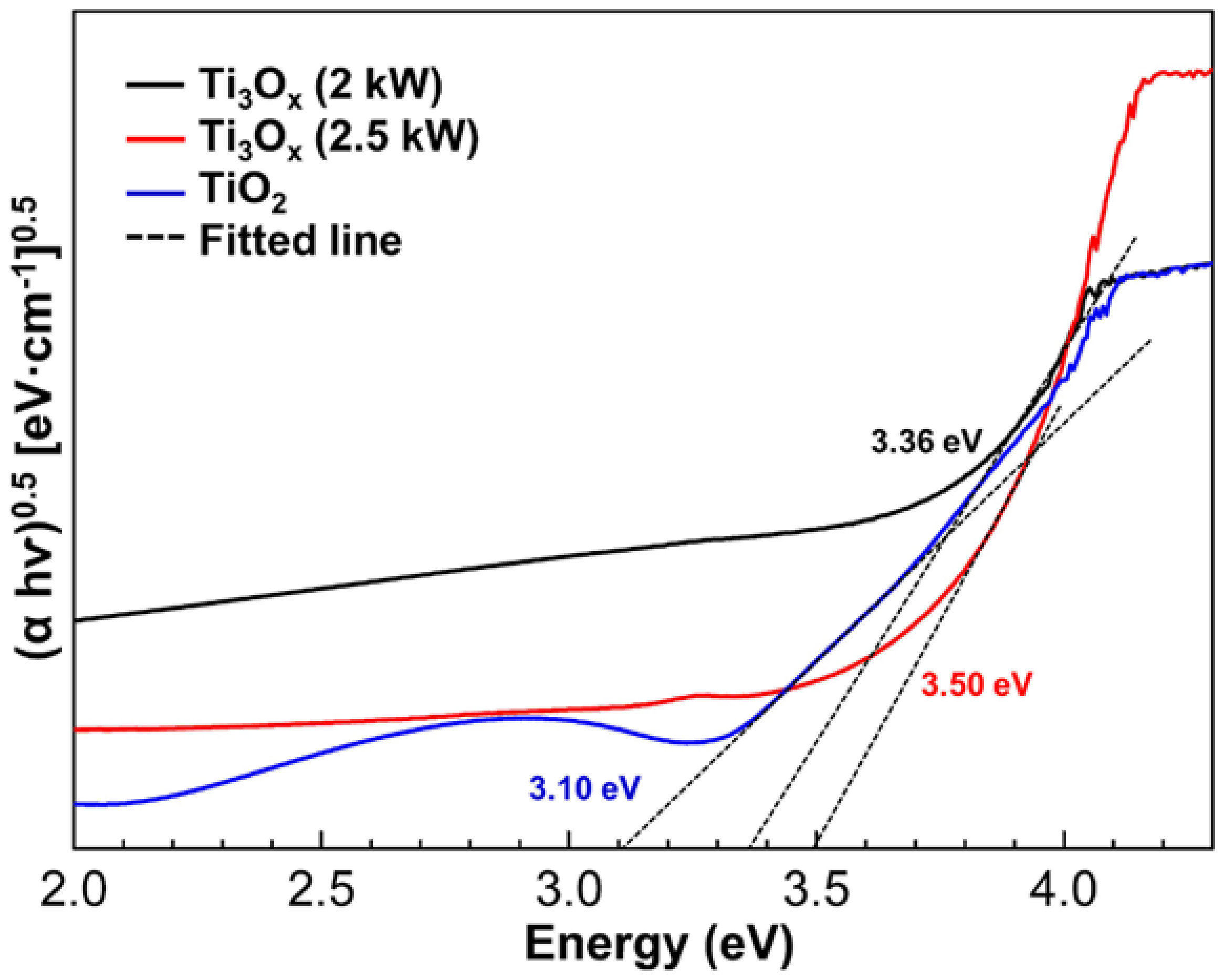
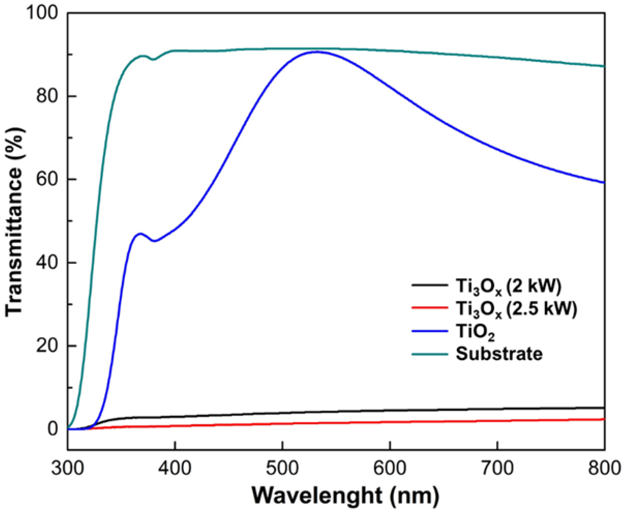
2.2. Optical Properties and Photocatalytic Activity of Ti3Ox Films

It is challenging to know the energy bandgap value for the Ti3Ox films, while TiO2 has an indirect bandgap with a value around 3.0–3.2 eV [24]. However, Ti3O and Ti3O5 are still unknown. The indirect bandgap α   h v = A   h v E g 0.5 was used to calculate the energy bandgap of Ti3Ox films. The energy bandgap of TiO2 films was used as a reference to compare with Ti3Ox films. The energy bandgap of the Ti3Ox films with different sputtering power is shown in Figure 6. The bandgap values of Ti3Ox films are 3.36 and 3.50 eV for 2 and 2.5 kW, respectively. In this study, increasing the sputtering power up to 2.5 kW will lead to a monotonically increasing energy bandgap.
Indirect optical band gap in the range 3.58–3.75 eV with an increase in sputtering pressure in TiO2 thin films being reported earlier by Prabitha et al. [45]. It is well known that the energy bandgap refers to the minimum energy needed to excite an electron from the bottom of the valence band to the top of the conduction band. The lower energy bandgap will lead to electrons excited into the conduction band from the valence band. However, it was found that the energy bandgap of Ti3Ox films is higher than TiO2 films (3.10 eV). Generally, the energy bandgap (Eg) of TiO2 varies with its structure, with the Eg of crystalline anatase and rutile phase being 3.2 and 3.0 eV, respectively [46].
The optical transmittance spectra of as-deposited Ti3Ox films were measured using UV–Vis spectroscopy. Transmittance measurements as a function of wavelength are illustrated in Figure 7. Note that the TiO2 films with 66 nm thickness were used as reference. The Ti3Ox films exhibit low transmission in the spectral range 300–800 nm wavelength compared to TiO2 films. The optical transmission spectra of the Ti3Ox films showed feeble transmittance (<10%), while the sample with high sputtering power (2.5 kW) pointed almost no light transmission. TiO2 films with equal thickness are highly transparent in the visible region, around 90%, and are relatively equal to glass substrate transmission spectra. According to microstructure analysis (Figure 2 and Figure 3) of Ti3Ox films, the intermixing of Ti3O and Ti3O5 crystalline phases was observed in the films. These crystalline phases imply a lack of oxygen content in the films that causes poor light transmission. Moreover, the transmission of thin films correlates with the energy bandgap value. The bandgap of Ti3Ox value is higher than TiO2 thin films created the light was absorbed not transmitted [39]. It has been reported that the oxygen vacancies in TiO2 film enhanced the absorption of visible light [25].
The photocatalytic activities of Ti3Ox films deposited at 2 and 2.5 kW were investigated by degradation of methylene blue solution as shown in Figure 8. When the samples were irradiated by UV light, the absorbance of MB decreased, which indicates that the films performed photocatalytic properties. TiO2 films with similar thickness and preparation were used to compare the photocatalytic activity. It can be observed that all the samples exhibited quick absorption in the dark. This could be attributed to the high specific surface and small particle size [47] as shown in Figure 8a, The methylene blue degradation of the substrate as control was ~18% (Figure 8a). After UV irradiation, the samples show has a good degradation performance with a range of 51–61% compared to the substrate.
The degradation percentage of methylene blue solution with Ti3Ox thin films with 2.5 kW sputtering power has higher photocatalytic activity than 2 kW. According to the HR-TEM and XRD analysis, Ti3O and Ti3O5 crystalline phases co-existed in the films, and the crystalline phases increased with increasing sputter power, as displayed in Figure 3. Interestingly, the Ti3Ox films with 2.5 kW sputtering power have a higher photocatalytic activity (61.0%) compared with TiO2 films (55.9%) at a similar thickness. This indicates that the Ti3Ox phases exhibit higher photocatalytic activity than TiO2 (rutile) films at similar thickness. Qi et al. [36] reported that UV light could stimulate the Ti3O5 to produce electron–hole pairs and help redox reactions. The photogenerated electrons will interact with molecular oxygen (O2) to generate superoxide radical anions (O2-), and the photo-generated holes react with water to form hydroxyl (·OH) radicals. Ti3Ox films prepared by this HiPIMS technique at 2.5 kW and low oxygen ratio (2.5%) demonstrated good photocatalytic activity.
According to first-order reaction kinetics (as shown in Figure 8b), the time-dependent ln(C/C0) terms are illustrated for all the film samples. The reaction rate constants (k) of the Ti3Ox films and TiO2 were calculated: Ti3Ox films at 2 kW, 2.5 kW, TiO2 film were 3.42 × 10⁻³ min−1,4.67 × 10−3 min−1, and 4.05 × 10⁻³ min−1, respectively. These results explain that a higher degradation rate can be obtained for Ti3Ox films sputtered at 2.5 kW. The reaction constants (k) gradually increased with an increasing the Ti3Ox film thickness. Influencing significant effects on the electronic, photonic, and photocatalytic properties of TiO2, oxygen deficiencies are critical features [48,49,50].
The schematic diagram of photocatalytic reaction for MB removal by Ti3Ox thin films is illustrated in Figure 9. The valence bandgap (vb) and conduction band (cb) of Ti3Ox thin films around 3.5 eV. When Ti3Ox is exposed to energy light (hv) that is larger than the thin film’s energy bandgap, the electron (e) in the vb will migrate to the cb, retaining a positive hole (h+) in the vb. These exciting holes and electrons have high energy potentials that can transfer to the semiconductor’s surface, producing strong reactive free radicals or recombining. Water (H2O) and oxygen (O2) molecules will be trapped on the semiconductor surface by an exciting hole in vb and an excited electron in cb, respectively. Finally, trapped H2O is oxidized into highly reactive hydroxyl radicals (OH•) at vb, whereas oxygen is reduced into superoxide radical ion (O2) at cb. These processes may accelerate the photocatalytic process and MB degradation [51].
Film thickness and energy bandgap are two key factors determining the photocatalytic performance of Ti3Ox thin films. The energy bandgap of Ti3Ox thin films is ~3.5 eV, which is higher than the rutile (3.0 eV) or anatase (3.2 eV) phases. According to Qutub et al. [52], the photocatalytic activity of semiconductor particles in aqueous solutions is conducted by electrons and holes produced by photo-excitation. As electrons prefer to recombine with positive holes, the photogenerated electrons should be efficiently separated from the holes to enhance photocatalytic efficiency. One of the effective approaches is to increase the semiconductor catalyst’s bandgap, which minimizes the recombination of electrons and holes. In addition, a higher bandgap also corresponds to promoting high redox ability [53]. As the Ti3Ox has a higher bandgap than rutile TiO2, its oxidizing ability should be stronger. Therefore, Ti3Ox has a higher energy bandgap promoting better photocatalytic performance than rutile TiO2 thin films. However, the Ti3Ox deposited at 2 kW showed lower photocatalytic activity than TiO2 even though it has a higher energy bandgap (3.36 eV). A possible reason is that the film thickness of Ti3Ox deposited at 2 kW is lower than TiO2 thin films.
These outcomes indicated that increasing the deposition power up to 2.5 kW can promote high thickness of Ti3Ox thin films with energy band gap of ~3.5 eV thus showing high photocatalytic performance. Furthermore, semiconducting photocatalysts mainly depend upon separating a photogenerated hole–electron pairs and transferring electrons from the photocatalyst into the organic pollutants within the oxygen vacancy defects on the surface [54,55].

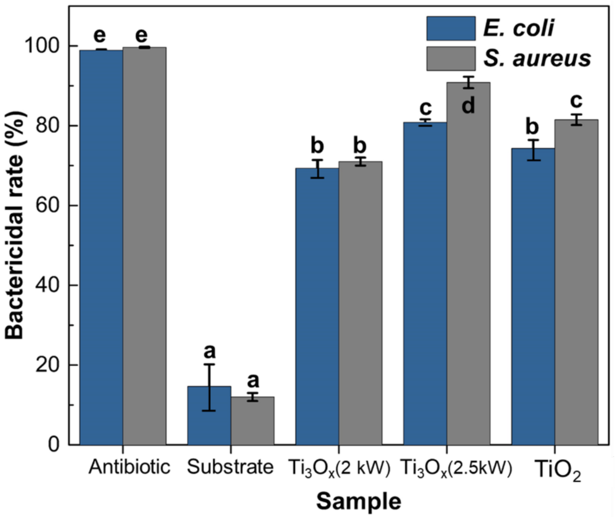
2.3. Antibacterial Activity of Ti3Ox Films

The results of the antibacterial activity of E. coli (Gram-negative) and S. aureus (Gram-positive) on Ti3Ox thin films compared to glass substrate and antibiotic are presented in Figure 10. The initial concentration of bacteria was 1 × 10⁵ CFU/mL. The glass substrate under UV light only showed a minor antibacterial activity on E. coli and S. aureus (less than 16%). Instead, the group treated with an antibiotic (100 μg/mL) as positive control showed high antibacterial inhibition for both bacteria. Notably, two kinds of Ti3Ox thin-film presented significant antibacterial effects against E. coli and S. aureus (p < 0.05) with bactericidal rates around 80 and 90% in the presence of UV light, respectively (Figure 9).
It could be seen that the bactericidal rate for both E. coli and S. aureus significantly increases remarkably with sputtering power increases up to 2.5 kW after 60 min of UV irradiation. The antibacterial effect of Ti3Ox thin films on E. coli and S. aureus can be ascribed both to the UV irradiation and photocatalytic activity of Ti3Ox thin films containing crystal structures of Ti3O5 and Ti3O. The thin films fabricated with higher sputtering power (2.5 kW) showed a higher bactericidal rate. The increasing thickness of Ti3Ox thin films is effective for the inactivation of both Gram-positive and Gram-negative bacteria, and the antibacterial ability of the films Ti3Ox (2.5 kW) is even better than the rutile TiO2 film (74%). The results indicate that the antimicrobial activity depended on the UV irradiation and sputtering power of Ti3Ox thin films.
Numerous studies have reported the antibacterial activity of TiO2 thin films. However, there has been limited published work that refers to the antibacterial mechanism on Ti3Ox thin films. According to Sunada et al. [56], the bactericidal process comprises low-rate photo-killing and high-rate photo-killing stages. The low-rate photo-killing stage is the partial destruction of the outer membrane of bacteria through robust reactive oxygen species (ROS) produced by TiO2. Several investigations have shown that ROS formation is the primary mechanism responsible for the antibacterial activities of TiO2-based materials [56,57,58,59]. Generation of reactive oxygen species (ROS) occurs due to the induction of electron–hole pairs under light radiation, which recombine and release energy as heat or dissociate due to the charge trapping, thus creating charge carriers for redox reactions in the photocatalytic process.
Photoexcited electron–hole pairs interact with adsorbed electron donors and acceptors (water and oxygen) on the TiO2 surface to generate highly reactive hydroxyl radicals (OH•) and superoxide ions (O2) during the photocatalytic process as described in Figure 9. These two reactive oxygen species are strong oxidants that can degrade and oxidize organic material, including bacteria. This process has significant effect on cell mortality. Furthermore, it alters cell permeability, allowing ROS to be more easily accessible to the cell membrane. As a result, ROS attach to the cell membrane, accelerating the peroxidation of the cell membrane’s polyunsaturated phospholipid layer. This process will result in the loss of respiratory activity and the leaking of cell components, which will eventually lead to cell death [60,61]. In addition, the formation of such Magnéli phase in a previous study could contribute to the enhanced antibacterial properties observed under irradiation light [31].
Moreover, the bactericidal activity on Ti3Ox thin films is statistically significantly higher on S. aureus (90.83%) than E. coli (80.83%), as presented in Figure 10. According to Russell [62], Gram-negative bacteria such as E. coli are less sensitive to ROS than Gram-positive bacteria (S. aureus). The structural differences in the bacterial cell membrane are one of the primary causes of the increased resistance. Cell membranes of Gram-positive bacteria are covered by a membrane predominantly composed of a peptidoglycan layer and teichoic and lipoteichoic acids. Gram-positive bacteria have a membrane surrounding the cell wall that is composed primarily of a thick peptidoglycan layer and teichoic and lipoteichoic acids but no outer lipid membrane. However, the cell membrane of Gram-negative bacteria is more complex due to an outer membrane composed mainly of lipopolysaccharide (LPS) and a thin peptidoglycan layer [63]. The outer membrane serves as the first barrier, and once it is breached, the cytoplasmic membrane is targeted, resulting in a reduction in cellular respiration and, finally, the death of cells. Consequently, the outer membrane of Gram-negative bacteria works as a permeability barrier, reducing the absorption of reactive oxygen species (ROS) into the cell [62].
These results indicated that deposition power plays crucial parameters for obtaining high photocatalytic performance and antibacterial activity of Ti3Ox films from our studies. Furthermore, sputtering power at 2.5 kW could provide a high thickness of Ti3Ox films with an energy bandgap of ~3.4 eV and exhibited higher photocatalytic activity than TiO2 thin films. Thus, the study demonstrated that the intermixing of the Ti3O and Ti3O5 phase on thin-film could promote a high photocatalytic and antibacterial activity that could be considered as an efficient and lasting antibacterial material for future antibacterial and biomedical application.

3. Materials and Methods

3.1. Preparation of Ti3Ox Thin Films

The Ti3Ox thin films were deposited onto the glass substrate using reactive HiPIMS. Glass substrate size was 76 × 25 mm and ultrasonically cleaned in acetone, ethanol, and deionized water for 30 min in sequence before the deposition process. Then, it was blown dry with air and put on holders. Titanium layers were deposited at sputtering power 2 and 2.5 kW using a 99.99% purity titanium target (Ultimate materials Technology Co., Ltd., Hsinchu, Taiwan) with dimensions 550 × 125 mm and thickness 6 mm. The target to substrate distance was kept constant at 150 mm. High-purity oxygen (99.99%) and argon (99.99%) were used as reactive and working gas, respectively. Reactive sputter deposition was operated at 2.7 × 10⁻³ Torr (0.36 pa) working pressure, the substrate temperature was controlled at 25 °C, and the flow rate of argon and oxygen was fixed at 200 and 5 sccm, respectively. All Ti3Ox thin film samples were prepared without a post-annealing process. Moreover, the TiO2 thin films sample was developed as a comparable sample for Ti3Ox thin films. TiO2 thin films with thickness around 65 nm were prepared from Ti3Ox thin films (2.5 kW; 5 sccm) samples that continue with post-annealing at 550 °C for 3 h. A summary of the parameters and deposition conditions of Ti3Ox thin film is given in Table 2.

3.2. Sample Characterization

The crystal phase was determined using X-ray diffraction (XRD; Bruker D8 Advance- AXS Gmbh, Am Studio 2D, Berlin, Germany) with monochromatic Cu-Kα radiation (λ = 1.541 Å), operated at 40 kV and 40 mA. The 2θ scan range is between 10° ≤ 2θ ≤ 60° with a grazing angle of 0.1° and a scan speed of 5 s. Microstructural and film thickness observation of the cross-sectional and plane-view morphology of the films deposited onto a glass substrate was observed using a Field Emission Gun Transmission Electron Microscopy (FEG-TEM; FEI E.O Tecnai F20, FEI Company—Thermo Fisher Scientific, Hillsboro, OR, USA) equipped with charge-coupled device (CCD) camera. Selected area diffraction (SAD) was employed at an acceleration voltage of 200 kV. The evaluation of high-resolution TEM (HR-TEM) and SAD patterns was conducted using the Gatan Digital Micrograph software package (version 2.11.1404.0) (Gatan Inc., Pleasanton, CA, USA). The interplanar distance in the real spaces (d-spacing) that were observed from software was compared with the crystallographic database of the Ti3O and Ti3O5 planes from JCPDS. The films’ quantitative composition on the surface and internal area was examined with X-ray photoelectron spectroscopy (XPS; JEOL, JPS-9010MX, JEOL Ltd, Tokyo, Japan). Internal profile experiments were performed by sputter etching rate 28 nm/min in the thin film for 20 s. Then, the analyzer was operated at voltage 10 kV and current 5 mA with Mg Kα radiation line. The X-ray photoelectron spectra were referenced to the C 1s peak (Eb = 285 eV) present on the sample surface.

3.3. Optical Properties Characterization

The absorbance and transmittance of Ti3Ox thin films were determined using UV-vis spectrophotometer (UV/Vis, U-3310, Hitachi Ltd., Tokyo, Japan) in the range wavelength 200–1000 nm and 300–800 nm, respectively. The energy bandgap of the thin films was estimated from the Tauc plots of the optical absorbance. The determination of the energy bandgap was obtained by Tauc’s Equation [64,65] Equation (1):
α   h v = A   h v E g n
where α is the absorbed coefficient, h is the Planck constant, ν is the photon’s frequency, A is a constant proportionality, Eg is the allowed energy gap, and n = 0.5 for allowed direct transition.
The photocatalytic activity of thin films was evaluated by measuring the degradation of Methylene Blue (MB Alfa Aesar; Thermo Fisher Scientific, Lancashire, UK) in the presence of UV light. Ti3Ox Thin-film samples with size 26 mm × 38 mm were immersed into 20 mL aqueous MB solution with an initial concentration (C) of 10 mg/L then irradiated using UV lamp 8 W (UV T8; Philip, Amsterdam, The Netherlands) for 3 h. The UV source was 9 cm from the thin films. Prior to UV radiation, the film was immersed in MB solution and kept under dark conditions for 10 min to achieve MB absorption equilibrium on the Ti3Ox and TiO2 layer. From the end of the dark-storage time (t = 0 min, C0), a sample of dye solution was analyzed every 30 min for 3 h (t = 30–180 min, C30 to C180). The Ti3Ox film photocatalytic activity was determined using UV–Vis spectrophotometer (UV/Vis, U-3310, Hitachi Ltd., Tokyo, Japan) based on the change in MB concentration following the UV irradiation time (the maximum absorption wavelength of the dye at 664 nm), as described in a previous report [4,28,66]. The degradation of methylene blue was estimated by calculating the percentage of the dye concentration that remained over (C0) the initial dye concentration (C), obtained from the absorbance standard curve. The degradation performance evaluated by considering the following Equation (2):
M e t h y l e n e   B l u e   D e g r a d a t i o n   % = 1 C 0 C × 100
The initial and final concentrations of methylene blue (mg/L) at reaction time (t) were C and C0, respectively. In the pseudo-first-order photodegradation kinetics, the ln(C/C0) linear fit slope in the function of time plot represents the kinetic constant (k) of the photodegradation, which can be found in Equation (3):
ln C C 0 = k t

3.4. Antibacterial Activity

The antibacterial activity of the Ti3Ox thin films was evaluated by the agar dilution method [67] against Gram-negative Escherichia coli (ATCC25922) and Gram-positive bacteria Staphylococcus aureus (ATCC10234). The bacteria were grown in Tryptic Soy Broth (DIFCOtm, Becton Dickirnson and Company sparks, Maryland, USA) at 37 °C for 12 h. The initial cell concentration used for antibacterial activity was about 1 × 10⁵ colony-forming units (CFU)/mL. Thin films samples with dimensions 1 × 1 cm were placed in sterilized 12 welled plates (SPL Life Sciences Co., Ltd., Gyeonggi-do, Korea). Furthermore, 40 μL of the bacterial solution was dropped on the surface of the thin films. The plates were sealed then UV-irradiated (UV-A, 8 W T8; λ = 365 nm, Philip, Amsterdam, The Netherlands) for 60 min at room temperature (25 ± 2 °C). After finishing, 10 μL of the remaining sample on the thin-film was taken, and serial dilution was performed. The aliquots of serially diluted suspensions were plated on Tryptic Soy Agar (DIFCOtm, Becton Dickirnson and Company sparks, MD, USA) plates and incubated at 37 °C for 24 h, and the number of colonies on the plates was counted. In order to compare antibacterial activity, substrate (glass) and antibiotic (100 μg/mL, kanamycin and ampicillin, Sigma-Aldrich, MO, USA)) were used as a negative and positive control. The same treatment was done under dark conditions. The experiment was done in triplicate. All of the experiment was conducted in a sterile environment. The bactericidal rate (K) was calculated by Equation (4):
K = A B A × 100
A and B are the numbers of original bacteria colonies grown in the culture medium corresponding to the sample after irradiation with UV light, respectively.
One-way analysis of variance (ANOVA) was performed by using ‘SPSS Statistics’ version 16 software (SPSS for Windows; IBM Corp, New York, NY, USA). Post hoc testing was done using Tukey HSD tests, which were done to determine significant group differences in antibacterial analysis, and means were considered statistically significant if p < 0.05. Data were expressed as mean ± standard deviation.

4. Conclusions

The Ti3Ox films can be prepared using the reactive High-Power Impulse Magnetron Sputtering (HiPIMS) method without post-thermal annealing. There are two phases (Ti3O and Ti3O5) that co-existed in the Ti3Ox films. The sputtering power played a role in influencing the structure, morphology, optical, and photocatalytic properties of Ti3Ox thin films. It was found that increasing the sputtering power up to 2.5 kW leads to increased crystalline intensity of Ti3O and Ti3O5. The Ti3Ox films with 2.5 kW sputtering power could promote higher photocatalytic and antibacterial activity against Gram-negative and Gram-positive bacteria compared with TiO2 films. Therefore, the Ti3Ox thin film photocatalyst is an attractive and long-lasting antibacterial material appropriate to future biomedical and bactericidal applications.

Author Contributions

Conceptualization, Y.-C.L. and J.-L.H.; methodology, F.-Y.X.; software, J.-H.J.; validation, C.-T.C.; formal analysis, E.W. and F.-Y.X.; investigation, E.W.; resources, Y.-C.L. and J.-L.H.; data curation, C.-T.C.; writing—original draft preparation, E.W.; writing—review and editing, E.W. and Y.-C.L.; visualization, E.W. and J.-H.J.; supervision, Y.-C.L. and J.-L.H.; funding acquisition, Y.-C.L. All authors have read and agreed to the published version of the manuscript.

Funding

This research received no external funding.

Data Availability Statement

Data are contained within the article.

Acknowledgments

The author wishes to thank the TaiwanICDF for Ph.D. scholarship funding; Douglas J.H. Shyu and Liyu Chiang from Dept. Biological Science and technology, NPUST, Taiwan, for providing a bacterial culture; and the National Sun Yat-sen University staff for assistance with XPS (Instrument ID: ESCA00002100) experiments. This research did not receive any specific grant from funding agencies in agencies in the public, commercial, or non-profit sectors.

Conflicts of Interest

The authors declare no conflict of interest.

References

  1. Huang, C.Y.; Selvaraj, P.; Senguttuvan, G.; Hsu, C.J. Electro-optical and dielectric properties of TiO2 nanoparticles in nematic liquid crystals with high dielectric anisotropy. J. Mol. Liq. 2019, 286, 110902. [Google Scholar] [CrossRef]
  2. Banerjee, S.; Dionysiou, D.D.; Pillai, S.C. Self-cleaning applications of TiO2 by photo-induced hydrophilicity and photocatalysis. Appl. Catal. B Environ. 2015, 176, 396–428. [Google Scholar] [CrossRef] [Green Version]
  3. Kathirvel, S.; Pedaballi, S.; Su, C.; Chen, B.-R.; Li, W.-R. Morphological control of TiO2 nanocrystals by solvothermal synthesis for dye-sensitized solar cell applications. Appl. Surf. Sci. 2020, 519, 146082. [Google Scholar] [CrossRef]
  4. Lee, P.-Y.; Widyastuti, E.; Lin, T.-C.; Chiu, C.-T.; Xu, F.-Y.; Tseng, Y.-T.; Lee, Y.-C. The phase evolution and photocatalytic properties of a Ti-TiO2 bilayer thin film prepared using thermal oxidation. Coatings 2021, 11, 808. [Google Scholar] [CrossRef]
  5. Lu, Y.; Matsuda, Y.; Sagara, K.; Hao, L.; Otomitsu, T.; Yoshida, H. Fabrication and Thermoelectric Properties of Magneli Phases by Adding Ti into TiO2. In Proceedings of the Advanced Materials Research; Trans Tech Publications Ltd: Zurich, Switzerland, 2012; Volume 415, pp. 1291–1296. [Google Scholar]
  6. Huang, S.-S.; Lin, Y.-H.; Chuang, W.; Shao, P.-S.; Chuang, C.-H.; Lee, J.-F.; Lu, M.-L.; Weng, Y.-T.; Wu, N.-L. Synthesis of high-performance titanium sub-oxides for electrochemical applications using combination of sol–gel and vacuum-carbothermic processes. ACS Sustain. Chem. Eng. 2018, 6, 3162–3168. [Google Scholar] [CrossRef]
  7. Xu, B.; Sohn, H.Y.; Mohassab, Y.; Lan, Y. Structures, preparation and applications of titanium suboxides. RSC Adv. 2016, 6, 79706–79722. [Google Scholar] [CrossRef]
  8. Pabón, B.M.; Beltrán, J.I.; Sánchez-Santolino, G.; Palacio, I.; López-Sánchez, J.; Rubio-Zuazo, J.; Rojo, J.M.; Ferrer, P.; Mascaraque, A.; Muñoz, M.C. Formation of titanium monoxide (001) single-crystalline thin film induced by ion bombardment of titanium dioxide (110). Nat. Commun. 2015, 6, 1–6. [Google Scholar] [CrossRef] [Green Version]
  9. Fusi, M.; Maccallini, E.; Caruso, T.; Casari, C.S.; Bassi, A.L.; Bottani, C.E.; Rudolf, P.; Prince, K.C.; Agostino, R.G. Surface electronic and structural properties of nanostructured titanium oxide grown by pulsed laser deposition. Surf. Sci. 2011, 605, 333–340. [Google Scholar] [CrossRef] [Green Version]
  10. Lackner, J.M.; Waldhauser, W.; Ebner, R.; Major, B.; Schöberl, T. Pulsed laser deposition of titanium oxide coatings at room temperature—Structural, mechanical and tribological properties. Surf. Coatings Technol. 2004, 180, 585–590. [Google Scholar] [CrossRef]
  11. Farstad, M.H.; Ragazzon, D.; Grönbeck, H.; Strømsheim, M.D.; Stavrakas, C.; Gustafson, J.; Sandell, A.; Borg, A. TiOx thin films grown on Pd (100) and Pd (111) by chemical vapor deposition. Surf. Sci. 2016, 649, 80–89. [Google Scholar] [CrossRef]
  12. Greczynski, G.; Petrov, I.; Greene, J.E.; Hultman, L. Paradigm shift in thin-film growth by magnetron sputtering: From gas-ion to metal-ion irradiation of the growing film. J. Vac. Sci. Technol. A 2019, 37, 60801. [Google Scholar] [CrossRef] [Green Version]
  13. Horprathum, M.; Eiamchai, P.; Chindaudom, P.; Pokaipisit, A.; Limsuwan, P. Oxygen partial pressure dependence of the properties of TiO2 thin films deposited by DC reactive magnetron sputtering. Procedia Eng. 2012, 32, 676–682. [Google Scholar] [CrossRef] [Green Version]
  14. Okimura, K. Low temperature growth of rutile TiO2 films in modified rf magnetron sputtering. Surf. Coatings Technol. 2001, 135, 286–290. [Google Scholar] [CrossRef]
  15. Gouttebaron, R.; Cornelissen, D.; Snyders, R.; Dauchot, J.P.; Wautelet, M.; Hecq, M. XPS study of TiOx thin films prepared by dc magnetron sputtering in Ar–O2 gas mixtures. Surf. Interface Anal. 2000, 30, 527–530. [Google Scholar] [CrossRef]
  16. Godfroid, T.; Gouttebaron, R.; Dauchot, J.P.; Leclere, P.; Lazzaroni, R.; Hecq, M. Growth of ultrathin Ti films deposited on SnO2 by magnetron sputtering. Thin Solid Films 2003, 437, 57–62. [Google Scholar] [CrossRef]
  17. Tiron, V.; Velicu, I.-L.; Dobromir, M.; Demeter, A.; Samoila, F.; Ursu, C.; Sirghi, L. Reactive multi-pulse HiPIMS deposition of oxygen-deficient TiOx thin films. Thin Solid Films 2016, 603, 255–261. [Google Scholar] [CrossRef]
  18. Greczynski, G.; Mráz, S.; Hans, M.; Primetzhofer, D.; Lu, J.; Hultman, L.; Schneider, J.M. Unprecedented Al supersaturation in single-phase rock salt structure VAlN films by Al+ subplantation. J. Appl. Phys. 2017, 121, 171907. [Google Scholar] [CrossRef] [Green Version]
  19. Zauner, L.; Ertelthaler, P.; Wojcik, T.; Bolvardi, H.; Kolozsvári, S.; Mayrhofer, P.H.; Riedl, H. Reactive HiPIMS deposition of Ti-Al-N: Influence of the deposition parameters on the cubic to hexagonal phase transition. Surf. Coatings Technol. 2020, 382, 125007. [Google Scholar] [CrossRef]
  20. Mareš, P.; Dubau, M.; Polášek, J.; Mates, T.; Kozák, T.; Vyskočil, J. High deposition rate films prepared by reactive HiPIMS. Vacuum 2021, 191, 110329. [Google Scholar] [CrossRef]
  21. Depla, D.; Heirwegh, S.; Mahieu, S.; De Gryse, R. Towards a more complete model for reactive magnetron sputtering. J. Phys. D Appl. Phys. 2007, 40, 1957. [Google Scholar] [CrossRef]
  22. Lou, B.-S.; Yang, Y.-C.; Qiu, Y.-X.; Diyatmika, W.; Lee, J.-W. Hybrid high power impulse and radio frequency magnetron sputtering system for TiCrSiN thin film depositions: Plasma characteristics and film properties. Surf. Coatings Technol. 2018, 350, 762–772. [Google Scholar] [CrossRef]
  23. Pansila, P.; Witit-Anun, N.; Chaiyakun, S. Influence of sputtering power on structure and photocatalyst properties of DC magnetron sputtered TiO2 thin film. Procedia Eng. 2012, 32, 862–867. [Google Scholar] [CrossRef] [Green Version]
  24. Kamble, S.S.; Radhakrishnan, J.K. Influence of O2 flow rate on the characteristics of TiO2 thin films deposited by RF reactive sputtering. Mater. Today Proc. 2020, 33, 709–715. [Google Scholar] [CrossRef]
  25. Sério, S.; Jorge, M.E.M.; Maneira, M.J.P.; Nunes, Y. Influence of O2 partial pressure on the growth of nanostructured anatase phase TiO2 thin films prepared by DC reactive magnetron sputtering. Mater. Chem. Phys. 2011, 126, 73–81. [Google Scholar] [CrossRef]
  26. Fan, Y.; Zhang, C.; Liu, X.; Lin, Y.; Gao, G.; Ma, C.; Yin, Y.; Li, X. Structure and transport properties of titanium oxide (Ti2O, TiO1+ δ, and Ti3O5) thin films. J. Alloys Compd. 2019, 786, 607–613. [Google Scholar] [CrossRef]
  27. Ohkoshi, S.; Tsunobuchi, Y.; Matsuda, T.; Hashimoto, K.; Namai, A.; Hakoe, F.; Tokoro, H. Synthesis of a metal oxide with a room-temperature photoreversible phase transition. Nat. Chem. 2010, 2, 539. [Google Scholar] [CrossRef]
  28. Shang, J.T.; Chen, C.M.; Cheng, T.C.; Lee, Y.C. Influences of annealing temperature on microstructure and properties for TiO2 films deposited by DC magnetron sputtering. Jpn. J. Appl. Phys. 2015, 54, 125501. [Google Scholar] [CrossRef]
  29. Starbova, K.; Yordanova, V.; Nihtianova, D.; Hintz, W.; Tomas, J.; Starbov, N. Excimer laser processing as a tool for photocatalytic design of sol–gel TiO2 thin films. Appl. Surf. Sci. 2008, 254, 4044–4051. [Google Scholar] [CrossRef]
  30. Domaschke, M.; Zhou, X.; Wergen, L.; Romeis, S.; Miehlich, M.E.; Meyer, K.; Peukert, W.; Schmuki, P. Magnéli-phases in anatase strongly promote cocatalyst-free photocatalytic hydrogen evolution. ACS Catal. 2019, 9, 3627–3632. [Google Scholar] [CrossRef] [Green Version]
  31. Morakul, S.; Otsuka, Y.; Ohnuma, K.; Tagaya, M.; Motozuka, S.; Miyashita, Y.; Mutoh, Y. Enhancement effect on antibacterial property of gray titania coating by plasma-sprayed hydroxyapatite-amino acid complexes during irradiation with visible light. Heliyon 2019, 5, e02207. [Google Scholar] [CrossRef] [Green Version]
  32. Abir, M.M.M.; Otsuka, Y.; Ohnuma, K.; Miyashita, Y. Effects of composition of hydroxyapatite/gray titania coating fabricated by suspension plasma spraying on mechanical and antibacterial properties. J. Mech. Behav. Biomed. Mater. 2021, 125, 104888. [Google Scholar] [CrossRef] [PubMed]
  33. Matsuya, T.; Morakul, S.; Otsuka, Y.; Ohnuma, K.; Tagaya, M.; Motozuka, S.; Miyashita, Y.; Mutoh, Y. Visible light-induced antibacterial effects of the luminescent complex of hydroxyapatite and 8-hydroxyquinoline with gray titania coating. Appl. Surf. Sci. 2018, 448, 529–538. [Google Scholar] [CrossRef]
  34. Ould-Hamouda, A.; Tokoro, H.; Ohkoshi, S.-I.; Freysz, E. Single-shot time resolved study of the photo-reversible phase transition induced in flakes of Ti3O5 nanoparticles at room temperature. Chem. Phys. Lett. 2014, 608, 106–112. [Google Scholar] [CrossRef] [Green Version]
  35. Liu, R.; Shang, J.-X.; Wang, F.-H. Electronic, magnetic and optical properties of β-Ti3O5 and λ-Ti3O5: A density functional study. Comput. Mater. Sci. 2014, 81, 158–162. [Google Scholar] [CrossRef]
  36. Qi, W.; Du, J.; Peng, Y.; Wang, Y.; Xu, Y.; Li, X.; Zhang, K.; Gong, C.; Luo, M.; Peng, H. Self-induced preparation of Ti3O5 nanorods by chemical vapor deposition. Vacuum 2017, 143, 380–385. [Google Scholar] [CrossRef]
  37. Sun, P.; Hu, X.; Wei, G.; Wang, R.; Wang, Q.; Wang, H.; Wang, X. Ti3O5 nanofilm on carbon nanotubes by pulse laser deposition: Enhanced electrochemical performance. Appl. Surf. Sci. 2021, 548, 149269. [Google Scholar] [CrossRef]
  38. Nupriyonok, I.S.; Chaplanov, A.M.; Shibko, A.N. Oxidation of titanium films irradiated with weak photon beam in the course of thermal treatment. In Proceedings of the 5th International Conference on Industrial Lasers and Laser Applications’ 95: International Society for Optics and Photonics, Moscow, Russia, 24–26 June 1995; Volume 2713, pp. 321–324. [Google Scholar]
  39. Zhou, B.; Jiang, X.; Liu, Z.; Shen, R.; Rogachev, A.V. Preparation and characterization of TiO2 thin film by thermal oxidation of sputtered Ti film. Mater. Sci. Semicond. Process. 2013, 16, 513–519. [Google Scholar] [CrossRef]
  40. Yeh, M.-Y.; Lee, P.-Y.; Shang, J.-T.; Lee, Y.-C. Effect of thermal oxidation temperatures on the phase evolution and photocatalytic property of tungsten-doped TiO2 thin film. Jpn. J. Appl. Phys. 2018, 57, 125801. [Google Scholar] [CrossRef]
  41. Pouilleau, J.; Devilliers, D.; Garrido, F.; Durand-Vidal, S.; Mahé, E. Structure and composition of passive titanium oxide films. Mater. Sci. Eng. B 1997, 47, 235–243. [Google Scholar] [CrossRef]
  42. Stegemann, C.; Moraes, R.S.; Duarte, D.A.; Massi, M. Thermal annealing effect on nitrogen-doped TiO2 thin films grown by high power impulse magnetron sputtering plasma power source. Thin Solid Films 2017, 625, 49–55. [Google Scholar] [CrossRef]
  43. Khan, H.; Bera, S.; Sarkar, S.; Jana, S. Fabrication, structural evaluation, optical and photoelectrochemical properties of soft lithography based 1D/2D surface patterned indium titanium oxide sol-gel thin film. Surf. Coat. Technol. 2017, 328, 410–419. [Google Scholar] [CrossRef]
  44. Abdullah, S.A.; Sahdan, M.Z.; Nayan, N.; Embong, Z.; Hak, C.R.C.; Adriyanto, F. Neutron beam interaction with rutile TiO2 single crystal (111): Raman and XPS study on Ti3+-oxygen vacancy formation. Mater. Lett. 2020, 263, 127143. [Google Scholar] [CrossRef]
  45. Nair, P.B.; Justinvictor, V.B.; Daniel, G.P.; Joy, K.; Ramakrishnan, V.; Thomas, P. V Effect of RF power and sputtering pressure on the structural and optical properties of TiO2 thin films prepared by RF magnetron sputtering. Appl. Surf. Sci. 2011, 257, 10869–10875. [Google Scholar] [CrossRef]
  46. Kunti, A.K.; Sekhar, K.C.; Pereira, M.; Gomes, M.J.M.; Sharma, S.K. Oxygen partial pressure induced effects on the microstructure and the luminescence properties of pulsed laser deposited TiO2 thin films. AIP Adv. 2017, 7, 15021. [Google Scholar] [CrossRef] [Green Version]
  47. Takahashi, K.; Yoshikawa, A.S. Fundamental Properties and Modern Photonic and Electronic Devices; Springer: New York, NY, USA, 2007. [Google Scholar]
  48. Bikondoa, O.; Pang, C.L.; Ithnin, R.; Muryn, C.A.; Onishi, H.; Thornton, G. Direct visualization of defect-mediated dissociation of water on TiO2 (110). Nat. Mater. 2006, 5, 189–192. [Google Scholar] [CrossRef]
  49. Minato, T.; Sainoo, Y.; Kim, Y.; Kato, H.S.; Aika, K.; Kawai, M.; Zhao, J.; Petek, H.; Huang, T.; He, W. The electronic structure of oxygen atom vacancy and hydroxyl impurity defects on titanium dioxide (110) surface. J. Chem. Phys. 2009, 130, 124502. [Google Scholar] [CrossRef]
  50. Shi, H.; Liu, Y.-C.; Zhao, Z.-J.; Miao, M.; Wu, T.; Wang, Q. Reactivity of the defective rutile TiO2 (110) surfaces with two bridging-oxygen vacancies: Water molecule as a probe. J. Phys. Chem. C 2014, 118, 20257–20263. [Google Scholar] [CrossRef]
  51. Obregón, S.; Rodríguez-González, V. Photocatalytic TiO2 thin films and coatings prepared by sol–gel processing: A brief review. J. Sol-Gel Sci. Technol. 2021, 58, 1–17. [Google Scholar] [CrossRef]
  52. Qutub, N.; Pirzada, B.M.; Umar, K.; Sabir, S. Synthesis of CdS nanoparticles using different sulfide ion precursors: Formation mechanism and photocatalytic degradation of Acid Blue-29. J. Environ. Chem. Eng. 2016, 4, 808–817. [Google Scholar] [CrossRef]
  53. Yu, J.C.; Zhang, L.; Zheng, Z.; Zhao, J. Synthesis and characterization of phosphated mesoporous titanium dioxide with high photocatalytic activity. Chem. Mater. 2003, 15, 2280–2286. [Google Scholar] [CrossRef]
  54. Pan, X.; Yang, M.-Q.; Fu, X.; Zhang, N.; Xu, Y.-J. Defective TiO2 with oxygen vacancies: Synthesis, properties and photocatalytic applications. Nanoscale 2013, 5, 3601–3614. [Google Scholar] [CrossRef]
  55. Stojadinović, S.; Tadić, N.; Radić, N.; Grbić, B.; Vasilić, R. Effect of Tb3+ doping on the photocatalytic activity of TiO2 coatings formed by plasma electrolytic oxidation of titanium. Surf. Coatings Technol. 2018, 337, 279–289. [Google Scholar] [CrossRef]
  56. Sunada, K.; Watanabe, T.; Hashimoto, K. Studies on photokilling of bacteria on TiO2 thin film. J. Photochem. Photobiol. A Chem. 2003, 156, 227–233. [Google Scholar] [CrossRef]
  57. Dasari, T.P.; Pathakoti, K.; Hwang, H.-M. Determination of the mechanism of photoinduced toxicity of selected metal oxide nanoparticles (ZnO, CuO, Co3O4 and TiO2) to E. coli bacteria. J. Environ. Sci. 2013, 25, 882–888. [Google Scholar] [CrossRef]
  58. Kubacka, A.; Diez, M.S.; Rojo, D.; Bargiela, R.; Ciordia, S.; Zapico, I.; Albar, J.P.; Barbas, C.; dos Santos, V.A.P.M.; Fernández-García, M. Understanding the antimicrobial mechanism of TiO2-based nanocomposite films in a pathogenic bacterium. Sci. Rep. 2014, 4, 1–9. [Google Scholar] [CrossRef] [Green Version]
  59. Van Acker, H.; Coenye, T. The role of reactive oxygen species in antibiotic-mediated killing of bacteria. Trends Microbiol. 2017, 25, 456–466. [Google Scholar] [CrossRef]
  60. Pant, B.; Park, M.; Park, S.-J. Recent advances in TiO2 films prepared by sol-gel methods for photocatalytic degradation of organic pollutants and antibacterial activities. Coatings 2019, 9, 613. [Google Scholar] [CrossRef] [Green Version]
  61. Phuinthiang, P.; Trinh, D.T.T.; Channei, D.; Ratananikom, K.; Sirilak, S.; Khanitchaidecha, W.; Nakaruk, A. Novel strategy for the development of antibacterial TiO2 thin film onto polymer substrate at room temperature. Nanomaterials 2021, 11, 1493. [Google Scholar] [CrossRef]
  62. Russell, A.D. Similarities and differences in the responses of microorganisms to biocides. J. Antimicrob. Chemother. 2003, 52, 750–763. [Google Scholar] [CrossRef] [Green Version]
  63. Epand, R.M.; Epand, R.F. Lipid domains in bacterial membranes and the action of antimicrobial agents. Biochim. Biophys. Acta (BBA) Biomembranes 2009, 1788, 289–294. [Google Scholar] [CrossRef] [Green Version]
  64. Vyas, S.; Tiwary, R.; Shubham, K.; Chakrabarti, P. Study the target effect on the structural, surface and optical properties of TiO2 thin film fabricated by RF sputtering method. Superlattices Microstruct. 2015, 80, 215–221. [Google Scholar] [CrossRef]
  65. Tauc, J. Optical Properties and Electronic Structure of Amorphous Semiconductors. In Optical Properties of Solids; Springer: Berlin/Heidelberg, Germany, 1969; pp. 123–136. [Google Scholar]
  66. Tayade, R.J.; Natarajan, T.S.; Bajaj, H.C. Photocatalytic degradation of methylene blue dye using ultraviolet light emitting diodes. Ind. Eng. Chem. Res. 2009, 48, 10262–10267. [Google Scholar] [CrossRef]
  67. Espitia, P.J.P.; de Soares, N.F.F.; dos Reis Coimbra, J.S.; de Andrade, N.J.; Cruz, R.S.; Medeiros, E.A.A. Zinc oxide nanoparticles: Synthesis, antimicrobial activity and food packaging applications. Food Bioprocess Technol. 2012, 5, 1447–1464. [Google Scholar] [CrossRef]
Figure 1. X-ray diffraction pattern of Ti3Ox thin film deposited at 2 and 2.5 kW. TiO2 thin films with the related thickness were used as a control.
Catalysts 11 01416 g001
Figure 2. Cross-sectional HR-TEM images of Ti3Ox thin-film sample deposit at 2 kW, (a) bright-field of the film with a thickness 55 nm. The image (inset A) was selected for electron diffraction analysis and (b) the selective area diffraction pattern of section “A” adjacent to Ti3O and Ti3O5.
Catalysts 11 01416 g002
Figure 3. Cross-sectional HR-TEM images of Ti3Ox thin-film sample deposit at 2.5 kW (a) bright-field of the film with a thickness 66 nm. The image (inset A) was selected for electron diffraction analysis, and (b) the selective area diffraction pattern of section “A” adjacent to Ti3O and Ti3O5.
Catalysts 11 01416 g003
Figure 4. Cross-sectional HR-TEM images of TiO2 thin-film (a) bright-field of the film with a thickness 65 nm. The image (inset A) was selected for electron diffraction analysis, and (b) the selective area diffraction pattern of section “A” adjacent to rutile TiO2.
Catalysts 11 01416 g004
Figure 5. X-ray photoelectron spectroscopy of (a) Ti2p and (b) O1s spectra obtained from Ti3Ox thin films deposit at 2 and 2.5 kW and TiO2 thin films as a reference.
Catalysts 11 01416 g005
Figure 6. Indirect energy band gap value of the Ti3Ox thin film deposited at 2 and 2.5 kW and TiO2 thin film as reference.
Catalysts 11 01416 g006
Figure 7. UV–Vis transmittance spectra of the Ti3Ox thin film deposited at 2 and 2.5 kW compared with TiO2 thin film.
Catalysts 11 01416 g007
Figure 8. Photocatalytic activity of Ti3Ox thin film samples in the function of the irradiation time in MB solution. (a) Methylene blue degradation percentage. (b) First-order reaction rate of thin films samples with MB.
Catalysts 11 01416 g008
Figure 9. Schematic diagram energy bandgap of Ti3Ox thin films showing the charge transportation process leading to light irradiation-driven photocatalytic degradation of methylene blue.
Catalysts 11 01416 g009
Figure 10. Effect of Ti3Ox thin films on bactericidal activity against E. coli and S. aureus compared to the substrate (negative control) and antibiotic (100 μg/mL, positive control) after 60 min UV light irradiation treatment. Means denoted by a different letter indicate significant differences between treatments (p < 0.05; Tukey HSD test). Data were expressed as mean ± standard deviation (n = 3).
Catalysts 11 01416 g010
Table 1. Chemical composition of Ti3Ox thin films.
SampleAtomic Concentration (%)Ti: O Ratio
TiO
Ti3Ox (2 kW)48.8651.140.95
Ti3Ox (2.5 kW)54.6745.331.20
TiO229.9470.060.42
Table 2. The deposition rate and thickness of Ti3Ox thin film deposited at 2- and 2.5 kW.
Sputtering Power (kW)Argon Flow (sccm)Oxygen Flow (sccm)Deposition Rate (nm/min)Thickness (nm)
220059955
2.5200511966
Publisher’s Note: MDPI stays neutral with regard to jurisdictional claims in published maps and institutional affiliations.

