Article

Photocatalytic Performance of Electrospun Silk Fibroin/ZnO Mats to Remove Pesticide Residues from Water under Natural Sunlight

1
Sustainability and Quality Group of Fruit and Vegetable Products, Murcia Institute of Agri-Food Research and Development, C/ Mayor s/n. La Alberca, 30150 Murcia, Spain
2
Biotechnology Group, Murcia Institute of Agri-Food Research and Development, C/ Mayor s/n. La Alberca, 30150 Murcia, Spain
3
Applied Technology Group to Environmental Health, Faculty of Health Science, Catholic University of Murcia, Campus de Los Jerónimos, s/n. Guadalupe, 30107 Murcia, Spain
4
Department of Agricultural Chemistry, Geology and Pedology, Faculty of Chemistry, University of Murcia, Campus Universitario de Espinardo, 30100 Murcia, Spain
*
Author to whom correspondence should be addressed.
Catalysts 2020, 10(1), 110; https://doi.org/10.3390/catal10010110
Submission received: 23 December 2019 / Revised: 9 January 2020 / Accepted: 10 January 2020 / Published: 12 January 2020
(This article belongs to the Special Issue Trends in Catalytic Advanced Oxidation Processes)

Abstract

:
We have evaluated the efficiency of silk fibroin (SF) coated with ZnO nanoparticles in the photocatalytic disappearance of one acaricide (etoxazole) and three fungicides (difenoconazole, myclobutanil and penconazole) in water exposed to sunlight irradiation. Electrospun SF/ZnO mats were successfully synthesized by electrospinning technique and characterized by XRD, FE-SEM, XPS, XDS, FTIR, and BET. The influence of catalyst loading on the degradation kinetics of the different pesticides was examined in order to gain knowledge of maximum degradation efficiency. A significant increment in degradation rates was observed with the addition of ZnO. SF mats with 25 mg of ZnO were finally selected since no significant differences (p < 0.05) were detected when the loading was enlarged from 25 to 50 mg for the majority of the compounds. In the experimental conditions, the half-lives ranged from 33 min to 93 min for etoxazole and myclobutanil, respectively. The comparison of SF materials coated with similar amount of TiO2 and ZnO showed that the later was slightly more efficient to remove pesticide residues. Hence, the use of electrospun SF/ZnO nanostructures would provide an environmentally friendly approach with photocatalytic activity to be applied in the reclamation of water polluted by pesticides.

1. Introduction

Agricultural activity depends heavily on the use of pesticides with the consequent risks for humans and the environment. Many of them are endocrine disruptors (EDs), compounds that alter the function(s) of the endocrine system and consequently cause adverse health effects in an intact organism, or its progeny, or sub-populations [1]. Owing to the required use of pesticides in intensive agriculture, their residues have important effects on the quality of aquatic ecosystems and drinking water resources. Recent studies have pointed to the presence of different pesticides in environmental (surface-, ground-, and seawater), waste- and drinking waters [2]. For this reason, the European Water Framework Directive proposes a strategy to fight against water pollution. Therefore, to protect its citizens from dangerous effects, the European Union (EU) has set quality standards (0.1 mg L−1 for individual pesticides and 0.5 mg L−1 for the sum of all pesticides). However, many of them are biorecalcitrants and are not amendable to microbial degradation. Consequently, other more effective technologies such as advanced oxidation processes (AOPs) have been proposed in the past years.
The development of AOPs constitute a significant alternative to traditional technologies such as adsorption, chemical coagulation/flocculation, precipitation, membrane filtration, ion exchange, or biological degradation because they get the removal of organic pollutants by mineralization instead of transferring them from water to other medium. Therefore, the research of photochemical techniques based on the use of sunlight in order to catalyze the degradation of these toxic compounds toward innocuous derivatives is becoming an important field of research nowadays [3,4,5]. These technologies are considered an advantageous option for the treatment of pesticide-polluted water because their effectiveness has been verified for different types of pesticides during the last decade [6]. In this context, binary semiconductors (SCs) like TiO2 and ZnO have been extensively used as photocatalysts because of their chemical and optical properties and their use to remove pollutants is being increasingly explored by scientific community [7,8,9,10,11,12]. ZnO is an excellent n-type semiconductor oxide that possesses excellent electrical, mechanical, and optical properties, similar to TiO2 although the influence of the synthesis parameters on the properties of the ZnO nanopowders is very important to improve their photocatalytic activity [13].
Typically, TiO2 and ZnO have been used in slurry form making their removal from the treated water cumbersome. To avoid this problem, different immobilized materials have been tested. In order to minimize the inherent risks of the systemic use of these materials and their prevalence in the environment, many attempts have been carried out to incorporate them into three-dimensional supports. In this sense, ZnO nanoparticles have been combined with cellulose supports [14] or matrices of fibroin [15,16], among others. On the other hand, TiO2 nanoparticles are also used for the removal of organic contaminants because of their activity and optimal electronic and optical behavior [17,18,19,20] and its insertion into 3D-matrices is being studied [10]. Some recent works expose the potential uses of materials combining SF and TiO2 in their composition. For example, Cai et al. [21] stated a new methodology to produce silk fibers with enhanced mechanical properties by means of the feeding of silkworms with nanoparticles of TiO2. Other authors have produced SF films [22] or porous scaffolds [23] containing them as biomaterials for tissue engineering, improving their mechanical and thermal properties. Fibrous mats of SF/TiO2 has also been manufactured as potential wound dressings [24] or in the field of self-cleaning textiles [9].
Electrospinning is a technique used to make fibrous scaffolds with different applications such as catalysts, filtration, electronic set ups or tissue engineering [25,26,27,28,29]. Silk fibroin (SF) is a protein produced by the silkworm (Bombyx mori), and it has been widely employed to produce electrospun materials [30,31,32], because of their excellent properties in terms of biocompatibility and mechanical behavior [33]. The electrospinning of this versatile protein allows the functionalization of the materials produced with different molecules of great interest. For instance, different growing factors [25,34,35], polymeric blends [26], conductive polymers [27,28,36], or even nanoparticles have been successfully included in the composition of electrospun SF-mats [9,37] with variety of applications. However, as far as we know, electrospun SF meshes have never been studied or produced as photocatalyst for the elimination of pesticides in polluted water.
With this aim, we propose a new methodology for manufacturing electrospun SF materials incorporating ZnO into their fibrillary network (SF/ZnO), analyzing their ability to degrade etoxazole (diphenyl oxazoline) and difenoconazole, myclobutanil and penconazole (triazole compounds) in water, pesticides commonly used in agriculture, by means of sunlight. In addition to this, their mechanical properties, infrared spectra, and appearance, using scanning electron microscopy, are also described. Finally, the photocatalytic efficiency of electrospun SF materials prepared with TiO2 (SF/TiO2) was also evaluated and compared with SF/ZnO for the reclamation of pesticide-polluted water.

2. Results and Discussion

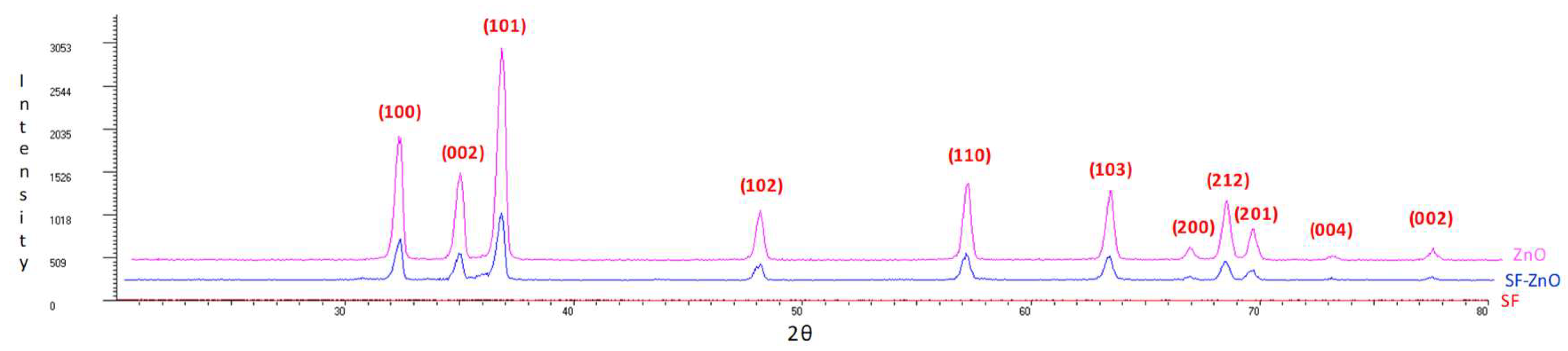
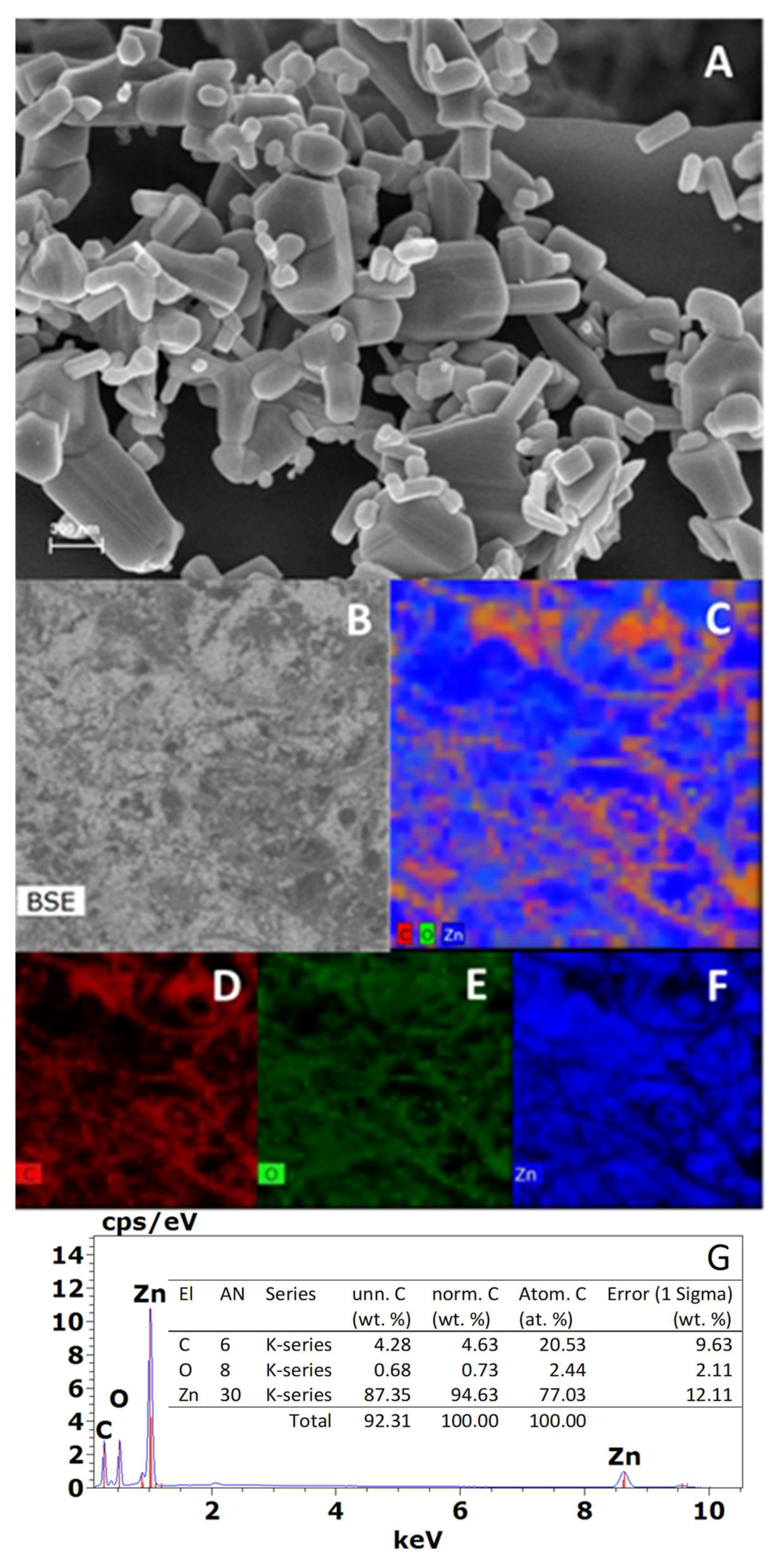
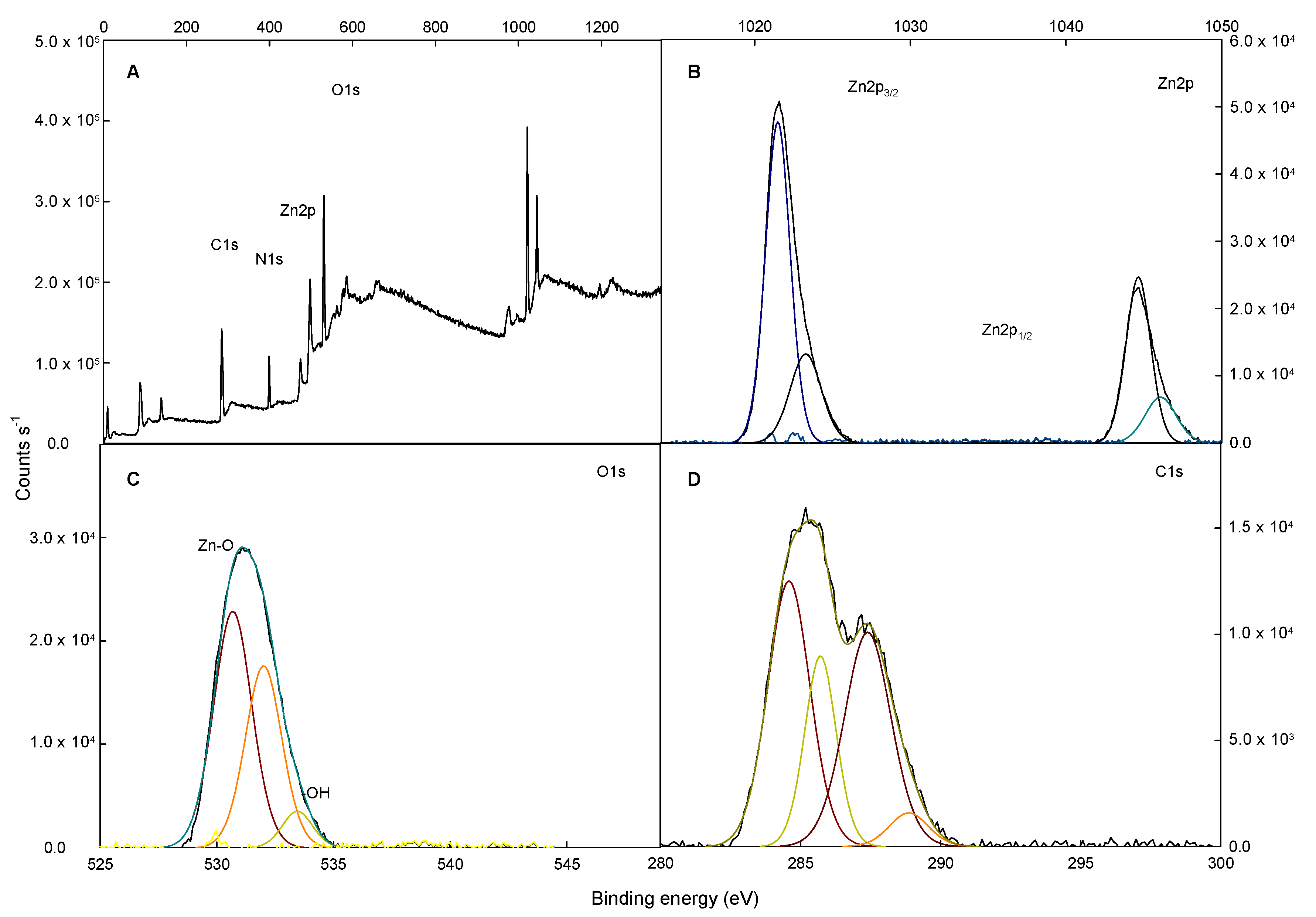
2.1. X-ray Diffraction (XRD), Field Emission Scanning Electron Microscopy (FESEM), X-ray Photoelectron Spectroscopy (XPS), and Energy Dispersive X-ray Spectroscopy Analysis (XDS) Characterization

SF/TiO2 and SF/ZnO samples were characterized following the methodology described by Fenoll et al. [38]. The characterization of SF/TiO2 samples was described in a previous paper [39]. In the case of SF/ZnO samples, the X-ray diffractograms are showed in Figure 1. The XRD of SF/ZnO exhibited the peaks attributed to zincite (hexagonal system). In accordance with the PDF2 database (International Centre for Diffraction Data), the pattern showed by the peaks is closely associated to JCPDS card no. 36-1451. The characteristic zincite peaks at 2θ diffraction angles of 32, 34, 36, 47, 57, 63, 66, 68, 69, 72, and 77·corresponding to the planes 100, 002, 101, 102, 110, 103, 200, 212, 201, 004, and 002 respectively, were clearly observed. Consequently, we considered that the surface coverage of electrospun SF materials prepared with ZnO was complete. Figure 2A shows the SEM images of SF/ZnO. It can be seen that a non-uniform ZnO layer around the SF and a cluster of aggregates of ZnO accumulated on the surface. In addition, it can be observed that the hexagonal structure corresponds to zincite as demonstrated by XRD. Concerning the elemental mapping of SF/ZnO (Figure 2C–F), the surface coverage of the SF with ZnO was almost complete. In addition, EDX analysis (Figure 2G) showed the presence of Zn with C in the sample supporting the elemental mapping and XPS data that Zn surface coverage was almost complete. The SEM image of the TiO2 coated SF also showed a cluster of aggregates of TiO2 deposited on the SF [39]. These clustered aggregates cannot be broken down into smaller particles by physical processing.
The XPS survey spectra for SF/ZnO is exposed in Figure 3A. The Zn2p spectrum for SF/ZnO shows two peaks (1021.5 and 1044.7 eV) that are coincident to Zn2p3/2 and Zn2p1/2, while in pure ZnO, these binding energies were 1021.1 and 1044.2 eV, respectively, according to Naumkin et al. 2012 [40]. Figure 3C illustrates the O1s spectra. O1s peaks show an asymmetric appearance that could be attributed to the existence of various oxygen bonds. The high intensity found in lower binding energy for O1s spectra suggests stronger Zn-O bond in the ZnO. The intermediary peak could be due to oxygen shortcomings of ZnO. The higher binding energy peak may be attributed to the adsorption of hydroxyl species [41]. These results are in concordance with those obtained from the XRD pattern. The estimated value of surface area (SBET) for SF/ZnO was 21 m2 g−1.

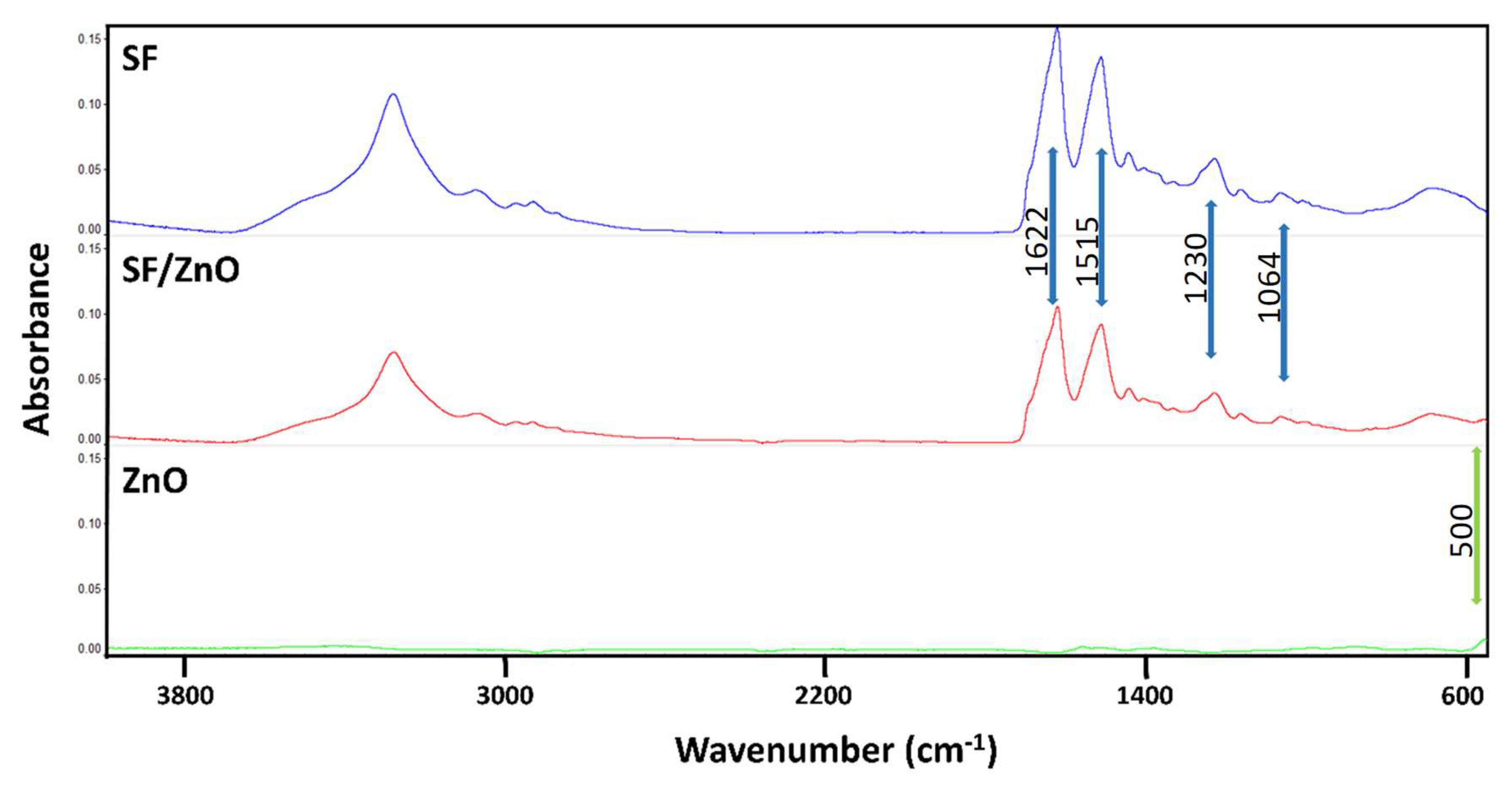
2.2. Attenuated Total Reflectance Fourier Transformed Infrared Spectroscopy (ATR-FTIR) Depiction

SF/ZnO mats revealed the typical peaks of fibroin corresponding to amide I (1622 cm−1), amide II (1515 cm−1), amide III (1230 cm−1), and amide IV (1064 cm−1), highlighted with blue arrows in Figure 4. These bands are described in the scientific literature for SF materials enriched in β-sheet conformation (non-soluble state in water) [42,43]. The non-presence of the peak at 1660 cm−1 also verified this fact [44].
The results of ATR-FTIR analysis also confirmed that the TiO2 and ZnO nanoparticles were successfully incorporated in the materials. Along the spectra obtained from the SF/TiO2 [39] and SF/ZnO mats (Figure 5) are observed both, the characteristic bands of SF (previously described) and the corresponding to TiO2 or ZnO. In the case of SF/TiO2 mats it can be observed a broad band at 700 cm−1 that can be allocated to the existence of TiO2; this band is also present in the spectrum of pure TiO2 nanoparticles, and is caused by the vibrations of (Ti-O-Ti) stretch [23,45]. SF/ZnO meshes displayed a spectrum almost identical to that of pure SF mats caused by the intense peaks of fibroin, these ones masked the presence of ZnO nanoparticles, which peaks presented a lower intensity. However, a slight difference can be observed at 500 cm−1 (Figure 4, green arrow), a band present in both SF/ZnO mats and ZnO nanoparticles, that can be attributed to the ZnO stretching modes, as stated by other authors [46,47].

2.3. Mechanical Properties and Fibre Diameters

The analysis of mechanical properties and diameter of the fibers of the materials obtained was performed in order to visualize the way in which these parameters could be affected by the deposition of either TiO2 or ZnO on the surface of the SF fibers.
As observed in Table 1 the mechanical properties of the different materials produced were not significantly affected by the covering of the fibers with the studied nanoparticles (ANOVA, p > 0.05). The results obtained for tensile strength (0.3–0.4 MPa), elastic modulus (21–24 MPa), and strain at break (1.9–2.3%) were in the range of the values described in previous studies using electrospun SF materials as scaffolds with different purposes [28,31,32,48,49]. However, the deposition of TiO2 gave rise to a homogeneous layer on the surface of the fibers that was responsible of a significant increase (p < 0.05) in the diameter of the fibers. This one was estimated around 2.8–2.9 µm for SF and SF/ZnO mats but in the case of SF/TiO2 meshes was 3.9 µm.

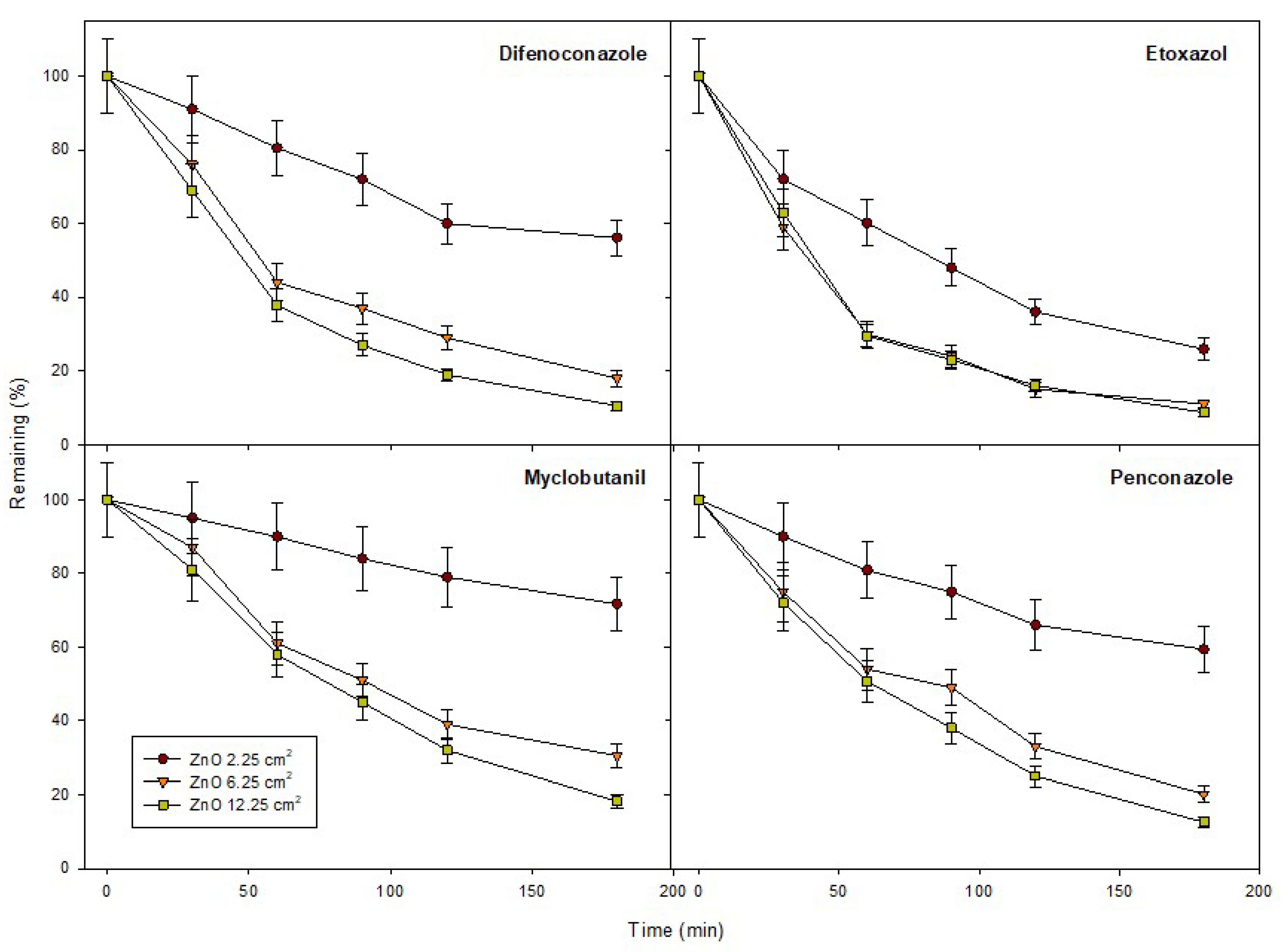
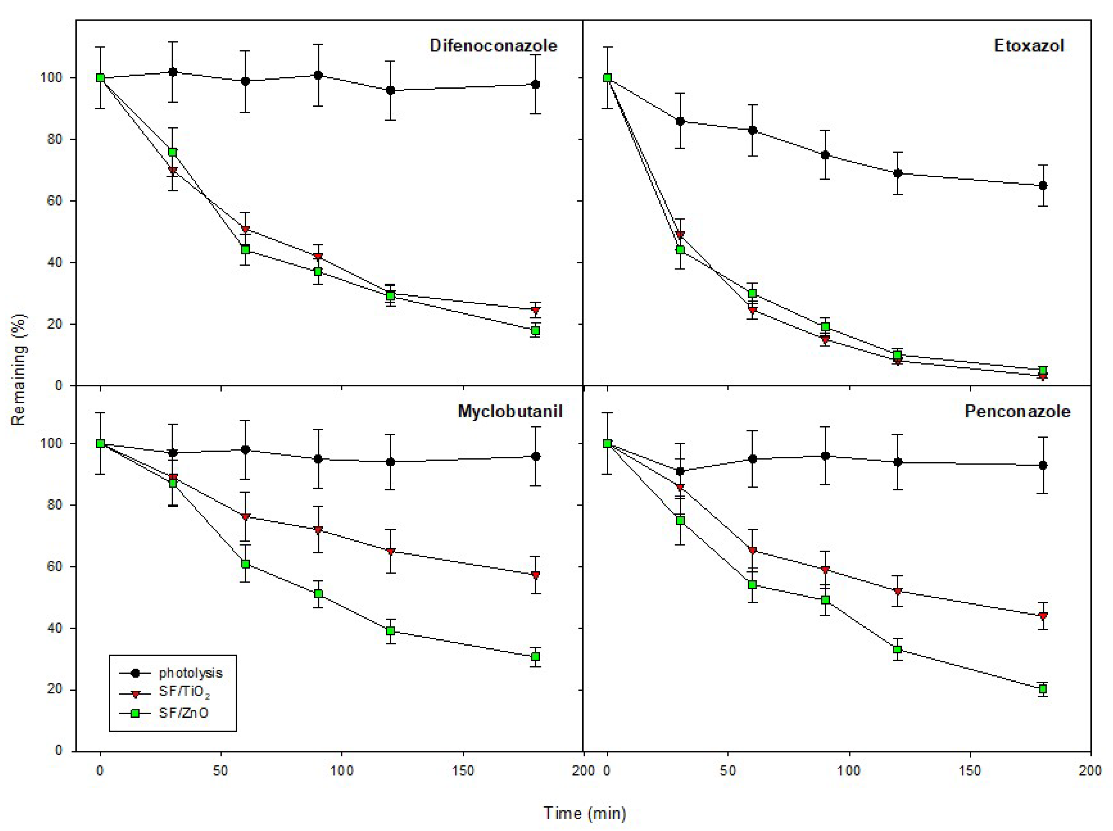
2.4. Photocatalytic Activity and Kinetics

The influence of ZnO loading on the elimination of the pesticides was assessed to gain knowledge on degradation efficiency. The essay was conducted with three fragments of SF/ZnO mats (2.25, 6.25, and 12.25 cm2) coated with different ZnO amounts (10, 25, and 50 mg, respectively). We noticed a significant increment in degradation rates when ZnO was added. Given that we did not observe significant differences (p < 0.05) when ZnO loading was increased from 25 to 50 mg for most of pesticides (Figure 5), SF mats with 25 mg of ZnO was the option finally selected. The photocatalytic degradation of pesticides was also assessed using SF/ZnO and SF/TiO2 (Figure 6). In the experiment carried out in the presence of SF/ZnO, the residual levels of difenoconazole, etoxazole, myclobutanil, and penconazole, after 180 min of irradiation were 18, 5, 30.5, and 20.1 µg L−1, respectively. For SF/TiO2, the residual levels once finished the experiment were 24.6, 3, 57.3, and 43.8 µg L−1, respectively. The experiments performed in the absence of catalyst showed a clear decrease in the photodecomposition rate and only a reduction between 65–98% of the initial mass was accomplished for etoxazole and difenoconazole, respectively, after 180 min of light exposure.
Regarding the efficiency for catalyzing the elimination of the pesticides studied, the comparison of electrospun SF materials (coated with similar amount of TiO2 and ZnO) showed that both catalysts were equally efficient for removing etoxazole while in the case of difenoconazole, myclobutanil, and penconazole, ZnO was slightly more efficient.
The adsorption in dark conditions after 30 min of incubation was insignificant (~4.6%) for the studied pesticides on both SF/TiO2 and SF/ZnO mats. The higher values found for SF/TiO2 (3.3–4.6%) with respect to SF/ZnO (1.2–2.6%) could be justified by the higher surface area of SF/TiO2. The mineralization degree was assessed by measuring DOC content. The initial content of DOC was 4.7 mg L−1. The percentages found at the end of the solar treatment in the SF/TiO2 and SF/ZnO systems were 25.4% and 13.2%, respectively. These lingering percentages could be attributed to the formation of recalcitrant intermediates during the experiment. In addition, coadjuvants presents in commercial products could also explain the values obtained.
Kinetic parameters of the photocatalytic degradation of pesticides for SF/TiO2 and SF/ZnO systems are listed in Table 2. Degradation followed a first-order behavior (CP = CP0 e−kapp*t) with R2 ranging from 0.96 to 0.99. The rate constants obtained for SF/TiO2 and SF/ZnO systems varied within 0.003–0.022 min−1 and 0.007–0.021 min−1, respectively.
The higher photocatalytic activity of SF/ZnO could be attributed to the fraction of solar spectrum that ZnO absorbs, since it is wider than the fraction absorbed by TiO2. Consequently, this system receives more light quanta [50,51]. In addition, other characteristics of the catalysts (surface area, crystallinity, particle size, optical properties, and impurities) would play an important role in the detected discrepancies between both systems [52]. Thus, a direct relation between the photocatalytic response of ZnO and the defects in its nanostructure can be established [13]. Furthermore, even though SF/ZnO has major particle size (~200 nm) and lower specific surface area (~21 m2 g−1) than SF/TiO2 (~20 nm and 31 m2 g−1), ZnO exhibits larger electronic mobility than TiO2 and therefore it shows a minor charge recombination [53]. In consequence, there is more unoccupied space for the absorption of pesticides, with the resulting enhancement of the photodegradation efficacy. The photodissolution for both, Ti and Zn into solution was observed to be inappreciable.
One crucial aspect that must be taken into account when using photocatalysts in practical industrial applications is their reusability. SF/TiO2 and SF/ZnO mats were recycled for another three times by simple washing and drying. After using them under same experimental conditions, we observed a slight reduction in the rate constants for pesticide degradation during the second, third, and fourth cycle ranged between 5% (3.6% for SF/TiO2 and 4.7% for SF/ZnO), 8% (5.9% for SF/TiO2 and 7.8% for SF/ZnO), and 12% (8.3% for SF/TiO2 and 11.5% for SF/ZnO), respectively.

3. Materials and Methods

3.1. Pesticides and Reagents

Pure standards (purity > 99%) were supplied by Ehrenstorfer GmBh (Augsburg, Germany). The physicochemical properties of the pesticides are listed in Table 3 [54]. The commercial formulations (Score 25% w/v—difenoconazole, Borneo 11% w/v—etoxazole, and Systhane Forte 24% w/v—myclobutanil and Furabel 10% w/v—penconazole) were supplied by Syngenta Agro, Kenogard, Dow Agrosciences Iberica, S.A. and Probelte, respectively. Catalyzers, zinc oxide (99.99%, 200 nm, BET 10 m2 g−1) and titanium dioxide P25 (99.5%, <21 nm, 50 m2 g−1) were purchased from Alfa-Aesar (Karlsruhe, Germany) and Nippon Aerosil Co Ltd. (Osaka, Japan), respectively. Deionized water was obtained from a Millipore Water Purification System (Bedford, MA, USA).

3.2. Silk Fibroin Processing

Cocoons of B. mori were chopped into four pieces and subsequently boiled in 0.02 M Na2CO3 during 30 min, in order to accomplish the elimination of the sericin. Later, the SF was washed with water and dried during 3 days at 22 ± 1 °C. It was dissolved using 9.3 M LiBr (Acros Organics, Belgium) for 3 h (60 °C), obtaining a 20 wt.% dissolution that was dialyzed with water for 3 days using Snakeskin Dialysis Tubing 3.5 KDa MWCO (Thermo Scientific, Waltham, MA, USA) carrying out eight changes of water (4 °C). The resultant 7 wt.% SF dissolution was retrieved and concentrated by means of dialysis in 30 wt.% polyethylene glycol (11,000 Da) for 24 h, in order to reach a concentration of 20–21 wt.% of SF, which is optimal for the electrospinning experiments.

3.3. Electrospinning and Post-Treatment of SF/TiO2 and SF/ZnO Mats

The electrospinning setup used in order to produce the materials was the same described in previous works of our research group [28,32,37,48]. The working conditions were adapted so that the Taylor cone was stable, being the voltage implemented to the capillary needle 19 kV and −1.5 kV to the metallic collector (~375 cm2). The distance between both elements of the setup was 42 cm and the selected injection rate of the SF dissolution was 1 mL h−1. Either ZnO or TiO2 nanoparticles (contained in methanol) were sprayed, using an atomizer, throughout the electrospinning experiments over the surface of the mats, as it is clarified below. Each mat was produced from 6 mL of 20% wt SF dissolution. Total of 30 mL of methanol containing 1.5 g of either TiO2 or ZnO nanoparticles were sprayed during the electrospinning experiments. With this purpose, seven rounds of atomization (periodically distributed) were carried out (4.3 mL per round), covering homogeneously the collector.
A total of 1.5 mL of SF dissolution was electrospun before the first atomization and after the last one, in order to guarantee the fabrication of a compact net, avoiding the loss of the nanoparticles included. Pure SF electrospun mats were produced as negative controls and the materials including TiO2 or ZnO in their composition were named as SF/TiO2 or SF/ZnO, respectively.
The annealing of all the materials produced was performed by an immersion step in absolute methanol during 30 min, subsequently the mats were dried locating them between two portions of filter paper.

3.4. XRD, FESEM, XPS, XDS and SBET

XRD, FESEM, XPS, and XDS were used to characterize the materials as previously published by Garrido et al. [20]. A Quadrasorb SI-MP (Quantachrome Instruments, Boynton Beach, FL, USA) was used to obtain the gas sorption (nitrogen, 77 K) surface area according to Brunauer–Emmett–Teller (BET) method. Outgassing was performed with a Masterprep Degasser (Quantachrome Instruments) at 120 °C for 12 h for SF/ZnO.

3.5. ATR-FTIR

ATR-FTIR was the technique used to confirm the structural changes of SF from a water soluble conformation to a non-soluble state, with a characteristic increase in β-sheet content. IR spectra were also used to visualize the presence of the nanoparticles in the materials, by means of the analysis of some characteristic bands of ZnO or TiO2. For these purposes, a Nicolet iS5 spectrometer equipped with an iD5 ATR accessory (Thermo Scientific, USA) was used. The instrument worked in absorbance mode (resolution of 4 cm−1, a spectral range of 4000–550 cm−1, and 64 scans).

3.6. Analysis of Mechanical Properties and Fibre Diameter

Tensile tests were conducted using a universal test frame machine (Qtest; MTS Systems, Eden Prairie, MN, USA). Fragments of mats (10 mm × 30 mm) were proven at a crosshead speed of 0.1 mm·s−1 with a load cell of 10 N. A digital micrometer (Mitutoyo, IL, USA) with 0–25 mm, resolution of 0.001 mm, and precision of ±2 μm was used to measure the width of each sample. The stress–strain curves obtained were used in order to calculate the tensile strength (MPa), the strain at break (%) and the elastic modulus (MPa) in the linear elastic part of the curves. The tests were replicated three times per condition. Micrographs obtained by SEM were acquired and used to measure the diameter of the fibers of the materials produced.

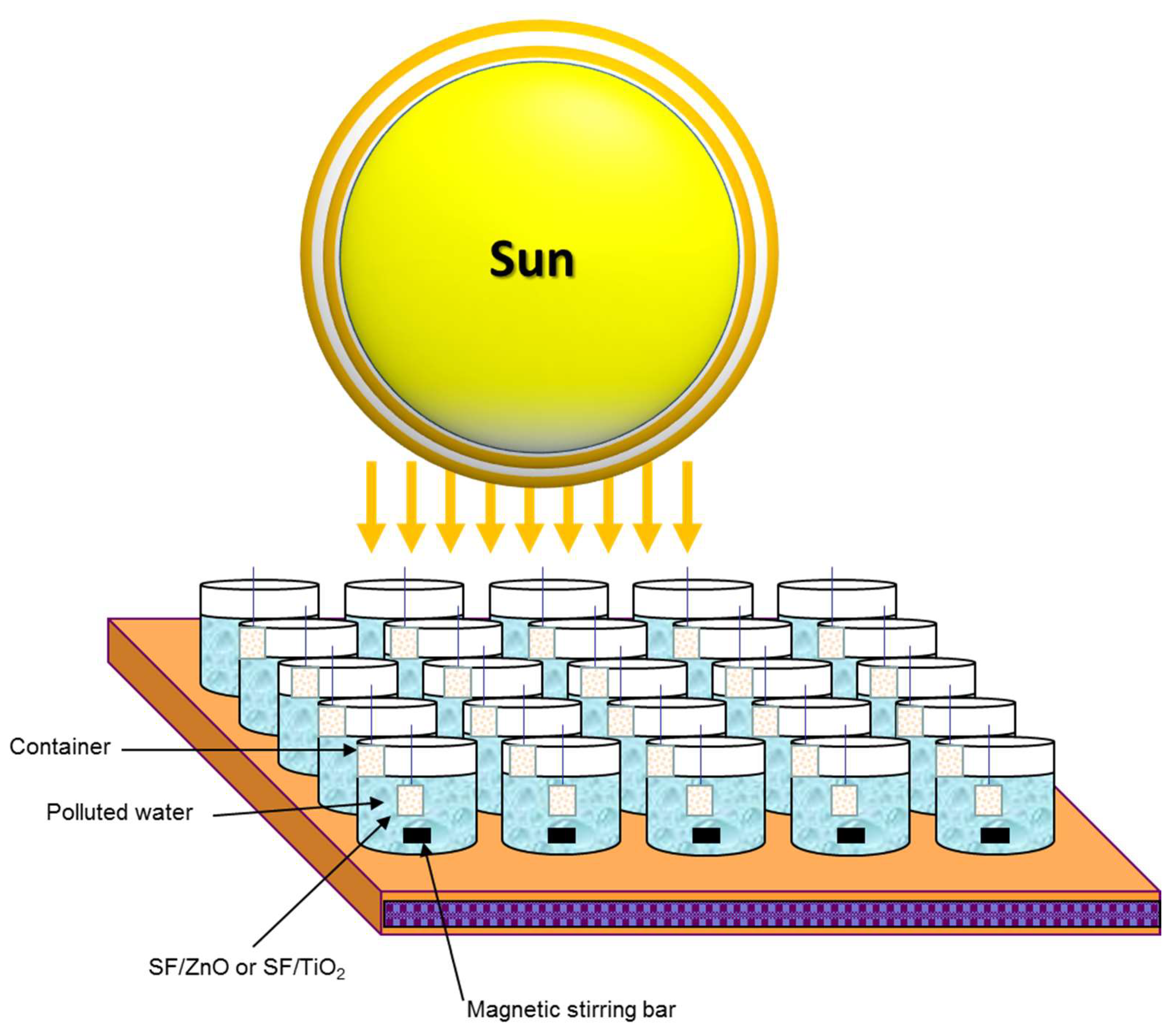
3.7. Photoreaction Setup

Photocatalytic and photolytic trials were performed during August 2018 following the procedure described by Fenoll et al. with some modification [38]. For both experiments we mixed Milli-RX water (pH 7.1, ORP 230 mV, conductivity 5.5 µS cm−1, and TOC < 30 µg L−1) with commercial formulations of the pesticides studied. Experiments were performed in vessels containing 100 mL of water and introducing a square fragment of SF/semiconductor oxide mats. The vessels were exposed to sunlight for 180 min (from 11 AM to 2 PM). Before starting experiments, the solution was stirred for 30 min in darkness and later a sample was collected to measure the adsorption of the pesticides onto each square fragment of SF/ZnO mats. Figure 7 illustrates a schematic picture of the experimental setup. 948.6 ± 52.4, 21.5 ± 2.3, and 1.7 ± 0.4 (all in W m−2) of VIS plus NIR, UVA, and UVB, respectively, were recorded at noon.

3.8. Analytical Determinations

Pesticide residues in water samples were analyzed by an HPLC system (Agilent Series 1200 Agilent Technologies, Santa Clara, CA, USA) and a G6410A triple quadrupole mass spectrometer provided with an ESI+ interface following the methodology previously published by Fenoll et al. [55]. Table 4 shows the analytical conditions of the studied pesticides. The presence of Ti and Zn in solution was determined using an Agilent 7900 ICP/MS system. The measure of dissolved organic carbon (DOC) content in water samples was performed by a Multi N/C 3100 TOC Analyzer (Analytic Jena AG, Jena, Germany) equipped with an NDIR detector (950 °C), following the procedure outlined by Fenoll et al. [38].

3.9. Statistical Analysis

Statistical analyses and curve fitting were performed by SigmaPlot version 13.0 statistical software (Systat, Software Inc., San Jose, CA, USA). IBM SPSS 25 statistics software was used to analyze statistics of mechanical properties and fiber diameter. ANOVA (p < 0.05) or Mann–Whitney (p < 0.05) tests were determined by the data accomplished normality and homogeneity of variance requirements or not, respectively.

4. Conclusions

One of the environmental issues that citizens and scientists are most concerned is the pollution of natural resources by pesticides residues. These substances are capable of polluting remote areas from the point where they were applied and remaining for years/decades. In this work, we have developed a new and simple method to obtain silk fibroin (SF) fibers coated with ZnO nanoparticles using electrospinning technique. ZnO nanostructures have been shown to be a potential candidate as photocatalyst for solar-driven photodegradation process of pesticides owing to its low production cost in comparison to TiO2, non-toxic, and ability to absorb larger fraction of solar spectrum compared to TiO2. The photocatalytic efficiency of these materials under natural sunlight exposure was studied on the removal of four pesticides in water. The results showed an increment in the degradation rates when the ZnO loading was increased. To promote the practical applicability of this methodology a study of reusability was carried out showing that the reduction in rate constants for photocatalytic degradation after four cycles was lower than 11%. The photocatalytic efficiency of electrospun SF fibers coated with similar amount of TiO2 and ZnO was slightly higher in the case of SF/ZnO. Thus, the obtained nanocomposite (SF/ZnO) has been shown to be a potential candidate as photocatalyst for solar-driven photocatalytic degradation of pesticide residues to minimize their potential harm to the environment and human health. In order to provide more information on the photocatalytic degradation of pesticides in larger scale of application, future research should be developed using SF/ZnO mats.

Author Contributions

Conceptualization, S.A.-C., J.L.C., S.N. and J.F.; methodology, I.G., M.A., N.V., M.J.Y.-G., S.N., and J.F.; validation, I.G., M.A., N.V., and M.J.Y-G.; formal analysis, S.A.-C., J.L.C., S.N., and J.F.; investigation, S.A.-C., J.L.C., S.N., and J.F.; resources, I.G., M.A., N.V., M.J.Y.-G., S.N., and J.F.; data curation, S.A.-C., S.N., and J.F.; writing—original draft preparation, I.G., S.A.-C., S.N., and J.F.; writing—review and editing, I.G., S.N., and J.F.; visualization, I.G., S.N., and J.F.; supervision, S.N. and J.F.; project administration, I.G.; funding acquisition, S.A.-C., J.L.C., S.N., and J.F. All authors have read and agreed to the published version of the manuscript and there are no other persons who satisfied the criteria for authorship but are not listed. We further confirm that all of us have approved the order of authors listed in the manuscript.

Funding

This work was financially supported by Operative Regional Program FEDER for Murcia 2014–2020, project RTA2015-00073-00-00, INIA 2017–2020 and with the aid of the Resolution of Presidency of State Research Agency (State Subprogram of Training of State Program of Promotion of Talent and its Employability), within the framework of the State Plan for Scientific and Technical Research and Innovation 2013–2016 co-funded by the European Social Fund. Salvador Aznar-Cervantes acknowledges the financial support of his research contract, program INIA-CCAA (DOC INIA 2015), announced by the National Institute for Agricultural and Food Research and Technology (INIA) and supported by The Spanish State Research Agency (AEI) under the Spanish Ministry of Economy, Industry and Competitiveness.

Acknowledgments

The authors are grateful to Herminia Jiménez, Inmaculada Garrido, Juana Cava, and María V. Molina for technical assistance.

Conflicts of Interest

The authors declare no conflict of interest associated with this manuscript.

References

  1. Mnif, W.; Hassine, A.I.H.; Bouaziz, A.; Bartegi, A.; Thomas, O.; Roig, B. Effect of endocrine disruptor pesticides: A review. Int. J. Environ. Res. Public Health 2011, 8, 2265–2303. [Google Scholar] [CrossRef] [PubMed] [Green Version]
  2. Vela, N.; Pérez-Lucas, G.; Fenoll, J.; Navarro, S. Recent overview on the abatement of pesticide residues in water by photocatalytic treatment using TiO2. In Applications of Titanium Dioxide; Janus, M., Ed.; IntechOpen: Rijeka, Croatia, 2017; pp. 147–177. [Google Scholar]
  3. Ribeiro, A.R.; Nunes, O.C.; Pereira, M.F.R.; Silva, A.M.T. An overview on the advanced oxidation processes applied for the treatment of water pollutants defined in the recently launched Directive 2013/39/EU. Environ. Int. 2015, 75, 33–51. [Google Scholar] [CrossRef] [PubMed] [Green Version]
  4. Fagan, R.; McCormack, E.; Dionysiou, D.D.; Pillai, S.C. A review of solar and visible light active TiO2 photocatalysis for treating bacteria, cyanotoxins and contaminants of emerging concern. Mater. Sci. Semicond. Process. 2016, 42, 2–14. [Google Scholar] [CrossRef] [Green Version]
  5. Miklos, D.B.; Remy, C.; Jekel, M.; Linden, K.G.; Drewes, J.E.; Hübner, U. Evaluation of advanced oxidation processes for water and wastewater treatment. A critical review. Water Res. 2018, 139, 118–131. [Google Scholar] [CrossRef] [PubMed]
  6. Quiroz, M.A.; Bandala, E.R.; Martínez-Huitle, C.A. Advanced Oxidation Processes (AOPs) for Removal of Pesticides from Aqueous Media. In Pesticides-Formulations, Effects, Fate; Stoytcheva, M., Ed.; IntechOpen: Rijeka, Croatia, 2011; pp. 685–730. [Google Scholar]
  7. Chen, X.; Wu, Z.; Liu, D.; Gao, Z. Preparation of ZnO photocatalyst for the efficient and rapid photocatalytic degradation of azo dyes. Nanoscale Res. Lett. 2017, 12, 4–13. [Google Scholar] [CrossRef] [PubMed] [Green Version]
  8. Bhatia, S.; Verma, N. Photocatalytic activity of ZnO nanoparticles with optimization of defects. Mater. Res. Bull. 2017, 95, 468–476. [Google Scholar] [CrossRef]
  9. Wu, M.C.; Chan, S.H.; Lin, T.H. Fabrication and photocatalytic performance of electrospun PVA/silk/TiO2 nanocomposite textile. Funct. Mater. Lett. 2015, 8, 1540013. [Google Scholar] [CrossRef]
  10. Nakata, K.; Fujishima, A. TiO2 photocatalysis: Design and applications. J. Photochem. Photobiol. C Photochem. Rev. 2012, 13, 169–189. [Google Scholar] [CrossRef]
  11. Ong, C.B.; Ng, L.Y.; Mohammad, A.W. A review of ZnO nanoparticles as solar photocatalysts: Synthesis, mechanisms and applications. Renew. Sustain. Energy Rev. 2018, 81, 536–551. [Google Scholar] [CrossRef]
  12. Wetchakun, K.; Wetchakun, N.; Sakulsermsuk, S. An overview of solar/visible light-driven heterogeneous photocatalysis for water purification: TiO2 and ZnO based photocatalysts used in suspension photoreactors. J. Ind. Eng. Chem. 2019, 71, 19–49. [Google Scholar] [CrossRef]
  13. Montero-Muñoz, M.; Ramos-Ibarra, J.E.; Rodríguez-Páez, J.E.; Teodoro, M.D.; Marques, G.E.; Sanabria, A.E.; Cajas, P.C.; Páez, C.A.; Heinrichs, B.; Coaquira, J.A.H. Role of defects on the enhancement of the photocatalytic response of ZnO nanostructures. Appl. Surf. Sci. 2018, 448, 646–654. [Google Scholar] [CrossRef]
  14. Wei, G.; Zuo, H.F.; Guo, Y.R.; Pan, Q.J. Synthesis of ZnO with enhanced photocatalytic activity: A novel approach using nanocellulose. BioResources 2016, 11, 6244–6253. [Google Scholar] [CrossRef] [Green Version]
  15. Xu, J.; Su, H.; Han, J.; Chen, Y.; Song, W.; Gu, Y.; Moon, W.J.; Zhang, D. In situ deposition of flower-like ZnO on silk fibroin fibers. Appl. Phys. A 2012, 108, 235–238. [Google Scholar] [CrossRef]
  16. Zhao, G.; Zhang, Y.; Zhang, L.; Ye, Z.G.; Ren, W.; Xu, F.; Wang, S.; Liu, M.; Zhang, X. 3D Conformal modification of electrospun silk nanofibers with nanoscaled ZnO deposition for enhanced photocatalytic activity. ACS Biomater. Sci. Eng. 2017, 3, 2900–2906. [Google Scholar] [CrossRef]
  17. Etacheri, V.; Di Valentin, C.; Schneider, J.; Bahnemann, D.; Pillai, S.C. Visible-light activation of TiO2 photocatalysts: Advances in theory and experiments. J. Photochem. Photobiol. C Photochem. Rev. 2015, 25, 1–29. [Google Scholar] [CrossRef] [Green Version]
  18. Ahmed, S.; Rasul, M.G.; Brown, R.; Hashib, M.A. Influence of parameters on the heterogeneous photocatalytic degradation of pesticides and phenolic contaminants in wastewater: A short review. J. Environ. Manag. 2011, 92, 311–330. [Google Scholar] [CrossRef] [PubMed] [Green Version]
  19. Fenoll, J.; Garrido, I.; Cava, J.; Hellín, P.; Flores, P.; Navarro, S. Photometabolic pathways of chlorantraniliprole in aqueous slurries containing binary and ternary oxides of Zn and Ti. Chem. Eng. J. 2015, 264, 720–727. [Google Scholar] [CrossRef]
  20. Garrido, I.; Pastor-Belda, M.; Campillo, N.; Viñas, P.; Yañez, M.J.; Vela, N.; Navarro, S.; Fenoll, J. Photooxidation of insecticide residues by ZnO and TiO2 coated magnetic nanoparticles under natural sunlight. J. Photochem. Photobiol. A Chem. 2019, 372, 245–253. [Google Scholar] [CrossRef]
  21. Cai, L.; Shao, H.; Hu, X.; Zhang, Y. Reinforced and ultraviolet resistant silks from silkworms fed with titanium dioxide nanoparticles. ACS Sustain. Chem. Eng. 2015, 3, 2551–2557. [Google Scholar] [CrossRef]
  22. Feng, X.X.; Zhang, L.L.; Chen, J.Y.; Guo, Y.H.; Zhang, H.P.; Jia, C.I. Preparation and characterization of novel nanocomposite films formed from silk fibroin and nano-TiO2. Int. J. Biol. Macromol. 2007, 40, 105–111. [Google Scholar] [CrossRef]
  23. Kim, J.H.; Sheikh, F.A.; Ju, H.W.; Park, H.J.; Moon, B.M.; Lee, O.J.; Park, C.H. 3D silk fibroin scaffold incorporating titanium dioxide (TiO2) nanoparticle (NPs) for tissue engineering. Int. J. Biol. Macromol. 2014, 68, 158–168. [Google Scholar] [CrossRef] [PubMed]
  24. Jao, W.C.; Yang, M.C.; Lin, C.H.; Hsu, C.C. Fabrication and characterization of electrospun silk fibroin/TiO2 nanofibrous mats for wound dressings. Polym. Adv. Technol. 2012, 23, 1066–1076. [Google Scholar] [CrossRef]
  25. Li, C.; Vepari, C.; Jin, H.J.; Kim, H.J.; Kaplan, D.L. Electrospun silk-BMP-2 scaffolds for bone tissue engineering. Biomaterials 2006, 27, 3115–3124. [Google Scholar] [CrossRef] [PubMed]
  26. Wang, J.; Sun, B.; Bhutto, M.A.; Zhu, T.; Yu, K.; Bao, J.; Morsi, Y.; El-Hamshary, H.; El-Newehy, M.; Mo, X. Fabrication and characterization of Antheraea pernyi silk fibroin-blended P(LLA-CL) nanofibrous scaffolds for peripheral nerve tissue engineering. Front. Mater. Sci. 2017, 11, 22–32. [Google Scholar] [CrossRef]
  27. Aznar-Cervantes, S.; Roca, M.I.; Martinez, J.G.; Meseguer-Olmo, L.; Cenis, J.L.; Moraleda, J.M.; Otero, T.F. Fabrication of conductive electrospun silk fibroin scaffolds by coating with polypyrrole for biomedical applications. Bioelectrochemistry 2012, 85, 36–43. [Google Scholar] [CrossRef] [PubMed]
  28. Aznar-Cervantes, S.; Pagán, A.; Martínez, J.G.; Bernabeu-Esclapez, A.; Otero, T.F.; Meseguer-Olmo, L.; Paredes, J.I.; Cenis, J.L. Electrospun silk fibroin scaffolds coated with reduced graphene promote neurite outgrowth of PC-12 cells under electrical stimulation. Mater. Sci. Eng. C 2017, 79, 315–325. [Google Scholar] [CrossRef]
  29. Martinez, J.G.; Aznar-Cervantes, S.; Abel Lozano-Pérez, A.; Cenis, J.L.; Otero, T.F. Graphene adsorbed on silk-fibroin meshes: Biomimetic and reversible conformational movements driven by reactions. Electrochim. Acta 2016, 209, 521–528. [Google Scholar] [CrossRef]
  30. Zhang, X.; Reagan, M.R.; Kaplan, D.L. Electrospun silk biomaterial scaffolds for regenerative medicine. Adv. Drug Deliv. Rev. 2009, 61, 988–1006. [Google Scholar] [CrossRef] [Green Version]
  31. Aznar-Cervantes, S.D.; Vicente-Cervantes, D.; Meseguer-Olmo, L.; Cenis, J.L.; Lozano-Pérez, A.A. Influence of the protocol used for fibroin extraction on the mechanical properties and fiber sizes of electrospun silk mats. Mater. Sci. Eng. C 2013, 33, 1945–1950. [Google Scholar] [CrossRef]
  32. Aznar-Cervantes, S.D.; Lozano-Pérez, A.A.; García Montalban, M.; Villora, G.; Vicente-Cervantes, D.; Cenis, J.L. Importance of refrigeration time in the electrospinning of silk fibroin aqueous solutions. J. Mater. Sci. 2015, 50, 4879–4887. [Google Scholar] [CrossRef]
  33. Kearns, V.; MacIntosh, A.C.; Crawford, A.; Hatton, P.V. Silk-based biomaterials for tissue engineering. Top. Tissue Eng. 2008, 4, 1–19. [Google Scholar]
  34. Du, G.Y.; He, S.W.; Sun, C.X.; Mi, L.D. Bone morphogenic protein-2 (rhBMP2)-loaded silk fibroin scaffolds to enhance the osteoinductivity in bone tissue engineering. Nanoscale Res. Lett. 2017, 12, 573. [Google Scholar] [CrossRef] [PubMed] [Green Version]
  35. Sahoo, S.; Toh, S.L.; Goh, J.C.H. A bFGF-releasing silk/PLGA-based biohybrid scaffold for ligament/tendon tissue engineering using mesenchymal progenitor cells. Biomaterials 2010, 31, 2990–2998. [Google Scholar] [CrossRef] [PubMed]
  36. Nalvuran, H.; Elçin, A.E.; Elçin, Y.M. Nanofibrous silk fibroin/reduced graphene oxide scaffolds for tissue engineering and cell culture applications. Int. J. Biol. Macromol. 2018, 114, 77–84. [Google Scholar] [CrossRef]
  37. López-Maya, E.; Montoro, C.; Rodríguez-Albelo, L.M.; Aznar Cervantes, S.D.; Lozano-Pérez, A.A.; Cenís, J.L.; Barea, E.; Navarro, J.A.R. Textile/metal—Organic-framework composites as self-detoxifying filters for chemical-warfare agents. Angew. Chem. Int. Ed. 2015, 54, 6790–6794. [Google Scholar] [CrossRef]
  38. Fenoll, J.; Garrido, I.; Pastor-Belda, M.; Camillo, N.; Viñas, P.; Yáñez, M.J.; Vela, N.; Navarro, S. Solar detoxification of water polluted with fungicide residues using ZnO coated magnetic particles. Chem. Eng. J. 2017, 330, 71–81. [Google Scholar] [CrossRef]
  39. Aznar-Cervantes, S.; Aliste, M.; Garrido, I.; Yáñez-Gascón, M.J.; Vela, N.; Cenis, J.L.; Navarro, S.; Fenoll, J. Electrospun silk fibroin/TiO2 mats. Preparation, characterization and efficiency for the photocatalytic solar treatment of pesticide polluted water. RSC Adv. 2020, 10, 1917–1924. [Google Scholar] [CrossRef] [Green Version]
  40. Naumkin, A.V.; Kraut-Vass, A.; Powell, C.J.; Gaarenstroom, S.W. NIST X-Ray Photoelectron Spectroscopy Database 20, Version 4.1. 2012. Available online: http://srdata.nist.gov/xps/ (accessed on 15 September 2019).
  41. Ökte, A.N. Characterization and photocatalytic activity of Ln (La, Eu, Gd, Dy and Ho) loaded ZnO nanocatalysts. Appl. Catal. A Gen. 2014, 475, 27–39. [Google Scholar] [CrossRef]
  42. Zhang, F.; Zuo, B.Q.; Bai, L. Study on the structure of SF fiber mats electrospun with HFIP and FA and cells behavior. J. Mater. Sci. 2009, 44, 5682–5687. [Google Scholar] [CrossRef]
  43. Wang, M.; Jin, H.J.; Kaplan, D.L.; Rutledge, G.C. Mechanical Properties of Electrospun Silk Fibers. Macromolecules 2004, 37, 6856–6864. [Google Scholar] [CrossRef]
  44. Wang, H.; Shao, H.; Hu, X. Structure of silk fibroin fibers made by an electrospinning process from a silk fibroin aqueous solution. J. Appl. Polym. Sci. 2006, 101, 961–968. [Google Scholar] [CrossRef]
  45. Ochoa, Y.; Ortegón, Y.; Vargas, M.; Rodríguez Páez, J.E. Síntesis de TiO2, fase anatasa, por el método Pechini. Suplemento de la Revista Latinoamericana de Metalurgia y Materiales 2009, 1, 931–937. [Google Scholar]
  46. Senthilkumaar, S.; Rajendran, K.; Banerjee, S.; Chini, T.K.; Sengodan, V. Influence of Mn doping on the microstructure and optical property of ZnO. Mater. Sci. Semicond. Process. 2008, 11, 6–12. [Google Scholar] [CrossRef] [Green Version]
  47. Djaja, N.F.; Montja, D.A.; Saleh, R. The effect of Co incorporation into ZnO nanoparticles. Adv. Mater. Phys. Chem. 2013, 3, 33–41. [Google Scholar] [CrossRef] [Green Version]
  48. Millán-Rivero, J.E.; Martínez, C.M.; Romecín, P.A.; Aznar-Cervantes, S.D.; Carpes-Ruiz, M.; Cenis, J.L.; Moraleda, J.M.; Atucha, N.M.; García-Bernal, D. Silk fibroin scaffolds seeded with Wharton’s jelly mesenchymal stem cells enhance re-epithelialization and reduce formation of scar tissue after cutaneous wound healing. Stem Cell Res. Ther. 2019, 6, 1–14. [Google Scholar] [CrossRef] [PubMed] [Green Version]
  49. Aznar-Cervantes, S.; Martínez, J.G.; Bernabeu-Esclapez, A.; Lozano-Pérez, A.A.; Meseguer-Olmo, L.; Otero, T.F.; Cenis, J.L. Fabrication of electrospun silk fibroin scaffolds coated with graphene oxide and reduced graphene for applications in biomedicine. Bioelectrochemistry 2016, 108, 36–45. [Google Scholar] [CrossRef] [PubMed]
  50. Malato, S.; Fernández-Ibañez, P.; Maldonado, M.I.; Blanco, J.; Gernjak, W. Decontamination and disinfection of water by solar photocatalysis: Recent overview and trends. Catal. Today 2009, 147, 1–59. [Google Scholar] [CrossRef]
  51. Sakthivel, S.; Neppolian, B.; Shankar, M.V.; Arabindoo, B.; Palanichamy, M.; Murugesan, V. Solar photocatalytic degradation of azo dye: Comparison of photocatalytic efficiency of ZnO and TiO2. Sol. Energ Mater. Sol. Cells 2003, 77, 65–82. [Google Scholar] [CrossRef]
  52. Salah, N.H.; Bouhelassa, M.; Bekkouche, S.; Boultif, A. Study of photocatalytic degradation of phenol. Desalination 2004, 166, 347–354. [Google Scholar] [CrossRef]
  53. Kitsiou, V.; Filippidis, N.; Mantzavinos, D.; Pouios, I. Heterogeneous and homogeneous photocatalytic degradation of the insecticide imidacloprid in aqueous solutions. Appl. Catal. B Eviron. 2009, 86, 27–35. [Google Scholar] [CrossRef]
  54. Lewis, K.A.; Tzilivakis, J.; Warner, D.J.; Green, A. An international database for pesticide risk assessments and management. Hum. Ecol. Risk Assess. Int. J. 2016, 22, 1050–1064. [Google Scholar] [CrossRef] [Green Version]
  55. Fenoll, J.; Hellín, P.; Martínez, C.M.; Flores, P.; Navarro, S. Determination of 48 pesticides and their main metabolites in water samples by employing sonication and liquid chromatography—Tandem mass spectrometry. Talanta 2011, 85, 975–982. [Google Scholar] [CrossRef] [PubMed]
Figure 1. XRD patterns of SF/ZnO, SF, and ZnO.
Catalysts 10 00110 g001
Figure 2. SEM image of SF/ZnO (A), back scattered electron (BSE) image (B), elemental mapping (CF), and EDX data (G) of a SF/ZnO zone.
Catalysts 10 00110 g002
Figure 3. XPS spectra of SF/ZnO sample: (A) survey spectrum, (BD) high-resolution binding energy spectra of Ti2p, O1s, and C1s, respectively.
Catalysts 10 00110 g003
Figure 4. Infrared spectra of pure fibroin electrospun meshes (SF), fibroin mats containing ZnO nanoparticles (SF/ZnO) and pure ZnO nanoparticles (ZnO).
Catalysts 10 00110 g004
Figure 5. Optimization of ZnO load for photocatalytic degradation of the pesticides (0.1 mg L−1). Error bars denote standard deviation (n = 3).
Catalysts 10 00110 g005
Figure 6. Pesticide decay (0.1 mg L−1) by photolysis and photocatalysis (SF/ZnO and SF/TiO2) during the photoperiod. Approximately 250 mg L−1 of ZnO or TiO2 in all cases. Error bars denote standard deviation (n = 3).
Catalysts 10 00110 g006
Figure 7. Schematic drawing of the experimental setup.
Catalysts 10 00110 g007
Table 1. Average values of fiber diameter and mechanical properties studied in SF/ZnO electrospun materials.
MatsFiber Diameter (nm)Tensile Strength (MPa)Elastic Modulus (MPa)Strain at Break (%)
SF2948 ± 7480.41 ± 0.1224.24 ± 8.132.28 ± 0.26
SF/ZnO2766 ± 5990.32 ± 0.1020.77 ± 10.212.27 ± 0.41
SF/TiO23858 ± 1167 *0.33 ± 0.0922.77 ± 5.061.85 ± 0.27
* statistically different values compared to negative control (p < 0.05). Values of SF and SF/TiO2 reported in Aznar-Cervantes et al. [40].
Table 2. Kinetic parameters for photocatalytic treatments (SF/TiO2 and SF/ZnO) of pesticides in water under natural sunlight.
FungicideSF/TiO2SF/ZnO
R2k (min−1)t½ (min)R2k (min−1)t½ (min)
Difenoconazole0.9789.416 × 10−373.60.9791.092 × 10−263.5
Etoxazole0.9982.258 × 10−230.70.9832.092 × 10−233.1
Myclobutanil0.9743.276 × 10−3211.60.9797.446 × 10−393.1
Penconazole0.9625.183 × 10−3133.70.9908.909 × 10−377.8
Table 3. Physical-chemical characteristics of the pesticides used in this study.
CompoundStructureMF aMW bLog KOW cVP dWS eGUS f
Difenoconazole Catalysts 10 00110 i001C19H17Cl2N3O3406.34.363.3 × 10−515.00.90
Etoxazole Catalysts 10 00110 i002C21H23FNO2359.45.520.0070.070.25
Myclobutanil Catalysts 10 00110 i003C15H17ClN4288.82.890.1981323.30
Penconazole Catalysts 10 00110 i004C13H15Cl2N3284.23.720.36673.01.36
a Molecular formula; b Molecular mass (g mol−1); c octanol/water partition coefficient (KOW); d Vapor pressure (mPa at 25 °C); e water solubility (mg L−1); f GUS (Groundwater Ubiquity Score) index.
Table 4. Analytical conditions of the studied pesticides.
CompoundtR (min)SRM1Fragmentor1 (V)Collision Energy1 (V)SRM2Fragmentor2 (V)Collision Energy2 (V)
Difenoconazole28.10406→25113020406→33713015
Etoxazole32.90360→14112030360→30412020
Myclobutanil25.60289→7013015289→12513040
Penconazole26.90284→7011030284→15911010
SRM: Selected Reaction Monitoring.

