Review

Non-Canonical Hh Signaling in Cancer—Current Understanding and Future Directions

Departments of Pediatrics, Biochemistry and Molecular Biology, Pharmacology and Toxicology, The Wells Center for Pediatrics Research, 1044 W, Walnut Street, Indianapolis, IN 46202, USA
*
Author to whom correspondence should be addressed.
Cancers 2015, 7(3), 1684-1698; https://doi.org/10.3390/cancers7030857
Submission received: 10 July 2015 / Revised: 17 August 2015 / Accepted: 24 August 2015 / Published: 27 August 2015

Abstract

:
As a major regulatory pathway for embryonic development and tissue patterning, hedgehog signaling is not active in most adult tissues, but is reactivated in a number of human cancer types. A major milestone in hedgehog signaling in cancer is the Food and Drug Administration (FDA) approval of a smoothened inhibitor Vismodegib for treatment of basal cell carcinomas. Vismodegib can block ligand-mediated hedgehog signaling, but numerous additional clinical trials have failed to show significant improvements in cancer patients. Amounting evidence indicate that ligand-independent hedgehog signaling plays an essential role in cancer. Ligand-independent hedgehog signaling, also named non-canonical hedgehog signaling, generally is not sensitive to smoothened inhibitors. What we know about non-canonical hedgehog signaling in cancer, and how should we prevent its activation? In this review, we will summarize recent development of non-canonical hedgehog signaling in cancer, and will discuss potential ways to prevent this type of hedgehog signaling.

1. Introduction

For a long time, since its discovery in Drosophila in 1980, hedgehog (Hh) signaling was regarded as an essential regulator for tissue polarity during development. It was not until 1996, when two independent research groups discovered somatic inactivating PTCH1 gene mutations in a genetic skin cancer disease, Gorlin syndrome, and subsequent discovery of activating mutations of smoothened (SMO) in skin cancer basal cell carcinomas [1,2,3]. It is now clear that there are at least four types of human cancer using activated hedgehog signaling to drive tumor development: basal cell carcinoma, medulloblastoma, rhabdomyosarcoma [4,5], and meningiomas [6,7,8].
The current understanding for hedgehog signaling activation in these tumors is described as the following: In normal situation, PTCH1 functions to suppress smoothened signaling but this suppressive effect can be relieved when hedgehog ligands bind PTCH1. In these tumors, somatic mutations of PTCH1 (inactivating) or smoothened (SMO) (activating) result in constitutive activation of the Hh pathway. Vismodegib, a specific inhibitor for smoothened, has been approved by FDA to treat locally advanced and metastatic basal cell carcinomas.
Mice with Ptch1 knocked out or with expression of oncogenic SmoM2 develop tumors similar to Gorlin syndrome patients, BCCs and medulloblastomas. These results provide convincing evidence for the driver role of Hh signaling for cancer development. In addition, rhabdomyosarcomas frequently occur in mice after SmoM2 expression or Ptch1 knockout [9,10,11,12].
In addition to medulloblastomas and rhabdomyosarcomas, there are numerous types of cancer associated with Hh signaling activation. However, in each tumor type, the mechanism underlying the hedgehog signaling activation differs. For example, Ptch1 knockout under the control myeloid lineage specific cre LysM results in the development of gastrointestinal stromal-like tumors (GIST) [13], supporting a dominant role of activated Hh signaling for GIST development. On the other hand, in a small cell lung cancer mouse model, although oncogenic SmoM2 expression is not sufficient to drive tumor formation, it increases the tumor number. Conversely, mice with Smo knockout have reduced tumor number [14]. In a Barrett’s esophagus model, Shh expression in the epithelium of esophagus results in elevated Hh signaling in the stroma [15,16]. The situation in pancreatic cancer is quite different. It has been shown that SmoM2 has no effects on Kras-induced pancreatic cancer development [17]. Similarly, prostate-specific expression of oncogenic SmoM2 does not lead to formation of prostate cancer [18]. It needs to be cautious to interpret the negative data because most mouse models only represent part of cancer progression. For example, the promoting effects on tumor metastasis are hard to determine because of lack of reliable and robust cancer metastasis models for most cancer types in mice.

2. Hedgehog Signaling Modes of Action in Cancer

We propose three major roles of Hh signaling for cancer development: tumor driving effect, tumor promoting role, or regulator for residual cancer cells. Numerous studies have demonstrated a tumor-driving role of activated Hh signaling in BCCs, medulloblastomas, rhabdomyosarcoma, GIST and Barrett’s esophagus [9,10,13,15,19,20]. In this situation, dysregulation of Hh signaling alone is able to drive tumor development. Thus, active Hh signaling in those tumors functions as a tumor driver. For small cell lung cancer (SCLC) at least in the mouse model, activated Hh signaling can increases the severity of the tumor but this pathway alone is not capable of driving tumor development [14]. In this situation, Hh signaling is mainly promoting tumor development. In Kras-driven pancreatic cancer, Hh signaling inhibition has no effects on tumor formation but may promote cancer metastasis [21,22,23,24,25,26,27,28]. In other situations, active Hh signaling can regulate cancer initiating cells or the tumor milieu [29,30]. Residual cancer cells are responsible for cancer recurrence after chemotherapy, and there is evidence to indicate that Hh signaling may be responsible for maintaining the residual cell population following therapy.
As to the hedgehog signaling modes of action, at least three types have been reported in human cancer. First, hedgehog signaling is constitutively activated through somatic gene mutations in genes like PTCH1, smoothened (SMO) or Su(Fu) (Mode 1). Furthermore, hedgehog signaling can be activated through over-expression of hedgehog ligand, leading to either paracrine Hh signaling (stromal cells with activated hedgehog signaling) (Mode 2) or autocrine Hh signaling (cancer cells with activated hedgehog signaling) (Mode 3). In addition, Hh signaling may also be activated via non-canonical signaling, through targeting Gli transcriptional factors (transcriptionally or epigenetically) or independent of Gli transcriptional factors (e.g., regulation of cyclin B1 through PTCH1 [31] or regulation of G-protein signaling via SMO) [32]. The best way to monitor Hh signaling activation is to detect expression of putative hedgehog target genes (e.g., GLI1, PTCH1, HIP or SFRP1). In addition, Hh signaling may synergize with other signaling pathways, including EGF signaling, to regulate certain target genes [33]. Since there are already several reviews on the first three modes of hedgehog signaling in cancer, our focus will be on the non-canonical signaling mechanisms.

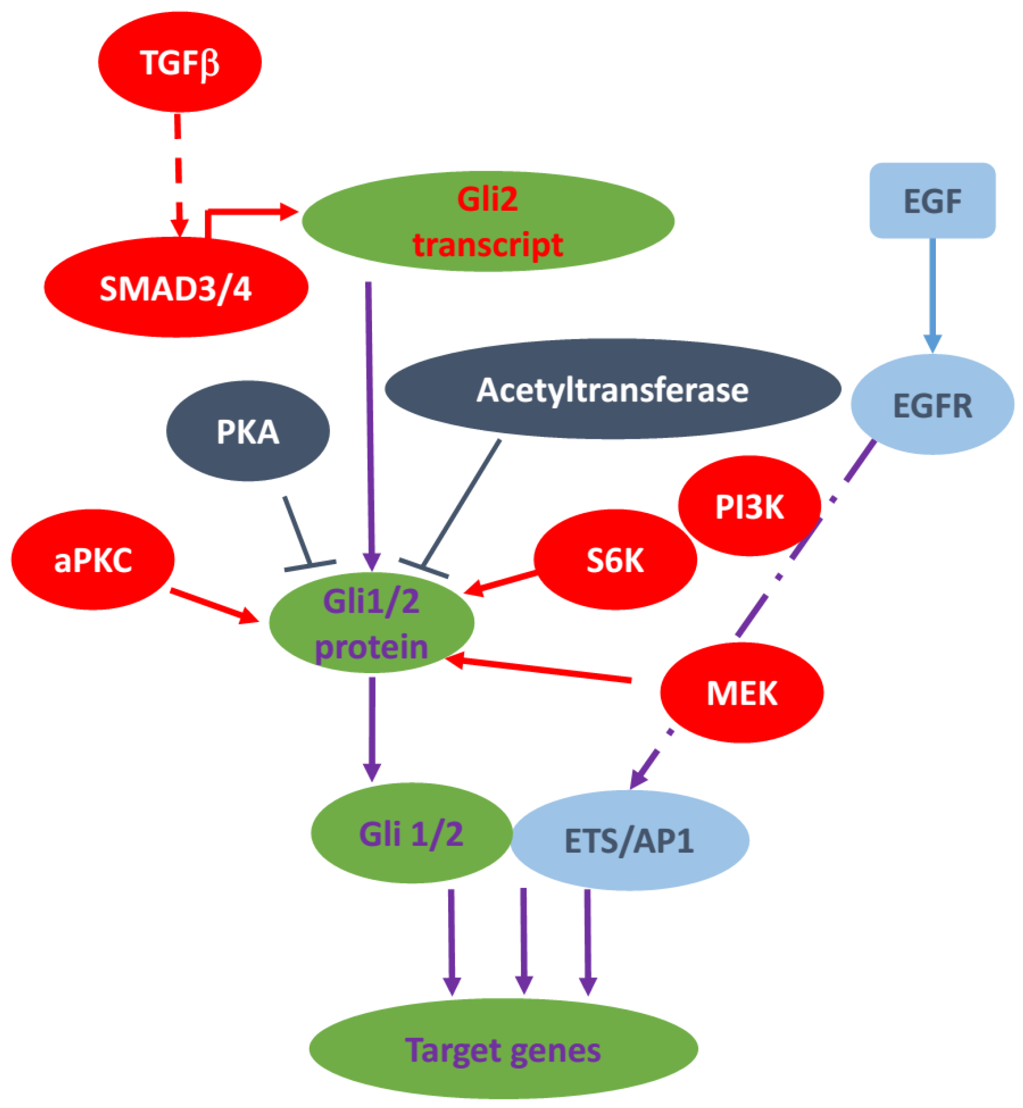
3. Non-Canonical Hedgehog Signaling in Cancer

In normal situation, the transcription factor Gli is activated through ligand binding of PTCH1 and activation of SMO and this is the canonical Hh signaling pathway. However, in some situations, the Gli transcription factors can be activated by other molecules/signaling independent of ligand and SMO. This type of Gli activation is called non-canonical Hh signaling. Non-canonical Hh signaling is mainly investigated in the context of malignant diseases. The molecules/signaling pathways that can bypass the ligand-receptor signaling axis to activate Hh signaling pathway include Kras signaling, TGFβ, PI3K, PKC and epigenetic regulators.

3.1. The RAS-RAF-MEK Signaling Axis

Hedgehog signaling plays a functionally important role in the genesis of Pancreatic ductal adenocarcinoma (PDAC) [34,35]. However, in the PDAC mouse model with oncogenic Kras expression, conditional deletion of Smo in ductal cells does not affect GLI1 expression in cancer cells and had no effects on the multistage development of PDAC tumors [36], indicating that ligand-receptor Hh signaling is dispensable in pancreatic ductal cells for PDAC progression, and GLI transcription in the neoplastic ductal cells is regulated through alternative manners. Since Kras mutation presents almost universally in PDA and is one of the earliest genetic alterations, it is reasonable to speculate that the RAS/RAF/MEK signaling plays a vital role in the non-canonical activation of Hh signaling in cancer cells.
Indeed, ectopic expression of oncogenic Kras in normal human pancreatic cell line HPDE-c7 can increase transcription activity of Gli molecules [37]. Similar results are also observed after overexpression of mutant Kras in BxPC3 cells, a pancreatic cancer cell line with wild type Kras [36]. Depletion of oncogenic Kras with Kras-targeted siRNAs resulted in a significant down-regulation of the transcription activity of GLI, as indicated by GLI1 and PTCH1 mRNAs [37] in PDAC cell lines in vitro. In gastric cancer cells, the KRAS-MEK-ERK cascade has a positive regulatory role in GLI transcriptional activity [38]. How RAS/RAF/MEK/MAPK cascade causes activation of Gli1 transcription remains to be elucidated. It has been reported that GLI transcriptional activity is regulated by the pattern of Gli phosphorylation [39]. In 3T3 cells, if co-transfected with an activated MEK construct, the activity of overexpressed GLI1 can be enhanced by 10 times [40], this enhancement was abolished in an N-terminal deletion mutant of Gli1 lacking the first 130 amino acids [40]; in another report, it was shown that the Ser130 of Gli1 transcription factor are bound and phosphorylated by MAPK cascade [41]. Based on these data, it is reasonable to speculate that the transcription activity of Gli protein could be directly regulated by phosphorylation of the KRAS-MEK-ERK cascade, but this hypothesis need to be confirmed by further study. On the other hand, Gli activity may also be regulated by the KRAS-MEK-ERK cascade by preventing Gli protein degradation. It has been reported that oncogenic Kras blocks proteasome-mediated GLI1 degradation and consequently leads to the activation of Hh signaling in pancreatic cancer cells [37]. In keratinocytes, ERK1/2 activated by EGFR also stabilizes GLI proteins, particularly GLI2, by preventing its degradation via the proteasome pathway [33] (see Figure 1).
Figure 1. A diagram of typical non-canonical Gli1/2 signaling. There are several ways for regulating Gli transcriptional factors independent of smoothened and Hh ligands. In melanomas, Gli2 transcription is regulated by TGFβ signaling. In esophageal cancer, Gli1 can be phosphorylated by S6K to enhance Gli1 transcriptional activity. In BCCs, a typical PKCi/l can phosphorylate Gli transcription factors to enhance their activities. Previously, it is reported that PI3K and MEK can regulate Gli transcription factors. In addition to the positive regulation, PKA and acetyltransferase are known to negatively regulate Gli protein transcriptional activity. Furthermore, Gli1/2 can also work with downstream effectors of EGF signaling to control transcription of certain target genes.
Cancers 07 00857 g001
Through the RAS/RAF/MEK cascade, it is possible that the up-stream growth factor receptor tyrosine kinase (RTK) can affect the activity of GLI in a SMO-independent manner or by potentiating the existing Hh Signaling. In human normal keratinocytes, EGFR can transduce signal through RAF/MEK/ERK to cooperate with GLI proteins to selectively regulate some target genes [33], and synergistically induce oncogenic transformation [42]. These findings have been further confirmed in a transgenic BCC mouse model and a subcutaneous xenograft model of pancreatic cancer by genetic and pharmacologic inhibition of EGFR signaling [43], as well as in medulloblastoma [44] and prostate cancer [45]. In NIH3T3 cells, bFGF stimulates GLI1-luciferase activity in a MEK-1-dependent manner [40], but in medulloblastoma, FGF is shown as a canonical shh signaling inhibitor in an ERK dependent manner [46], indicating a significant effect of cellular context in the output of signal interaction (see more in Figure 1).

3.2. TGFβ Signaling Pathway

TGFβ and its receptors are widely expressed in human body, and its signaling plays a major role in human diseases. During the progression of malignant disorders, TGFβ signaling acts both as a tumor suppressor and as a tumor promoter depending on tumor context (see review [47]). There is increasing evidence to show that TGFβ signal transduction is able to interact with the HH pathway downstream of SMO. Firstly, TGF-β/smad3 cascade was identified as a potent inducer of GLI2 expression and consequently GLI1 expression in various cancer cell lines, normal fibroblasts and in keratinocytes [48].
Moreover, GLI2 induction by TGFβ is independent from the Hh/Ptch/Smo axis and de novo protein synthesis [48]. In SMO-deleted pancreatic cancer cell line from mouse, TGFβ treatment leads to marked elevation of GLI1 and GLI3, even when GLI2 expression is undetectable [36]. In a bone metastasis model of breast cancer cell MDA-231, Gli2 in cancer cell induces secretion of PTHrP, an important osteolytic factor, and promotes bone destruction [49]. In this MDA-231 cell line, the SMO mRNA is not detectable, and GLI2 expression was not repressed by SMO inhibitor cyclopamine but regulated by TGFβ signaling (see Figure 1). GLI2 expression and osteolytic ability of metastatic tumor can be inhibited by abolishing TGFβ signaling, indicating SMO-downstream regulation of GLI2 via TGFβ cascade [49]. In a series of bladder cancer cell lines, it was found that Hh-independent GLI2 expression and function contributes to invasiveness. The authors suggested that non-canonical GLI2 activity may be attributed to RAS and TGF-β, both of these pathways being critical for bladder cancer development [50]. In agreement of these studies, we found TGFβ signaling is critical for Hh signaling-mediated biological functions and carcinogenesis [51].
All these studies demonstrated that TGFβ signaling is a regulator of Hh cascade independent of SMO function. To address the mechanism of non-canonical regulation of GLI by TGFβ signaling, Dennler and colleagues cloned and analyzed human GLI2 promoter and identified a 91 bp region harboring SMAD and lymphoid enhancer factor/T cell factor binding sites [52]. This study indicates that in response to TGF-β stimulation, SMAD3 and β-catenin are able to be recruited to the promoter and drive GLI2 gene transcription. In summary, all these data demonstrate that TGF-β signaling can regulate Gli2 activity by promoting its transcription directly.

3.3. PKC Signaling

Protein kinase C (PKC) is a family of widely distributed phospholipid-dependent serine/threonine kinases, which can be divided into 3 groups: conventional or classic PKC isozymes (cPKCs; α, βI, βII, and γ), novel or non-classic PKC isozymes (nPKCs; δ, ε, η, and θ), and atypical PKC isozymes (aPKCs; ζ, ι, and λ). As a receptor for the tumor-promoting phorbol esters, the function of PKC in cancer has been intensely investigated in the context of cancer [53]. The crosstalk between Hh signaling and PKC signaling has been reviewed [54].
Here, we mainly discuss the studies about how PKC signaling activates Gli transcription factors without going through SMO. The role of PKC in Gli activity is controversial. By using LIGHT2 cells, as well as NIH/3T3 cells, Riobo et al. found that PMA, a classical PKC and novel PKC stimulator, can induce GLI-luciferase reporter activity in this cell line through PKCδ [40]. This activation can be blocked by the selective MEK-1 inhibitor PD98059 or the dual MEK-1/2 inhibitor U0126, indicating that the PKC/MEK/ERK axis drives the activation of GLI upon PMA stimulation. Neill and colleagues also found that PKCδ promotes GLI1 expression and PKCα decrease GLI1 expression in 293 T cells independent of MAPK signaling [55]. Cai and colleagues reported that PKCα increases Gli1 activity via MEK/ERK pathway and PKCδ negatively regulates Gli1 expression through transcriptional down-regulation of GLI1 mRNA in NIH/3T3 cells and human Hepatoma Hep3B Cells [56]. In mouse BCC cell lines, aPKC-ι/λ was identified as a novel GLI regulator. Although functioning downstream of SMO, aPKC-ι/λ phosphorylates and activates GLI1, resulting in maximal DNA binding and transcriptional activation [57]. In addition, aPKCι is also an Hh target gene, and is required for maximal sustained Hh signaling [57]. In summary, the function of PKC signaling the activation of non-canonical Hh signaling is complicated and might be cell type dependent or subtype of PKC dependent (see Figure 1).

3.4. AKT/PI3K Signaling

Riobo and colleagues demonstrated that activation of PI3-kinase/Akt increases Shh-induced Gli transcriptional activity through antagonizing PKA-dependent Gli2 inactivation in several experimental systems [58]. Since loss-of-function mutations in the PTEN, a negative regulator of PI3-kinase activity, occur frequently in human cancer and overexpression of IGF I exists in malignant disease, this study indicated that activated AKT/PI3K may facilitate Hh signaling pathway and promote tumor formation. In human PTEN-deficient glioblastomas, Hh signaling is activated [59]. Compensatory up-regulation of IGF-1R-PI3K is a candidate for the development of Smo antagonist resistant medulloblastoma, and combination of PI3K inhibitor NVP-BKM120 or the dual PI3K-mTOR inhibitor NVP-BEZ235 with the Smo antagonist markedly delayed the development of resistance [60].
In mouse melanoma cells, COS7 cells, human prostate cancer LNCaP cells and human glioma U87 cells, the acquisition of oncogenic RAS-RAF-MEK and/or AKT signaling could be responsible for cell-intrinsic enhancement of GLI1 transcriptional activity driven by enhanced nuclear localization and transcriptional activity [61].

3.5. TNFα/mTOR Pathway

In a recent study in esophageal adenocarcinomas, it was revealed that GLI1 activity can be enhanced by TNFα/mTOR pathway independent of SMO activity [62]. TNFα induces mTOR activation, and then mTOR phosphorylates its target S6K1. GLI1 protein can be phosphorylated by activated S6K1 at ser84, and this phosphorylation enhances GLI1 function by attenuating SuFu-mediated Gli1 inhibition [62]. This study also confirmed the importance of phosphorylation in the regulation of GLI proteins function.

3.6. Epigenetic Regulation of GLI

Cancer epigenetics is a rapidly developing field of cancer research since more and more epigenetic regulators are found mutated in malignant diseases and more and more evidence is showing the important role of epigenetic dysregulation in cancer growth and metastasis. Not surprising, many compounds have been reported targeting components of the epigenetic machineries for cancer therapy.
Recently there are some data showing that Hh pathway can be regulated epigenetically during cancer development. SNF5, a tumor suppressor, is core component of the SWI-SNF chromatin remodeling complex. Jagani and coworkers found that SNF5 interacts with GLI1 and is enriched at Gli1-regulated promoter region [63]. Inactivation of SNF5 results in constitutive up-regulation of GLI1 transcriptional activity in malignant rhabdoid tumors cells in vivo and in vitro [63]. Although detailed mechanisms remain unclear, this study provided a link between abnormal activation of the Hh pathway and deregulated chromatin remodeling in tumor development.
Multiple endocrine neoplasia type 1 (MEN-1 syndrome), a hereditary condition associated with tumors of the endocrine (hormone producing) glands, and the tumor suppressor gene MEN1 is frequently mutated in this disease. Gurung and colleagues found that menin combines with Gli1 promoter, and recruits protein arginine methyltransferase 5 (PRMT5) to promote repressive histone arginine methylation, leading to decreased Gli1 expression independent of the canonical GAS1/PTCH1/SMO-mediated Hh signaling [64]. This study revealed a novel non-canonical manner controlling the levels of GLI1 via a menin-mediated epigenetic pathway, and provided a possible mechanism of tumor development of MEN-1 disease, and a rationale for directly inhibiting GLI1 for treating these tumors. Malatesta et al. reported that p300/CREB-binding protein-associated factor (PCAF), a histone acetyltransferase, interacts with GLI1 and is recruited to the promoter of GLI1 responsive gene leading to H3K9 acetylation of Hh target gene promoters and their activation in medulloblastoma and glioblastoma cells, and PCAF downregulation in these cells leads to decreased proliferation and increased apoptosis [65]. However, in hepatocellular carcinoma (HCC) cells, PCAF suppresses Hh signaling by directly acetylating cytoplasmic GLI1 protein at lysine 518, preventing its nuclear translocation and promoter occupancy [66], whereas deacetylation of Gli1 and Gli2 by histone deacetylases (HDAC) promotes their transcriptional activation in medulloblastoma [67].
BRD4, a conserved member of the bromodomain and extraterminal (BET) family of chromatin readers, plays a critical role in transcription by binding acetylated histones and recruiting transcriptional complexes [68]. Recently, two groups revealed that BRD4 and other BET members can occupy GLI1 and GLI2 promoters directly, and enhance their transcription [69,70]. BRD4 inhibitor JQ1 or I-BET151 treatment inhibits GLI1 and GLI2 expression and growth of hedgehog-driven tumors in vitro and in vivo, such as basal cell carcinoma, medulloblastoma and atypical teratoid rhabdoid tumor [69,70]. However, the details how BRD4 is regulated and involves in Gli activation, and whether BRD4 facilitates Hh independent non-canonical Gli regulation are needed for further investigation.

3.7. Other Non-Canonical Regulations of Hedgehog Signaling

Ewing sarcoma family of tumors (ESFT) is characterized by producing an aberrant fusion gene EWS-FLI1 resulting from the chromosomal translocation t(11;22). EWS-FLI1 can induce GLI1 transcription by direct binding to the Gli1 promoter, and overexpressed GLI1 is an important mediator in ESFT development [71]. Earlier study has revealed direct binding of PTCH1 with cyclinB1 in cancer cells, leading to delayed cell cycling [31]. Several studies have indicated signaling of SMO to G-proteins, which is independent of Gli transcription factors [32,72]. How much this G-protein coupling is involved in cancer development is still an unanswered question.
Gli1 activity is also regulated by energy sensor, AMP-activated protein kinase (AMPK). Recently, Li et al. reported that in the situation of insufficient cellular energy, AMPK is activated by accumulated ADP and AMP, and then phosphorylates directly GLI1 at serines 102 and 408 and threonine 1074, leading to suppression of GLI1 transcription activity. In this manner, when energy stores are inadequate, cells or organs slow or postpone developmental steps driven by Hh signaling in order to enhance the chance of survival of a growing animal [73]. In medulloblastoma, this mechanism of Gli1 regulation also exists, and mutation of these three phosphorylation sites may prevent phosphorylation by AMPK and leads to increased GLI1 protein stability, transcriptional activity, and oncogenic ability.

4. Conclusions

It is clear that regulation of Gli transcription factors is not only restricted to the canonical Hh/SMO-dependent cascade but also through the convergence of multiple SMO-independent pathways. These findings not only explain the ineffectiveness and failed clinical trials using SMO antagonists, and also encourage developing new strategies to suppress non-canonical Hh signaling.
Unlike the canonical pathway, there are different ways by which non-canonical Hh signaling can occur. In some cancer types, more than one type of non-canonical Hh signaling may co-exist. Thus, there is a need of paradigm shift in designing strategies to block hedgehog signaling in cancer.
First, canonical Hh signaling in cancer is quite limited to tumors with gene mutations in PTCH1 and SMO, which will be sensitive to SMO signaling inhibitors. In most cancer cells, one or more non-canonical Hh signaling modes of action play a critical role in cancer development. Thus, there is an urgent need to understand the specific non-canonical signaling mechanism in a given tumor type before novel mitigation strategies can be established.
Second, due to the co-existence of activation of multiple signaling pathways in a given cancer types, as well as different mechanisms of non-canonical Hh signaling, combined target therapy will be more effective in suppressing tumor progression.
Third, despite the existence of multiple aberrant signaling in a given cancer cells, each alteration may affect different stages of cancer development. Thus, physiologically relevant mouse models will be needed to test the role of each aberrant signaling.
Taken together, we believe that the unsuccessful clinical trials using SMO signaling inhibitors have revealed novel mechanisms underlying Hh signaling activation in cancer, particularly non-canonical Hh signaling. Thus, inhibition of Hh signaling at the level of GLI transcription factors, rather than the level of SMO, might be a more effective way to combat cancer [74,75,76,77]. Our efforts in understanding non-canonical Hh signaling may lead to novel strategies to treat cancer.

Acknowledgments

Our research was supported by National Cancer Institute (R01Ca155086), The Wells Center for Pediatric Research, Riley Children’s Foundation and Jeff Gordon Children’s Foundation.

Author Contributions

Dongsheng Gu wrote the draft, Jingwu Xie edited the manuscript and drew Figure 1.

Conflicts of Interest

The authors declare no conflict of interest

References

  1. Hahn, H.; Wicking, C.; Zaphiropoulous, P.G.; Gailani, M.R.; Shanley, S.; Chidambaram, A.; Vorechovsky, I.; Holmberg, E.; Unden, A.B.; Gillies, S.; et al. Mutations of the human homolog of drosophila patched in the nevoid basal cell carcinoma syndrome. Cell 1996, 85, 841–851. [Google Scholar] [CrossRef]
  2. Johnson, R.L.; Rothman, A.L.; Xie, J.; Goodrich, L.V.; Bare, J.W.; Bonifas, J.M.; Quinn, A.G.; Myers, R.M.; Cox, D.R.; Epstein, E.H., Jr.; et al. Human homolog of patched, a candidate gene for the basal cell nevus syndrome. Science 1996, 272, 1668–1671. [Google Scholar] [CrossRef] [PubMed]
  3. Epstein, E., Jr. Genetic determinants of basal cell carcinoma risk. Med. Pediatr. Oncol. 2001, 36, 555–558. [Google Scholar] [CrossRef] [PubMed]
  4. Tostar, U.; Malm, C.J.; Meis-Kindblom, J.M.; Kindblom, L.G.; Toftgard, R.; Unden, A.B. Deregulation of the hedgehog signalling pathway: A possible role for the ptch and sufu genes in human rhabdomyoma and rhabdomyosarcoma development. J. Pathol. 2006, 208, 17–25. [Google Scholar] [CrossRef] [PubMed]
  5. Pressey, J.G.; Anderson, J.R.; Crossman, D.K.; Lynch, J.C.; Barr, F.G. Hedgehog pathway activity in pediatric embryonal rhabdomyosarcoma and undifferentiated sarcoma: A report from the children’s oncology group. Pediatr. Blood Cancer 2011, 57, 930–938. [Google Scholar] [CrossRef] [PubMed]
  6. Clark, V.E.; Erson-Omay, E.Z.; Serin, A.; Yin, J.; Cotney, J.; Ozduman, K.; Avsar, T.; Li, J.; Murray, P.B.; Henegariu, O.; et al. Genomic analysis of non-NF2 meningiomas reveals mutations in TRAF7, KLF4, AKT1, and SMO. Science 2013, 339, 1077–1080. [Google Scholar] [CrossRef] [PubMed]
  7. Aavikko, M.; Li, S.P.; Saarinen, S.; Alhopuro, P.; Kaasinen, E.; Morgunova, E.; Li, Y.; Vesanen, K.; Smith, M.J.; Evans, D.G.; et al. Loss of SUFU function in familial multiple meningioma. Am. J. Hum. Genet. 2012, 91, 520–526. [Google Scholar] [CrossRef] [PubMed]
  8. Kijima, C.; Miyashita, T.; Suzuki, M.; Oka, H.; Fujii, K. Two cases of nevoid basal cell carcinoma syndrome associated with meningioma caused by a PTCH1 or SUFU germline mutation. Familial Cancer 2012, 11, 565–570. [Google Scholar] [CrossRef] [PubMed]
  9. Hahn, H.; Wojnowski, L.; Zimmer, A.M.; Hall, J.; Miller, G.; Zimmer, A. Rhabdomyosarcomas and radiation hypersensitivity in a mouse model of Gorlin syndrome. Nat. Med. 1998, 4, 619–622. [Google Scholar] [CrossRef] [PubMed]
  10. Hatley, M.E.; Tang, W.; Garcia, M.R.; Finkelstein, D.; Millay, D.P.; Liu, N.; Graff, J.; Galindo, R.L.; Olson, E.N. A mouse model of rhabdomyosarcoma originating from the adipocyte lineage. Cancer Cell 2012, 22, 536–546. [Google Scholar] [CrossRef] [PubMed]
  11. Ignatius, M.S.; Chen, E.; Elpek, N.M.; Fuller, A.Z.; Tenente, I.M.; Clagg, R.; Liu, S.; Blackburn, J.S.; Linardic, C.M.; Rosenberg, A.E.; et al. In vivo imaging of tumor-propagating cells, regional tumor heterogeneity, and dynamic cell movements in embryonal rhabdomyosarcoma. Cancer Cell 2012, 21, 680–693. [Google Scholar] [CrossRef] [PubMed]
  12. Nitzki, F.; Zibat, A.; Frommhold, A.; Schneider, A.; Schulz-Schaeffer, W.; Braun, T.; Hahn, H. Uncommitted precursor cells might contribute to increased incidence of embryonal rhabdomyosarcoma in heterozygous Patched1-mutant mice. Oncogene 2011, 30, 4428–4436. [Google Scholar] [CrossRef] [PubMed]
  13. Pelczar, P.; Zibat, A.; van Dop, W.A.; Heijmans, J.; Bleckmann, A.; Gruber, W.; Nitzki, F.; Uhmann, A.; Guijarro, M.V.; Hernando, E.; et al. Inactivation of Patched1 in mice leads to development of gastrointestinal stromal-like tumors that express Pdgfralpha but not kit. Gastroenterology 2013, 144, 134–144. [Google Scholar] [CrossRef] [PubMed]
  14. Park, K.S.; Martelotto, L.G.; Peifer, M.; Sos, M.L.; Karnezis, A.N.; Mahjoub, M.R.; Bernard, K.; Conklin, J.F.; Szczepny, A.; Yuan, J.; et al. A crucial requirement for hedgehog signaling in small cell lung cancer. Nat. Med. 2011, 17, 1504–1508. [Google Scholar] [CrossRef] [PubMed]
  15. Wang, D.H.; Clemons, N.J.; Miyashita, T.; Dupuy, A.J.; Zhang, W.; Szczepny, A.; Corcoran-Schwartz, I.M.; Wilburn, D.L.; Montgomery, E.A.; Wang, J.S.; et al. Aberrant epithelial-mesenchymal hedgehog signaling characterizes barrett’s metaplasia. Gastroenterology 2010, 138, 1810–1822. [Google Scholar] [CrossRef] [PubMed]
  16. Yang, L.; Wang, L.S.; Chen, X.L.; Gatalica, Z.; Qiu, S.; Liu, Z.; Stoner, G.; Zhang, H.; Weiss, H.; Xie, J. Hedgehog signaling activation in the development of squamous cell carcinoma and adenocarcinoma of esophagus. Int. J. Biochem. Mol. Biol. 2012, 3, 46–57. [Google Scholar] [PubMed]
  17. Tian, H.; Callahan, C.A.; DuPree, K.J.; Darbonne, W.C.; Ahn, C.P.; Scales, S.J.; de Sauvage, F.J. Hedgehog signaling is restricted to the stromal compartment during pancreatic carcinogenesis. Proc. Natl. Acad. Sci. USA 2009, 106, 4254–4259. [Google Scholar] [CrossRef] [PubMed]
  18. Mao, J.; Ligon, K.L.; Rakhlin, E.Y.; Thayer, S.P.; Bronson, R.T.; Rowitch, D.; McMahon, A.P. A novel somatic mouse model to survey tumorigenic potential applied to the hedgehog pathway. Cancer Res. 2006, 66, 10171–10178. [Google Scholar] [CrossRef] [PubMed]
  19. Aszterbaum, M.; Epstein, J.; Oro, A.; Douglas, V.; LeBoit, P.E.; Scott, M.P.; Epstein, E.H., Jr. Ultraviolet and ionizing radiation enhance the growth of bccs and trichoblastomas in patched heterozygous knockout mice. Nat. Med. 1999, 5, 1285–1291. [Google Scholar] [CrossRef] [PubMed]
  20. Goodrich, L.V.; Milenkovic, L.; Higgins, K.M.; Scott, M.P. Altered neural cell fates and medulloblastoma in mouse patched mutants. Science 1997, 277, 1109–1113. [Google Scholar] [CrossRef] [PubMed]
  21. Feldmann, G.; Dhara, S.; Fendrich, V.; Bedja, D.; Beaty, R.; Mullendore, M.; Karikari, C.; Alvarez, H.; Iacobuzio-Donahue, C.; Jimeno, A.; et al. Blockade of hedgehog signaling inhibits pancreatic cancer invasion and metastases: A new paradigm for combination therapy in solid cancers. Cancer Res. 2007, 67, 2187–2196. [Google Scholar] [CrossRef] [PubMed]
  22. Bailey, J.M.; Swanson, B.J.; Hamada, T.; Eggers, J.P.; Singh, P.K.; Caffery, T.; Ouellette, M.M.; Hollingsworth, M.A. Sonic hedgehog promotes desmoplasia in pancreatic cancer. Clin. Cancer Res. 2008, 14, 5995–6004. [Google Scholar] [CrossRef] [PubMed]
  23. Chang, Q.; Foltz, W.D.; Chaudary, N.; Hill, R.P.; Hedley, D.W. Tumor-stroma interaction in orthotopic primary pancreatic cancer xenografts during hedgehog pathway inhibition. Int. J. Cancer 2013, 133, 225–234. [Google Scholar] [CrossRef] [PubMed]
  24. Tang, S.N.; Fu, J.; Nall, D.; Rodova, M.; Shankar, S.; Srivastava, R.K. Inhibition of sonic hedgehog pathway and pluripotency maintaining factors regulate human pancreatic cancer stem cell characteristics. Int. J. Cancer 2012, 131, 30–40. [Google Scholar] [CrossRef] [PubMed]
  25. Feldmann, G.; Fendrich, V.; McGovern, K.; Bedja, D.; Bisht, S.; Alvarez, H.; Koorstra, J.B.; Habbe, N.; Karikari, C.; Mullendore, M.; et al. An orally bioavailable small-molecule inhibitor of hedgehog signaling inhibits tumor initiation and metastasis in pancreatic cancer. Mol. Cancer Ther. 2008, 7, 2725–2735. [Google Scholar] [CrossRef] [PubMed]
  26. Gu, D.; Liu, H.; Su, G.H.; Zhang, X.; Chin-Sinex, H.; Hanenberg, H.; Mendonca, M.S.; Shannon, H.E.; Chiorean, E.G.; Xie, J. Combining hedgehog signaling inhibition with focal irradiation on reduction of pancreatic cancer metastasis. Mol. Cancer Ther. 2013, 12, 1038–1048. [Google Scholar] [CrossRef] [PubMed]
  27. Bailey, J.M.; Mohr, A.M.; Hollingsworth, M.A. Sonic hedgehog paracrine signaling regulates metastasis and lymphangiogenesis in pancreatic cancer. Oncogene 2009, 28, 3513–3525. [Google Scholar] [CrossRef] [PubMed]
  28. Olive, K.P.; Jacobetz, M.A.; Davidson, C.J.; Gopinathan, A.; McIntyre, D.; Honess, D.; Madhu, B.; Goldgraben, M.A.; Caldwell, M.E.; Allard, D.; et al. Inhibition of hedgehog signaling enhances delivery of chemotherapy in a mouse model of pancreatic cancer. Science 2009, 324, 1457–1461. [Google Scholar] [CrossRef] [PubMed]
  29. Zhao, C.; Chen, A.; Jamieson, C.H.; Fereshteh, M.; Abrahamsson, A.; Blum, J.; Kwon, H.Y.; Kim, J.; Chute, J.P.; Rizzieri, D.; et al. Hedgehog signalling is essential for maintenance of cancer stem cells in myeloid leukaemia. Nature 2009, 458, 776–779. [Google Scholar] [CrossRef] [PubMed]
  30. Boyd, A.L.; Salci, K.R.; Shapovalova, Z.; McIntyre, B.A.; Bhatia, M. Nonhematopoietic cells represent a more rational target of in vivo hedgehog signaling affecting normal or acute myeloid leukemia progenitors. Exp. Hematol. 2013, 41, 858–869.e4. [Google Scholar] [CrossRef] [PubMed]
  31. Barnes, E.A.; Kong, M.; Ollendorff, V.; Donoghue, D.J. Patched1 interacts with cyclin B1 to regulate cell cycle progression. EMBO J. 2001, 20, 2214–2223. [Google Scholar] [CrossRef] [PubMed]
  32. Ogden, S.K.; Fei, D.L.; Schilling, N.S.; Ahmed, Y.F.; Hwa, J.; Robbins, D.J. G protein galphai functions immediately downstream of smoothened in hedgehog signalling. Nature 2008, 456, 967–970. [Google Scholar] [CrossRef] [PubMed]
  33. Kasper, M.; Schnidar, H.; Neill, G.W.; Hanneder, M.; Klingler, S.; Blaas, L.; Schmid, C.; Hauser-Kronberger, C.; Regl, G.; Philpott, M.P.; et al. Selective modulation of Hedgehog/GLI target gene expression by epidermal growth factor signaling in human keratinocytes. Mol. Cell. Biol. 2006, 26, 6283–6298. [Google Scholar] [CrossRef] [PubMed]
  34. Berman, D.M.; Karhadkar, S.S.; Maitra, A.; Montes De Oca, R.; Gerstenblith, M.R.; Briggs, K.; Parker, A.R.; Shimada, Y.; Eshleman, J.R.; Watkins, D.N.; et al. Widespread requirement for hedgehog ligand stimulation in growth of digestive tract tumours. Nature 2003, 425, 846–851. [Google Scholar] [CrossRef] [PubMed]
  35. Morton, J.P.; Mongeau, M.E.; Klimstra, D.S.; Morris, J.P.; Lee, Y.C.; Kawaguchi, Y.; Wright, C.V.; Hebrok, M.; Lewis, B.C. Sonic hedgehog acts at multiple stages during pancreatic tumorigenesis. Proc. Natl. Acad. Sci. USA 2007, 104, 5103–5108. [Google Scholar] [CrossRef] [PubMed]
  36. Nolan-Stevaux, O.; Lau, J.; Truitt, M.L.; Chu, G.C.; Hebrok, M.; Fernandez-Zapico, M.E.; Hanahan, D. GLI1 is regulated through smoothened-independent mechanisms in neoplastic pancreatic ducts and mediates PDAC cell survival and transformation. Genes Dev. 2009, 23, 24–36. [Google Scholar] [CrossRef] [PubMed]
  37. Ji, Z.; Mei, F.C.; Xie, J.; Cheng, X. Oncogenic KRAS activates hedgehog signaling pathway in pancreatic cancer cells. J. Biol. Chem. 2007, 282, 14048–14055. [Google Scholar] [CrossRef] [PubMed]
  38. Seto, M.; Ohta, M.; Asaoka, Y.; Ikenoue, T.; Tada, M.; Miyabayashi, K.; Mohri, D.; Tanaka, Y.; Ijichi, H.; Tateishi, K.; et al. Regulation of the hedgehog signaling by the mitogen-activated protein kinase cascade in gastric cancer. Mol. Carcinogen. 2009, 48, 703–712. [Google Scholar] [CrossRef] [PubMed]
  39. Niewiadomski, P.; Kong, J.H.; Ahrends, R.; Ma, Y.; Humke, E.W.; Khan, S.; Teruel, M.N.; Novitch, B.G.; Rohatgi, R. Gli protein activity is controlled by multisite phosphorylation in vertebrate hedgehog signaling. Cell Rep. 2014, 6, 168–181. [Google Scholar] [CrossRef] [PubMed]
  40. Riobo, N.A.; Haines, G.M.; Emerson, C.P., Jr. Protein kinase C-delta and mitogen-activated protein/extracellular signal-regulated kinase-1 control GLI activation in hedgehog signaling. Cancer Res. 2006, 66, 839–845. [Google Scholar] [CrossRef] [PubMed]
  41. Whisenant, T.C.; Ho, D.T.; Benz, R.W.; Rogers, J.S.; Kaake, R.M.; Gordon, E.A.; Huang, L.; Baldi, P.; Bardwell, L. Computational prediction and experimental verification of new MAP kinase docking sites and substrates including Gli transcription factors. PLoS Comput. Biol. 2010, 6. [Google Scholar] [CrossRef] [PubMed]
  42. Schnidar, H.; Eberl, M.; Klingler, S.; Mangelberger, D.; Kasper, M.; Hauser-Kronberger, C.; Regl, G.; Kroismayr, R.; Moriggl, R.; Sibilia, M.; et al. Epidermal growth factor receptor signaling synergizes with Hedgehog/GLI in oncogenic transformation via activation of the MEK/ERK/JUN pathway. Cancer Res. 2009, 69, 1284–1292. [Google Scholar] [CrossRef] [PubMed]
  43. Eberl, M.; Klingler, S.; Mangelberger, D.; Loipetzberger, A.; Damhofer, H.; Zoidl, K.; Schnidar, H.; Hache, H.; Bauer, H.C.; Solca, F.; et al. Hedgehog-EGFR cooperation response genes determine the oncogenic phenotype of basal cell carcinoma and tumour-initiating pancreatic cancer cells. EMBO Mol. Med. 2012, 4, 218–233. [Google Scholar] [CrossRef] [PubMed] [Green Version]
  44. Gotschel, F.; Berg, D.; Gruber, W.; Bender, C.; Eberl, M.; Friedel, M.; Sonntag, J.; Rungeler, E.; Hache, H.; Wierling, C.; et al. Synergism between Hedgehog-Gli and EGFR signaling in Hedgehog-responsive human medulloblastoma cells induces downregulation of canonical Hedgehog-target genes and stabilized expression of GLI1. PLoS ONE 2013, 8, e65403. [Google Scholar] [CrossRef] [PubMed]
  45. Zhu, G.; Zhou, J.; Song, W.; Wu, D.; Dang, Q.; Zhang, L.; Li, L.; Wang, X.; He, D. Role of GLI-1 in epidermal growth factor-induced invasiveness of ARCaPE prostate cancer cells. Oncol. Rep. 2013, 30, 904–910. [Google Scholar] [PubMed]
  46. Fogarty, M.P.; Emmenegger, B.A.; Grasfeder, L.L.; Oliver, T.G.; Wechsler-Reya, R.J. Fibroblast growth factor blocks sonic hedgehog signaling in neuronal precursors and tumor cells. Proc. Natl. Acad. Sci. USA 2007, 104, 2973–2978. [Google Scholar] [CrossRef] [PubMed]
  47. Massague, J. Tgfbeta in cancer. Cell 2008, 134, 215–230. [Google Scholar] [CrossRef] [PubMed]
  48. Dennler, S.; Andre, J.; Alexaki, I.; Li, A.; Magnaldo, T.; ten Dijke, P.; Wang, X.J.; Verrecchia, F.; Mauviel, A. Induction of sonic hedgehog mediators by transforming growth factor-beta: Smad3-dependent activation of Gli2 and Gli1 expression in vitro and in vivo. Cancer Res. 2007, 67, 6981–6986. [Google Scholar] [CrossRef] [PubMed]
  49. Johnson, R.W.; Nguyen, M.P.; Padalecki, S.S.; Grubbs, B.G.; Merkel, A.R.; Oyajobi, B.O.; Matrisian, L.M.; Mundy, G.R.; Sterling, J.A. TGF-beta promotion of Gli2-induced expression of parathyroid hormone-related protein, an important osteolytic factor in bone metastasis, is independent of canonical hedgehog signaling. Cancer Res. 2011, 71, 822–831. [Google Scholar] [CrossRef] [PubMed]
  50. Mechlin, C.W.; Tanner, M.J.; Chen, M.; Buttyan, R.; Levin, R.M.; Mian, B.M. Gli2 expression and human bladder transitional carcinoma cell invasiveness. J. Urol. 2010, 184, 344–351. [Google Scholar] [CrossRef] [PubMed]
  51. Fan, Q.; He, M.; Sheng, T.; Zhang, X.; Sinha, M.; Luxon, B.; Zhao, X.; Xie, J. Requirement of TGFbeta signaling for SMO-mediated carcinogenesis. J. Biol. Chem. 2010, 285, 36570–36576. [Google Scholar] [CrossRef] [PubMed]
  52. Dennler, S.; Andre, J.; Verrecchia, F.; Mauviel, A. Cloning of the human GLI2 promoter: Transcriptional activation by transforming growth factor-beta via SMAD3/beta-catenin cooperation. J. Biol. Chem. 2009, 284, 31523–31531. [Google Scholar] [CrossRef] [PubMed]
  53. Kang, J.H. Protein kinase c (PKC) isozymes and cancer. New J. Sci. 2014, 2014, 36. [Google Scholar] [CrossRef]
  54. Li, J.; Evers, B.M. Hedgehog and Protein Kinase C Signaling. In Hedgehog Signaling Activation in Human Cancer and Its Clinical Implications; Xie, J., Ed.; Springer: New York, NY, USA, 2011; pp. 77–83. [Google Scholar]
  55. Neill, G.W.; Ghali, L.R.; Green, J.L.; Ikram, M.S.; Philpott, M.P.; Quinn, A.G. Loss of protein kinase Calpha expression may enhance the tumorigenic potential of Gli1 in basal cell carcinoma. Cancer Res. 2003, 63, 4692–4697. [Google Scholar] [PubMed]
  56. Cai, Q.; Li, J.; Gao, T.; Xie, J.; Evers, B.M. Protein kinase Cdelta negatively regulates hedgehog signaling by inhibition of Gli1 activity. J. Biol. Chem. 2009, 284, 2150–2158. [Google Scholar] [CrossRef] [PubMed]
  57. Atwood, S.X.; Li, M.; Lee, A.; Tang, J.Y.; Oro, A.E. Gli activation by atypical protein kinase C iota/lambda regulates the growth of basal cell carcinomas. Nature 2013, 494, 484–488. [Google Scholar] [CrossRef] [PubMed]
  58. Riobo, N.A.; Lu, K.; Ai, X.; Haines, G.M.; Emerson, C.P., Jr. Phosphoinositide 3-kinase and Akt are essential for Sonic Hedgehog signaling. Proc. Natl. Acad. Sci. USA 2006, 103, 4505–4510. [Google Scholar] [CrossRef] [PubMed]
  59. Filbin, M.G.; Dabral, S.K.; Pazyra-Murphy, M.F.; Ramkissoon, S.; Kung, A.L.; Pak, E.; Chung, J.; Theisen, M.A.; Sun, Y.; Franchetti, Y.; et al. Coordinate activation of Shh and PI3K signaling in PTEN-deficient glioblastoma: New therapeutic opportunities. Nat. Med. 2013, 19, 1518–1523. [Google Scholar] [CrossRef] [PubMed]
  60. Buonamici, S.; Williams, J.; Morrissey, M.; Wang, A.; Guo, R.; Vattay, A.; Hsiao, K.; Yuan, J.; Green, J.; Ospina, B.; et al. Interfering with resistance to smoothened antagonists by inhibition of the PI3K pathway in medulloblastoma. Sci. Transl. Med. 2010, 2, 51–70. [Google Scholar] [CrossRef] [PubMed]
  61. Stecca, B.; Mas, C.; Clement, V.; Zbinden, M.; Correa, R.; Piguet, V.; Beermann, F.; Ruiz, I.A.A. Melanomas require Hedgehog-GLI signaling regulated by interactions between GLI1 and the RAS-MEK/AKT pathways. Proc. Natl. Acad. Sci. USA 2007, 104, 5895–5900. [Google Scholar] [CrossRef] [PubMed]
  62. Wang, Y.; Ding, Q.; Yen, C.J.; Xia, W.; Izzo, J.G.; Lang, J.Y.; Li, C.W.; Hsu, J.L.; Miller, S.A.; Wang, X.; et al. The crosstalk of mTOR/S6K1 and hedgehog pathways. Cancer Cell 2012, 21, 374–387. [Google Scholar] [CrossRef] [PubMed]
  63. Jagani, Z.; Mora-Blanco, E.L.; Sansam, C.G.; McKenna, E.S.; Wilson, B.; Chen, D.; Klekota, J.; Tamayo, P.; Nguyen, P.T.; Tolstorukov, M.; et al. Loss of the tumor suppressor Snf5 leads to aberrant activation of the Hedgehog-Gli pathway. Nat. Med. 2010, 16, 1429–1433. [Google Scholar] [CrossRef] [PubMed]
  64. Gurung, B.; Feng, Z.; Hua, X. Menin directly represses Gli1 expression independent of canonical hedgehog signaling. Mol. Cancer Res. 2013, 11, 1215–1222. [Google Scholar] [CrossRef] [PubMed]
  65. Malatesta, M.; Steinhauer, C.; Mohammad, F.; Pandey, D.P.; Squatrito, M.; Helin, K. Histone acetyltransferase PCAF is required for Hedgehog-Gli-dependent transcription and cancer cell proliferation. Cancer Res. 2013, 73, 6323–6333. [Google Scholar] [CrossRef] [PubMed]
  66. Gai, X.; Tu, K.; Li, C.; Lu, Z.; Roberts, L.R.; Zheng, X. Histone acetyltransferase PCAF accelerates apoptosis by repressing a GLI1/BCL2/BAX axis in hepatocellular carcinoma. Cell Death Dis. 2015, 6, e1712. [Google Scholar] [CrossRef] [PubMed]
  67. Canettieri, G.; di Marcotullio, L.; Greco, A.; Coni, S.; Antonucci, L.; Infante, P.; Pietrosanti, L.; de Smaele, E.; Ferretti, E.; Miele, E.; et al. Histone deacetylase and Cullin3-REN(KCTD11) ubiquitin ligase interplay regulates hedgehog signalling through Gli acetylation. Nat. Cell Biol. 2010, 12, 132–142. [Google Scholar] [CrossRef] [PubMed]
  68. Musselman, C.A.; Lalonde, M.E.; Cote, J.; Kutateladze, T.G. Perceiving the epigenetic landscape through histone readers. Nat. Struct. Mol. Biol. 2012, 19, 1218–1227. [Google Scholar] [CrossRef] [PubMed]
  69. Long, J.; Li, B.; Rodriguez-Blanco, J.; Pastori, C.; Volmar, C.H.; Wahlestedt, C.; Capobianco, A.; Bai, F.; Pei, X.H.; Ayad, N.G.; et al. The BET bromodomain inhibitor I-BET151 acts downstream of smoothened protein to abrogate the growth of hedgehog protein-driven cancers. J. Biol. Chem. 2014, 289, 35494–35502. [Google Scholar] [CrossRef] [PubMed]
  70. Tang, Y.; Gholamin, S.; Schubert, S.; Willardson, M.I.; Lee, A.; Bandopadhayay, P.; Bergthold, G.; Masoud, S.; Nguyen, B.; Vue, N.; et al. Epigenetic targeting of hedgehog pathway transcriptional output through BET bromodomain inhibition. Nat. Med. 2014, 20, 732–740. [Google Scholar] [CrossRef] [PubMed]
  71. Beauchamp, E.; Bulut, G.; Abaan, O.; Chen, K.; Merchant, A.; Matsui, W.; Endo, Y.; Rubin, J.S.; Toretsky, J.; Uren, A. GLI1 is a direct transcriptional target of EWS-FLI1 oncoprotein. J. Biol. Chem. 2009, 284, 9074–9082. [Google Scholar] [CrossRef] [PubMed]
  72. Zhan, X.; Wang, J.; Liu, Y.; Peng, Y.; Tan, W. GPCR-like signaling mediated by smoothened contributes to acquired chemoresistance through activating Gli. Mol. Cancer 2014, 13, 4. [Google Scholar] [CrossRef] [PubMed]
  73. Li, Y.H.; Luo, J.; Mosley, Y.Y.; Hedrick, V.E.; Paul, L.N.; Chang, J.; Zhang, G.; Wang, Y.K.; Banko, M.R.; Brunet, A.; et al. AMP-activated protein kinase directly phosphorylates and destabilizes hedgehog pathway transcription factor GLI1 in medulloblastoma. Cell Rep. 2015, 12, 599–609. [Google Scholar] [CrossRef] [PubMed]
  74. Tostar, U.; Toftgard, R.; Zaphiropoulos, P.G.; Shimokawa, T. Reduction of human embryonal rhabdomyosarcoma tumor growth by inhibition of the hedgehog signaling pathway. Genes Cancer 2010, 1, 941–951. [Google Scholar] [CrossRef] [PubMed]
  75. Mazumdar, T.; Devecchio, J.; Agyeman, A.; Shi, T.; Houghton, J.A. Blocking hedgehog survival signaling at the level of the GLI genes induces DNA damage and extensive cell death in human colon carcinoma cells. Cancer Res. 2011, 71, 5904–5914. [Google Scholar] [CrossRef] [PubMed]
  76. Wickstrom, M.; Dyberg, C.; Shimokawa, T.; Milosevic, J.; Baryawno, N.; Fuskevag, O.M.; Larsson, R.; Kogner, P.; Zaphiropoulos, P.G.; Johnsen, J.I. Targeting the hedgehog signal transduction pathway at the level of GLI inhibits neuroblastoma cell growth in vitro and in vivo. Int. J. Cancer. 2013, 132, 1516–1524. [Google Scholar] [CrossRef] [PubMed]
  77. Huang, L.; Walter, V.; Hayes, D.N.; Onaitis, M. Hedgehog-GLI signaling inhibition suppresses tumor growth in squamous lung cancer. Clin. Cancer Res. 2014, 20, 1566–1575. [Google Scholar] [CrossRef] [PubMed]

