Review

The Interactions of microRNA and Epigenetic Modifications in Prostate Cancer

by
Prashant Kumar Singh
* and
Moray J. Campbell
Department of Pharmacology & Therapeutics, Roswell Park Cancer Institute, Elm and Carlton Streets, Buffalo, NY 14263, USA
*
Author to whom correspondence should be addressed.
Cancers 2013, 5(3), 998-1019; https://doi.org/10.3390/cancers5030998
Submission received: 26 June 2013 / Revised: 16 July 2013 / Accepted: 24 July 2013 / Published: 9 August 2013
(This article belongs to the Special Issue Cancer Epigenetics)

Abstract

:
Epigenetic modifiers play important roles in fine-tuning the cellular transcriptome. Any imbalance in these processes may lead to abnormal transcriptional activity and thus result in disease state. Distortions of the epigenome have been reported in cancer initiation and progression. DNA methylation and histone modifications are principle components of this epigenome, but more recently it has become clear that microRNAs (miRNAs) are another major component of the epigenome. Interactions of these components are apparent in prostate cancer (CaP), which is the most common non-cutaneous cancer and second leading cause of death from cancer in the USA. Changes in DNA methylation, altered histone modifications and miRNA expression are functionally associated with CaP initiation and progression. Various aspects of the epigenome have also been investigated as biomarkers for different stages of CaP detection, though with limited success. This review aims to summarize key aspects of these mechanistic interactions within the epigenome and to highlight their translational potential as functional biomarkers. To this end, exploration of TCGA prostate cancer data revealed that expression of key CaP miRNAs inversely associate with DNA methylation. Given the importance and prevalence of these epigenetic events in CaP biology it is timely to understand further how different epigenetic components interact and influence each other.

1. Epigenetic Contributions to Cancer

1.1. An Epigenetic Basis for Prostate Cancer

Prostate cancer (CaP) is most common noncutaneous cancer diagnosed and second leading cause of death in men from cancer in the United States [1,2]. This cancer is highly heterogeneous, ranging from asymptomatic to a rapidly fatal systemic malignancy. Several genetic and epigenetic alterations are highly prevalent and appear to be important factors in the tumorigenesis and progression of CaP. Most common genetic changes associated with CaP includes deletions involving NK3 homeobox 1 (NKX3.1) [3] and phosphatase and tensin homologue tumor suppressor genes (PTEN) [4,5], amplifications of the androgen receptor (AR) [6] and MYC [7] genes and more recently described translocations that lead to TMPRSS2:ERG and SLC45A3:ERG gene fusions [8,9]. Recently, tumor analyses with next generation sequencing approaches have identified several additional novel somatic mutations, including MED12, FOXA1 and SPOP. The E3 ubiquitin ligase adaptor speckle-type poxvirus and zinc finger (POZ) domain protein (SPOP) showed recurrent mutations in 6–15% of tumors across multiple independent cohorts [10].
The complex nature of cancer phenotypes however cannot be explained by genetic components alone [11]. Epigenetic modifications appear to contribute significantly to transformation. These events can be defined as heritable changes to the expression and regulation of gene expression that are not associated with alteration in the DNA sequences [12]. These epigenetic events including DNA methylation and histone modifications appear to play distinct yet complementary roles to genetic events and add to a fuller explanation for the basis of cancer. For example, many cancers show gene-specific and global changes in DNA CpG methylation and/or altered histone modification patterns [13,14].
Among the epigenetic modifications, altered DNA methylation and histone modification patterns have been frequently reported in CaP [15,16,17]. These epigenetic modifications govern tissues specific gene regulation by a complex machinery of several DNA and histone modifying enzymes including DNA methyltransferases (DNMTs) [18], histone acetyltransferases (HATs) [19], histone deacetylases (HDACs) [20], histone methyltransferases (HMTs) [21], histone demethylases (HDMTs) [22], and chromatin remodeling enzymes (reviewed in [13]).
One of the most prominent epigenetic modifications reported in CaP is hypermethylation of glutathione S-transferase (GSTP1) gene. GSTP1 is involved in detoxification of genotoxic and electrophilic compounds. Hypermethylation of GSTP1 is reported in 70 to 90% of patients with CaP but not in benign hyperplastic prostate tissue [23]. Similarly, up-regulation of HMT enhancer of zeste homolog 2 (EZH2) is reported in metastatic CaP, and localized CaP with higher expression of EZH2 show poorer prognosis [24]. Also, high expression of EZH2 has been reported in hormone-refractory CaP compared to benign hyperplastic prostate tissue [24,25]. EZH2 catalyzes the trimethylation of histone H3 on Lys 27 (H3K27) and is involved in gene repression and contribute to CaP tumorigenesis through silencing of tumor suppressor genes.
Recently published ENCODE project data has greatly enhanced the understanding of transcriptional landscape of the genome. Analysis across multiple cell lines suggested that as much as 75% of the genome is transcribed into different types of RNA molecules e.g., protein coding, long non-coding, pseudogenes and small RNA genes [26]. Although this percentage is the subject of significant debate [27,28], it is clear that the rate, magnitude and diversity of RNA transcription is greater than previously suspected. Of these RNA species, the microRNAs (miRNAs) have emerged as major biological regulators. MiRNAs are small (about 19–22 nucleotides) non-coding RNA molecules and modulate the expression of many, if not all, genes in the human genome. They act by inhibition of protein translation and/or degradation of mRNA transcripts [29]. Understanding their function has contributed significantly to the knowledge of how genomic information is interpreted in physiology and corrupted in malignancy [30].
It is important to understand the interplay of DNA methylation, histone modifications and miRNA and their cumulative effect on CaP initiation and progression. In last five years considerable work has been undertaken and has begun to reveal the true extent of epigenetic dis-regulation of the genome in cancer. In this review we particularly focus on how miRNAs and DNA/histone modifications interact and regulate each other and how they can be used in CaP diagnosis and treatment.

1.2. An Emergent Understanding on the Role of miRNA in Prostate Cancer

In parallel to the well-established understanding on the role of classical epigenetic events, the role of non-coding miRNA in epigenetic regulation of gene expression has also emerged. The four types of small RNA genes, namely, small nuclear (sn)RNAs, small nucleolar (sno)RNAs, miRNAs and transfer (t)RNAs correspond to 85% of total small RNAs (7,053) annotated by GENCODE. Importantly, ENCODE data revealed the wide range expression of miRNAs in different normal and cancer cell lines [26]. At any given time point 28% of miRNAs (497 of 1,756) were expressed in at least one cell line. Interestingly 30% (147 of 497) of these miRNAs were detected in all 12 cell lines analyzed [26]. This observation strengthens the idea that miRNAs are important components of gene regulation networks and expressed differentially in different disease models. Furthermore, de-regulation of miRNAs has been shown for a variety of solid tumors, including breast, colon, and prostate [31,32,33]. Thus, these studies have established an important role of miRNAs in the control of gene regulation and they are frequently de-regulated in many cancers [34,35].
Although miRNAs represent only ~1% of the genome but they are estimated to target 30% of genes [36]. The relatively small number of miRNAs and their wide coverage of protein coding genes make them very attractive to be used as disease biomarkers. Compared to mRNA signatures miRNAs have better and strong biomarker properties [37]. It has been estimated that miRNAs provide 20 times more power in biomarker studies as compared to mRNAs (when comparing 20,000 mRNAs to ~1,000 miRNAs), which makes them ideal candidates for studies with small sample size [37].
In CaP, several miRNAs have been reported to be differentially regulated and act as both tumor suppressor and onco-miRs. For instance, miR-221 and miR-222 have been shown to be upregulated in castration-resistant prostate cancer (CRPC) cells compared to androgen-dependent CaP cell line and significantly affect the response to DHT [38]. In general, oncogenic properties of miR-221 and miR-222 are attributed to their control of the cyclin dependent kinase (Cdk) inhibitors p27KIP1 and p57KIP2, and thus control of G1 to S phase transition [39,40], PI3K and PTEN signaling [41] and other targets implicated in malignant transformation, including CX43 [42], RECK [43], and ERα [44,45].
Lin et al. observed that aggressive tumors (Gleason grade ≥7) had elevated expression of miR-221 and miR-222 compared to less aggressive tumor tissues (Gleason grade <7), further suggesting an oncogenic role of these miRNAs in CaP development [46]. Conversely, others have shown the loss of miR221/222 in early stage [47] as well as in aggressive CaP [48,49]. Most recently, a miRnome wide scan also found miR-221 and miR-222 to be downregulated when comparing 20 matched pairs of microdissected tissue samples of prostate cancer and non-tumor tissue [50]. These conflicting results perhaps indicate that expression levels are differentially modulated at divergent disease stages.
Similarly, miR-125b has been identified in CaP along with miR-143 to be upregulated in metastatic CaP serum samples as compared to normal individuals [51]. Expression of miR-125b in serum of CaP patients is reported to be upregulated as compared to normal controls [51] whereas other studies reported it to be downregulated in CaP as compared to normal or benign prostatic hyperplastic (BPH) samples [47,52,53]. MiR-125 regulates cell proliferation in prostate cancer cell lines [54], and it has been suggested to be upregulated by androgen signaling [56]. Functionally in CaP, miR-125b has been reported to target BAK1 [55] (a pro-apoptotic member of the BCL-2 gene family) and EIF4EBP1 (Eukaryotic translation initiation factor 4E-binding protein 1), a gene that encodes one member of a family of translation repressor proteins [52].
Other important miRNAs involved in CaP includes miR-21 which is an AR-regulated miRNA and overexpressed in CRPC compared to adjacent normal tissue [56]. Overexpression of miR-21 was also observed in docetaxel resistant variant of PC3 cells, additionally, ectopic expression of miR-21 in wild type PC3 cells increased the resistance to docetaxel [57]. The oncogenic and drug resistance properties of miR-21 were attributed to its control of downstream target, programmed cell death 4 (PDCD4) [57,58].
Several miRNAs have also been identified as tumor suppressors in Cap including miR-143, miR-145 and miR-200 family. Expression of miR-143 and miR-145 are significantly suppressed in CaP and negatively associate with metastasis [59]. These miRs contribute to CaP progression through epithelial-mesenchymal transition (EMT) [59] and loss of their repressive effect on EGFR/RAS/MAPK pathway [60]. MiR-205 and miR-200 family miRNAs are also downregulated in CaP and have been shown to regulate EMT by targeting ZEB1 and ZEB2 in CaP [43,60,61].

2. Interplay between Histone Modifications, DNA Methylation and miRNA

Critically, the three different epigenetic modifications i.e., DNA methylation, histone modifications and miRNAs interact and influence each other [62]. For example, expression of miRNAs can be regulated by DNA methylation in their respective promoter regions [63]. Similarly miRNAs can directly target chromatin remodeling enzymes [64] and DNA methyltransferases (DNMTs) [65] and in turn modulate the downstream effects.

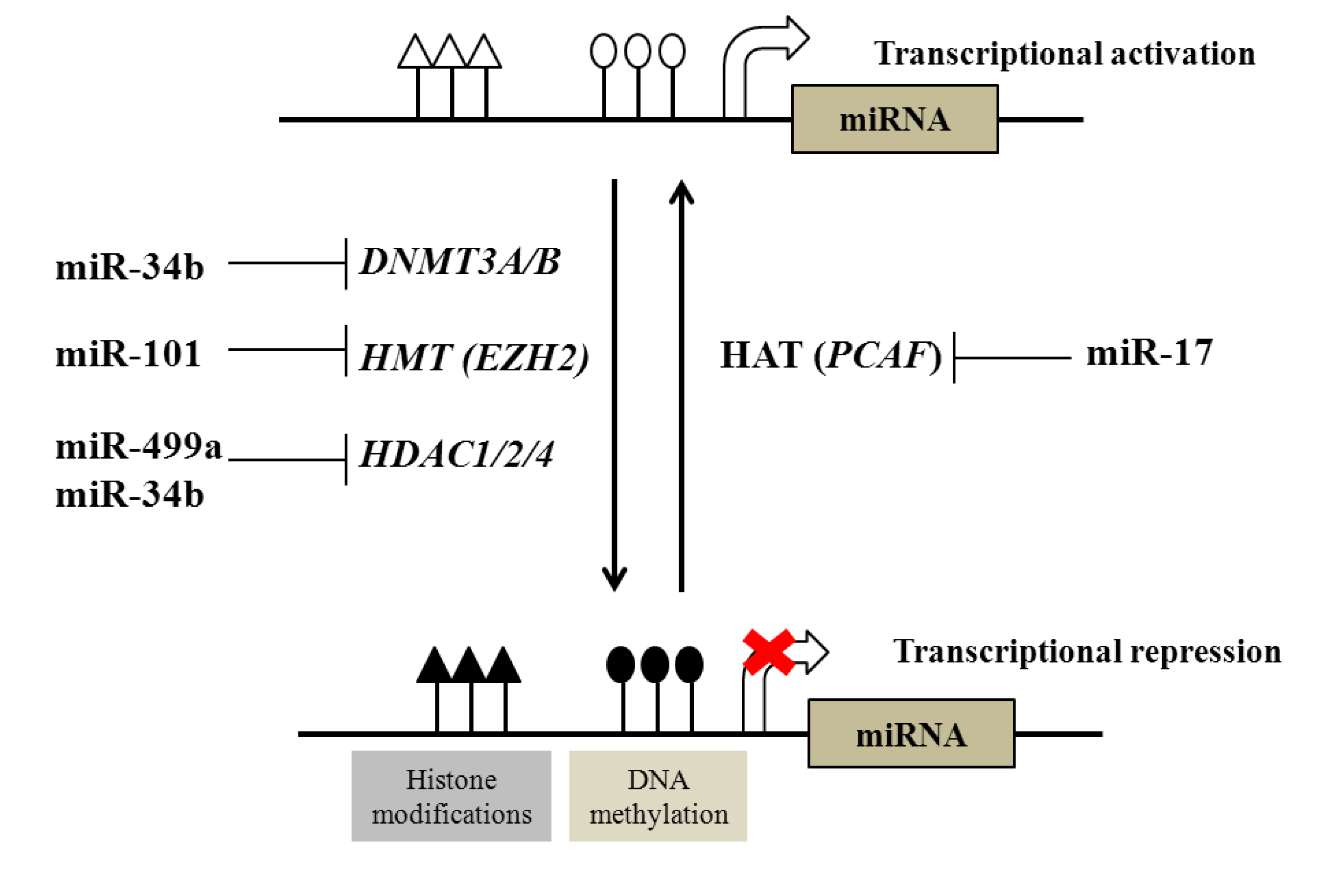
2.1. Regulation of DNA and Histone Modifications under the Control of miRNAs in Prostate Cancer

MiRNAs control gene expression post-transcriptionally by translational inhibition when base pairing between miRNA and target sequence is imperfect, while perfect or near perfect complementarity can induce the degradation of the target mRNA [66]. One miRNA can target several genes and one gene can similarly be targeted by several miRNAs [66]. The latest release of miRBase (miRBase 19) has 1,595 primary transcripts and 2,038 mature miRNA sequences. The wide range of miRNA targets makes them essential in the majority of biological processes, including cell cycle regulation, differentiation, development, metabolism and aging.
Although the biogenesis of miRNA has been well described, the regulation of miRNA expression remains less well explored. Several miRNAs have well-defined promoters but for many the regulatory elements are not as clear. They are often present in introns of protein coding genes and the promoters for the genes are considered to be promoters for the intragenic miRNAs [66,67]. Interestingly, there is evidence for differential regulation between the host mRNA and miRNA and may suggest an emergent role for alternative regulation processes.
Like their target genes, miRNAs can also directly or indirectly target key effectors of the epigenetic machinery (Figure 1). For example, miR-29 family members (29a, 29b, and 29c) are downregulated in several cancers and they are predicted to target DNA methyltransferase DNMT3A, DNMT3B and DNMT1. Indeed, enforced expression of miR-29 family members resulted in the loss of DNA methylation in lung cancer and acute myeloid leukemia and re-expression of methylation silenced genes [68,69]. Recently, miR-34b has been shown to target DNMT1, HDAC1, HDAC2 and HDAC4 in CaP cell lines [70]. Interestingly, miR-34b was also epigenetically silenced by DNA methylation and ectopic expression of miR-34b in CaP cells resulted in partial demethylation of 5' upstream sequence of the miR-34b gene and also showed enrichment of trimethylated histone H3 lysine 4 (H3K4me3), mark for an active chromatin [70]. These data provide an insight into interplay between miRNA and DNA/histone marks in CaP.
Figure 1. Interactions of DNA/histone modification and miRNA. Epigenetic regulatory mechanisms such as DNA methylation (controlled by DNMTs) and histone modifications modulate epigenetic states and transcriptional responsiveness of both mRNA and miRNA target genes. These actions are controlled by antagonistic classes of enzymes, for example by histone deacetylases (HDACs) and histone acetyl transfereases (HATs). There is good evidence for control of their expression by miRNA. For example, miR-101 targets include EZH2 [71] and miR-499a targets include HDAC1 [72], whereas the HAT (p300/CBP-associated factor (PCAF)) is targeted by miR-17-5p [73].
Cancers 05 00998 g001
A similar miRNA-mediated epigenetic cross-talk occurs with key histone modifications. Polycomb proteins form chromatin-modifying complexes that induces gene silencing by catalyzing tri-methylation of histone H3K27. There are two polycomb repressor complexes (PRCs), PRC1 and PRC2 and deregulation of these complexes has been reported to associate with many cancers [74,75]. For example, EZH2, is a highly conserved catalytic subunit within PRC2, and has been reported to be elevated in CaP and correlate with metastatic CaP and poor prognosis in localized CaP [17]. One of the important mechanisms for altered PRC2 activity arises due to elevated EZH2 level that occurs through down-regulation of miR-101 [76]; expression of miR-101 is frequently lost in CaP [34,35]. Increased EZH2 expression is associated with CaP progression and loss of miR-101 may be responsible for increased EZH2 expression in CaP [71]. MiR-101 also plays important roles in control of cell proliferation, and its expression correlates inversely with aggressiveness in CaP cell lines models [71]. Re-expression of miR-101 inhibits the expression of EZH2 and attenuates cell proliferation and tumor invasiveness [71]. More recently, knock-down of Dicer, a key protein required for miRNA processing, resulted in increased expression of other PRC components e.g., EZH2, EED, & SUZ12 (PRC2 components) and BMI1 and RING2 (PRC1 components). Together these data suggest miRNA control of the PRC complex occurs through multiple targets [77].
Upon H3K27 methylation by PRC2 proteins, PRC1 proteins are recruited to chromatin to maintain stable gene silencing by catalyzing ubiquitinylation of histone H2A. BMI1 is negatively regulated by miR-203, 200b/c while RING2 is negatively regulated by miR-181a/b and miR-200b/c [77]. Downregulation of BMI1 and RING2 by these miRNAs also resulted in decreased global ubiquityl-H2A. Interestingly, expression of these miRNAs was in turn regulated by EZH2 and silencing of EZH2 or expression of miR-101 resulted in overexpression of these miRs, and reflects the intricate co-regulation of miRNA and mRNA in feed forward loop structures [67,77,78,79,80,81,82,83]. These finding illustrated the complex interplay of epigenetic machinery and miRNA in controlling gene silencing in CaP.
MiRNA also regulate the expression of histone deacetylases (HDACs) that control gene silencing by catalyzing deacetylation of key histone residues. The class I HDACs are the most frequently overexpressed in cancers, particularly HDAC1, which is found to be overexpressed in CaP [84]. HDAC1 is a direct target of miR-499a in PC-3 cells [72], and in turn miR-449a is involved in cell-cycle regulation and is frequently downregulated in CaP. Overexpression of miR-449a in CaP cell lines leads to downregulation of HDAC1 and cell cycle arrest. The locus specific versus global effects of HDACs are emerging and interestingly the growth arrest induced by miR-449a mediates repression of HDAC1 leading to the induction of p27(kip1) [72], again suggesting the combinatorial epigenetic regulation of key growth regulators.
Another class of important histone modifiers are the histone acetyltransferases (HATs). They acetylate core histones, which is associated with easily accessible chromatin for the transcriptional machinery. In CaP p300/CBP-associated factor (PCAF), which has HAT activity, has been shown to act as a co-activator for androgen receptor (AR) [85]. Gong et al. showed that PCAF was upregulated in CaP cell lines compared to normal prostate epithelium cells and promotes DHT-stimulated AR transcriptional activity. Interestingly, they also showed that PCAF is a direct target of miR-17-5p, and it attenuated DHT-induced expression of the PSA gene and inhibited DHT-induced cell growth in LNCaP cells [73]. Taken together these observations suggest that miRNAs play an important role in fine tuning of both silencing and activating epigenetic marks in CaP (Table 1).
Table 1. Interaction of miRNA and epigenetic modifications.
Examples of miRNAs regulated by DNA methylation
DNA methylationmiR-145, miR-193b, miR-34a, miR-34b, miR-200c, miR-141, miR-205, miR-196b, miR-21, miR-615
Examples of epigenetic modifiers regulated by miRNAs
Epigenetic modifiersTargeting miRNA
DNA methyltransferases (DNTMTs)DNMT3A and DNMT3BmiR-29 family
DNMT1miR-34b
Histone methyltransferases (HMTs)EZH2miR-101
Histone deacetylases (HDACs)HDAC1, HDAC2 and HDAC4miR-34b, miR-499a
Histone acetyltransferases (HATs)PCAFmiR-17-5p

2.2. Regulation of microRNAs by DNA and Histone Modification in Prostate Cancer

Genes encoding miRNAs are as tightly regulated as any other gene in the genome and thus miRNA expression is also subjected to similar epigenetic regulation (Figure 1). Many miRNAs are present in introns of protein coding genes and they are regulated by the promoter of host gene. In this manner the epigenetic regulation of protein-coding genes can also affect the expression of miRNA. Also, many miRNAs are located in clusters in the genome and therefore more than one miRNA can be regulated in parallel by a single transcriptional event. In the genome, distribution of CpG islands around miRNA genes is similar to protein coding genes [86]. For example, mapping to the genome of differentially regulated miRNAs after DNA methyltransferase inhibition with 5-azacytidine (5-aza) showed that between 13 and 24% of human miRNA genes are located within 3 and 10 kb of a CpG island, respectively [86].
Several miRNAs are frequently deregulated in different cancers and many of them are epigenetically regulated. In CaP, miR-200 family members and miR-205 specifically are frequently altered and regulate EMT and cell migration/invasion [60,87]. Members of the miR-200 family include five miRNAs transcribed from two clusters, one on chromosome 1 encoding miR-200b, miR-200a, and miR-429 and other on chromosome 12 encoding miR-200c and miR-141. Members of miR-200 family combined with miR-205 regulate EMT and cell migration/invasion by directly targeting ZEB1 and ZEB2 [61]. MiR-205 is downregulated in the majority of cancers including CaP [88]. One of the proposed mechanisms for downregulation of miR-205 is through methylation of its promoter, and has been shown to be associated with chemotherapy drug resistance [89]. Specifically, downregulation of miR-205 arises due to a hypermethylated promoter, and the expression of miR-205 increased when cells were treatment with 5-aza. Similarly, miR-200c and miR-141 are downregulated in PC-3 cells as compared to LNCaP, and showed hypermethylation of the promoter CpG region in PC-3 and unmethylated promoter CpG in LNCaP cells. Indeed treatment with 5-aza increased the expression of miR-200c and miR-141 in PC-3 cells [90]. These findings suggest repression of miR-200c/miR-141 is epigenetically controlled in CaP.
Another miRNA that is frequently downregulated in cancers is miR-34a [91,92]. The expression of miR-34a is regulated by p53, and perhaps reflecting this, it induces apoptosis and cell-cycle arrest [93,94]. Again, miR-34a is downregulated in many CaP cell lines and the promoter region of miR-34a is hypermethylated in malignant CaP cell lines as compared to non-malignant or benign prostate hyperplasia [92]. Interestingly, similar to hypermethylation observed in CaP cell lines, the promoter of miR-34a has also been demonstrated to be methylated in the majority (79%) of primary CaP tumors as compared to normal prostate stroma [92]. Additionally, miR-34a is inactivated by aberrant CpG methylation in many other types of cancer cell lines including lung (29.1%), breast (25%), colon (13%), kidney (21.4%), bladder (33.3%) and pancreatic carcinoma (15.7%) [92]. Intriguingly, miR-34a has been shown to regulate cancer stem cell (CSCs) properties in CaP and downregulation of miR-34a in prostate cancer stem cells may contribute to metastasis by regulating migratory, invasive and metastatic properties of CSCs [95].
Other workers have investigated repressed miRNA in CaP. For example, Rauhala et al. searched for miRNAs silenced by DNA methylation in CaP cell lines and identified miR-193b to be silenced in 22Rv1 and VCaP cell lines. Expression of miR-193b was increased after treatment with 5-aza and trichostatin A (TSA) [96]. Also, miR-193b expression was significantly reduced in CaP tumors compared to adjacent normal tissue. Methylation levels at a CpG island located ~1 kb upstream of the mature miR-193b locus were higher in CaP cell lines compared to a normal prostate epithelial cell line. The methylation was partially removed after treatment with 5aza in combination with TSA, and expression of miR-193b was restored in 22Rv1 cells [96]. Overexpression of miR-193b caused reduction in cell growth and an inhibition of anchorage independent growth suggesting that miR-193b is an epigenetically silenced putative tumor suppressor in CaP [96].
Similarly, miR-145 has been reported as tumor suppressor and is frequently downregulated in many cancers including CaP [97]. MiR-145 is also regulated by p53 and inhibits tumor cell growth and invasion by targeting several genes such as c-MYC [98,99]. In CaP, miR-145 is down regulated in cell lines as well as 81% of primary tumors compared to adjacent normal tissue [100,101]. The promoter of miR-145 was hypermethylated in CaP cell lines and proximal CpG sites were completely methylated in all three cell lines tested (PC3, LNCaP and DU145), whereas distal CpG’s showed complete methylation in PC3 and partial methylation in other two cell lines [100]. The miR-145 promoter was most methylated in PC3 cells and treatment with 5-aza increased the expression of miR-145 in all three cell lines [100]. Another study from the same group revealed that the promoter of miR-145 was methylated in CaP tumors and the expression of miR-145 also correlated with mutation status of p53 [101]. Of all the CaP tumor samples tested, 81% showed downregulation of miR-145 of which 35% had both methylation of miR-145 promoter and p53 mutation, 29% had p53 mutation only and 18% had methylation only. Interestingly, four CaP samples with no change in miR-145 expression had neither p53 mutation nor hypermethylation of miR-145 and 9 of 10 BPH samples with high expression of miR-145 showed no hypermethylation of miR-145 [101]. They also showed that methylation of miR-145 promoter inhibits the binding of p53 and regulation of miR-145 by p53. Renewal of miR-145 promoter activity and p53 binding led to increased apoptosis [101].
Whilst these candidate approaches are illustrative of concepts, genome-wide approaches are required to reveal the full significance of interaction between miRNA and epigenetic modifiers. Hulf and co-workers performed integrative analysis combining primary transcription, genome-wide DNA methylation and H3K9Ac marks with miRNA expression to identify epigenetically regulated miRNA in cancer [102]. They identified three epigenetically silenced miRNAs and one activated miRNA in CaP by using the normal prostate epithelial (PrEC) and LNCaP cells with three criterion; primary transcript expression, DNA methylation loss and H3K9Ac gain, and identified miR-205, miR-196b and miR-21 to be epigenetically repressed and miR-615 to be epigenetically activated. As discussed above, miR-205 has been shown to be epigenetically silenced in CaP [89]. Also, miR-21 has previously been shown to be epigenetically regulated in ovarian cancer [103], and may be similarly regulated in CaP. The role of miR-196b and miR-615 is not very clear in CaP but it is interesting to note that in LNCaP cells, the HOXC cluster on chromosome 13, where miR-615 is transcribed from, is epigenetically regulated [104].

3. Clinical Exploitation of Epigenetic States in Prostate Cancer

3.1. Epigenetic Modifications as Biomarkers in Prostate Cancer

Although, the commonly used prostate-specific antigen (PSA) test has significantly increased the detection of clinically localized tumors, there are many concerns regarding sensitivity and specificity of the PSA test [105]. Therefore, there is an urgent need to identify either more accurate biomarkers for CaP or to establish markers that can be used in combination with PSA for more accurate CaP diagnosis. Understanding the molecular changes during CaP development and progression may help identify accurate biomarkers.
Accumulating evidence for the role of epigenetic modifications and miRNAs during CaP development suggests that they can be used in more accurate diagnosis of CaP initiation and progression and in turn be translated as a functional biomarker. DNA methylation has been frequently studied in CaP and contributes to both the disease initiation and progression [106,107]. As discussed previously, methylation of GSTP1 promoter is most frequent epigenetic modification in CaP. Hypermethylation of GSTP1 promoter is present in 90% of adenocarcinomas and 70% of high-grade prostatic intraepithelial neoplasia (high-grade PIN) lesions but not in normal prostate epithelium or hyperplastic epithelium [23]. Analysis of methylation in tumor samples is highly invasive, which is not an ideal biomarker property. However, GSTP1 methylation can be detected in body fluids, i.e., urine and serum with high specificity (86.8–100%) but low sensitivity in both urine (18.8–38.9%) and serum/plasma (13.0–72.5%) [108,109,110]. Hence, analysis of multiple gene methylation patterns along with GSTP1 may provide increased specificity. Hoque et al. used promoter methylation patterns of nine genes in urine sediment DNA to distinguish normal and CaP individuals [108]. In general, methylation patterns in urine samples showed correlation with the methylation status in the primary tumor. A combination of only four genes (p16, ARF, MGMT, and GSTP1) was able to detect 87% of prostate cancers with 100% specificity [108]. Similarly, Rouprêt et al. showed that the promoter methylation pattern of four genes, GSTP1, RASSF1a, RARbeta2, and APC, was able to differentiate malignant from nonmalignant cases with 86% sensitivity and 89% specificity [111]. A similar combination of four genes GSTP1, PTGS2, RPRM, and TIG1 was reported using serum samples [112]. Individually, methylation status of GSTP1 was most specific (98%) in discriminating CaP and BPH with low sensitive (42–47%). Combination of methylation patterns of four genes provides slightly high specificity (AUC = 0.699) as biomarker [112].
Another class of epigenetic modifications which has been utilized to separate CaP from normal tissue is histone modifications. In a tissue microarray analysis of 113 CaP and 23 non-malignant prostate tissues significant reduction of H3K4me1, H3K9me2, H3K9me3, H3Ac, and H4Ac were observed [113]. Also, levels of H3Ac and H3K9me2 were able to separate CaP and non-malignant prostate tissue with high specificity (>91%) and sensitivity (>78%) [113]. Histone modifications in body fluids have not been studied in detail but one study used ELISA to study global levels of H3K27Me3. This study showed significant decrease in H3K27Me3 in metastatic disease (n = 28) compared to localized disease (n = 33) [114].

3.2. Diagnostic and Prognostic miRNA Expression Patterns

Tumor-specific miRNA patterns are emerging as highly attractive biomarkers of cancer risk and progression. Given miRNAs are secreted into body fluids [115] and can be reliably extracted and measured [116], miRNAs offer significant clinical potential as highly sensitive serum-borne prognostic indicators [51,117]. There have been several studies for miRNAs as biomarkers using serum, plasma and urine samples (as reviewed in [118]). Mitchell et al. for the first time reported the serum expression of miR-141 was highly elevated in advanced CaP (n = 25) as compare to healthy men (n = 25) [51]. Expression of miR-141 was also correlated with serum PSA levels and could detect individuals with advanced PCa with 60% sensitivity at 100% specificity [51]. Expression of miR-21 has been shown to be elevated in hormone-refractory prostate cancer especially in those resistant to docetaxel-based chemotherapy [119]. In another approach Selth et al. identified miRNA signatures in TRAMP model and then validated four miRNAs in CaP serum. They identified miR-141, miR-298, and miR-375 to be upregulated in advance CaP [120]. In a candidate approach Yaman Agaoglu et al. identified miR-21 (AUC, 88%) and miR-221 (AUC, 83%) as markers to differentiate normal and CaP individuals while miR-141 was able to distinguish localized/local advanced disease (AUC = 0.755) [121]. In a similar candidate approach four miRs (miR-26a, miR-32, miR-195, miR-let7i) were tested in CaP serum samples and showed increased diagnostic accuracy in combination (sensitivity: 78.4%; specificity: 66.7%; AUC: 0.758) [122].
More recently, analysis of 742 miRNAs in plasma-derived circulating microvesicles of 78 prostate cancer patients and 28 normal control individuals identified 12 miRNAs differentially expressed in prostate cancer patients compared to controls [125]. They also confirmed the association of miR-141 and miR-375 with metastatic prostate cancer in a separate cohort of patients. Interestingly, five miRNAs identified in serum samples were also detected in urine samples. miR-107 and miR-574-3p were measured at significantly higher concentrations in the urine of men with prostate cancer compared to controls [123]. MiRNA expression has also been reported to correlate with other clinical parameters. miR-141, miR-200b and miR-375 showed upregulation in CaP and correlate with increasing tumor stage and Gleason score [124]. All together these studies show high potential of miRNAs to be non-invasive biomarkers of CaP. Additionally miRNAs can also help predict disease progression and drug response.

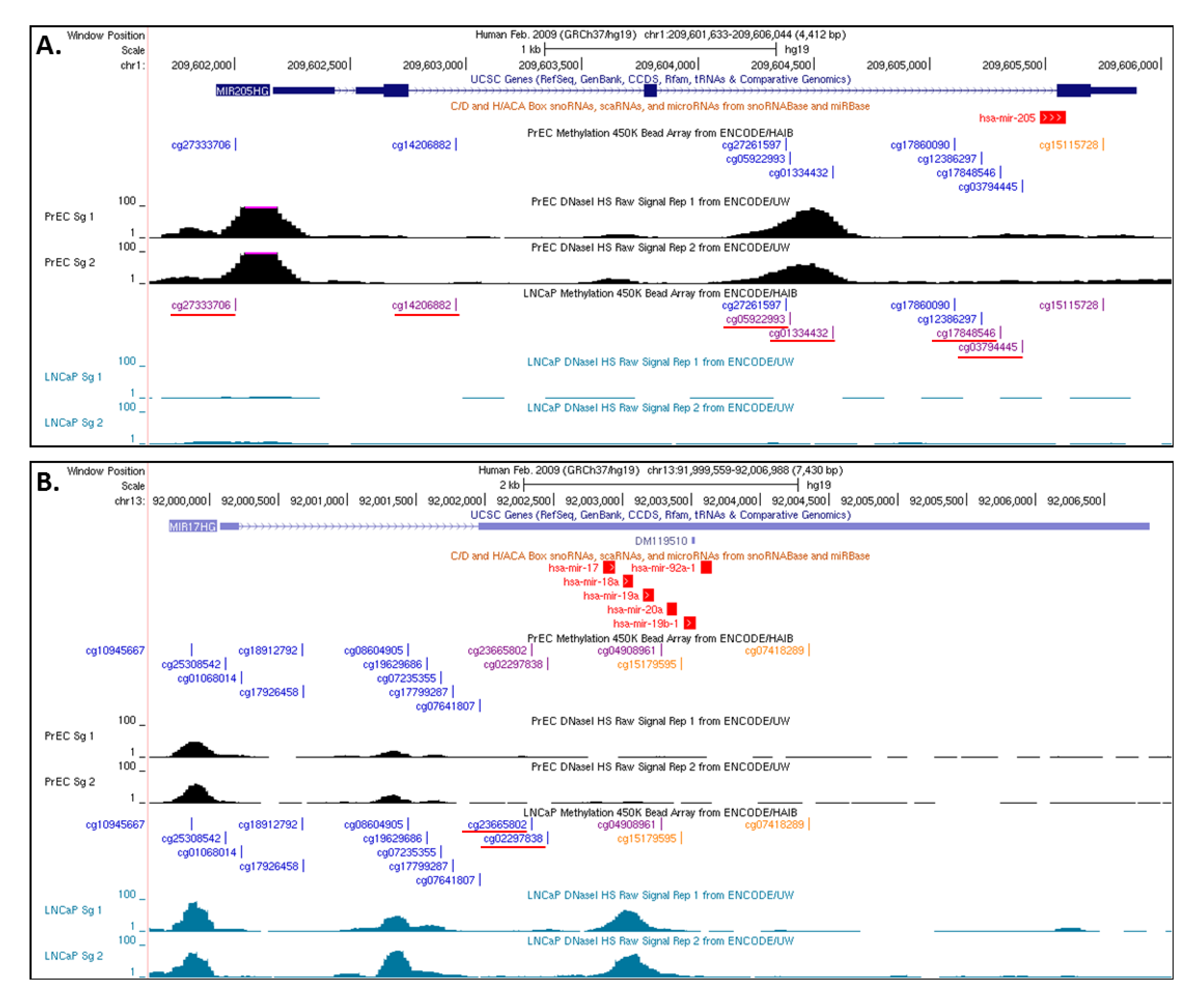
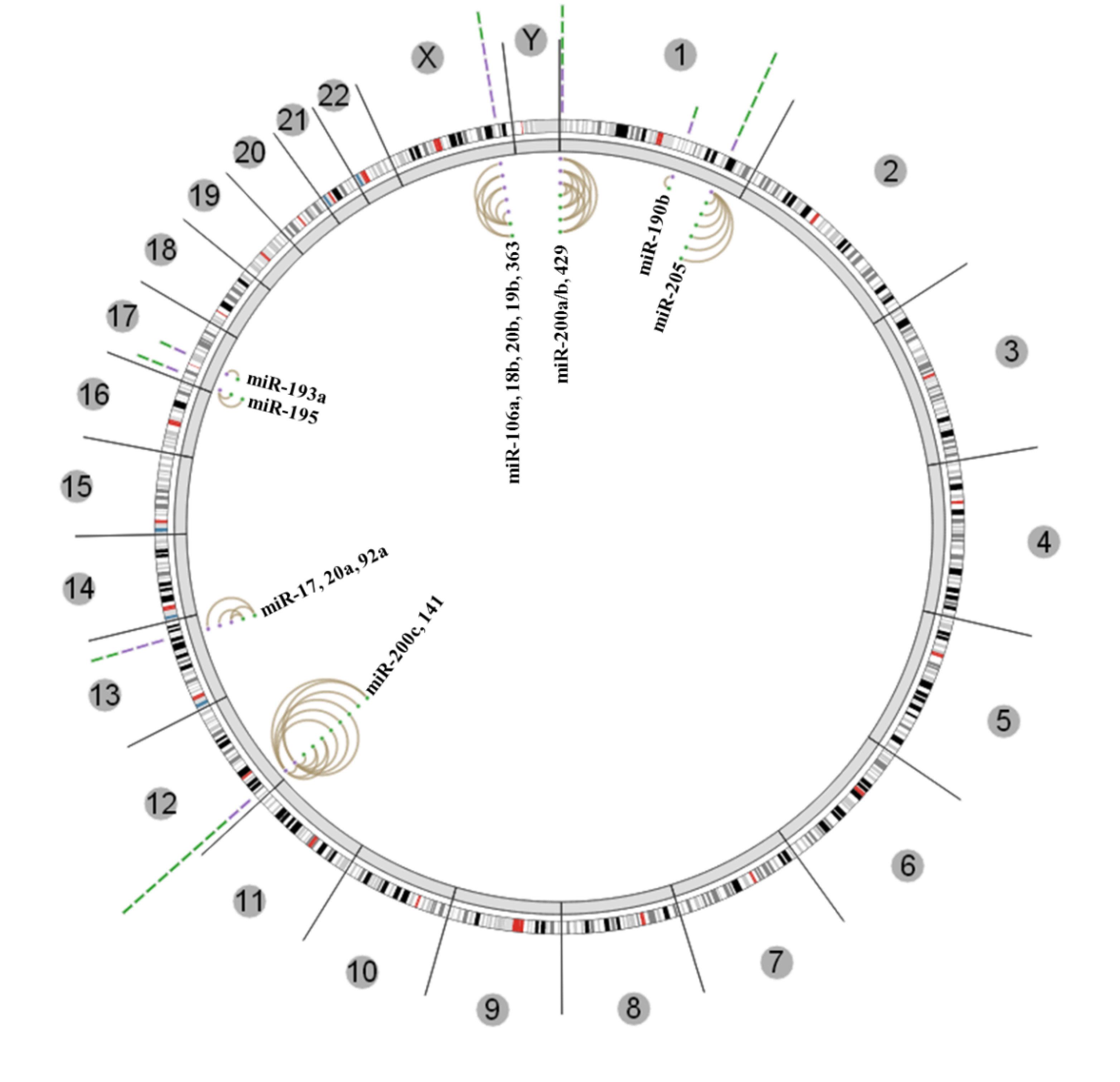
3.3. Insights from the Cancer Genome Atlas (TCGA) and ENCODE

We explored TCGA data for associations between DNA methylation and miRNA expression in CaP tumors. An integrated tool of all the data types generated by TCGA have been developed to understand the systems level interaction between different data features. Statistically significant association can be identified and visualized using Regulome Explorer [125]. Regulome Explorer has been used for integrated analysis in colon, rectal and breast cancers [126,127]. We used Regulome Explorer to identify statistically significant correlations between miRNA expression (RNA-seq) and status of DNA methylation (Illumina 450k methylation arrays). We specifically searched for CpG sites showing negative correlation with miRNA expression on the same chromosome (using “cis” setting in distance filter and p-value cutoff of −log10(p) ≥ 6). This analysis identified a total of 27 CpGs negatively correlated with expression of 17 miRNAs from 5 different chromosomes (Figure 2). Notably corroborating the discussion above, all five miRNAs of miR-200 family (miR-200a/b/429, miR-200c/141) and miR-205 showed strong negative correlation between miRNA expression and CpG methylation [89,90]. Many of miRNAs with important function in CaP did not show significant correlation in this analysis which can be partially because of the allocation of CpGs to miRNAs in Illumina 450K array annotation. This preliminary exploration of TCGA data strengthens the observation that miRNAs are epigenetically silenced in CaP. It will be interesting to interrogate this resource in further detail to identify how other genomic data from TCGA correlates with miRNA expression and DNA methylation.
Figure 2. Significant negative correlation between altered miRNA expression and DNA methylation pattern in primary prostate cancer tumors from the TCGA data-set. Circos plot showing miRNAs where there is a significant correlation with CpGs near miRNA gene locations. Outer ring represents all the chromosomes. MiRNA genes are represented by purple lines near their chromosomal locations. Green lines represent CpGs in the region. Lines connecting two dots represent the statistically significant correlation between two features selected, miRNA expression and CpG methylation in this case. (Figures generated from [125] using latest version of data release for Prostate cancer “PRAD-13-March-2013”).
Cancers 05 00998 g002
Figure 3. UCSC genome browser tracks showing CpG methylation and DNase I cleavage status around miRNAs. Top panel track shows the genomic location of the miRNA. Middle track shows the CpGs in genomic fragment and color of each CpG represent the beta values (ENCODE description—Orange, beta value equal to or greater than 0.6 was considered fully methylated; Purple, Beta values between 0.2 and 0.6 were considered to be partially methylated; Blue, beta value equal to or less than 0.2 was considered to be fully unmethylated). Bottom panel shows the DNaseI sensitivity measured in PrEC (black) and LNCaP (blue). DNaseI hypersensitive sites shown as peaks suggest accessible chromatin; conversely no DNaseI hypersensitive sites suggest closed chromatin. (A) Shows the status of miR-205. Most of the CpGs around miR-205 are partially methylated in LNCaP and un-methylated in PrEC. Supportively, DNaseI sensitivity shows open areas in PrEC cells and closed chromatin in LNCaP and. (B) Shows the status of a miR-17-92 cluster. Two CpGs around this cluster are partially methylated in PrEC cells and un-methylated in LNCaP. Supportively, DNaseI sensitivity shows open chromatin in LNCaP and closed in PrEC cells. Differentially methylated CpGs are underlined.
Cancers 05 00998 g003
Recently published large scale ENCODE ChIP-seq data for various epigenetic features (e.g., histone marks, chromatin accessibility and DNA methylation) provide a key opportunity to study these associations in various cancer models including CaP [26,128]. Additionally, as an example, we also examined the chromatin accessibility around the miRNAs identified from regulome analysis in LNCaP and PrEC cells from ENCODE. As shown in Figure 3, miRNAs with higher expression in CaP tumors (miR-17, miR-20a and miR-92a) which were associated with low methylation also showed low methylation and open chromatin in LNCaP as compared to PrEC cells. Similarly, miRNA with low expression and high methylation in CaP tumors showed more methylation and closed chromatin in LNCaP (e.g., miR-205).

4. Conclusions

The de-regulated cross-talk between epigenetic states and miRNA transcription and action is functionally important in the progression of CaP. Studies utilizing components of epigenetic machinery may offer highly accurate, and accessible, information about CaP diagnosis and prognosis. For example, urine-borne DNA methylation patterns in combination with serum miRNA patterns offer the opportunity to be exploited as highly accurate functional biomarkers. Also, they can be used to understand the underlying epigenetic status to be prognostic of CaP disease progression risks. These components have a strong likelihood to be exploited as integrated non-invasive biomarker of CaP progression and drug resistance.

Conflict of Interest

The authors declare no conflict of interest.

References

  1. Schroder, F.H.; Hugosson, J.; Roobol, M.J.; Tammela, T.L.; Ciatto, S.; Nelen, V.; Kwiatkowski, M.; Lujan, M.; Lilja, H.; Zappa, M.; et al. Screening and prostate-cancer mortality in a randomized european study. N. Engl. J. Med. 2009, 360, 1320–1328. [Google Scholar] [CrossRef]
  2. Andriole, G.L.; Crawford, E.D.; Grubb, R.L., 3rd; Buys, S.S.; Chia, D.; Church, T.R.; Fouad, M.N.; Gelmann, E.P.; Kvale, P.A.; Reding, D.J.; et al. Mortality results from a randomized prostate-cancer screening trial. N. Engl. J. Med. 2009, 360, 1310–1319. [Google Scholar] [CrossRef]
  3. He, W.W.; Sciavolino, P.J.; Wing, J.; Augustus, M.; Hudson, P.; Meissner, P.S.; Curtis, R.T.; Shell, B.K.; Bostwick, D.G.; Tindall, D.J.; et al. A novel human prostate-specific, androgen-regulated homeobox gene (nkx3.1) that maps to 8p21, a region frequently deleted in prostate cancer. Genomics 1997, 43, 69–77. [Google Scholar] [CrossRef]
  4. Cairns, P.; Okami, K.; Halachmi, S.; Halachmi, N.; Esteller, M.; Herman, J.G.; Jen, J.; Isaacs, W.B.; Bova, G.S.; Sidransky, D. Frequent inactivation of pten/mmac1 in primary prostate cancer. Cancer Res. 1997, 57, 4997–5000. [Google Scholar]
  5. Li, J.; Yen, C.; Liaw, D.; Podsypanina, K.; Bose, S.; Wang, S.I.; Puc, J.; Miliaresis, C.; Rodgers, L.; McCombie, R.; et al. Pten, a putative protein tyrosine phosphatase gene mutated in human brain, breast, and prostate cancer. Science 1997, 275, 1943–1947. [Google Scholar] [CrossRef]
  6. Visakorpi, T.; Hyytinen, E.; Koivisto, P.; Tanner, M.; Keinanen, R.; Palmberg, C.; Palotie, A.; Tammela, T.; Isola, J.; Kallioniemi, O.P. In vivo amplification of the androgen receptor gene and progression of human prostate cancer. Nat. Genet. 1995, 9, 401–406. [Google Scholar] [CrossRef]
  7. Taylor, B.S.; Schultz, N.; Hieronymus, H.; Gopalan, A.; Xiao, Y.; Carver, B.S.; Arora, V.K.; Kaushik, P.; Cerami, E.; Reva, B.; et al. Integrative genomic profiling of human prostate cancer. Cancer Cell 2010, 18, 11–22. [Google Scholar] [CrossRef]
  8. Perner, S.; Demichelis, F.; Beroukhim, R.; Schmidt, F.H.; Mosquera, J.M.; Setlur, S.; Tchinda, J.; Tomlins, S.A.; Hofer, M.D.; Pienta, K.G.; et al. Tmprss2:Erg fusion-associated deletions provide insight into the heterogeneity of prostate cancer. Cancer Res. 2006, 66, 8337–8341. [Google Scholar] [CrossRef]
  9. Tomlins, S.A.; Laxman, B.; Dhanasekaran, S.M.; Helgeson, B.E.; Cao, X.; Morris, D.S.; Menon, A.; Jing, X.; Cao, Q.; Han, B.; et al. Distinct classes of chromosomal rearrangements create oncogenic ets gene fusions in prostate cancer. Nature 2007, 448, 595–599. [Google Scholar] [CrossRef]
  10. Barbieri, C.E.; Baca, S.C.; Lawrence, M.S.; Demichelis, F.; Blattner, M.; Theurillat, J.P.; White, T.A.; Stojanov, P.; van Allen, E.; Stransky, N.; et al. Exome sequencing identifies recurrent spop, foxa1 and med12 mutations in prostate cancer. Nat. Genet. 2012, 44, 685–689. [Google Scholar] [CrossRef]
  11. Sandoval, J.; Esteller, M. Cancer epigenomics: Beyond genomics. Curr. Opin. Genet. Dev. 2012, 22, 50–55. [Google Scholar] [CrossRef]
  12. Holliday, R. The inheritance of epigenetic defects. Science 1987, 238, 163–170. [Google Scholar]
  13. Esteller, M. Cancer epigenomics: DNA methylomes and histone-modification maps. Nat. Rev. Genet. 2007, 8, 286–298. [Google Scholar] [CrossRef]
  14. Campbell, M.J.; Turner, B.M. Altered histone modifications in cancer. Adv. Exp. Med. Biol. 2013, 754, 81–107. [Google Scholar] [CrossRef]
  15. Chiam, K.; Ricciardelli, C.; Bianco-Miotto, T. Epigenetic biomarkers in prostate cancer: Current and future uses. Cancer Lett. 2012. [Google Scholar] [CrossRef]
  16. Jeronimo, C.; Bastian, P.J.; Bjartell, A.; Carbone, G.M.; Catto, J.W.; Clark, S.J.; Henrique, R.; Nelson, W.G.; Shariat, S.F. Epigenetics in prostate cancer: Biologic and clinical relevance. Eur. Urol. 2011, 60, 753–766. [Google Scholar] [CrossRef]
  17. Shen, M.M.; Abate-Shen, C. Molecular genetics of prostate cancer: New prospects for old challenges. Genes Dev. 2010, 24, 1967–2000. [Google Scholar] [CrossRef]
  18. Denis, H.; Ndlovu, M.N.; Fuks, F. Regulation of mammalian DNA methyltransferases: A route to new mechanisms. EMBO Rep. 2011, 12, 647–656. [Google Scholar] [CrossRef]
  19. Lee, K.K.; Workman, J.L. Histone acetyltransferase complexes: One size doesn’t fit all. Nat. Rev. Mol. Cell Biol. 2007, 8, 284–295. [Google Scholar] [CrossRef]
  20. Kim, H.J.; Bae, S.C. Histone deacetylase inhibitors: Molecular mechanisms of action and clinical trials as anti-cancer drugs. Am. J. Transl. Res. 2011, 3, 166–179. [Google Scholar]
  21. Albert, M.; Helin, K. Histone methyltransferases in cancer. Semin. Cell Dev. Biol. 2010, 21, 209–220. [Google Scholar] [CrossRef]
  22. Kooistra, S.M.; Helin, K. Molecular mechanisms and potential functions of histone demethylases. Nat. Rev. Mol. Cell Biol. 2012, 13, 297–311. [Google Scholar] [CrossRef]
  23. Nakayama, M.; Bennett, C.J.; Hicks, J.L.; Epstein, J.I.; Platz, E.A.; Nelson, W.G.; de Marzo, A.M. Hypermethylation of the human glutathione s-transferase-pi gene (gstp1) cpg island is present in a subset of proliferative inflammatory atrophy lesions but not in normal or hyperplastic epithelium of the prostate: A detailed study using laser-capture microdissection. Am. J. Pathol. 2003, 163, 923–933. [Google Scholar] [CrossRef]
  24. Varambally, S.; Dhanasekaran, S.M.; Zhou, M.; Barrette, T.R.; Kumar-Sinha, C.; Sanda, M.G.; Ghosh, D.; Pienta, K.J.; Sewalt, R.G.; Otte, A.P.; et al. The polycomb group protein ezh2 is involved in progression of prostate cancer. Nature 2002, 419, 624–629. [Google Scholar] [CrossRef]
  25. Saramaki, O.R.; Tammela, T.L.; Martikainen, P.M.; Vessella, R.L.; Visakorpi, T. The gene for polycomb group protein enhancer of zeste homolog 2 (ezh2) is amplified in late-stage prostate cancer. Genes Chromosomes Cancer 2006, 45, 639–645. [Google Scholar] [CrossRef]
  26. Djebali, S.; Davis, C.A.; Merkel, A.; Dobin, A.; Lassmann, T.; Mortazavi, A.; Tanzer, A.; Lagarde, J.; Lin, W.; Schlesinger, F.; et al. Landscape of transcription in human cells. Nature 2012, 489, 101–108. [Google Scholar] [CrossRef]
  27. Graur, D.; Zheng, Y.; Price, N.; Azevedo, R.B.; Zufall, R.A.; Elhaik, E. On the immortality of television sets: “Function” in the human genome according to the evolution-free gospel of encode. Genome Biol. Evol. 2013, 5, 578–590. [Google Scholar] [CrossRef]
  28. Doolittle, W.F. Is junk DNA bunk? A critique of encode. Proc. Natl. Acad. Sci. USA 2013, 110, 5294–5300. [Google Scholar] [CrossRef]
  29. Bartel, D.P. Micrornas: Genomics, biogenesis, mechanism, and function. Cell 2004, 116, 281–297. [Google Scholar] [CrossRef]
  30. Calin, G.A.; Croce, C.M. Microrna signatures in human cancers. Nat. Rev. Cancer 2006, 6, 857–866. [Google Scholar] [CrossRef]
  31. Iorio, M.V.; Croce, C.M. Microrna dysregulation in cancer: Diagnostics, monitoring and therapeutics. A comprehensive review. EMBO Mol. Med. 2012, 4, 143–159. [Google Scholar] [CrossRef]
  32. Manikandan, J.; Aarthi, J.J.; Kumar, S.D.; Pushparaj, P.N. Oncomirs: The potential role of non-coding micrornas in understanding cancer. Bioinformation 2008, 2, 330–334. [Google Scholar] [CrossRef]
  33. Esquela-Kerscher, A.; Slack, F.J. Oncomirs—Micrornas with a role in cancer. Nat. Rev. Cancer 2006, 6, 259–269. [Google Scholar] [CrossRef]
  34. Volinia, S.; Calin, G.A.; Liu, C.G.; Ambs, S.; Cimmino, A.; Petrocca, F.; Visone, R.; Iorio, M.; Roldo, C.; Ferracin, M.; et al. A microrna expression signature of human solid tumors defines cancer gene targets. Proc. Natl. Acad. Sci. USA 2006, 103, 2257–2261. [Google Scholar] [CrossRef]
  35. Lu, J.; Getz, G.; Miska, E.A.; Alvarez-Saavedra, E.; Lamb, J.; Peck, D.; Sweet-Cordero, A.; Ebert, B.L.; Mak, R.H.; Ferrando, A.A.; et al. Microrna expression profiles classify human cancers. Nature 2005, 435, 834–838. [Google Scholar] [CrossRef]
  36. Lewis, B.P.; Burge, C.B.; Bartel, D.P. Conserved seed pairing, often flanked by adenosines, indicates that thousands of human genes are microrna targets. Cell 2005, 120, 15–20. [Google Scholar] [CrossRef]
  37. Lussier, Y.A.; Stadler, W.M.; Chen, J.L. Advantages of genomic complexity: Bioinformatics opportunities in microrna cancer signatures. J. Am. Med. Inform. Assoc. 2012, 19, 156–160. [Google Scholar] [CrossRef]
  38. Sun, T.; Wang, Q.; Balk, S.; Brown, M.; Lee, G.S.; Kantoff, P. The role of microrna-221 and microrna-222 in androgen-independent prostate cancer cell lines. Cancer Res. 2009, 69, 3356–3363. [Google Scholar] [CrossRef]
  39. Medina, R.; Zaidi, S.K.; Liu, C.G.; Stein, J.L.; van Wijnen, A.J.; Croce, C.M.; Stein, G.S. Micrornas 221 and 222 bypass quiescence and compromise cell survival. Cancer Res. 2008, 68, 2773–2780. [Google Scholar]
  40. Mercatelli, N.; Coppola, V.; Bonci, D.; Miele, F.; Costantini, A.; Guadagnoli, M.; Bonanno, E.; Muto, G.; Frajese, G.V.; de Maria, R.; et al. The inhibition of the highly expressed mir-221 and mir-222 impairs the growth of prostate carcinoma xenografts in mice. PLoS One 2008, 3, e4029. [Google Scholar] [CrossRef] [Green Version]
  41. Wang, L.; Tang, H.; Thayanithy, V.; Subramanian, S.; Oberg, A.L.; Cunningham, J.M.; Cerhan, J.R.; Steer, C.J.; Thibodeau, S.N. Gene networks and micrornas implicated in aggressive prostate cancer. Cancer Res. 2009, 69, 9490–9497. [Google Scholar] [CrossRef]
  42. Hao, J.; Zhang, C.; Zhang, A.; Wang, K.; Jia, Z.; Wang, G.; Han, L.; Kang, C.; Pu, P. Mir-221/222 is the regulator of cx43 expression in human glioblastoma cells. Oncol. Rep. 2012, 27, 1504–1510. [Google Scholar]
  43. Tucci, P.; Agostini, M.; Grespi, F.; Markert, E.K.; Terrinoni, A.; Vousden, K.H.; Muller, P.A.; Dotsch, V.; Kehrloesser, S.; Sayan, B.S.; et al. Loss of p63 and its microrna-205 target results in enhanced cell migration and metastasis in prostate cancer. Proc. Natl. Acad. Sci. USA 2012, 109, 15312–15317. [Google Scholar] [CrossRef]
  44. Zhao, J.J.; Lin, J.; Yang, H.; Kong, W.; He, L.; Ma, X.; Coppola, D.; Cheng, J.Q. Microrna-221/222 negatively regulates estrogen receptor alpha and is associated with tamoxifen resistance in breast cancer. J. Biol. Chem. 2008, 283, 31079–31086. [Google Scholar]
  45. Miller, T.E.; Ghoshal, K.; Ramaswamy, B.; Roy, S.; Datta, J.; Shapiro, C.L.; Jacob, S.; Majumder, S. Microrna-221/222 confers tamoxifen resistance in breast cancer by targeting p27kip1. J. Biol. Chem. 2008, 283, 29897–29903. [Google Scholar]
  46. Lin, D.; Cui, F.; Bu, Q.; Yan, C. The expression and clinical significance of gtp-binding ras-like 3 (arhi) and microrna 221 and 222 in prostate cancer. J. Int. Med. Res. 2011, 39, 1870–1875. [Google Scholar] [CrossRef]
  47. Tong, A.W.; Fulgham, P.; Jay, C.; Chen, P.; Khalil, I.; Liu, S.; Senzer, N.; Eklund, A.C.; Han, J.; Nemunaitis, J. Microrna profile analysis of human prostate cancers. Cancer Gene Ther. 2009, 16, 206–216. [Google Scholar]
  48. Spahn, M.; Kneitz, S.; Scholz, C.J.; Stenger, N.; Rudiger, T.; Strobel, P.; Riedmiller, H.; Kneitz, B. Expression of microrna-221 is progressively reduced in aggressive prostate cancer and metastasis and predicts clinical recurrence. Int. J. Cancer 2010, 127, 394–403. [Google Scholar]
  49. Martens-Uzunova, E.S.; Jalava, S.E.; Dits, N.F.; van Leenders, G.J.; Moller, S.; Trapman, J.; Bangma, C.H.; Litman, T.; Visakorpi, T.; Jenster, G. Diagnostic and prognostic signatures from the small non-coding rna transcriptome in prostate cancer. Oncogene 2012, 31, 978–991. [Google Scholar] [CrossRef]
  50. Wach, S.; Nolte, E.; Szczyrba, J.; Stohr, R.; Hartmann, A.; Orntoft, T.; Dyrskjot, L.; Eltze, E.; Wieland, W.; Keck, B.; et al. Microrna profiles of prostate carcinoma detected by multiplatform microrna screening. Int. J. Cancer 2012, 130, 611–621. [Google Scholar] [CrossRef]
  51. Mitchell, P.S.; Parkin, R.K.; Kroh, E.M.; Fritz, B.R.; Wyman, S.K.; Pogosova-Agadjanyan, E.L.; Peterson, A.; Noteboom, J.; O’Briant, K.C.; Allen, A.; et al. Circulating micrornas as stable blood-based markers for cancer detection. Proc. Natl. Acad. Sci. USA 2008, 105, 10513–10518. [Google Scholar] [CrossRef]
  52. Ozen, M.; Creighton, C.J.; Ozdemir, M.; Ittmann, M. Widespread deregulation of microrna expression in human prostate cancer. Oncogene 2008, 27, 1788–1793. [Google Scholar] [CrossRef]
  53. Porkka, K.P.; Pfeiffer, M.J.; Waltering, K.K.; Vessella, R.L.; Tammela, T.L.; Visakorpi, T. Microrna expression profiling in prostate cancer. Cancer Res. 2007, 67, 6130–6135. [Google Scholar]
  54. Lee, Y.S.; Kim, H.K.; Chung, S.; Kim, K.S.; Dutta, A. Depletion of human micro-rna mir-125b reveals that it is critical for the proliferation of differentiated cells but not for the down-regulation of putative targets during differentiation. J. Biol. Chem. 2005, 280, 16635–16641. [Google Scholar]
  55. Shi, X.B.; Xue, L.; Yang, J.; Ma, A.H.; Zhao, J.; Xu, M.; Tepper, C.G.; Evans, C.P.; Kung, H.J.; deVere White, R.W. An androgen-regulated mirna suppresses bak1 expression and induces androgen-independent growth of prostate cancer cells. Proc. Natl. Acad. Sci. USA 2007, 104, 19983–19988. [Google Scholar] [CrossRef]
  56. Ribas, J.; Ni, X.; Haffner, M.; Wentzel, E.A.; Salmasi, A.H.; Chowdhury, W.H.; Kudrolli, T.A.; Yegnasubramanian, S.; Luo, J.; Rodriguez, R.; et al. Mir-21: An androgen receptor-regulated microrna that promotes hormone-dependent and hormone-independent prostate cancer growth. Cancer Res. 2009, 69, 7165–7169. [Google Scholar] [CrossRef]
  57. Shi, G.H.; Ye, D.W.; Yao, X.D.; Zhang, S.L.; Dai, B.; Zhang, H.L.; Shen, Y.J.; Zhu, Y.; Zhu, Y.P.; Xiao, W.J.; et al. Involvement of microrna-21 in mediating chemo-resistance to docetaxel in androgen-independent prostate cancer pc3 cells. Acta Pharmacol. Sin. 2010, 31, 867–873. [Google Scholar] [CrossRef]
  58. Lu, Z.; Liu, M.; Stribinskis, V.; Klinge, C.M.; Ramos, K.S.; Colburn, N.H.; Li, Y. Microrna-21 promotes cell transformation by targeting the programmed cell death 4 gene. Oncogene 2008, 27, 4373–4379. [Google Scholar] [CrossRef]
  59. Peng, X.; Guo, W.; Liu, T.; Wang, X.; Tu, X.; Xiong, D.; Chen, S.; Lai, Y.; Du, H.; Chen, G.; et al. Identification of mirs-143 and -145 that is associated with bone metastasis of prostate cancer and involved in the regulation of emt. PLoS One 2011, 6, e20341. [Google Scholar]
  60. Kong, D.; Li, Y.; Wang, Z.; Banerjee, S.; Ahmad, A.; Kim, H.R.; Sarkar, F.H. Mir-200 regulates pdgf-d-mediated epithelial-mesenchymal transition, adhesion, and invasion of prostate cancer cells. Stem Cells 2009, 27, 1712–1721. [Google Scholar] [CrossRef]
  61. Gregory, P.A.; Bert, A.G.; Paterson, E.L.; Barry, S.C.; Tsykin, A.; Farshid, G.; Vadas, M.A.; Khew-Goodall, Y.; Goodall, G.J. The mir-200 family and mir-205 regulate epithelial to mesenchymal transition by targeting zeb1 and sip1. Nat. Cell Biol. 2008, 10, 593–601. [Google Scholar] [CrossRef]
  62. Liep, J.; Rabien, A.; Jung, K. Feedback networks between micrornas and epigenetic modifications in urological tumors. Epigenetics 2012, 7, 315–325. [Google Scholar] [CrossRef]
  63. Saito, Y.; Jones, P.A. Epigenetic activation of tumor suppressor micrornas in human cancer cells. Cell Cycle 2006, 5, 2220–2222. [Google Scholar] [CrossRef]
  64. Chen, J.F.; Mandel, E.M.; Thomson, J.M.; Wu, Q.; Callis, T.E.; Hammond, S.M.; Conlon, F.L.; Wang, D.Z. The role of microrna-1 and microrna-133 in skeletal muscle proliferation and differentiation. Nat. Genet. 2006, 38, 228–233. [Google Scholar] [CrossRef]
  65. Duursma, A.M.; Kedde, M.; Schrier, M.; le Sage, C.; Agami, R. Mir-148 targets human dnmt3b protein coding region. RNA 2008, 14, 872–877. [Google Scholar] [CrossRef]
  66. Filipowicz, W.; Bhattacharyya, S.N.; Sonenberg, N. Mechanisms of post-transcriptional regulation by micrornas: Are the answers in sight? Nat. Rev. Genet. 2008, 9, 102–114. [Google Scholar]
  67. Thorne, J.L.; Maguire, O.; Doig, C.L.; Battaglia, S.; Fehr, L.; Sucheston, L.E.; Heinaniemi, M.; O’Neill, L.P.; McCabe, C.J.; Turner, B.M.; et al. Epigenetic control of a vdr-governed feed-forward loop that regulates p21(waf1/cip1) expression and function in non-malignant prostate cells. Nucleic Acids Res. 2011, 39, 2045–2056. [Google Scholar] [CrossRef]
  68. Garzon, R.; Liu, S.; Fabbri, M.; Liu, Z.; Heaphy, C.E.; Callegari, E.; Schwind, S.; Pang, J.; Yu, J.; Muthusamy, N.; et al. Microrna-29b induces global DNA hypomethylation and tumor suppressor gene reexpression in acute myeloid leukemia by targeting directly dnmt3a and 3b and indirectly dnmt1. Blood 2009, 113, 6411–6418. [Google Scholar] [CrossRef]
  69. Fabbri, M.; Garzon, R.; Cimmino, A.; Liu, Z.; Zanesi, N.; Callegari, E.; Liu, S.; Alder, H.; Costinean, S.; Fernandez-Cymering, C.; et al. Microrna-29 family reverts aberrant methylation in lung cancer by targeting DNA methyltransferases 3a and 3b. Proc. Natl. Acad. Sci. USA 2007, 104, 15805–15810. [Google Scholar] [CrossRef]
  70. Majid, S.; Dar, A.A.; Saini, S.; Shahryari, V.; Arora, S.; Zaman, M.S.; Chang, I.; Yamamura, S.; Tanaka, Y.; Chiyomaru, T.; et al. Mirna-34b inhibits prostate cancer through demethylation, active chromatin modifications, and akt pathways. Clin. Cancer Res. 2013, 19, 73–84. [Google Scholar] [CrossRef]
  71. Cao, P.; Deng, Z.; Wan, M.; Huang, W.; Cramer, S.D.; Xu, J.; Lei, M.; Sui, G. Microrna-101 negatively regulates ezh2 and its expression is modulated by androgen receptor and hif-1alpha/hif-1beta. Mol. Cancer 2010, 9, 108. [Google Scholar] [CrossRef] [Green Version]
  72. Noonan, E.J.; Place, R.F.; Pookot, D.; Basak, S.; Whitson, J.M.; Hirata, H.; Giardina, C.; Dahiya, R. Mir-449a targets hdac-1 and induces growth arrest in prostate cancer. Oncogene 2009, 28, 1714–1724. [Google Scholar] [CrossRef]
  73. Gong, A.Y.; Eischeid, A.N.; Xiao, J.; Zhao, J.; Chen, D.; Wang, Z.Y.; Young, C.Y.; Chen, X.M. Mir-17-5p targets the p300/cbp-associated factor and modulates androgen receptor transcriptional activity in cultured prostate cancer cells. BMC Cancer 2012, 12, 492. [Google Scholar] [CrossRef]
  74. Margueron, R.; Reinberg, D. The polycomb complex prc2 and its mark in life. Nature 2011, 469, 343–349. [Google Scholar] [CrossRef]
  75. Bracken, A.P.; Helin, K. Polycomb group proteins: Navigators of lineage pathways led astray in cancer. Nat. Rev. Cancer 2009, 9, 773–784. [Google Scholar] [CrossRef]
  76. Varambally, S.; Cao, Q.; Mani, R.S.; Shankar, S.; Wang, X.; Ateeq, B.; Laxman, B.; Cao, X.; Jing, X.; Ramnarayanan, K.; et al. Genomic loss of microrna-101 leads to overexpression of histone methyltransferase ezh2 in cancer. Science 2008, 322, 1695–1699. [Google Scholar] [CrossRef]
  77. Cao, Q.; Mani, R.S.; Ateeq, B.; Dhanasekaran, S.M.; Asangani, I.A.; Prensner, J.R.; Kim, J.H.; Brenner, J.C.; Jing, X.; Cao, X.; et al. Coordinated regulation of polycomb group complexes through micrornas in cancer. Cancer Cell 2011, 20, 187–199. [Google Scholar] [CrossRef]
  78. Cohen, E.E.; Zhu, H.; Lingen, M.W.; Martin, L.E.; Kuo, W.L.; Choi, E.A.; Kocherginsky, M.; Parker, J.S.; Chung, C.H.; Rosner, M.R. A feed-forward loop involving protein kinase calpha and micrornas regulates tumor cell cycle. Cancer Res. 2009, 69, 65–74. [Google Scholar] [CrossRef]
  79. Brosh, R.; Shalgi, R.; Liran, A.; Landan, G.; Korotayev, K.; Nguyen, G.H.; Enerly, E.; Johnsen, H.; Buganim, Y.; Solomon, H.; et al. P53-repressed mirnas are involved with e2f in a feed-forward loop promoting proliferation. Mol. Syst. Biol. 2008, 4, 229. [Google Scholar]
  80. Polioudakis, D.; Bhinge, A.A.; Killion, P.J.; Lee, B.K.; Abell, N.S.; Iyer, V.R. A myc-microrna network promotes exit from quiescence by suppressing the interferon response and cell-cycle arrest genes. Nucleic Acids Res. 2013, 41, 2239–2254. [Google Scholar] [CrossRef]
  81. Hassan, M.Q.; Maeda, Y.; Taipaleenmaki, H.; Zhang, W.; Jafferji, M.; Gordon, J.A.; Li, Z.; Croce, C.M.; van Wijnen, A.J.; Stein, J.L.; et al. Mir-218 directs a wnt signaling circuit to promote differentiation of osteoblasts and osteomimicry of metastatic cancer cells. J. Biol. Chem. 2012, 287, 42084–42092. [Google Scholar] [CrossRef]
  82. McInnes, N.; Sadlon, T.J.; Brown, C.Y.; Pederson, S.; Beyer, M.; Schultze, J.L.; McColl, S.; Goodall, G.J.; Barry, S.C. Foxp3 and foxp3-regulated micrornas suppress satb1 in breast cancer cells. Oncogene 2012, 31, 1045–1054. [Google Scholar] [CrossRef]
  83. Da Costa Martins, P.A.; Salic, K.; Gladka, M.M.; Armand, A.S.; Leptidis, S.; el Azzouzi, H.; Hansen, A.; Coenen-de Roo, C.J.; Bierhuizen, M.F.; van der Nagel, R.; et al. Microrna-199b targets the nuclear kinase dyrk1a in an auto-amplification loop promoting calcineurin/nfat signalling. Nat. Cell Biol. 2010, 12, 1220–1227. [Google Scholar] [CrossRef]
  84. Weichert, W.; Roske, A.; Gekeler, V.; Beckers, T.; Stephan, C.; Jung, K.; Fritzsche, F.R.; Niesporek, S.; Denkert, C.; Dietel, M.; et al. Histone deacetylases 1, 2 and 3 are highly expressed in prostate cancer and hdac2 expression is associated with shorter psa relapse time after radical prostatectomy. Br. J. Cancer 2008, 98, 604–610. [Google Scholar] [CrossRef]
  85. Nagy, Z.; Tora, L. Distinct gcn5/pcaf-containing complexes function as co-activators and are involved in transcription factor and global histone acetylation. Oncogene 2007, 26, 5341–5357. [Google Scholar] [CrossRef]
  86. Dudziec, E.; Miah, S.; Choudhry, H.M.; Owen, H.C.; Blizard, S.; Glover, M.; Hamdy, F.C.; Catto, J.W. Hypermethylation of cpg islands and shores around specific micrornas and mirtrons is associated with the phenotype and presence of bladder cancer. Clin. Cancer Res. 2011, 17, 1287–1296. [Google Scholar] [CrossRef]
  87. Gandellini, P.; Folini, M.; Longoni, N.; Pennati, M.; Binda, M.; Colecchia, M.; Salvioni, R.; Supino, R.; Moretti, R.; Limonta, P.; et al. Mir-205 exerts tumor-suppressive functions in human prostate through down-regulation of protein kinase cepsilon. Cancer Res. 2009, 69, 2287–2295. [Google Scholar] [CrossRef]
  88. Iorio, M.V.; Croce, C.M. Micrornas in cancer: Small molecules with a huge impact. J. Clin. Oncol. 2009, 27, 5848–5856. [Google Scholar] [CrossRef]
  89. Bhatnagar, N.; Li, X.; Padi, S.K.; Zhang, Q.; Tang, M.S.; Guo, B. Downregulation of mir-205 and mir-31 confers resistance to chemotherapy-induced apoptosis in prostate cancer cells. Cell Death Dis. 2010, 1, e105. [Google Scholar] [CrossRef]
  90. Vrba, L.; Jensen, T.J.; Garbe, J.C.; Heimark, R.L.; Cress, A.E.; Dickinson, S.; Stampfer, M.R.; Futscher, B.W. Role for DNA methylation in the regulation of mir-200c and mir-141 expression in normal and cancer cells. PLoS One 2010, 5, e8697. [Google Scholar] [CrossRef]
  91. Hermeking, H. The mir-34 family in cancer and apoptosis. Cell Death Differ. 2010, 17, 193–199. [Google Scholar] [CrossRef]
  92. Lodygin, D.; Tarasov, V.; Epanchintsev, A.; Berking, C.; Knyazeva, T.; Korner, H.; Knyazev, P.; Diebold, J.; Hermeking, H. Inactivation of mir-34a by aberrant cpg methylation in multiple types of cancer. Cell Cycle 2008, 7, 2591–2600. [Google Scholar] [CrossRef]
  93. He, L.; He, X.; Lim, L.P.; de Stanchina, E.; Xuan, Z.; Liang, Y.; Xue, W.; Zender, L.; Magnus, J.; Ridzon, D.; et al. A microrna component of the p53 tumour suppressor network. Nature 2007, 447, 1130–1134. [Google Scholar] [CrossRef]
  94. Chang, T.C.; Wentzel, E.A.; Kent, O.A.; Ramachandran, K.; Mullendore, M.; Lee, K.H.; Feldmann, G.; Yamakuchi, M.; Ferlito, M.; Lowenstein, C.J.; et al. Transactivation of mir-34a by p53 broadly influences gene expression and promotes apoptosis. Mol. Cell 2007, 26, 745–752. [Google Scholar] [CrossRef]
  95. Liu, C.; Kelnar, K.; Liu, B.; Chen, X.; Calhoun-Davis, T.; Li, H.; Patrawala, L.; Yan, H.; Jeter, C.; Honorio, S.; et al. The microrna mir-34a inhibits prostate cancer stem cells and metastasis by directly repressing cd44. Nat. Med. 2011, 17, 211–215. [Google Scholar] [CrossRef]
  96. Rauhala, H.E.; Jalava, S.E.; Isotalo, J.; Bracken, H.; Lehmusvaara, S.; Tammela, T.L.; Oja, H.; Visakorpi, T. Mir-193b is an epigenetically regulated putative tumor suppressor in prostate cancer. Int. J. Cancer 2010, 127, 1363–1372. [Google Scholar] [CrossRef]
  97. Sachdeva, M.; Mo, Y.Y. Mir-145-mediated suppression of cell growth, invasion and metastasis. Am. J. Transl. Res. 2010, 2, 170–180. [Google Scholar]
  98. Sachdeva, M.; Zhu, S.; Wu, F.; Wu, H.; Walia, V.; Kumar, S.; Elble, R.; Watabe, K.; Mo, Y.Y. P53 represses c-myc through induction of the tumor suppressor mir-145. Proc. Natl. Acad. Sci. USA 2009, 106, 3207–3212. [Google Scholar]
  99. Sachdeva, M.; Mo, Y.Y. Microrna-145 suppresses cell invasion and metastasis by directly targeting mucin 1. Cancer Res. 2010, 70, 378–387. [Google Scholar] [CrossRef]
  100. Zaman, M.S.; Chen, Y.; Deng, G.; Shahryari, V.; Suh, S.O.; Saini, S.; Majid, S.; Liu, J.; Khatri, G.; Tanaka, Y.; et al. The functional significance of microrna-145 in prostate cancer. Br. J. Cancer 2010, 103, 256–264. [Google Scholar] [CrossRef]
  101. Suh, S.O.; Chen, Y.; Zaman, M.S.; Hirata, H.; Yamamura, S.; Shahryari, V.; Liu, J.; Tabatabai, Z.L.; Kakar, S.; Deng, G.; et al. Microrna-145 is regulated by DNA methylation and p53 gene mutation in prostate cancer. Carcinogenesis 2011, 32, 772–778. [Google Scholar] [CrossRef]
  102. Hulf, T.; Sibbritt, T.; Wiklund, E.D.; Bert, S.; Strbenac, D.; Statham, A.L.; Robinson, M.D.; Clark, S.J. Discovery pipeline for epigenetically deregulated mirnas in cancer: Integration of primary mirna transcription. BMC Genomics 2011, 12, 54. [Google Scholar] [CrossRef]
  103. Iorio, M.V.; Visone, R.; di Leva, G.; Donati, V.; Petrocca, F.; Casalini, P.; Taccioli, C.; Volinia, S.; Liu, C.G.; Alder, H.; et al. Microrna signatures in human ovarian cancer. Cancer Res. 2007, 67, 8699–8707. [Google Scholar] [CrossRef]
  104. Coolen, M.W.; Stirzaker, C.; Song, J.Z.; Statham, A.L.; Kassir, Z.; Moreno, C.S.; Young, A.N.; Varma, V.; Speed, T.P.; Cowley, M.; et al. Consolidation of the cancer genome into domains of repressive chromatin by long-range epigenetic silencing (lres) reduces transcriptional plasticity. Nat. Cell Biol. 2010, 12, 235–246. [Google Scholar]
  105. Cookson, M.S.; Aus, G.; Burnett, A.L.; Canby-Hagino, E.D.; D’Amico, A.V.; Dmochowski, R.R.; Eton, D.T.; Forman, J.D.; Goldenberg, S.L.; Hernandez, J.; et al. Variation in the definition of biochemical recurrence in patients treated for localized prostate cancer: The american urological association prostate guidelines for localized prostate cancer update panel report and recommendations for a standard in the reporting of surgical outcomes. J. Urol. 2007, 177, 540–545. [Google Scholar] [CrossRef]
  106. Goering, W.; Kloth, M.; Schulz, W.A. DNA methylation changes in prostate cancer. Methods Mol. Biol. 2012, 863, 47–66. [Google Scholar] [CrossRef]
  107. Perry, A.S.; Watson, R.W.; Lawler, M.; Hollywood, D. The epigenome as a therapeutic target in prostate cancer. Nat. Rev. Urol. 2010, 7, 668–680. [Google Scholar] [CrossRef]
  108. Hoque, M.O.; Topaloglu, O.; Begum, S.; Henrique, R.; Rosenbaum, E.; van Criekinge, W.; Westra, W.H.; Sidransky, D. Quantitative methylation-specific polymerase chain reaction gene patterns in urine sediment distinguish prostate cancer patients from control subjects. J. Clin. Oncol. 2005, 23, 6569–6575. [Google Scholar] [CrossRef]
  109. Gonzalgo, M.L.; Pavlovich, C.P.; Lee, S.M.; Nelson, W.G. Prostate cancer detection by gstp1 methylation analysis of postbiopsy urine specimens. Clin. Cancer Res. 2003, 9, 2673–2677. [Google Scholar]
  110. Jeronimo, C.; Usadel, H.; Henrique, R.; Silva, C.; Oliveira, J.; Lopes, C.; Sidransky, D. Quantitative gstp1 hypermethylation in bodily fluids of patients with prostate cancer. Urology 2002, 60, 1131–1135. [Google Scholar] [CrossRef]
  111. Roupret, M.; Hupertan, V.; Yates, D.R.; Catto, J.W.; Rehman, I.; Meuth, M.; Ricci, S.; Lacave, R.; Cancel-Tassin, G.; de la Taille, A.; et al. Molecular detection of localized prostate cancer using quantitative methylation-specific pcr on urinary cells obtained following prostate massage. Clin. Cancer Res. 2007, 13, 1720–1725. [Google Scholar] [CrossRef]
  112. Ellinger, J.; Haan, K.; Heukamp, L.C.; Kahl, P.; Buttner, R.; Muller, S.C.; von Ruecker, A.; Bastian, P.J. Cpg island hypermethylation in cell-free serum DNA identifies patients with localized prostate cancer. Prostate 2008, 68, 42–49. [Google Scholar] [CrossRef]
  113. Ellinger, J.; Kahl, P.; von der Gathen, J.; Rogenhofer, S.; Heukamp, L.C.; Gutgemann, I.; Walter, B.; Hofstadter, F.; Buttner, R.; Muller, S.C.; et al. Global levels of histone modifications predict prostate cancer recurrence. Prostate 2010, 70, 61–69. [Google Scholar] [CrossRef]
  114. Deligezer, U.; Yaman, F.; Darendeliler, E.; Dizdar, Y.; Holdenrieder, S.; Kovancilar, M.; Dalay, N. Post-treatment circulating plasma bmp6 mrna and h3k27 methylation levels discriminate metastatic prostate cancer from localized disease. Clin. Chim. Acta 2010, 411, 1452–1456. [Google Scholar] [CrossRef]
  115. Cortez, M.A.; Bueso-Ramos, C.; Ferdin, J.; Lopez-Berestein, G.; Sood, A.K.; Calin, G.A. Micrornas in body fluids-the mix of hormones and biomarkers. Nat. Rev. Clin. Oncol. 2011, 8, 467–477. [Google Scholar] [CrossRef]
  116. Chen, X.; Ba, Y.; Ma, L.; Cai, X.; Yin, Y.; Wang, K.; Guo, J.; Zhang, Y.; Chen, J.; Guo, X.; et al. Characterization of micrornas in serum: A novel class of biomarkers for diagnosis of cancer and other diseases. Cell Res. 2008, 18, 997–1006. [Google Scholar] [CrossRef]
  117. El-Hefnawy, T.; Raja, S.; Kelly, L.; Bigbee, W.L.; Kirkwood, J.M.; Luketich, J.D.; Godfrey, T.E. Characterization of amplifiable, circulating rna in plasma and its potential as a tool for cancer diagnostics. Clin. Chem. 2004, 50, 564–573. [Google Scholar] [CrossRef]
  118. Selth, L.A.; Tilley, W.D.; Butler, L.M. Circulating micrornas: Macro-utility as markers of prostate cancer? Endocr. Relat. Cancer 2012, 19, R99–R113. [Google Scholar] [CrossRef]
  119. Zhang, H.L.; Yang, L.F.; Zhu, Y.; Yao, X.D.; Zhang, S.L.; Dai, B.; Zhu, Y.P.; Shen, Y.J.; Shi, G.H.; Ye, D.W. Serum mirna-21: Elevated levels in patients with metastatic hormone-refractory prostate cancer and potential predictive factor for the efficacy of docetaxel-based chemotherapy. Prostate 2011, 71, 326–331. [Google Scholar] [CrossRef]
  120. Selth, L.A.; Townley, S.; Gillis, J.L.; Ochnik, A.M.; Murti, K.; Macfarlane, R.J.; Chi, K.N.; Marshall, V.R.; Tilley, W.D.; Butler, L.M. Discovery of circulating micrornas associated with human prostate cancer using a mouse model of disease. Int. J. Cancer 2012, 131, 652–661. [Google Scholar] [CrossRef]
  121. Yaman Agaoglu, F.; Kovancilar, M.; Dizdar, Y.; Darendeliler, E.; Holdenrieder, S.; Dalay, N.; Gezer, U. Investigation of mir-21, mir-141, and mir-221 in blood circulation of patients with prostate cancer. Tumour Biol. 2011, 32, 583–588. [Google Scholar] [CrossRef]
  122. Mahn, R.; Heukamp, L.C.; Rogenhofer, S.; von Ruecker, A.; Muller, S.C.; Ellinger, J. Circulating micrornas (mirna) in serum of patients with prostate cancer. Urology 2011, 77, 1265.e9–1265.e16. [Google Scholar] [CrossRef]
  123. Bryant, R.J.; Pawlowski, T.; Catto, J.W.; Marsden, G.; Vessella, R.L.; Rhees, B.; Kuslich, C.; Visakorpi, T.; Hamdy, F.C. Changes in circulating microrna levels associated with prostate cancer. Br. J. Cancer 2012, 106, 768–774. [Google Scholar] [CrossRef]
  124. Brase, J.C.; Johannes, M.; Schlomm, T.; Falth, M.; Haese, A.; Steuber, T.; Beissbarth, T.; Kuner, R.; Sultmann, H. Circulating mirnas are correlated with tumor progression in prostate cancer. Int. J. Cancer 2011, 128, 608–616. [Google Scholar] [CrossRef]
  125. Regulome Explorer. Available online: http://explorer.cancerregulome.org/ (accessed on 16 July 2013).
  126. Cancer Genome Atlas Network. Comprehensive molecular portraits of human breast tumours. Nature 2012, 490, 61–70. [CrossRef]
  127. Cancer Genome Atlas Network. Comprehensive molecular characterization of human colon and rectal cancer. Nature 2012, 487, 330–337. [CrossRef]
  128. Thurman, R.E.; Rynes, E.; Humbert, R.; Vierstra, J.; Maurano, M.T.; Haugen, E.; Sheffield, N.C.; Stergachis, A.B.; Wang, H.; Vernot, B.; et al. The accessible chromatin landscape of the human genome. Nature 2012, 489, 75–82. [Google Scholar]

