Automated Quantitative Analysis of p53, Cyclin D1, Ki67 and pERK Expression in Breast Carcinoma Does Not Differ from Expert Pathologist Scoring and Correlates with Clinico-Pathological Characteristics

Abstract
:1. Introduction
2. Results
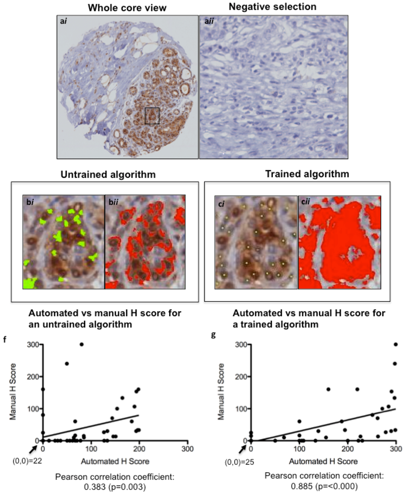
2.1. Comparison of Manual Versus Automated Scoring

| Biomarker | Pearson Correlation Coefficient (95% CI) | Kappa Statistic (95% CI) | Proportion Positive + |
|---|---|---|---|
| pERK | 0.89 (0.75–0.97) | 0.92 (0.80–1.00) | 18/58 (31%) |
| p53 | 0.80 (0.65–0.92) | 0.75 (0.56–0.95) | 16/56 (29%) |
| Cyclin D1 | 0.85 (0.71–0.94) | 0.73 (0.55–0.92) | 37/57 (65%) |
| Ki67 | 0.81 (0.71–0.91) | 0.55 (0.36–0.74) | 17/56 (30%) |
| HER2 | 0.90 (0.83–0.95) | 0.62 (0.40–0.84) | 14/56 (25%) |
2.2. Associations of Automated Scoring Between Biomarkers
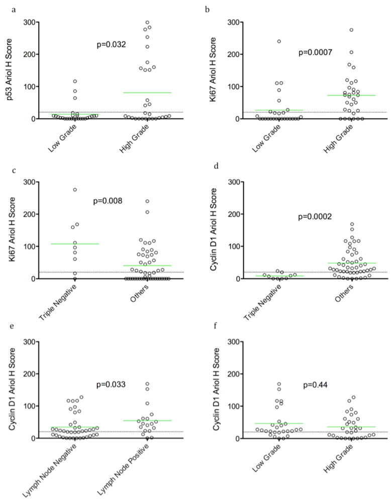
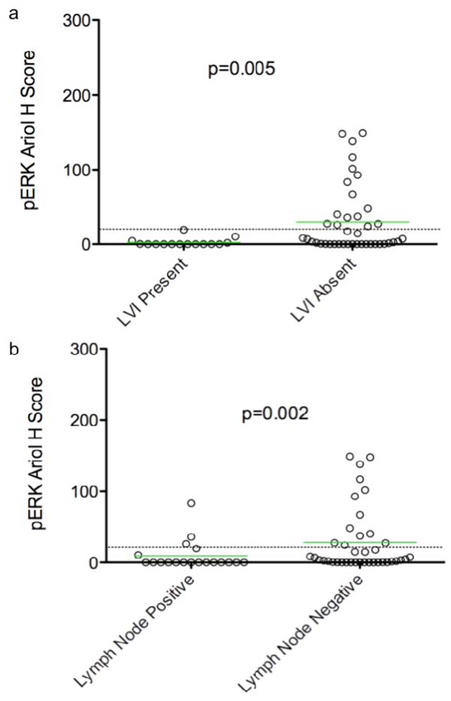
2.3. Associations of Biomarkers with Clinico-Pathological Parameters

| Biomarker (Ariol Score) | Clinical Parameter | n † | Original continuous score | Score dichotomized as positive >20 | |||
|---|---|---|---|---|---|---|---|
| Concordance index a | p-value | Odds Ratio (exact 95% CI) b | p-value | ||||
| pERK | LVI (present) | 56 | 0.25 c | 0.005 *,c | 0.00 (0.00–0.41) c | 0.0028 *,c | |
| Lymph node status (+) | 58 | 0.28 c | 0.002 *,c | NS | 0.22 | ||
| SBR score d (8 or 9) | 58 | NS | 0.37 | NS | 0.38 | ||
| ER/PR/HER2-ve (TN) | 58 | NS | 0.89 | NS | 0.44 | ||
| Recurrence (yes) | 51 | NS | 0.47 | NS | 1 | ||
| p53 | LVI (present) | 54 | NS | 0.21 | NS | 0.74 | |
| Lymph node status (+) | 56 | NS | 1.00 | NS | 1.00 | ||
| SBR score (8 or 9) | 56 | 0.67 | 0.032 | 6.5 (1.4–40) | 0.0074 * | ||
| ER/PR/HER2-ve (TN) | 56 | NS | 0.11 | NS | 0.26 | ||
| Recurrence (yes) | 48 | NS | 0.92 | NS | 1.00 | ||
| cyclin D1 | LVI (present) | 55 | NS | 0.36 | NS | 1 | |
| Lymph node status (+) | 57 | NS | 0.033 | 4.3 (0.96–26.1) | 0.041 | ||
| SBR score (8 or 9) | 57 | NS | 0.44 | NS | 0.17 | ||
| ER/PR/HER2-ve (TN) | 57 | 0.15 c | 0.0002 **,c | 0.038 (0.001–0.34) c | 0.0003 **,c | ||
| Recurrence (yes) | 50 | NS | 0.69 | NS | 0.72 | ||
| Ki67 | LVI (present) | 55 | NS | 0.42 | NS | 0.55 | |
| Lymph node status (+) | 57 | NS | 0.36 | NS | 0.57 | ||
| SBR score (8 or 9) | 57 | 0.75 | 0.0007 ** | 9.4 (2.4–38) | 0.0002 ** | ||
| ER/PR/HER2-ve (TN) | 57 | NS | 0.008 * | NS | 0.083 | ||
| Recurrence (yes) | 50 | NS | 0.2 | NS | 0.15 | ||
| HER2 | LVI (present) | 54 | NS | 0.16 | NS | 0.11 | |
| Lymph node status (+) | 56 | NS | 0.38 | NS | 0.76 | ||
| SBR score (8 or 9) | 56 | NS | 0.13 | NS | 0.16 | ||
| ER/PR/HER2-ve (TN) | 56 | NS | 0.24 | NS | 0.47 | ||
| Recurrence (yes) | 49 | 0.65 | 0.096 | NS | 0.26 | ||
3. Discussion
4. Experimental Section
4.1. Patients
| Parameter | Status | Number (%) |
|---|---|---|
| Age | <30 | 1 (2.1) |
| (Median: 45) | 30–40 | 11 (22.9) |
| (Range: 29–49) | 41–49 | 36 (75) |
| Tumor Stage | stage 1 | 26 (54.2) |
| stage 2 | 16 (33.3) | |
| stage 3 | 1 (2.1) | |
| stage 4 | 1 (2.1) | |
| Unknown | 4 (8.3) | |
| Tumor Grade a | Grade I | 8 (12.7) |
| Grade II | 23 (36.5) | |
| Grade III | 32 (50.8) | |
| LVI | Absent | 42 (64.3) |
| Present | 15 (35.7) | |
| Number of positive lymph nodes | 0 | 21 (60) |
| 1–3 | 11 (31.4) | |
| 4–10 | 1 (2.9) | |
| >10 | 2 (5.7) | |
| ER Status | Negative | 14 (29.2) |
| Positive | 34 (70.8) | |
| PR Status | Negative | 12 (25) |
| Positive | 36 (75) | |
| HER2 Status b | Negative | 36 (75) |
| Positive | 9 (18.8) | |
| Missing value | 3 (6.2) | |
| ER/PR/HER2 Status | Triple negative | 10 (14) |
| Others | 53 (86) | |
| Survival | Positive | 11 (17) |
| Negative | 43 (68) | |
| Missing value | 9 (15) |
4.2. Tissue Microarray Construction
4.3. Immunohistochemistry (IHC)
4.4. Manual Scoring
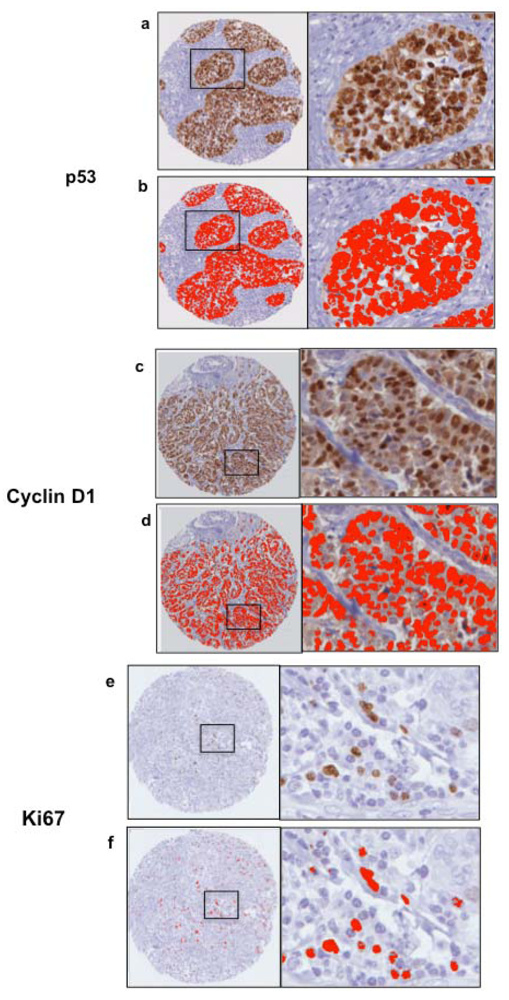
4.5. Automated Scoring

4.6. Statistical Analysis
5. Conclusions
Acknowledgments
References
- Dancey, J.E.; Chen, H.X. Strategies for optimizing combinations of molecularly targeted anticancer agents. Nat. Rev. Drug Discov. 2006, 8, 649–659. [Google Scholar] [CrossRef]
- Sotiriou, C.; Pusztai, L. Gene-expression signatures in breast cancer. N. Engl. J. Med. 2009, 360, 790–800. [Google Scholar] [CrossRef]
- Camp, R.L.; Neumeister, V.; Rimm, D.L. A decade of tissue microarrays: Progress in the discovery and validation of cancer biomarkers. J. Clin. Oncol. 2008, 26, 5630–5637. [Google Scholar] [CrossRef]
- Pleşan, D.M.; Georgescu, C.V.; Pătrană, N.; Pleşan, C.; Stoica, D. Immunohistochemical study of p53 and Ki67 in a group of patients with mammary carcinoma. Rom. J. Morphol. Embryol. 2010, 51, 459–465. [Google Scholar]
- Liu, C.; Chen, B.; Zhu, J.; Zhang, R.; Yao, F.; Jin, F.; Xu, H.; Lu, P. Clinical implications for nestin protein expression in breast cancer. Cancer Sci. 2009, 101, 815–819. [Google Scholar]
- Cho, E.Y.; Han, J.J.; Choi, Y.L.; Kim, K.M.; Oh, Y.L. Comparison of Her-2, EGFR and cyclin D1 in primary breast cancer and paired metastatic lymph nodes: An immunohistochemical and chromogenic in situ hybridization study. J. Korean Med. Sci. 2008, 6, 1053–1061. [Google Scholar]
- Frogne, T.; Laenkholm, A.V.; Lyng, M.B.; Henriksen, K.L.; Lykkesfeldt, A.E. Determination of HER2 phosphorylation at tyrosine 1221/1222 improves prediction of poor survival for breast cancer patients with hormone receptor-positive tumors. Breast Cancer Res. 2009, 11, R11. [Google Scholar] [CrossRef]
- Öhlschlegel, C.; Zahel, K.; Kradolfer, D.; Hell, M.; Jochum, W. HER2 genetic heterogeneity in breast carcinoma. J. Clin. Pathol. 2011, 64, 1112–1116. [Google Scholar] [CrossRef]
- Ridolfi, R.; Jamehdor, M.; Arber, J. HER-2/neu testing in breast carcinoma: A combined immunohistochemical and fluorescence in situ hybridization approach. Mod. Pathol. 2000, 13, 866–873. [Google Scholar] [CrossRef]
- Bobrovnikova-Marjon, E.; Grigoriadou, C.; Pytel, D.; Zhang, F.; Ye, J.; Koumenis, C.; Cavener, D.; Diehl, J. pERK promotes cancer cell proliferation and tumor growth by limiting oxidative DNA damage. Oncogene 2010, 27, 3881–3895. [Google Scholar]
- Montagut, C.; Settleman, J. Targeting the RAF-MEK-ERK pathway in cancer therapy. Cancer Lett. 2009, 2, 125–134. [Google Scholar] [CrossRef]
- Hadzisejdić, I.; Mustać, E.; Jonjić, N.; Petković, M.; Grahovac, B. Nuclear EGFR in ductal invasive breast cancer: Correlation with cyclin-D1 and prognosis. Mod. Pathol. 2010, 3, 392–403. [Google Scholar]
- Courjal, F.; Cuny, M.; Simony-Lafontaine, J.; Louason, G.; Speiser, P.; Zeillinger, R.; Rodriguez, C.; Theillet, C. Mapping of DNA amplifications at 15 chromosomal localizations in 1875 breast tumors: Definition of phenotypic groups. Cancer Res. 1997, 19, 4360–4367. [Google Scholar]
- Borrás, C.; Gómez-Cabrera, M.C.; Viña, J. The dual role of p53: DNA protection and antioxidant. Free Radic. Res. 2011, 45, 643–652. [Google Scholar] [CrossRef]
- Tennis, M.; Krishnan, S.; Bonner, M.; Ambrosone, C.B.; Vena, J.E.; Moysich, K.; Swede, H.; McCann, S.; Hall, P.; Shields, P.; et al. p53 Mutation analysis in breast tumors by a DNA microarray method. Cancer Epidemiol. Biomarkers Prev. 2006, 15, 80–85. [Google Scholar]
- Li, X.R.; Liu, M.; Zhang, Y.J.; Wang, J.D.; Zheng, Y.Q.; Li, J.; Ma, B.; Song, X. CK5/6, EGFR, Ki-67, cyclin D1, and nm23-H1 protein expressions as predictors of pathological complete response to neoadjuvant chemotherapy in triple-negative breast cancer patients. Med. Oncol. 2011, 28, 129–134. [Google Scholar]
- Millar, E.K.; Graham, P.H.; McNeil, C.M. Prediction of outcome of early ER+ breast cancer is improved using a biomarker panel, which includes Ki-67 and p53. Br. J. Cancer 2011, 105, 272–280. [Google Scholar] [CrossRef]
- Turashvili, G.; Leung, S.; Turbin, D.; Montgomery, K.; Gilks, B.; West, R.; Carrier, M.; Huntsman, D.; Aparicio, S. Inter-observer reproducibility of HER2 immunohistochemical assessment and concordance with fluorescent in situ hybridization (FISH): Pathologist assessment compared to quantitative image analysis. BMC Cancer 2009, 9, 165–177. [Google Scholar]
- Turbin, D.A.; Leung, S.; Cheang, M.; Kennecke, H.; Montgomery, K.; McKinney, S.; Treaba, D.; Boyd, N.; Goldstein, L.; Badve, S. Automated quantitative analysis of estrogen receptor expression in breast carcinoma does not differ from expert pathologist scoring: A tissue microarray study of 3,484 cases. Breast Cancer Res. Treat. 208, 3, 417–426. [Google Scholar]
- Bolton, K.L.; Garcia-Closas, M.; Pfeiffer, R.M.; Duggan, M.; Howat, W.; Hewitt, S.; Yang, X.; Cornelison, R.; Anzick, S.; Meltzer, P.; et al. Assessment of automated image analysis of breast cancer tissue microarrays for epidemiologic studies. Cancer Epidemiol. Biomarkers Prev. 2010, 4, 992–999. [Google Scholar]
- Rajput, A.; Hu, N.; Varma, S.; Chen, C.H.; Ding, K.; Park, P.C.; Chapman, J.A.; SenGupta, S.K.; Madarnas, Y.; Elliott, B.E.; et al. Immunohistochemical assessment of expression of centromere protein-A (CENPA) in human invasive breast cancer. Cancers 2011, 3, 4212–4227. [Google Scholar]
- Parissenti, A.M.; Chapman, J.A.; Kahn, H.J.; Guo, B.; Han, L.; O’Brien, P.; Clemons, M.P.; Jong, R.; Dent, R.; Fitzgerald, B.; et al. Association of low tumor RNA integrity with response to chemotherapy in breast cancer patients. Breast Cancer Res. Treat. 2010, 119, 347–356. [Google Scholar] [CrossRef]
- Milde-Langosch, K.; Bamberger, A.M.; Rieck, G.; Grund, D.; Hemminger, G.; Müller, V.; Löning, T. Expression and prognostic relevance of activated extracellular-regulated kinases (ERK1/2) in breast cancer. Br. J. Cancer 2005, 12, 2206–2215. [Google Scholar]
- Whyte, J.; Bergin, O.; Bianchi, A.; McNally, S.; Martin, F. Key signaling nodes in mammary gland development and cancer. Mitogen-activated protein kinase signaling in experimental models of breast cancer progression and in mammary gland development. Breast Cancer Res. 2009, 11, 209–222. [Google Scholar] [CrossRef]
- Torii, S.; Yamamoto, T.; Tsuchiya, Y.; Nishida, E. ERK MAP kinase in G1 cell cycle progression and cancer. Cancer Sci. 2006, 97, 697–702. [Google Scholar] [CrossRef]
- Whyte, J.; Bergin, O.; Bianchi, A.; McNally, S.; Martin, F. Key signalling nodes in mammary gland development and cancer. Mitogen-activated protein kinase signalling in experimental models of breast cancer progression and in mammary gland development. Breast Cancer Res. 2009, 11, 209–223. [Google Scholar] [CrossRef]
- Agarwal, R.; Gonzalez-Angulo, A.; Myhre, S.; Carey, M.; Lee, J.; Overgaard, J.; Alsner, J.; Stemke-Hale, K.; Lluch, A.; Neve, R.; et al. Integrative analysis of cyclin protein levels identifies cyclin b1 as a classifier and predictor of outcomes in breast cancer. Clin. Cancer Res. 2009, 11, 3654–3662. [Google Scholar]
- Boström, P.; Söderström, M.; Palokangas, T.; Vahlberg, T.; Collan, Y.; Carpen, O.; Hirsimäki, P. Analysis of cyclins A, B1, D1 and E in breast cancer in relation to tumor grade and other prognostic factors. BMC Res. Notes 2009, 2, 140–148. [Google Scholar] [CrossRef]
- Caldon, C.; Sutherland, R.; Musgrove, E. Cell cycle proteins in epithelial cell differentiation: Implications for breast cancer. Cell Cycle 2010, 10, 1918–1928. [Google Scholar]
- McShane, L.M.; Altman, D.G.; Sauerbrei, W.; Taube, S.E.; Gion, M.; Clark, G.M. Reporting recommendations for tumor MARKer prognostic studies (REMARK). Breast Cancer Res. Treat. 2006, 2, 229–235. [Google Scholar]
- Booy, E.P.; Henson, E.S.; Gibson, S.B. Epidermal growth factor regulates Mcl-1 expression through the MAPK-Elk-1 signaling pathway contributing to cell survival in breast cancer. Oncogene 2011, 30, 2367–2378. [Google Scholar] [CrossRef]
- Wolff, H.C.; Hammond, M.; Schwartz, J.; Hagerty, K.; Allred, D.; Cote, R.; Dowsett, M.; Fitzgibbons, P.; Hanna, W.; Langer, A.; et al. American Society of Clinical Oncology/College of American Pathologists guideline recommendations for human epidermal growth factor receptor 2 testing in breast cancer. Arch. Pathol. Lab. Med. 2007, 131, 18–43. [Google Scholar]
- Hanley, J.A.; McNeil, B.J. The meaning and use of the area under a receiver operating characteristic (Roc) Curve. Radiology 1982, 143, 29–36. [Google Scholar]
- Hu, J.X.; Zhao, H.; Zhou, H.H. False discovery rate control with groups. J. Am. Stat. Assoc. 2010, 105, 1215–1227. [Google Scholar] [CrossRef]
© 2012 by the authors; licensee MDPI, Basel, Switzerland. This article is an open access article distributed under the terms and conditions of the Creative Commons Attribution license (http://creativecommons.org/licenses/by/3.0/).
Share and Cite
Cass, J.D.; Varma, S.; Day, A.G.; Sangrar, W.; Rajput, A.B.; Raptis, L.H.; Squire, J.; Madarnas, Y.; SenGupta, S.K.; Elliott, B.E. Automated Quantitative Analysis of p53, Cyclin D1, Ki67 and pERK Expression in Breast Carcinoma Does Not Differ from Expert Pathologist Scoring and Correlates with Clinico-Pathological Characteristics. Cancers 2012, 4, 725-742. https://doi.org/10.3390/cancers4030725
Cass JD, Varma S, Day AG, Sangrar W, Rajput AB, Raptis LH, Squire J, Madarnas Y, SenGupta SK, Elliott BE. Automated Quantitative Analysis of p53, Cyclin D1, Ki67 and pERK Expression in Breast Carcinoma Does Not Differ from Expert Pathologist Scoring and Correlates with Clinico-Pathological Characteristics. Cancers. 2012; 4(3):725-742. https://doi.org/10.3390/cancers4030725
Chicago/Turabian StyleCass, Jamaica D., Sonal Varma, Andrew G. Day, Waheed Sangrar, Ashish B. Rajput, Leda H. Raptis, Jeremy Squire, Yolanda Madarnas, Sandip K. SenGupta, and Bruce E. Elliott. 2012. "Automated Quantitative Analysis of p53, Cyclin D1, Ki67 and pERK Expression in Breast Carcinoma Does Not Differ from Expert Pathologist Scoring and Correlates with Clinico-Pathological Characteristics" Cancers 4, no. 3: 725-742. https://doi.org/10.3390/cancers4030725
APA StyleCass, J. D., Varma, S., Day, A. G., Sangrar, W., Rajput, A. B., Raptis, L. H., Squire, J., Madarnas, Y., SenGupta, S. K., & Elliott, B. E. (2012). Automated Quantitative Analysis of p53, Cyclin D1, Ki67 and pERK Expression in Breast Carcinoma Does Not Differ from Expert Pathologist Scoring and Correlates with Clinico-Pathological Characteristics. Cancers, 4(3), 725-742. https://doi.org/10.3390/cancers4030725