Article

Robotic Rectal Cancer Surgery: Perioperative and Long-Term Oncological Outcomes of a Single-Center Analysis Compared with Laparoscopic and Open Approach

by
Shachar Laks
1,2,
Michael Goldenshluger
1,3,
Alexander Lebedeyev
3,
Yasmin Anderson
1,
Ofir Gruper
1 and
Lior Segev
1,3,*
1
Faculty of medicine, Tel-Aviv University, Tel-Aviv 6997801, Israel
2
Department of Surgery, Wolfson Medical Center, Holon 5822012, Israel
3
Division of Surgery, The Chaim Sheba Medical Center, Tel-Hashomer 5266202, Israel
*
Author to whom correspondence should be addressed.
Cancers 2025, 17(5), 859; https://doi.org/10.3390/cancers17050859
Submission received: 6 January 2025 / Revised: 16 February 2025 / Accepted: 26 February 2025 / Published: 2 March 2025
(This article belongs to the Special Issue Robotic Surgery for Gastrointestinal (GI) Malignancies)

Simple Summary

Robotic-assisted surgery is a promising option with distinct advantages in rectal cancer surgery, which were shown mainly regarding the immediate postoperative outcomes such as decreasing the length of hospital stay, lowering wound infection rates, readmission rates, conversions rates, and even improving the wellbeing and work ergonomics of the operating surgeon. However, the evidence concerning the efficacy of the robotic approach in rectal cancer clearance and long-term oncological outcomes is still conflicting, and therefore, the optimal surgical approach for rectal cancer is still questionable. Hence, this study aimed to compare the short- and long-term outcomes of robotic-assisted rectal cancer surgery with the conventional laparoscopic-assisted approach and the open approach.

Abstract

Background/Objectives: Robotic-assisted surgery is an attractive and promising option with unique advantages in rectal cancer surgery, but the optimal surgical approach is still debatable. Therefore, we aimed to compare the short- and long-term outcomes of the robotic-assisted approach with the laparoscopic-assisted and open approaches. Methods: A single referral center in Israel retrospectively reviewed all patients that underwent an elective rectal resection for primary non-metastatic rectal cancer between 2010 and 2020. The cohort was separated into three groups according to the surgical approach: robotic, laparoscopic, or open. Results: The cohort included 526 patients with a median age of 64 years (range 31–89), of whom 103 patients were in the robotic group, 144 in the open group, and 279 patients in the laparoscopic group. The robotic group had significantly more lower rectal tumors (24.3% versus 12.7% and 6%, respectively, p < 0.001), more locally advanced tumors (65.6% versus 51.2% and 50.2%, respectively, p = 0.004), and higher rates of neoadjuvant radiotherapy (70.9% versus 54.2% and 39.5%, respectively, p < 0.001). Conversion to an open laparotomy was more common in the laparoscopy group (23.1% versus 6.8%, respectively, p = 0.001). The open approach had higher rates of intraoperative complications (23.2% compared with 10.7% and 13.5% in the robotic and laparoscopic groups, respectively, p = 0.011), longer hospital stays (10 days compared with 7 and 8 days, respectively, p < 0.001), and higher rates of postoperative complications (76% compared with 68.9% and 59.1%, respectively, p = 0.002). The groups were similar in the number of harvested lymph nodes (14) and the incidence of positive resection margins (2.1%). The 5-year overall survival in the robotic group was 92.3% compared with 90.5% and 88.3% in the laparoscopic and open groups, respectively (p = 0.12). The 5-year disease-free survival in the robotic group was 68% compared with 71% and 63%, respectively (p = 0.2). Conclusions: The robotic, laparoscopic, and open approaches had similar histopathological outcomes and long-term oncological outcomes. The open approach was associated with higher rates of perioperative morbidity. These findings suggest that the robotic approach is safe and effective in rectal cancer surgery.

1. Introduction

Despite evolving oncological neoadjuvant and adjuvant treatments, surgery still remains the mainstay curative option for patients with rectal cancer, and total mesorectal excision (TME) is the gold standard technique in rectal cancer surgery, as it provides improved control of local recurrence and overall survival [1]. Although it has been traditionally approached through open laparotomy, minimally invasive approaches are increasingly utilized in patients with rectal cancer, and randomized clinical trials including the COLOR II and the COREAN trials have demonstrated laparoscopic rectal cancer surgery to have similar or better short-term surgical outcomes and similar long-term oncological outcomes compared with open rectal cancer surgery [2,3,4]. However, laparoscopic rectal cancer surgery might be very challenging due to the confines of the narrow bony pelvis and the restricted flexibility of the rigid laparoscopic arms, hence raising concerns regarding tumor clearance and controlling distal and circumferential resection margins. The recent ACOSOG Z6051 and ALaCaRT trials failed to demonstrate the non-inferiority of laparoscopy compared with open surgery concerning pathological outcomes, questioning the true oncological safety of laparoscopic surgery for rectal cancer [5,6]. Those limitations of the laparoscopic platform could be potentially overcome by the robotic approach with its articulating instruments, three-dimensional depth of field view, stable camera platform, and possibly more precise tissue dissection [7]. Robotic-assisted TME was first described in 2006 [8], and since then, different retrospective studies compared between the robotic and laparoscopic surgical approach, and the majority of them showed no significant differences in postoperative morbidity and oncological outcomes [9,10,11]. The ROLARR randomized clinical trial comparing robotic-assisted surgery with laparoscopic surgery for rectal cancer failed to show clear superiority of the robotic approach in terms of conversion rates [12], and the COLARAR randomized trial concluded that robotic-assisted surgery did not significantly improve the TME quality compared with conventional laparoscopic surgery [13]. Despite the growing utilization of robotic surgery in rectal cancer treatment, the question of whether it offers substantial clinical benefits over the more traditional laparoscopic or open approaches remains unanswered. A recent meta-analysis comparing the robotic and laparoscopic approach for rectal cancers found no significant difference in overall survival and postoperative complications between the groups [14]. Most of the previous literature comparing rectal cancer surgery approach outcomes was limited to pairwise comparison only, and just a few studies have compared the open, laparoscopic, and robotic approaches together. Therefore, the aim of this study was to present a contemporary large cohort long-term comprehensive analysis of robotic rectal cancer surgery compared with laparoscopic and open approach outcomes.

2. Materials and Methods

Study design/population: This was a single-center retrospective study. After the approval of the institutional review board, we searched our electronic medical records to extract all patients who underwent radical rectal resection between the years 2010 and 2020 using all appropriate surgery codes including anterior resection of rectum, proctectomy with coloanal anastomosis, and abdomino-perineal resection (APR). From this database, we chose only patients with a diagnosis of primary non-metastatic rectal adenocarcinoma, designated clinically as stage I-III according to the American Joint Committee on Cancer Guidelines 7th edition [15]. We excluded non-elective, emergency cases, and palliative procedures. The final cohort was divided into three groups according to the primary surgical approach: robotic (Da Vinci Si or Xi Surgical System Intuitive Surgical, Sunnyvale, CA, USA), laparoscopic, or open approach. The decision regarding the surgical approach was taken according to the surgeon’s discretion. Intraoperative conversion to a different surgical approach was analyzed as the original intention to treat. Study variables and outcome measures: Patients’ characteristics and demographics included age, gender, body mass index (BMI), smoking status, personal surgical history, comorbidities, and American Society of Anesthesiologists score (ASA). Preoperative disease characteristics included presenting symptoms, disease work-up and imaging, tumor location in the rectum (lower rectum ≤ 5 cm, mid rectum 5–10 cm, upper rectum > 10 cm), pretreatment clinical staging, neoadjuvant oncological therapy, and preoperative laboratory values. Operative characteristics included the extent of resection performed, such as abdomino-perineal resection (APR), low anterior resection of rectum (with TME), and anterior resection of rectum (with tumor-specific TME), type of stoma constructed (which was performed at the discretion of the surgeon), additional procedure undertaken during surgery, type of anastomosis, skin incision location, conversion to open procedure, and intraoperative complications. Postoperative surgical outcomes included length of hospital stay (LOS), 30-day readmission rates and in hospital or 30-day postoperative complications, and their severity grading according to the Clavien–Dindo classification system [16]. A Clavien–Dindo grade > 2 was defined as a major complication. Histopathological surgical results included tumor size, differentiation, pathological stage, number of harvested lymph nodes, and distance of tumor from distal margins. Long-term oncological outcomes included details about adjuvant radiation therapy or adjuvant chemotherapy, the presence of local or distant disease recurrence, and mortality data. Recurrence-free survival (RFS) was defined as the period from the date of surgery to the date of the first recurrence. If tumor recurrence was not recorded, RFS was defined as the time between the date of surgery and the date of the last follow-up. Overall survival (OS) was calculated from the date of surgery to either the date of death or the date of the last follow-up visit. Statistical analysis: For numerical variables, we reported the median and range for each group and tested their difference using the Wilcoxon rank test. For categorical variables, we used the chi-squared test of independence using bootstrap to obtain a more accurate p-value when there were categories with low frequencies. p value < 0.05 was considered statistically significant. Univariate and multivariate analyses for risk factors for major complications were performed using the logistic regression model. The multivariate analysis was adjusted for the covariables which had a p < 0.3 in the univariate analysis (age, gender, tumor distance from anal verge, surgical approach, neoadjuvant radiation, and stoma creation). We used the Kaplan–Meier procedure for the estimated survival curves and their 95% confidence intervals. Univariate and multivariate analyses for disease recurrence were based on the Cox proportional hazard regression. The multivariate analysis for disease recurrence was adjusted for the covariables which had a p < 0.3 in the univariate analysis (age, surgical approach, tumor distance from anal verge, and positive margins).

3. Results

3.1. Patients Characteristics and Demographics

The cohort included 526 patients with a median age of 64 years (range 31–89), of whom 103 patients were in the robotic group, 144 in the open group, and 279 patients in the laparoscopic group. All three groups were similar in relation to their demographics and baseline characteristics including comorbidities, ASA score, and prior abdominal operations (Table 1).

3.2. Disease Presentation and Preoperative Work-Up

The robotic group had significantly more patients with lower rectum tumors compared with the other groups (24.3% versus 12.7% in the open group and 6% in the laparoscopic group, p < 0.001) and significantly fewer patients with upper rectal tumors (38.8% versus 49.3% and 66.5%, respectively, p < 0.001). Consequently, the median tumor distance from the anal verge was the lowest among the robotic group (8 cm versus 9 cm and 12 cm, respectively, p < 0.001). The robotic group had significantly more patients with locally advanced tumors as could be expressed by their higher rates of clinical stage 3 tumors (65.6% versus 51.2% and 50.2%, respectively, p = 0.004) and their lower rates of clinical stage 1 tumors (11.5% versus 22.5% and 30.5%, respectively, p = 0.004). Accordingly, the rates of neoadjuvant radiotherapy (long or short course) were significantly higher among the robotic group (70.9% versus 54.2% and 39.5%, respectively, p < 0.001) (Table 2).

3.3. Surgical Procedure

APR was significantly more prevalent among the robotic group (17.6% versus 2.1% and 4.3%, respectively, p < 0.001). Consequently, the robotic group had higher rates of a permanent end colostomy compared with the open and laparoscopic group (18.4% versus 7.7% and 7.8%, respectively, p < 0.001). An additional surgical intervention during surgery was much more prevalent among the open group, and salpingo-ophorectomy was the most common of these (19.7% in the open group compared with 1.9% in the robotic group and 3.6% in the laparoscopic group, p < 0.001). With regard to the anastomosis, a hand-sewn coloanal anastomosis was significantly more prevalent in the open group (12.9% compared with 3.6% in the robotic group and 4.2% in the laparoscopic group, p < 0.001). Conversion rates to an open laparotomy were significantly higher among the laparoscopic group compared to the robotic group (23.1% versus 6.8%, respectively, p = 0.001). Intraoperative complications were significantly more prevalent within the open group (23.2% compared with 10.7% among the robotic group and 13.5% among the laparoscopic group, p = 0.011) (Table 3).

3.4. Postoperative Surgical Outcomes

The open group patients had significantly longer hospital stay (LOS) compared with the other groups (10 days compared with 7 and 8 days among the robotic and laparoscopic groups, respectively, p < 0.001). The postoperative overall complication rate was significantly higher among the open group (76% compared with 68.9% among the robotic group and 59.1% among the laparoscopic group, p = 0.002), and surgical site infection was the main morbidity differentiating the open groups from the other groups (28.9% compared with 7.8% and 10.5%, respectively, p < 0.001). Additionally, major complications were also more prevalent among the open group (23.9% versus 13.6% in the robotic group and 12.8% in the laparoscopic group, p = 0.01) (Table 4).
A logistic regression model to test for risk factors for major complications found male gender (OR, 1.676; 95% CI, 1.004–2.798; p = 0.048), open approach (OR, 1.959; 95% CI, 1.132–3.390; p = 0.026), and loop ileostomy construction (OR, 3.416; 95% CI, 1.570–7.431; p = 0.007) to be significantly associated with postoperative major complications both in univariate and multivariate analyses. Shorter tumor distance from the anal verge was associated with major complications in univariate analysis but not after multivariate analysis. The robotic and laparoscopic approach, age, BMI, smoking, and preoperative radiation were not found to be associated with major complications (Table 5).

3.5. Histopathological Results

The three groups were similar in the rates of complete pathological response, in the pathological stage distribution, number of harvested lymph nodes, lympho-vascular invasion rate, perineural invasion, tumor differentiation, mucinous tumors, and signet-ring cell features.
The distal margin was significantly larger among the robotic group (3.5 cm compared with 1.9 cm in the open group and 2.5 cm in the laparoscopic group, p = 0.006). The rate of involved margins (distal or radial margins) was 2.1% (11 patients) of the entire cohort, with no significant differences between the groups (Table 6).

3.6. Long-Term Oncological Outcomes

After a median follow-up time of 59 months (range of 1–171 months), no significant differences were noted between the groups in overall survival (OS) and recurrence-free survival (RFS) (Table 7, Figure 1).
The 5-year overall survival in the robotic group was 92.3% compared with 90.5% and 88.3% in the laparoscopic and open groups, respectively (p = 0.12). The 5-year disease-free survival in the robotic group was 68% compared with 71% and 63% in the laparoscopic and open groups, respectively (p = 0.2). In addition, there were no differences between the groups in OS and RFS after stratifying the cohort by clinical stage (Figure 2).
The Cox regression model to asses for risk factors for disease recurrence has found shorter tumor distance from anal verge (OR, 0.954; 95% CI, 0.914–0.996; p = 0.034) and involved distal/radial surgical margins (OR, 3.599; 95% CI, 1.320–9.812; p = 0.037) to be significantly associated with disease recurrence both in univariate and multivariate analyses. The surgical approach was not found to be associated with disease recurrence (Table 8).

3.7. Subgroup Analysis

In order to minimize the bias related to heterogeneous patients included in our cohort, we have conducted a subgroup analysis including only patients with mid and low rectal cancer who have received neoadjuvant radiation therapy. This analysis included 197 patients (82 lap, 57 open, and 58 robot), and the three groups were similar in terms of demographics, disease presentation, and preoperative work-up (including tumor location and clinical stage distribution) (Table S1). There were still no significant differences between the groups in OS and DFS. The 5-year DFS in the robotic group was 66.9% compared with 69% and 65% in the laparoscopic and open groups, p = 0.92 (Figure S1). An additional subgroup analysis including only patients with upper rectal cancer was performed consisting of 297 patients (186 lap, 71 open, and 40 robot) (Table S2). This subgroup analysis also showed similar long-term outcomes with 5-year DFSs of 67.6%, 72.9%, and 61.4% in the robotic, laparoscopic, and open groups, respectively, p = 0.17 (Figure S1). The 5-year DFS for stage 3 mid and low rectal cancer was 68.1%, 67.7%, and 59.9%, respectively, p = 0.49.

4. Discussion

There is still a continuous unsolved debate regarding the optimal surgical approach to treat rectal cancer [3,4,5,6]. Therefore, this present study aimed to retrospectively review the short- and long-term outcomes of robotic-assisted approach compared with the laparoscopic-assisted approach and the open approach.
Interestingly, in our study, the robotic group included more patients with distal and clinically locally advanced tumors compared with the other two groups. This was probably the reason for the higher rates of neoadjuvant radiation treatment observed among the robotic group, and also for the higher proportion of abdomino-perineal resections among the robotic group. Nevertheless, those differences did not translate into worse oncological outcomes as could possibly be expected. Similarly, in a study analyzing the short-term outcomes of 2114 consecutive patients in a single center in Korea, the robotic group also had the most distal tumors among the three groups, and the laparoscopic group included less patients with advanced and lower rectal tumors. The authors concluded that laparoscopic-assisted rectal cancer surgery tends to be preferred for upper rectal cancers and less advanced tumors, irrespective of the surgeon’s competence, and that the robotic procedure was probably chosen to overcome surgical complexity in patients with locally advanced and lower rectal cancers [17].
Despite operating on more distal challenging tumors, our robotic group had significantly lower conversion rates to an open procedure compared with the laparoscopic approach. This is in concordance with multiple prior studies that also described significant lower conversion rates among the robotic approach [17,18], while some studies have reported similar conversion rates between the laparoscopic and robotic approach [19,20]. This has clinical significance, as patients converted from the minimally invasive approach to open surgery were found to be at a greater risk for perioperative morbidity and to have worse oncological outcomes [21]. We speculate whether those lower conversion rates among the robotic group are partially related to better patients’ selection, and maybe surgeons using the robotic platform early on in their learning curve have a higher threshold for conversion compared with traditional laparoscopy. In addition, the higher proportion of neoadjuvant radiation among the robotic group, which is known to cause fibrosis and tissue edema that can make surgery technically more difficult [22], has not translated into higher rates of intraoperative complications, nor in intraoperative bleeding, both of which were significantly less prevalent among the robotic approach. Previous meta-analyses and systematic reviews have also observed lower intraoperative blood loss during robotic-assisted rectal cancer surgery compared with laparoscopic and open surgery [20,23]. This could be theoretically explained by the technical advantages of the robotic platform, including three-dimensional high-definition visualization, providing a detailed, stable, and magnified view of the surgical field, instrumental dexterity, and precise and stable dissection, all of which could potentially lead to decreased surgical blood loss, yet we acknowledge the need for cautious interpretation of these results. Still, higher blood loss during surgery has been associated with poor prognosis in colorectal cancer [24]; therefore, these findings suggest that robotic surgery may indirectly improve the prognosis of patients undergoing rectal surgery for cancer.
Consistent with previous studies, we also found a significant shorter hospital stay in the robotic group compared with the other two groups [20,25], and early postoperative complications, specifically ileus and wound infection, occurred more frequently in the open group than in the robotic and laparoscopic groups [17,26,27]. Moreover, major complications (Clavien–Dindo grade > 2) were more prevalent among the open group as well, and this is clinically meaningful as major complications after proctectomy for cancer are associated with earlier disease recurrence, ultimately leading to decreased survival [28]. Our multivariate analysis has found loop ileostomy creation, open surgery, and male gender to be associated with major complications. The latter two factors may be related to limited visibility and a confined narrow workspace, eventually leading to a more technically difficult surgery, possibly dissecting along non-anatomical planes, increasing blood loss, and altogether resulting in an increased postoperative morbidity. In a similar manner, Kim et al. reported that anastomotic complications, including leakage, abscess, fistula, and stricture, were significantly associated with male patients in multivariate analysis (OR, 1.85; 95 % CI, 1.049–3.263; p = 0.034), and so performed postoperative ileus (OR, 1.775; 95 % CI, 1.092–2.887; p = 0.021) [17]. This strengthens the notion that rectal cancer surgery in male patients is different than in females and could be very challenging and associated with an increased potential for major morbidity. Surgeons and oncologists should consider these issues upon deciding on preoperative treatment, while considering and planning surgery in male patients differently from females. For example, it should be considered to lower the “threshold” for non-operative management in male patients that seem to have a complete clinical response following neoadjuvant therapy.
Our overall postoperative complication rates were relatively high compared with previous studies [23,26]. We believe that this may be related to the definition and classification applied to postoperative morbidity documentation. We followed a very broad interpretation and included in our postoperative complication recordings any deviation from the normal postoperative course (such as electrolyte disturbances).
In line with the previous literature [17,26], the three groups did not differ in the number of retrieved lymph nodes, nor in terms of positive distal/radial margins, both of which serve as benchmarks for surgical oncology quality in rectal cancer surgery. On the other hand, a systematic review by Khajeh et al. found that the robotic approach had significantly higher rates of negative radial margins and a higher number of harvested lymph nodes than the open approach did, and also higher rates of negative radial margins than the laparoscopic approach [23]. The authors stated that this might be related to better visualization and improved access to the pelvis, utilizing the robotic platform. However, the superior histopathological outcomes of the robotic approach have not translated into differences in OS or DFS in this meta-analysis. Our robotic group did show significantly longer distal margins compared with the two other groups, which might also be related to the higher proportions of abdomino-perineal resections performed among this group. Unfortunately, our pathological reports lack reference regarding the completeness of mesorectal excision, which also serves as a surrogate marker for quality rectal cancer surgery.
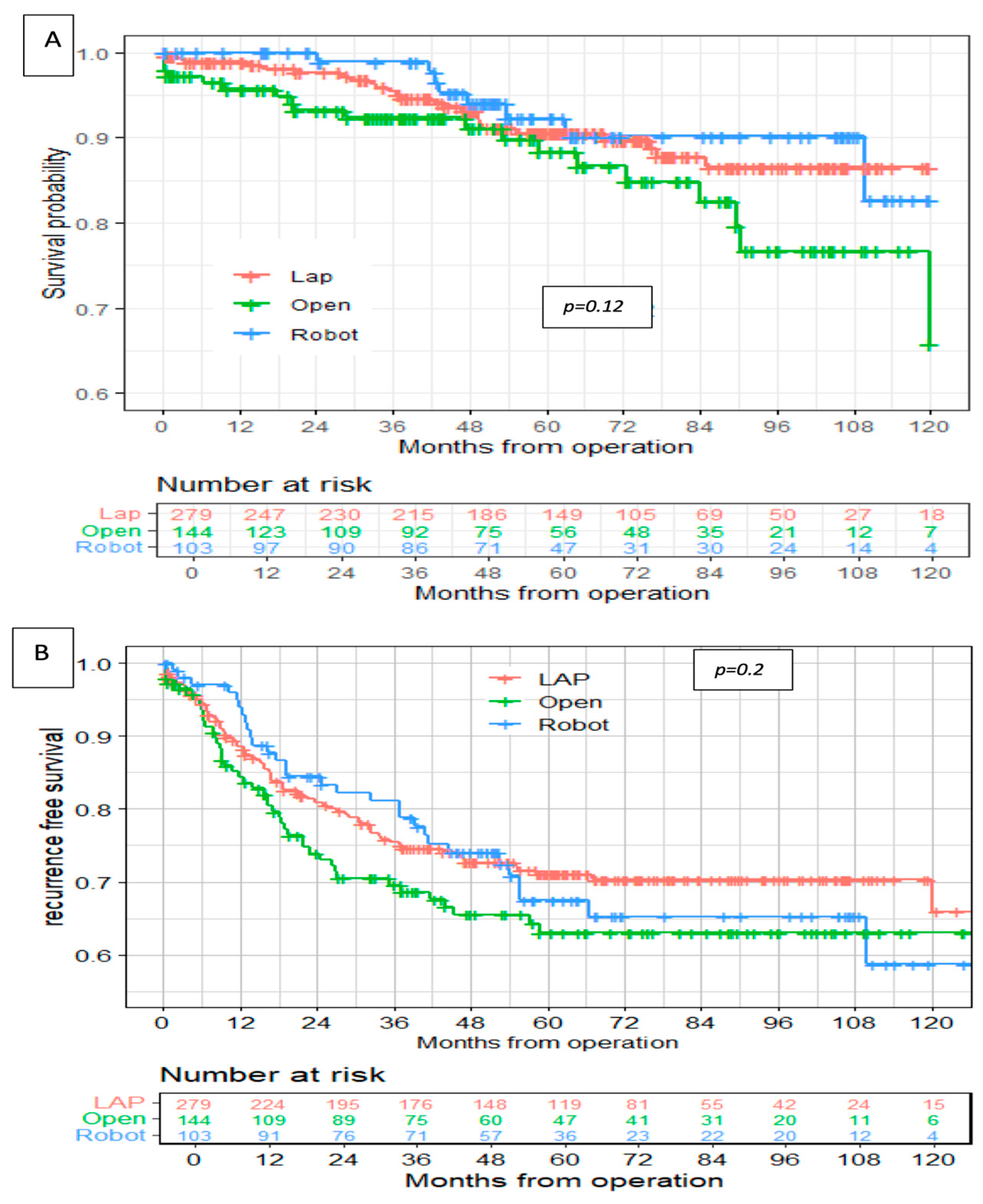
In this study, the robotic group showed similar long-term oncological outcomes compared with the laparoscopic and open approaches, and there were no significant differences between the groups in overall survival and disease-free survival rates. Those results are similar to previously published studies [23,26,29]. Our multivariate analysis has found shorter tumor distances from the anal verge and involved distal/radial surgical margins to be significantly associated with disease relapse. Similarly, Kim et al. found the local recurrence rates to be closely related to postoperative hemorrhage (OR, 14.02; 95% CI, 2.592–75.84; p = 0.002), DRM+ (OR, 13.4; 95% CI, 2.319– 77.439; p = 0.004), anastomotic complications (OR, 5.514; 95% CI, 1.416–21.475; p = 0.014), and tumor location (OR, 0.364; 95% CI, 0.134–0.989; p = 0.047) in multivariate analysis, but their study had a limited follow-up period [17]. The minimal acceptable DRM is controversial, but clearly, a threatened DRM is associated with local recurrence, and approximately three times more local recurrence was reported in patients with DRM <2 cm than in those with DRM >2 cm in one study [30]. This again emphasizes the importance of precise surgical techniques and controlling the distal resection margins in relation to disease-free survival and long-term oncological outcomes. Although we have not found differences in the margin positivity rate, the robotic group in our study presented significantly larger DRMs compared with the other groups, and this can imply that the robotic platform might be better in clear and safe distal resection margins compared with the open and laparoscopic approaches. However, Mirza et al. found that robotic and open TME were associated with higher margin positivity rates (8.2% versus 6.6% versus 1.9%, respectively, p = 0.17) compared with laparoscopic TME, and they thought it was related to the higher percentage of low rectal cancers in the robotic and open cohorts [29].
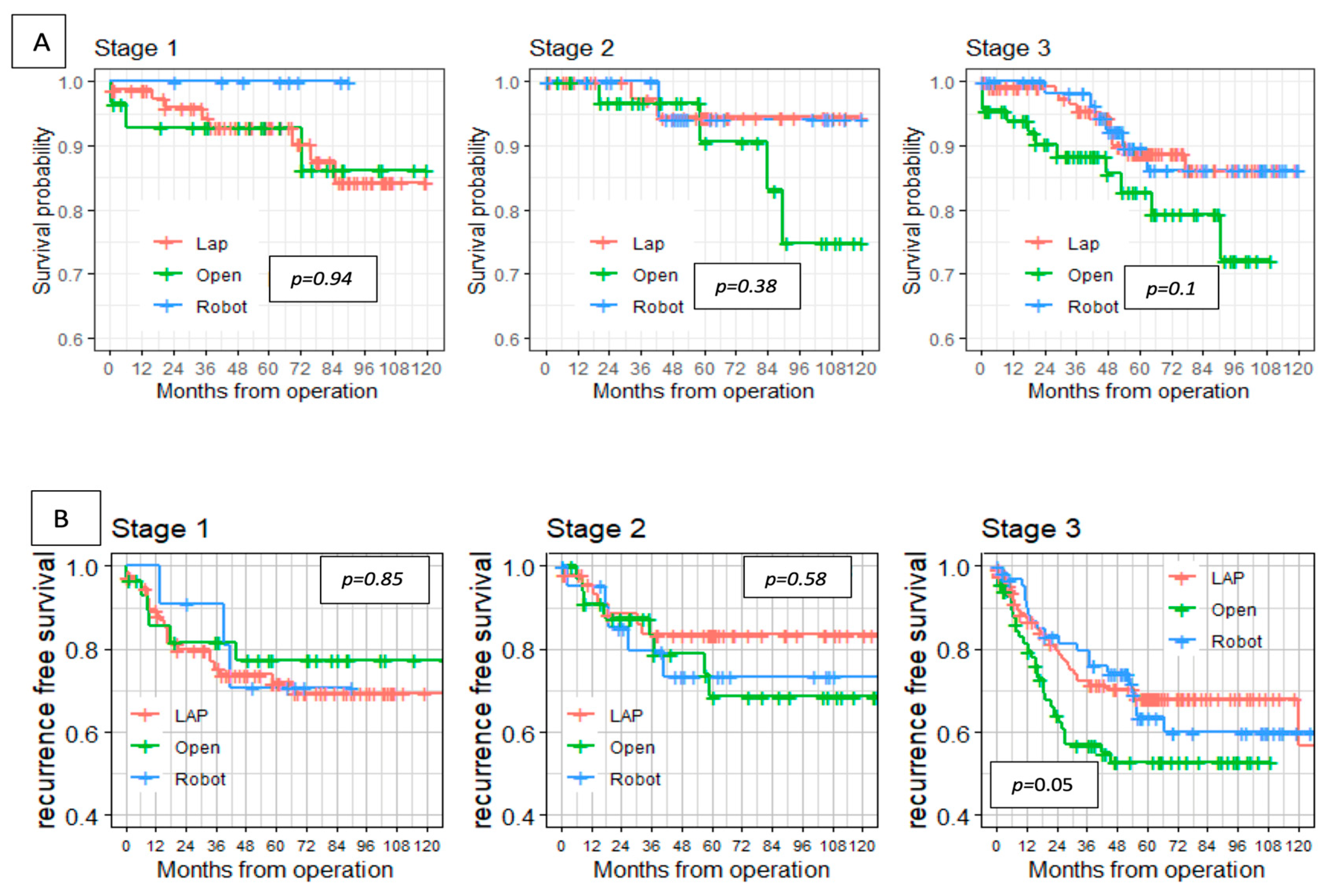
Our study has some limitations. First of all, the non-randomized, retrospective nature of our study is subject to a selection bias due to the surgeon’s preference for each procedure. In addition, this was a single-institution study, which limits the ability to generalize our results. The impact of the learning curve on surgical outcomes, specifically in minimally invasive approaches, such as robotic and laparoscopic, is substantial and should be considered. Our analysis included heterogenous populations of the respective groups regarding tumor location in the rectum, tumor stage, and preoperative radiation therapy, all of which could have an effect on surgical and oncological outcomes. However, this was confronted by subgroup analysis. Despite comprehensive data collection, our report lacks information on the completeness of mesorectal excision, estimated blood loss, and length of surgery, all of which are important markers in comparing the different surgical approaches.

5. Conclusions

In summary, our study found no significant differences in pathological outcomes and in long-term oncological outcomes between the robotic, laparoscopic, and open approaches. Additionally, we have demonstrated that minimally invasive approaches may be superior to the open approach in perioperative recovery, and given the lower conversion to open rates of robotic surgery, we believe that the robotic approach should be the preferred approach for rectal cancer, since it allows more patients to benefit from the advantages of the minimally invasive technique.

Supplementary Materials

The following supporting information can be downloaded at: https://www.mdpi.com/article/10.3390/cancers17050859/s1, Figure S1: Overall survival (A) and recurrence-free survival (B) after subgroup analysis for low and mid rectal cancer treated with neoadjuvant therapy, and upper rectal cancer; Table S1. Subgroup analysis for mid and low rectal cancer patients with neoadjuvant radiation; Table S2. Subgroup analysis for upper rectal cancer patients.

Author Contributions

Conceptualization, L.S. and M.G.; methodology, S.L.; software, Y.A. and O.G.; validation, L.S., S.L. and M.G.; formal analysis, Y.A.; investigation, L.S. and O.G.; resources, O.G.; data curation, S.L. and A.L.; writing—original draft preparation, L.S. and S.L.; writing—review and editing, L.S.; visualization, M.G.; supervision, L.S.; project administration, A.L. and Y.A.; funding acquisition, L.S. All authors have read and agreed to the published version of the manuscript.

Funding

This research received no external funding.

Institutional Review Board Statement

The study was conducted in accordance with the Declaration of Helsinki and approved by the Institutional Review Board of Sheba medical center (protocol code 9618-22-SMC, date of approval 12 December 2022).

Informed Consent Statement

Patient consent was waived since it was a retrospective study analyzing the electronic medical records.

Data Availability Statement

The data presented in this study are available upon request from the corresponding author due to privacy and ethical reasons.

Conflicts of Interest

The authors declare no conflicts of interest.

Abbreviations

The following abbreviations are used in this manuscript:
TMETotal Mesorectal Excision
APRAbdomino Perineal Resection
BMIBody Mass Index
ASAAmerican Society of Anesthesiologists
LOSLength Of Stay
RFSRecurrence-Free Survival
OSOverall Survival
CRCColorectal Cancer
TIATransient Ischemic Attack
CVACerebral Vascular Accident
COPDChronic Obstructive Pulmonary Disease
IHDIschemic Heart Disease
CHFCongestive Heart Failure
DMDiabetes Melitus
CKDChronic Kidney Disease
HTNHypertension
TRUSTrans Rectal Ultrasound
EMVIExtramural Vascular Invasion
BSOBilateral Salpingo-Oophorectomy
SSISurgical Site Infection
SBOSmall Bowel Obstruction
UTIUrinary Tract Infection
DVTDeep Vein Thrombosis
DLIDiverting Loop Ileostomy
AVAnal Verge

References

  1. Heald, R.; Ryall, R. Recurrence and survival after total mesorectal excision for rectal cancer. Lancet 1986, 327, 1479–1482. [Google Scholar] [CrossRef]
  2. van der Pas, M.H.; Haglind, E.; ACuesta, M.; Fürst, A.; Lacy, A.M.; Hop, W.C.; Bonjer, H.J. Laparoscopic versus open surgery for rectal cancer (COLOR II): Short-term outcomes of a randomised, phase 3 trial. Lancet Oncol. 2013, 14, 210–218. [Google Scholar] [CrossRef] [PubMed]
  3. Knudson, A.G., Jr. Mutation and cancer: Statistical study of retinoblastoma. Proc. Natl. Acad. Sci. USA 1971, 68, 820–823. [Google Scholar] [CrossRef] [PubMed]
  4. Bonjer, H.J.; Deijen, C.L.; Abis, G.A.; Cuesta, M.A.; Van Der Pas, M.H.G.M.; de Lange-de Klerk, E.S.M.; Lacy, A.M.; Bemelman, W.A.; Andersson, J.; Angenete, E.; et al. A Randomized Trial of Laparoscopic versus Open Surgery for Rectal Cancer. N. Engl. J. Med. 2015, 372, 1324–1332. [Google Scholar] [CrossRef]
  5. Fleshman, J.; Branda, M.; Sargent, D.J.; Boller, A.M.; George, V.; Abbas, M.; Peters, W.R., Jr.; Maun, D.; Chang, G.; Herline, A.; et al. Effect of Laparoscopic-Assisted Resection vs. Open Resection of Stage II or III Rectal Cancer on Pathologic Outcomes: The ACOSOG Z6051 Randomized Clinical Trial. JAMA 2015, 314, 1346–1355. [Google Scholar] [CrossRef] [PubMed]
  6. Stevenson, A.R.L.; Solomon, M.J.; Lumley, J.W.; Hewett, P.; Clouston, A.D.; Gebski, V.J.; Davies, L.; Wilson, K.M.; Hague, W.; Simes, J. Effect of Laparoscopic-Assisted Resection vs Open Resection on Pathological Outcomes in Rectal Cancer: The ALaCaRT Randomized Clinical Trial. JAMA 2015, 314, 1356–1363. [Google Scholar] [CrossRef]
  7. Park, S.; Kim, N.K. The Role of Robotic Surgery for Rectal Cancer: Overcoming Technical Challenges in Laparoscopic Surgery by Advanced Techniques. J. Korean Med. Sci. 2015, 30, 837–846. [Google Scholar] [CrossRef]
  8. Pigazzi, A.; Ellenhorn, J.D.I.; Ballantyne, G.H.; Paz, I.B. Robotic-assisted laparoscopic low anterior resection with total mesorectal excision for rectal cancer. Surg. Endosc. 2006, 20, 1521–1525. [Google Scholar] [CrossRef]
  9. Lam, J.; Tam, M.S.; Rettig, R.L.; McLemore, E.C. Robotic Versus Laparoscopic Surgery for Rectal Cancer: A Comprehensive Review of Oncological Outcomes. Perm. J. 2021, 25, 21.050. [Google Scholar] [CrossRef]
  10. Lim, D.R.; Bae, S.U.; Hur, H.; Min, B.S.; Baik, S.H.; Lee, K.Y.; Kim, N.K. Long-term oncological outcomes of robotic versus laparoscopic total mesorectal excision of mid–low rectal cancer following neoadjuvant chemoradiation therapy. Surg. Endosc. 2016, 31, 1728–1737. [Google Scholar] [CrossRef]
  11. Kim, Y.S.; Kim, M.J.; Park, S.C.; Sohn, D.K.; Kim, D.Y.; Chang, H.J.; Nam, B.-H.; Oh, J.H. Robotic Versus Laparoscopic Surgery for Rectal Cancer after Preoperative Chemoradiotherapy: Case-Matched Study of Short-Term Outcomes. Cancer Res. Treat. 2016, 48, 225–231. [Google Scholar] [CrossRef] [PubMed]
  12. Jayne, D.; Pigazzi, A.; Marshall, H.; Croft, J.; Corrigan, N.; Copeland, J.; Quirke, P.; West, N.; Rautio, T.; Thomassen, N.; et al. Effect of Robotic-Assisted vs Conventional Laparoscopic Surgery on Risk of Conversion to Open Laparotomy Among Patients Undergoing Resection for Rectal Cancer: The ROLARR Randomized Clinical Trial. JAMA 2017, 318, 1569–1580. [Google Scholar] [CrossRef] [PubMed]
  13. Park, J.S.; Lee, S.M.; Choi, G.-S.; Park, S.Y.; Kim, H.J.; Song, S.H.; Min, B.S.; Kim, N.K.; Kim, S.H.; Lee, K.Y. Comparison of Laparoscopic Versus Robot-Assisted Surgery for Rectal Cancers. Ann. Surg. 2023, 278, 31–38. [Google Scholar] [CrossRef]
  14. Safiejko, K.; Tarkowski, R.; Koselak, M.; Juchimiuk, M.; Tarasik, A.; Pruc, M.; Smereka, J.; Szarpak, L. Robotic-Assisted vs. Standard Laparoscopic Surgery for Rectal Cancer Resection: A Systematic Review and Meta-Analysis of 19,731 Patients. Cancers 2021, 14, 180. [Google Scholar] [CrossRef] [PubMed]
  15. Edge, S.B.; Compton, C.C. The American Joint Committee on Cancer: The 7th Edition of the AJCC Cancer Staging Manual and the Future of TNM. Ann. Surg. Oncol. 2010, 17, 1471–1474. [Google Scholar] [CrossRef]
  16. Clavien, P.A.; Barkun, J.; De Oliveira, M.L.; Vauthey, J.N.; Dindo, D.; Schulick, R.D.; de Santibañes, E.; Pekolj, J.; Slankamenac, K.; Bassi, C.; et al. The clavien-dindo classification of surgical complications: Five-year experience. Ann. Surg. 2009, 250, 187–196. [Google Scholar] [CrossRef]
  17. Kim, J.C.; Yu, C.S.; Lim, S.-B.; Park, I.J.; Kim, C.W.; Yoon, Y.S. Comparative analysis focusing on surgical and early oncological outcomes of open, laparoscopy-assisted, and robot-assisted approaches in rectal cancer patients. Int. J. Color. Dis. 2016, 31, 1179–1187. [Google Scholar] [CrossRef]
  18. Speicher, P.J.; Englum, B.R.; Ganapathi, A.M.; Nussbaum, D.P.; Mantyh, C.R.; Migaly, J. Robotic low anterior resection for rectal cancer: A national perspective on short-term oncologic outcomes. Ann. Surg. 2015, 262, 1040–1045. [Google Scholar] [CrossRef]
  19. Tang, B.; Lei, X.; Ai, J.; Huang, Z.; Shi, J.; Li, T. Comparison of robotic and laparoscopic rectal cancer surgery: A meta-analysis of randomized controlled trials. World J. Surg. Oncol. 2021, 19, 38. [Google Scholar] [CrossRef]
  20. Simillis, C.; Lal, N.; Thoukididou, S.N.; Kontovounisios, C.; Smith, J.J.; Hompes, R.; Adamina, M.; Tekkis, P.P. Open Versus Laparoscopic Versus Robotic Versus Transanal Mesorectal Excision for Rectal Cancer: A Systematic Review and Network Meta-analysis. Ann. Surg. 2019, 270, 59–68. [Google Scholar] [CrossRef]
  21. Allaix, M.E.; Furnée, E.J.; Mistrangelo, M.; Arezzo, A.; Morino, M. Conversion of laparoscopic colorectal resection for cancer: What is the impact on short-term outcomes and survival? World J. Gastroenterol. 2016, 22, 8304–8313. [Google Scholar] [CrossRef]
  22. Marijnen, C.A.M.; Kapiteijn, E.; van de Velde, C.J.H.; Martijn, H.; Steup, W.H.; Wiggers, T.; Kranenbarg, E.K.; Leer, J.W.H.; Cooperative Investigators of the Dutch Colorectal Cancer Group. Acute side effects and complications after short-term preoperative radi-otherapy combined with total mesorectal excision in primary rectal cancer: Report of a multicenter randomized trial. J. Clin. Oncol. 2002, 20, 817–825. [Google Scholar] [CrossRef] [PubMed]
  23. Khajeh, E.; Aminizadeh, E.; Moghadam, A.D.; Nikbakhsh, R.; Goncalves, G.; Carvalho, C.; Parvaiz, A.; Kulu, Y.; Mehrabi, A. Outcomes of Robot-Assisted Surgery in Rectal Cancer Compared with Open and Laparoscopic Surgery. Cancers 2023, 15, 839. [Google Scholar] [CrossRef]
  24. Mörner, M.E.; Gunnarsson, U.; Jestin, P.; Svanfeldt, M. The importance of blood loss during colon cancer surgery for long-term survival: An epidemiological study based on a population-based register. Ann. Surg. 2012, 255, 1126–1128. [Google Scholar] [CrossRef] [PubMed]
  25. Zhang, C.; Tan, H.; Xu, H.; Ding, J. The role of robotic-assisted surgery in the management of rectal cancer: A systematic review and meta-analysis. Int. J. Surg. 2024, 110, 6282–6296. [Google Scholar] [CrossRef] [PubMed]
  26. Kim, C.H.; Baik, S.H. Outcomes of Robotic-Assisted Colorectal Surgery Compared with Laparoscopic and Open Surgery: A Systematic Review. J. Gastrointest. Surg. 2014, 18, 816–830. [Google Scholar] [CrossRef]
  27. Krieg, A.; Kolbe, E.W.; Kaspari, M.; Krieg, S.; Loosen, S.H.; Roderburg, C.; Kostev, K. Trends and outcomes in colorectal cancer surgery: A multicenter cross-sectional study of minimally invasive versus open techniques in Germany. Surg. Endosc. 2024, 38, 6338–6346. [Google Scholar] [CrossRef]
  28. Gamboa, A.C.; Lee, R.M.; Turgeon, M.K.; Varlamos, C.; Regenbogen, S.E.; Hrebinko, K.A.; Holder-Murray, J.; Wiseman, J.T.; Ejaz, A.; Feng, M.P.; et al. Impact of post-operative complications on oncologic outcomes after rectal cancer surgery: An analysis of the US rectal cancer consortium. Ann. Surg. Oncol. 2021, 28, 1712–1721. [Google Scholar] [CrossRef]
  29. Mirza, M.B.; Gamboa, A.C.; Irlmeier, R.; Hopkins, B.; Regenbogen, S.E.; Hrebinko, K.A.; Holder-Murray, J.; Wiseman, J.T.; Ejaz, A.; Wise, P.E.; et al. Association of Surgical Approaches and Outcomes in Total Mesorectal Excision and Margin Status for Rectal Cancer. J. Surg. Res. 2024, 300, 494–502. [Google Scholar] [CrossRef]
  30. Law, W.L.; Chu, K.W. Anterior resection for rectal cancer with mesorectal excision: A prospective evaluation of 622 patients. Ann. Surg. 2004, 240, 260–268. [Google Scholar] [CrossRef]
Figure 1. Overall survival (A) and recurrence-free survival (B).
Cancers 17 00859 g001
Figure 2. Overall survival (A) and recurrence-free survival (B) stratified by stage.
Cancers 17 00859 g002
Table 1. Patients’ characteristics and demographics.
VariableAll Cohort (n = 526)LAP (n = 279)Open (n = 144)Robot (n = 103)p-Value
Age, median (range)64 (31–89)64 (31–89)66 (32–88)64 (31–83)0.078
Gender 0.623
Female n (%)231 (43.9%)124 (44.4%)66 (45.8%)41 (39.8%)
Male n (%)295 (56.1%)155 (55.6%)78 (54.2%)62 (60.2%)
BMI median (range)26.3 (15.6–45.6)26.1 (16.8–42.5)26.9 (15.6–45.6)25.9 (19.5–41.1)0.214
Smoking 0.203
No326 (62%)179 (64.2%)90 (62.5%)57 (55.3%)
Past smoker103 (19.6%)57 (20.4%)27 (18.8%)19 (18.4%)
Current smoker97 (18.4%)43 (15.4%)27 (18.8%)27 (26.2%)
Family history of CRC123 (27.5%)67 (28.2%)26 (23%)30 (31.2%)0.251
Yes 1st degree81 (18.1%)43 (18.1%)21 (18.6%)17 (17.7%)
Yes non-1st degree42 (9.4%)24 (10.1%)5 (4.4%)13 (13.5%)
Blood thinners139 (26.5%)78 (28%)40 (27.8%)21 (20.4)0.629
Antiaggregating agents114 (21.7%)63 (22.6%)33 (22.9%)18 (17.5%)
Anticoagulation25 (4.8%)15 (5.4%)7 (4.9%)3 (2.9%)
History of abdominal surgery212 (40.3)110 (39.4)56 (38.9%)46 (44.7%)0.282
Prior non-bowel abdominal surgery154 (29.3%)83 (29.7%)35 (24.3%)36 (35%)
Prior bowel resection58 (11%)27 (9.7%)21 (14.6%)10 (9.7%)
Comorbidity any415 (78.9%)220 (78.9%)119 (82.6%)76 (73.8%)0.256
TIA/CVA18 (3.4%)6 (2.2%)10 (6.9%)2 (1.9%)0.024
Asthma/COPD48 (9.1%)28 (10%)11 (7.6%)9 (8.7%)0.712
IHD/CHF74 (14.1%)39 (14%)24 (16.7%)11 (10.7%)0.418
Arrhythmia29 (5.5%)16 (5.7%)7 (4.9%)6 (5.8%)0.937
DM117 (22.2%)56 (20.1%)37 (25.7%)24 (23.3%)0.415
CKD17 (3.2%)7 (2.5%)8 (5.6%)2 (1.9%)0.19
HTN234 (44.5%)126 (45.2%)72 (50%)36 (35%)0.065
Dyslipidemia183 (34.8%)101 (36.2%)51 (35.4%)31 (30.1%)0.535
Hypothyroidism39 (7.4%)24 (8.6%)7 (4.9%)8 (7.8%)0.397
ASA score median (range)3 (1–5)3 (1–4)3 (1–5)3 (1–4)0.328
ASA score 0.127
119 (4.3%)13 (5.4%)3 (2.6%)3 (3.5%)
2153 (34.5%)77 (31.8%)39 (33.6%)37 (43%)
3257 (57.9%)148 (61.2%)67 (57.8%)42 (48.8%)
414 (3.2%)4 (1.7%)6 (5.2%)4 (4.7%)
BMI—body mass index, CRC—colorectal cancer, TIA—transient ischemic attack, CVA—cerebrovascular accident, COPD—chronic obstructive pulmonary disease, IHD—ischemic heart disease, CHF—congestive heart failure, DM—diabetes mellitus, CKD—chronic kidney disease, HTN—hypertension, and ASA—American Society of Anesthesiologists, Bold—statistically significant (p < 0.05).
Table 2. Disease presentation and preoperative work-up.
VariableAll cohort (n = 526)LAP (n = 279)Open (n = 144)Robot (n = 103)p-Value
Pre-diagnosis symptoms422 (80.2%)216 (77.4%)120 (83.3%)86 (83.5%)0.236
Abdominal pain93 (17.7%)42 (15.1%)33 (22.9%)18 (17.5%)0.137
Anemia47 (8.9%)26 (9.3%)16 (11.1%)5 (4.9%)0.231
Weight loss108 (20.5%)50 (17.9%)38 (26.4%)20 (19.4%)0.12
Change in bowel movements222 (42.2%)119 (42.7%)64 (44.4%)39 (37.9%)0.58
Rectal bleeding304 (57.8%)168 (60.2%)72 (50%)64 (62.1%)0.082
Preoperative CT scan441 (83.8%)244 (87.5%)118 (81.9%)79 (76.7%)0.031
Preoperative PET scan203 (38.6%)95 (34.1%)57 (39.6%)51 (49.5%)0.022
Preoperative TRUS362 (68.8%)215 (77.1%)90 (62.5%)57 (55.3%)<0.001
Preoperative pelvic MRI260 (49.4%)153 (54.8%)69 (47.9%)38 (36.9%)0.037
Colonoscopy522 (99.2%)278 (99.6%)142 (98.6%)102 (99%)0.694
Tumor location <0.001
Lower rectum (<5 cm)60 (11.4%)17 (6.1%)18 (12.5%)25 (24.3%)
Mid rectum (5 ≤ X ≤ 10 cm)169 (32.1%)76 (27.2%)55 (38.2%)38 (36.9%)
Upper rectum (>10 cm)297 (56.5%)186 (66.7%)71 (49.3%)40 (38.8%)
Distance from anal verge, median (range)10 (0–15)12 (0–15)9 (1–15)8 (0–15)<0.001
Clinical stage 0.003
Stage 1119 (24.6%)79 (30.7%)29 (22.1%)11 (11.5%)
Stage 2106 (21.9%)49 (19.1%)35 (26.7%)22 (22.9%)
Stage 3259 (53.5%)129 (50.2%)67 (51.1%)63 (65.6%)
Clinical T stage <0.001
T147 (9.7%)32 (12.5%)10 (7.6%)5 (5.2%)
T2101 (20.9%)65 (25.3%)25 (19.1%)11 (11.5%)
T3300 (62%)155 (60.3%)74 (56.5%)71 (74%)
T436 (7.4%)5 (1.9%)22 (16.8%)9 (9.4%)
Clinical N stage 0.108
N0235 (47.6%)134 (51%)65 (49.2%)36 (36.4%)
N1187 (37.9%)89 (33.8%)51 (38.6%)47 (47.5%)
N272 (14.6%)40 (15.2%)16 (12.1%)16 (16.2%)
Clinical EMVI9 (4%)4 (3.2%)1 (1.9%)4 (8.9%)0.151
Preoperative Albumin, median g/dL (range)4.1 (2.4–5.1)4.2 (2.5–5.1)4 (2.4–4.9)4.1 (2.5–4.8)0.06
Preoperative Hgb, median g/dL (range)12.7 (7.8–17.2)12.8 (8.7–17.2)12.5 (7.8–16)12.8 (9–16)0.272
Preoperative Creatinine, median (range)0.8 (0.2–2.5)0.8 (0.4–2.5)0.8 (0.2–1.9)0.8 (0.4–1.4)0.986
Preop CEA, median (range)2 (0–175)1.8 (0–175)2.2 (0–140.9)2 (0–78.3)0.057
Preop CA 19-9, median (range)9.3 (0–248)8.9 (0–248)10.8 (0–188)9.2 (0–92.1)0.077
Neoadjuvant radiation261 (49.7%)109 (39.1%)79 (54.8%)73 (70.9%)<0.001
Short-course radiotherapy34 (6.5%)10 (3.6%)11 (7.6%)13 (12.6%)
Long-course chemo-radiation227 (43.2%)99 (35.5%)68 (47.2%)60 (58.3%)
TRUS—transrectal ultrasound, EMVI—extramural vascular invasion, Hgb—hemoglobin, and CEA—carcinoembryonic antigen, Bold—statistically significant (p < 0.05).
Table 3. Operative details.
VariableAll Cohort (n = 526)LAP (n = 279)Open (n = 144)Robot (n = 103)p-Value
Procedure <0.001
Abdomino-perineal resection33 (6.3%)12 (4.3%)3 (2.1%)18 (17.6%)
Anterior resection175 (33.4%)113 (40.6%)43 (29.9%)19 (18.6%)
Low anterior resection (TME)316 (60.3%)153 (55%)98 (68.1%)65 (63.7%)
Stoma created321 (61%)146 (52.3%)98 (68%)77 (74.7)<0.001
Diverting loop ileostomy269 (51.1%)124 (44.4%)87 (60.4%)58 (56.3%)
End colostomy52 (9.9%)22 (7.9%)11 (7.6%)19 (18.4%)
Stoma reversed222 (69.2%)104 (71.2%)68 (69.4%)50 (64.9%)0.64
Additional procedure81 (15.4%)27 (9.7%)46 (31.9%)8 (7.8%)<0.001
Additional anastomosis17 (3.2%)5 (1.8%)12 (8.3%)0 (0%)0.001
BSO40 (7.6%)10 (3.6%)28 (19.4%)2 (1.9%)<0.001
Hysterectomy10 (1.9%)3 (1.1%)6 (4.2%)1 (1%)0.077
Hernia repair6 (1.1%)2 (0.7%)2 (1.4%)2 (1.9%)0.646
Cholecystectomy3 (0.6%)2 (0.7%)1 (0.7%)0 (0%)0.829
Type of anastomosis <0.001
Colonic pouch13 (2.7%)4 (1.6%)5 (3.7%)4 (4.8%)
End-to-end362 (76.2%)215 (83.7%)72 (53.7%)75 (89.3%)
Hand-sewn coloanal31 (6.5%)11 (4.3%)17 (12.7%)3 (3.6%)
Side-to-end69 (14.5%)27 (10.5%)40 (29.9%)2 (2.4%)
Skin incision 0.001
Left lower quadrant7 (1.8%)7 (2.5%)-0 (0%)
Midline laparotomy66 (17.3%)60 (21.5%)-6 (5.8%)
Natural orifice50 (13.1%)30 (10.8%)-20 (19.4%)
Periumbilical20 (5.2%)16 (5.7%)-4 (3.9%)
Pfanensteil239 (62.6%)166 (59.5%)-73 (70.9%)
Conversion 70 (18.3%)63 (22.6%)-7 (6.8%)0.001
Intraoperative complication82 (15.6%)38 (13.6%)33 (22.9%)11 (10.7%)0.015
Bleeding27 (5.1%)9 (3.2%)15 (10.4%)3 (2.9%)0.004
Ureter/Urethra injury4 (0.8%)2 (0.7%)2 (1.4%)0 (0%)0.443
Enterotomy23 (4.4%)10 (3.6%)11 (7.6%)2 (1.9%)0.065
Splenic injury7 (1.3%)2 (0.7%)5 (3.5%)0 (0%)0.018
Vaginal injury8 (1.5%)3 (1.1%)1 (0.7%)4 (3.9%)0.105
Anastomosis disruption8 (1.5%)6 (2.2%)1 (0.7%)1 (1%)0.566
TME—total mesorectal excision; BSO—bilateral salpingo-ophorectomy, Bold—statistically significant (p < 0.05).
Table 4. Postoperative immediate surgical outcomes.
VariableAll Cohort (n = 526)LAP (n = 279)Open (n = 144)Robot (n = 103)p-Value
LOS, days, median (range)8 (3–98)8 (3–86)10 (4–98)7 (4–53)<0.001
Postoperative complications, overall345 (65.6%)164 (58.8%)110 (76.4%)71 (68.9%)0.001
SSI78 (14.8%)29 (10.4%)41 (28.5%)8 (7.8%)<0.001
Intra-abdominal abscess37 (7%)20 (7.2%)11 (7.6%)6 (5.8%)0.854
Ileus/SBO113 (21.5%)53 (19%)44 (30.6%)16 (15.5%)0.007
Anastomotic leak44 (8.4%)20 (7.2%)18 (12.5%)6 (5.8%)0.106
Bleeding74 (14.1%)38 (13.6%)26 (18.1%)10 (9.7%)0.173
Pneumonia12 (2.3%)6 (2.2%)3 (2.1%)3 (2.9%)0.927
UTI33 (6.3%)12 (4.3%)15 (10.4%)6 (5.8%)0.053
DVT3 (0.6%)1 (0.4%)2 (1.4%)0 (0%)0.43
MI/arrhythmia19 (3.6%)6 (2.2%)9 (6.2%)4 (3.9%)0.105
Wound dehiscence10 (1.9%)3 (1.1%)5 (3.5%)2 (1.9%)0.251
Electrolyte disturbances/ARF182 (34.6%)79 (28.3%)58 (40.3%)45 (43.7%)0.006
Urinary retention38 (7.2%)17 (6.1%)8 (5.6%)13 (12.6%)0.064
Clavien–Dindo score, median (range)1 (0–5)1 (0–5)1 (0–5)1 (0–4)<0.001
Major complications (Clavien–Dindo > 2)84 (16%)35 (12.5%)35 (24.3%)14 (13.6%)0.007
Surgery-related 30d readmission92 (17.5%)43 (15.4%)32 (22.2%)17 (16.5%)0.215
30d mortality6 (1.1%)2 (0.7%)4 (2.8%)0 (0%)0.074
LOS—length of stay, SSI—surgical site infection, SBO—small bowel obstruction, UTI—urinary tract infection, DVT—deep vein thrombosis, and ARF—acute renal failure.
Table 5. Univariate (A) and multivariate (B) logistic regression model for variables associated with major postoperative complications.
VariableORLower CIUpper CIPr(>|z|)p-Value
A.
Univariate analysis
Age1.01140.99131.03180.26940.2658
Gender male1.69871.04002.77490.03430.0312
BMI <= 201
BMI 21–251.35000.37614.84560.64530.9628
BMI 26–301.34620.38084.75840.6445
BMI >= 311.41770.37735.32680.6053
No smoking1
Past smoker0.85860.46161.59690.63010.8784
Active smoker0.92140.49411.71810.7968
Tumor distance from AV0.91530.86520.96830.00210.0020
Surgical approach: LAP1
Open approach2.23851.33073.76570.00240.0079
Robotic approach1.09660.56362.13370.7859
No preoperative radiation1
Preop RAD short course1.95730.82264.65720.12890.2322
Preop RAD long course1.36070.83362.22100.2180
No stoma1
Stoma DLI3.10561.72315.59760.00020.0001
Stoma end colostomy3.54371.55688.06670.0026
B.
Multivariate analysis
Age1.0060.9841.0290.6070.606
Gender male1.6761.0042.7980.0480.048
Tumor distance from AV0.9570.8801.0420.3100.309
Open approach 1.9591.1323.3900.0160.026
Robotic approach0.8970.4411.8240.764
Preop RAD short course0.6970.2561.8990.4800.155
Preop RAD long course0.5150.2621.0110.054
Stoma DLI3.4161.5707.4310.0020.007
Stoma end colostomy3.9111.29511.8150.016
OR—odds ratio, CI—confidence interval, BMI—body mass index, AV—anal verge, LAP—laparoscopy, RAD—radiation, and DLI—diverting loop ileostomy. Pr(>|z|)—significance (relative to reference category), Bold—statistically significant (p < 0.05).
Table 6. Pathological results.
VariableAll Cohort (n = 526)LAP (n = 279)Open (n = 144)Robot (n = 103)p-Value
Complete pathological response54 (10.4%)32 (11.6%)11 (7.9%)11 (10.7%)0.512
Tumor size, cm, median (range)2.8 (0–11)2.8 (0–11)3 (0.1–8)2.2 (0.2–10.2)0.054
Tumor differentiation 0.993
Well differentiated 135 (36%)72 (35.5%)38 (35.8%)25 (37.9%)
Moderately differentiated 219 (58.4%)119 (58.6%)62 (58.5%)38 (57.6%)
Poorly differentiated 21 (5.6%)12 (5.9%)6 (5.7%)3 (4.5%)
Pathological stage 0.57
054 (10.4%)32 (11.5%)11 (7.9%)11 (10.8%)
I173 (33.3%)99 (35.6%)42 (30%)32 (31.4%)
II126 (24.2%)58 (20.9%)39 (27.9%)29 (28.4%)
III161 (31%)85 (30.6%)46 (32.9%)30 (29.4%)
IV6 (1.2%)4 (1.4%)2 (1.4%)0 (0%)
Pathological T stage 0.085
T055 (10.6%)33 (11.9%)11 (7.9%)11 (10.8%)
T162 (12%)42 (15.2%)10 (7.2%)10 (9.8%)
T2139 (26.8%)73 (26.4%)40 (28.8%)26 (25.5%)
T3252 (48.6%)126 (45.5%)72 (51.8%)54 (52.9%)
T410 (1.9%)3 (1.1%)6 (4.3%)1 (1%)
Lymph nodes harvested, n (range)14 (0–113)14 (0–113)16 (2–65)14 (0–54)0.558
Pathological N stage 0.755
N0353 (67.9%)190 (68.3%)92 (65.7%)71 (69.6%)
N1a (<2 LNs)45 (8.7%)25 (9%)15 (10.7%)5 (4.9%)
N1b (2–3 LNs)49 (9.4%)25 (9%)14 (10%)10 (9.8%)
N1c39 (7.5%)18 (6.5%)11 (7.9%)10 (9.8%)
N2a (4–6 LNs)18 (3.5%)13 (4.7%)3 (2.1%)2 (2%)
N2b (>7 LNs)14 (2.7%)6 (2.2%)5 (3.6%)3 (2.9%)
Lympho-vascular invasion39 (7.4%)18 (6.5%)15 (10.4%)6 (5.8%)0.271
Perineural invasion40 (7.6%)22 (7.9%)11 (7.6%)7 (6.8%)0.954
Distal margin, cm, median (range)2.5 (0.1–9)2.5 (0.1–9)1.9 (0.2–5.5)3.5 (0.3–7)0.008
Involved radial/distal margins11 (2.1%)5 (1.8%)5 (3.5%)1 (1%)0.373
Signet ring11 (2.1%)3 (1.1%)4 (2.8%)4 (3.9%)0.193
Mucinous tumors88 (16.7%)42 (15.1%)26 (18.1%)20 (19.4%)0.533
Bold—statistically significant (p < 0.05).
Table 7. Long-term oncological outcomes.
VariableAll Cohort (n = 526)LAP (n = 279)Open (n = 144)Robot (n = 103)p-Value
Median follow-up time, months (range)59 (1–171)61.6 (1–171.4)50 (1–153)56 (1–143)0.05
Adjuvant chemotherapy252 (47.9%)130 (46.6%)60 (41.7%)62 (60.2%)0.014
Disease recurrence132 (25.1%)64 (22.9%)39 (27.1%)29 (28.2%)0.279
Local recurrence43 (8.2%)19 (6.8%)17 (11.8%)7 (6.8%)
Distant recurrence89 (16.9%)45 (16.1%)22 (15.3%)22 (21.4%)
Overall mortality during follow-up time52 (9.9%)25 (9%)19 (13.2%)8 (7.8%)0.288
Bold—statistically significant (p < 0.05).
Table 8. Cox regression model for risk factors associated with disease recurrence.
VariableHRLower CIUpper CIPr(>|z|)p-Value
A.
Univariate analysis
Age1.0050.9901.020 0.4990.497
Gender male1.0460.7401.4770.8000.800
BMI 21–251.0050.4312.3430.9900.171
BMI 26–300.6460.2761.5150.315
BMI >= 310.8550.3512.0830.730
Past smoker0.9250.5951.4360.7270.596
Active smoker0.7890.4931.2620.323
Tumor distance from AV0.9510.9120.9910.0170.017
Surgical approach: LAP1
Open approach1.3050.8761.9440.1900.411
Robotic approach1.1740.7571.8220.473
Major complications1.2120.7671.9150.4110.421
Positive pathological margins3.6751.3579.9520.0100.034
Positive lymph nodes1.0000.9851.0150.9710.971
B.
Multivariate analysis
Age1.0060.9911.0210.4600.458
Tumor distance from AV0.9540.9140.9960.0340.034
Open approach1.1990.8001.7970.3790.682
Robotic approach1.0620.6691.6840.800
Positive pathological margins3.5991.3209.8120.0120.037
Bold—statistically significant (p < 0.05).
Disclaimer/Publisher’s Note: The statements, opinions and data contained in all publications are solely those of the individual author(s) and contributor(s) and not of MDPI and/or the editor(s). MDPI and/or the editor(s) disclaim responsibility for any injury to people or property resulting from any ideas, methods, instructions or products referred to in the content.

