Article

Analysis of the Performance and Accuracy of a PSA and PSA Ratio-Based Nomogram to Predict the Probability of Prostate Cancer in a Cohort of Patients with PIRADS 3 Findings at Multiparametric Magnetic Resonance Imaging

1
Department of Urology, ASST Fatebenefratelli-Sacco Hospital, 20157 Milan, Italy
2
Pediatric Department, Buzzi Children’s Hospital, 20154 Milan, Italy
3
Department of Urology, ASST Santi Paolo e Carlo, University of Milan, 20142 Milan, Italy
4
University of Milan, 20122 Milan, Italy
*
Authors to whom correspondence should be addressed.
Cancers 2024, 16(17), 3084; https://doi.org/10.3390/cancers16173084
Submission received: 6 August 2024 / Revised: 29 August 2024 / Accepted: 3 September 2024 / Published: 5 September 2024
(This article belongs to the Special Issue Contemporary Diagnosis and Management of Prostate Cancer)

Abstract

:

Simple Summary

Prostate cancer (PCa) detection and risk stratification in patients with PIRADS 3 represents a challenge, since it depicts a clinical scenario in which the chance of unnecessary biopsy procedures and overdiagnosis is still high. For these reasons, in this study, we analyzed and evaluated the performance of a nomogram previously developed by our group and based on age, total PSA and PSA ratio to estimate the probability of harboring PCa and help with discriminating both PCa with ISUP < 3 and ISUP ≥ 3 in patients scheduled for prostate biopsy at our institution and with a recent finding of PIRADS 3 at multiparametric MRI (mpMRI).

Abstract

Background: PIRADS score 3 represents a challenge in prostate cancer (PCa) detection with MRI. Our study aimed to evaluate the application of a nomogram on a cohort of patients with PIRADS 3. Methods: We analyzed 286 patients undergoing fusion prostate biopsy from January 2020 to February 2024. Only PIRADS 3 patients were included. Two nomograms, previously developed and based on clinical variables such as age, total PSA (specifically 2–10 ng/mL) and PSA ratio were applied to estimate the probability (Nomograms A and B) for PCa Grade Group (GG) > 3 and GG < 3. Results: Out of the 70 patients available for analysis, 14/70 patients (20%) had PCa, 4/14 were GG 1 (28.6%), 1/14 were GG 2 (7.1%), 5/14 were GG 3 (35.8%), 2/14 were GG 4 (14.3%) and 2/14 were GG 5 (14.3%). The median probability of PCa GG > 3 and GG < 3 was 5% and 33%, respectively. A significant difference (p = 0.033) was found between patients with negative versus positive biopsy for Nomogram B. There was a significant difference (p = 0.029) for Nomogram B comparing patients with GG < 3 and GG > 3. Using a cut-off of 40% for Nomogram B, sensitivity and specificity were 70% and 80%, respectively. Conclusions: This cohort has a low probability of harboring PCa especially ISUP > 3. Nomogram B has good accuracy for discriminating patients with PCa from those with negative biopsy.

1. Introduction

Prostate cancer (PCa) is one of the most common cancers in men, and early detection is crucial for effective management [1]. Prostate-specific antigen (PSA) testing has been widely used as a screening tool for prostate cancer, but its accuracy has been the subject of ongoing debate [2]. While PSA is a useful marker, it has limitations in differentiating between benign and malignant prostate conditions, leading to both the over-diagnosis and under-diagnosis of clinically significant disease [3,4]. Accurate prediction of disease behavior is critical, because radical treatment is associated with high morbidity [3]. One promising approach to improve the accuracy of PCa detection is the use of Multiparametric Magnetic Resonance Imaging (mpMRI) of the prostate, which has shown promising results in identifying clinically significant PCa [4,5]. In fact, with the introduction of mpMRI in clinical practice, several studies and trials have demonstrated an improvement in PCa detection and a reduction in the overdiagnosis phenomenon [6,7,8,9,10]. The Prostate Imaging Reporting and Data System (PIRADS), currently at its version 2.1 [11], is widely employed to standardize the interpretation of mpMRI findings, categorizing lesions on a scale from 1 to 5 based on their likelihood of being clinically significant PCa. Among these categories, PIRADS 3 lesions represent a particular diagnostic challenge. These lesions are classified as having an intermediate probability of malignancy with studies reporting different cancer detection rates ranging between 21% and 16% [12]. The uncertainty associated with PIRADS 3 lesions complicates clinical decision making, as it is unclear whether to proceed with biopsy, active surveillance, or other management strategies. This uncertainty is given by the fact that PIRADS 3 lesions often yield inconclusive or non-definitive results at biopsy, leading to the potential for both over-diagnosis and under-diagnosis of clinically significant PCa. In response to this challenge, an increasing need has been raised to develop additional tools and methods to stratify better the risk associated with PIRADS 3 findings, thereby enhancing diagnostic accuracy and patient management [12,13]. Among the proposed tools, advancements have been made with the implementation of Prostate-Specific Membrane Antigen (PSMA)–Positron Emission Tomography (PET)-based imaging and targeting; however, mpMRI remains the cornerstone for the diagnosis of PCa [14,15].
In this study, we aimed to evaluate the use and accuracy of a PSA and PSA ratio-based nomogram for predicting the probability of PCa in a cohort of patients with PIRADS 3 findings on multiparametric MRI.

2. Materials and Methods

We retrospectively analyzed data from 286 patients who underwent, at our institution, mpMRI of the prostate followed by targeted biopsy between January 2020 and March 2024. Patients were eligible for inclusion if they had a Prostate-Specific Antigen (PSA) level between 2 and 10 ng/mL and one or more PIRADS 3 lesions on mpMRI. Patients with a history of prior prostate biopsy or treatment for PCa were excluded. Patients with PIRADS > 3 were equally excluded.
Demographic and clinical data were collected for all patients, including age, total PSA, free PSA, PSA ratio, and PSA density. MpMRI findings, including prostate volume, number of PIRADS 3 lesions, lesion diameter, and lesion location, were also recorded.
Total and free PSA were measured by one assay (Roche), since a large body of literature has shown that PSA assays, in particular for free PSA, are poorly harmonized, and mixing results obtained by different assays may introduce a bias in patients’ classification [16].
All patients underwent targeted biopsy of the PIRADS 3 lesions identified on mpMRI with additional random systematic biopsies performed as well. Pathological analysis was conducted by an expert genitourinary pathologist, who determined the Gleason score and International Society of Urologic Pathologists (ISUP) Grade Group for both the targeted and random biopsies.
All prostate biopsies were performed using a transrectal ultrasound-guided fusion technique with the aid of software for the superimposition of the multiparametric MRI images of the prostate. For each biopsy procedure, 4 samples were taken from the lesions identified on the MRI, which was followed by a standard 12-core random prostate sampling. All the biopsies were performed by two surgeons specifically trained in fusion prostate biopsy and mpMRI reading, and the patients’ prostate MRI scans were reported and/or reviewed by an expert uroradiologist before the biopsy procedure.
An institutional developed nomogram (Figure 1), described elsewhere [17], was used to estimate the individual risk of high-grade (ISUP > 3) and low-grade (ISUP < 3) PCa for each patient based on age, total PSA and PSA ratio. Specifically, as described in the previous study by our group [16], the nomograms were conceived in such a way: Nomogram A was designed to predict the risk of advanced prostate cancer (ISUP grade ≥ 3), helping in differentiating between advanced PCa and ISUP < 3 PCa or no cancer cases. Nomogram B was developed to discriminate between ISUP < 3 PCa and no cancer cases. Moreover, the Area Under the Curve (AUC) was 0.80 and 0.63 for Nomograms A and B, respectively. Regarding the calibration, Nomogram A seemed to underestimate the risk when the estimated probability was higher than 30%, whilst Nomogram B was shown to overestimate the risk of PCa when the calculated probability was greater than 50%. Taking into account the Hosmer–Lemeshow Goodness-of-Fit test, both Nomograms A and B showed good fitness with a reported p-value of 0.35 and 0.62, respectively. Summarizing, Nomogram A demonstrated to be reliable and particularly useful for identifying patients at risk of advanced PCa. Meanwhile, Nomogram B seemed to perform less well, but it could still be useful in specific clinical context cases, such as patients with PIRADS 3 findings [17].
Categorical variables were reported with numbers and percentages, while continuous variables were reported with median and interquartile range (25th–75th percentile). Descriptive statistics were used to summarize and analyze the patient characteristics and biopsy results such as the pre-biopsy variables, the data of the MRI, and the final pathologic report. Univariate comparative statistical analysis was performed using the Mann–Whitney U test for continuous variables. Discrete variables were compared using Chi-squared (χ2) analysis. Statistical significance was set at p < 0.05. The Mann–Whitney U test and the chi-square test were used for statistical analysis with a p-value < 0.05 considered statistically significant. Receiver Operating Characteristic (ROC) curve analysis was used to evaluate the Area Under the Curve (AUC) of the nomograms. All statistical analyses were performed using SPSS version 29.0 (SPSS Inc., Chicago, IL, USA).

3. Results

The clinical and demographic characteristics of the patients are summarized in Table 1.
The median age of the patients at the time of biopsy was 66 years (range: 61–73). The median PSA value was 5.50 ng/mL (IQR: 3.9–6.9 ng/mL), while the PSA ratio was 17% (IQR: 12–23%). The median PSA density, on the other hand, was 0.10 ng/mL/mL (IQR: 0.06–0.12 ng/mL/mL). The median prostate volume was 63 cc (IQR: 49–100 cc). Overall, 77.1% of the patients (54/70) had a single PIRADS = 3 lesion, while the remaining 22.9% (16/70) had a maximum of two PIRADS = 3 lesions. The median diameter of the lesions was 8 mm (IQR: 6–10.5 mm). A positive biopsy was found in 20% of the patients (14/70), specifically 28.6% (4/14) were ISUP GG 1, 7.1% (1/14) was ISUP GG 2, 35.8% (5/14) were ISUP GG 3, 14.3% (2/14) were ISUP GG 4 and 14.3% (2/14) were ISUP GG 5. Data regarding the estimation of PCa risk according to the nomogram are shown in Table 2.
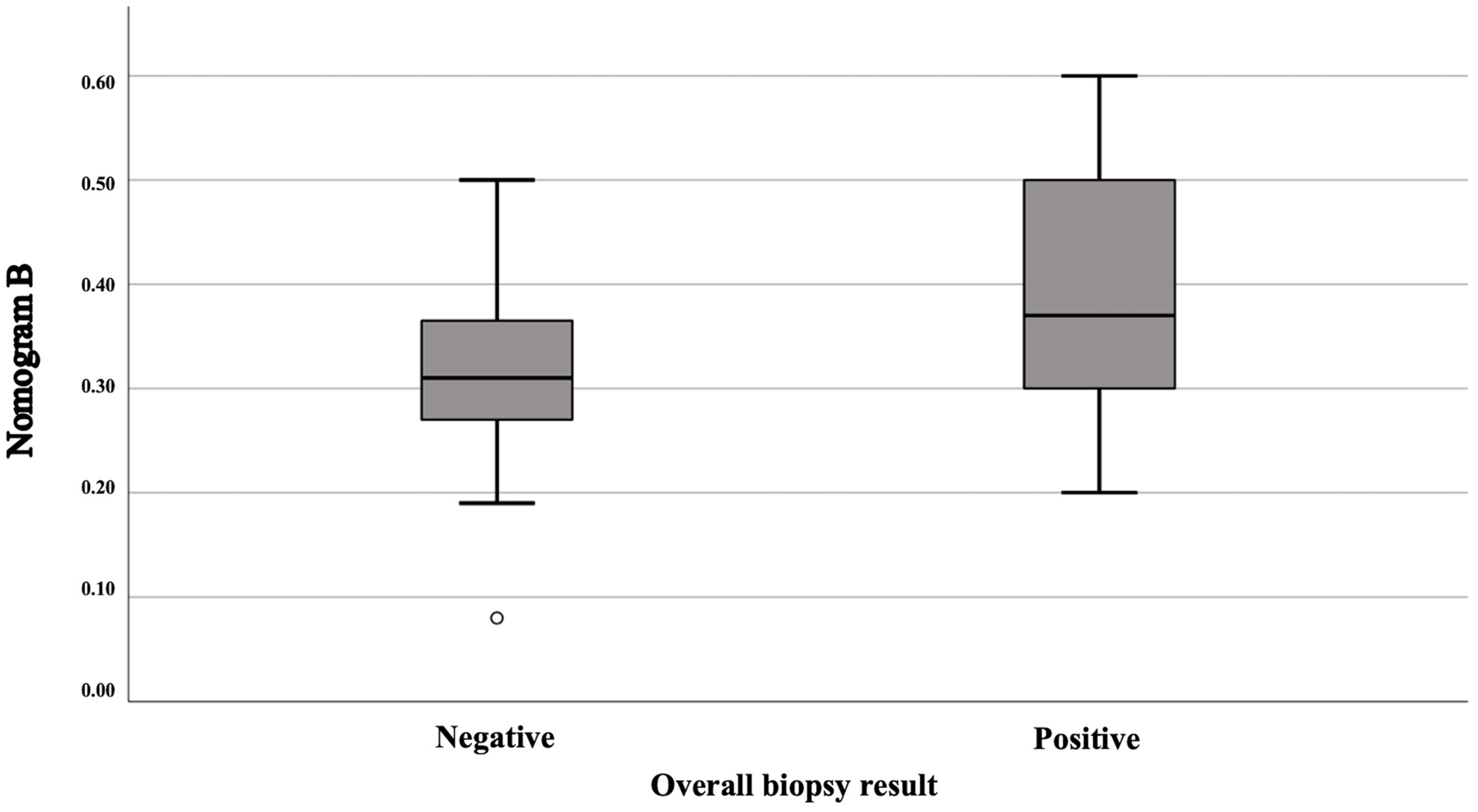
Applying our nomogram, as expected, the median probability of finding a PCa ISUP GG ≥ 3 (Nomogram A) was 5% (IQR: 0–15%), while for a PCa ISUP GG < 3 (Nomogram B), it was 33% (IQR: 27.5–40%). Looking in more detail, for patients with a positive biopsy, the median values of Nomogram A and Nomogram B were 8% (IQR: 0–20%) and 37% (IQR: 29.5–50.75%) respectively; for patients with a negative biopsy, on the other hand, the median was 5% (IQR: 2–13%) for Nomogram A and 31% (IQR: 27–36.75%) for Nomogram B. This last probability calculated with Nomogram B was statistically different (Figure 2) in patients with a positive biopsy compared to patients with a negative biopsy (p = 0.033), while no statistically significant difference was found for the probability calculated with Nomogram A in the two groups (p = 0.74).
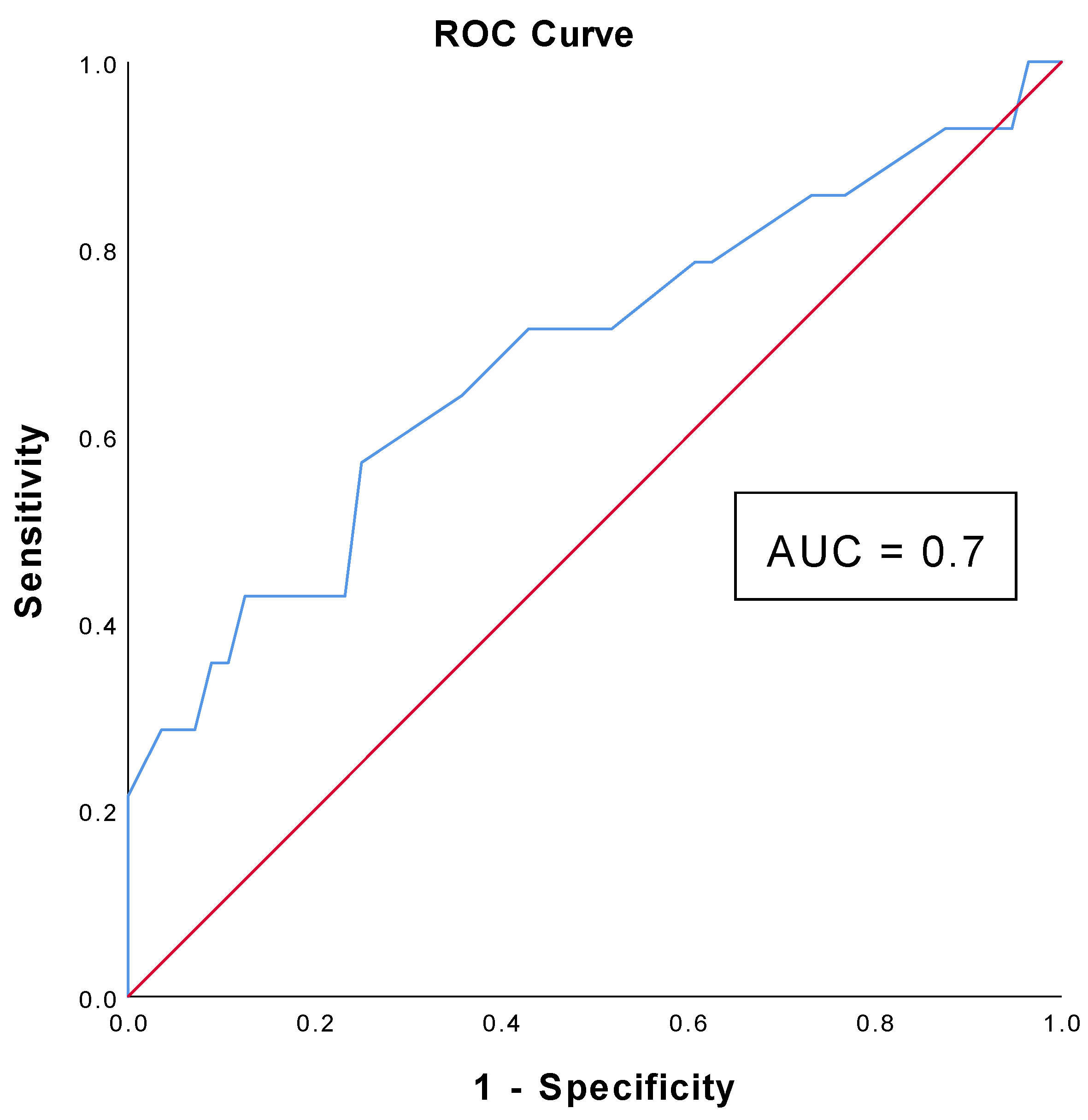
Regarding Nomogram B, a significant difference was also found when comparing patients with ISUP < 3 and with ISUP ≥ 3 (p = 0.029). The AUC of Nomogram B is equal to 0.685 (CI: 0.511–0.858) and using a cut-off of 40%, the nomogram shows a sensitivity of 70% and a specificity of 80% (Figure 3). Moreover, using this cut-off for Nomogram B, around 63% (44/70) of the patients would have been spared an unnecessary biopsy procedure without missing any cancer.
In patients with PSA density > 0.15 (5/14 patients with positive biopsy), the median values of Nomogram A and Nomogram B were 18% (IQR: 0–29%) and 35% (IQR: 26.50–54.50%) respectively.

4. Discussion

The cohort of patients examined in this study shows a prevalence of positive cases for PCa of 20%, whilst the negative cases are 80%; these data are consistent with the results of other studies in which the incidence of PCa was analyzed based on the categories of the PIRADS v2.1 score [18]. Some studies have reported cancer detection rates ranging from 15% to 31% for PIRADS 3 category [19,20]. For example, a study by Schoots et al. reported a cancer detection rate of approximately 22% for PIRADS 3 lesions, suggesting that while these lesions carry a moderate risk, the majority do not represent clinically significant disease [21]. Moreover, Thompson et al. found that most cancers detected in PIRADS 3 lesions were low grade (GG 1 or 2) with fewer cases of clinically significant cancer (GG ≥ 3) [22]. This study demonstrates how the use of the nomograms we evaluated, particularly for Nomogram B, can discriminate with significant accuracy patients with positive and negative biopsy results as well as between GG < 3 and GG > 3 cancers. The evaluation with this nomogram could therefore allow us to better evaluate patients in whom a prostate biopsy is truly indicated to confirm the diagnosis of PCa and avoid unnecessary procedures. This finding is supported by similar studies that have explored the application of nomograms and other predictive models in prostate cancer risk assessment [23,24]. As known, the PIRADS 3 category represents a clinical challenge in the interpretation of mpMRI results, for which several studies have proposed the use of different clinical variables and nomograms to guide the clinician in the diagnostic pathway of these patients [25]. In the context of assessing the risk of PCa, one of the most widely used clinical parameters is the PSA density, as also proposed in the European Association of Urology (EAU) guidelines [26]. According to the EAU guidelines, in the presence of a PIRADS 3 lesion and with a PSA density value higher than 0.15 ng/mL/mL, the probability of diagnosing a clinically significant PCa (ISUP > 2) is equal to 30%. This threshold of probability, therefore, suggests the need to perform a prostate biopsy. Using our nomogram, in patients with this cut-off of PSA density and therefore diagnosed with PCa, the probability of identifying a high-grade PCa was 18%, while for a low-grade tumor, it was 35%: these results indicate that our nomogram could be able not only to help in selecting patients who should undergo a biopsy but also to stratify the aggressiveness of cancer itself.
Historically, the European Randomized Study for Screening of Prostate Cancer (ERSPC) risk calculator was the first nomogram designed to predict the risk of prostate cancer after a prostate biopsy. The nomogram used the Prostate-Specific Antigen (PSA) level, the findings of a digital rectal examination (DRE), and the prostate volume to predict the probability of prostate cancer after a systematic biopsy [27]. Later, Thompson et al. developed a separate nomogram based on men who participated in the Prostate Cancer Prevention Trial (PCPT). In the latter, the variables included in this nomogram were total PSA, free PSA, DRE, a family history of PCa, and African American race, accounting also for the subsequent updates [28]. A more recent nomogram was implemented by the Prostate Biopsy Collaborative Group (PBCG). This calculator includes age, PSA, DRE, family history, race, and a prior negative biopsy [29]. The AUC for these models ranges from 0.65 to 0.75, 0.70 to 0.79 and 0.74 to 0.81 for PCPT, ERSPC and PBCG nomograms, respectively [30,31]. However, these nomograms did not focus mainly on a cohort of patients with PIRADS 3; hence, a direct comparison of this aspect is not possible. Nevertheless, as shown in a previous paper [17], the AUC for the nomograms we used was 0.80 and 0.63 for Nomogram A and Nomogram B, respectively. Taking these data into consideration, we believe in this specific cohort of patients with a PIRADS 3 at mpMRI, Nomogram B was found to perform better than Nomogram A.
A study by Mehralivand et al. evaluated the performance of a predictive model that combined clinical variables with mpMRI findings and found that such models could significantly enhance the risk stratification of patients with indeterminate PIRADS scores. Their model showed high predictive accuracy, particularly in identifying clinically significant cancers, which aligns with the current study’s results for Nomogram B. Specifically, including the mpMRI parameters alongside the traditional clinical variables improved the AUC from 64% to 84%. In this study, the authors included 400 patients in the development cohort and 251 patients in the validation cohort and stated that by applying their model, around 38% of biopsies could have been omitted with a 20% risk threshold. However, a specific analysis on PIRADS 3 patients was not performed [32].
A 2021 retrospective study led to the development and validation of a nomogram to predict the risk of prostate neoplasia before biopsy in patients with PSA < 20 ng/mL, using parameters such as age, PSA, free PSA, and prostate volume. This approach showed good results in terms of diagnostic accuracy (AUC = 0.857). Using a threshold value of 15%, the sensitivity and specificity of the nomogram were 95.6% and 42.5%, respectively, while the negative predictive value was 93.4%. Using this cut-off, it would have been possible to avoid 25% of biopsies, missing the diagnosis of PCa in only 4.4% of patients [33]. However, it is important to consider that measuring prostate volume requires imaging techniques such as transrectal ultrasound or magnetic resonance imaging, which can involve high costs, invasiveness, and limited availability, potentially leading to delays in patient evaluation and diagnosis [34,35].
In our study, the sensitivity and specificity of 70% and 80%, respectively, for Nomogram B at a 40% cut-off are within the range reported in other studies. Previously, Van Leeuwen et al. reported similar metrics in their evaluation of a predictive model for PIRADS 3 lesions, highlighting the potential of nomograms to serve as valuable decision-support tools in clinical practice [36]. Other nomograms, on the other hand, make use of additional biomarkers such as the 4K score, which is based on a kallikrein whose value is correlated with an increased risk of developing prostate cancer. In this retrospective study, the authors included 574 men with only 68 (20%) reported PIRADS 3 at mpMRI. The nomogram used demonstrates high accuracy (AUC = 0.84) in predicting the presence of prostate cancer. It was also estimated that in the overall cohort of patients examined, it was possible to avoid 10% of the biopsies performed without missing the diagnosis of any clinically insignificant tumors and only 1% of clinically significant ones. Compared to our nomograms, the 4K score integrated with mpMRI has similar accuracy to our Nomogram A based on the value of AUC, performing better in high-grade PCa [37].
Several other biomarkers have been proposed over the years and integrated with mpMRI to improve diagnostic accuracy and patients’ stratification risk. Among them, ExoDx Prostate Intelliscore (EPI) has been investigated and has shown promising results. Recently, King et al. performed a study on 226 patients who underwent both EPI and mpMRI (with a PIRADS score ≥ 3). They reported that the integration of EPI and mpMRI showed a sensitivity of 96% compared to 91% and 90% for EPI and mpRMI alone, respectively. Moreover, the AUC for EPI and mpMRI combined was 0.8, whilst the values of the AUC for EPI and mpMRI alone were 0.57 and 0.78, respectively. In this study, they also reported that using the integration of EPI and mpMRI, 43% of the patients could have avoided the biopsy procedure [38]. In our study, the percentage of patients that could have been spared a biopsy was found to be around 58% with a cut-off for Nomogram B set at 40% for the risk of PCa. However, the cited study included patients with a PIRADS score ≥ 3 and no data of a subgroup analysis regarding specifically patients with PIRADS 3 only was performed. Thus, such a population compared with our study has a higher pre-test prevalence of harboring PCa. These considerations should be taken into account while analyzing the value of sensitivity. To date, we have found no study exploring these biomarkers and focusing on a population of patients exclusively with PIRADS 3 [39].
Despite the promising results, its clinical use could be compromised by the cost of the test and limited availability [37]. Conversely, the variables included in our nomogram allow the use of easily available clinical parameters and therefore to stratify the patient potentially already at the time of the first clinical consultation without the need for diagnostic imaging investigations. However, our study is not devoid of limitations. Firstly, the number of patients included may be insufficient to ensure a representation of the study population, particularly regarding the number of patients with positive biopsy results. Second, it was an internal validation of the nomograms. However, we decided to include patients from our single center to pay particular attention to minimize the different sources of variability which may be associated with the use of different PSA immunoassays and to patients retrieved from different centers. In fact, for these reasons, we decided to include only patients with total and free PSA measured by one assay (Roche). These decisions also affected the relatively small sample size. Accordingly, this study served as an internal validation, providing important evidence relevant to proceed to an external validation study. Thus, an external validation will be the aim of our future research regarding the application of these nomograms.

5. Conclusions

The assessment of PCa risk represents a crucial challenge in clinical practice, especially considering the variety of factors involved in its diagnosis and management.
The results obtained with our nomogram clearly show that the population of patients with a PSA range of 2–10 ng/mL and a PIRADS 3 score have a low probability of developing PCa especially a high-risk tumor with an ISUP score greater than 3.
Furthermore, Nomogram B shows a good ability to discriminate between patients with PCa and those negative for biopsy, indicating a potential supporting role in selecting candidates for prostate biopsy.
Even more significant is the observation that Nomogram B demonstrates excellent accuracy in distinguishing between PCa with ISUP scores of 1–2 and those with ISUP scores greater than 3, thus providing valuable guidance in identifying patients at higher risk of having a more aggressive form of PCa.
The use of such nomograms could therefore facilitate the stratification of PCa risk in this specific category of patients, contributing significantly to the decision-making process regarding the need to undergo prostate biopsy.
In conclusion, the nomogram used in this study presents itself as a valuable additional tool in the diagnostic repertoire of PCa, offering a more detailed and personalized overview of individual risk and thus contributing to improving the care of patients suffering from this disease.

Author Contributions

Conceptualization, F.P., S.F. and A.G.; methodology, V.L., C.N. and M.T.; software, V.L. and C.N.; validation, A.G. and A.M.G.; formal analysis, V.L., R.L. and F.P.; investigation, F.P. and F.M.; resources, V.M.; data curation, P.T., R.L. and V.M.; writing—original draft preparation, V.L. and R.L.; writing—review and editing, F.P. and A.G.; supervision, B.R. and M.C.S. All authors have read and agreed to the published version of the manuscript.

Funding

This research received no external funding.

Institutional Review Board Statement

This retrospective study was conducted according to the guidelines and principles of the Declaration of Helsinki and standard ethical conduct for research involving humans. Ethical review and approval were waived for this study according to the Italian Law and the Italian Data Protection Authority, it is allowed the use of the data collected as part of research studies once you have acquired written consent from the patients. All patients signed an informed consent agreeing to supply their own anonymous data for this and future studies.

Informed Consent Statement

Informed consent was obtained from all subjects involved in the study.

Data Availability Statement

The original contributions presented in the study are included in the article; further inquiries can be directed to the corresponding author.

Acknowledgments

The authors thank all the staff of the Department of Urology of the ASST Fatebenefratelli-Sacco Hospitals for their help in the everyday practice.

Conflicts of Interest

The authors declare no conflicts of interest.

References

  1. Bray, F.; Laversanne, M.; Sung, H.; Ferlay, J.; Siegel, R.L.; Soerjomataram, I.; Jemal, A. Global Cancer Statistics 2022: GLOBOCAN Estimates of Incidence and Mortality Worldwide for 36 Cancers in 185 Countries. CA Cancer J. Clin. 2024, 74, 229–263. [Google Scholar] [CrossRef]
  2. Punglia, R.S.; D’Amico, A.V.; Catalona, W.J.; Roehl, K.A.; Kuntz, K.M. Effect of Verification Bias on Screening for Prostate Cancer by Measurement of Prostate-Specific Antigen. N. Engl. J. Med. 2003, 349, 335–342. [Google Scholar] [CrossRef]
  3. Cuzick, J.; Berney, D.M.; Fisher, G.; Mesher, D.; Møller, H.; Reid, J.E.; Perry, M.; Park, J.; Younus, A.; Gutin, A.; et al. Transatlantic Prostate Group. Prognostic Value of a Cell Cycle Progression Signature for Prostate Cancer Death in a Conservatively Managed Needle Biopsy Cohort. Br. J. Cancer 2012, 106, 1095–1099. [Google Scholar] [CrossRef]
  4. Thompson, J.E.; Moses, D.; Shnier, R.; Brenner, P.; Delprado, W.; Ponsky, L.; Pulbrook, M.; Böhm, M.; Haynes, A.-M.; Hayen, A.; et al. Multiparametric Magnetic Resonance Imaging Guided Diagnostic Biopsy Detects Significant Prostate Cancer and Could Reduce Unnecessary Biopsies and over Detection: A Prospective Study. J. Urol. 2014, 192, 67–74. [Google Scholar] [CrossRef]
  5. Stabile, A.; Giganti, F.; Rosenkrantz, A.B.; Taneja, S.S.; Villeirs, G.; Gill, I.S.; Allen, C.; Emberton, M.; Moore, C.M.; Kasivisvanathan, V. Multiparametric MRI for Prostate Cancer Diagnosis: Current Status and Future Directions. Nat. Rev. Urol. 2020, 17, 41–61. [Google Scholar] [CrossRef] [PubMed]
  6. Ahmed, H.U.; Bosaily, A.E.-S.; Brown, L.C.; Gabe, R.; Kaplan, R.; Parmar, M.K.; Collaco-Moraes, Y.; Ward, K.; Hindley, R.G.; Freeman, A.; et al. Diagnostic Accuracy of Multi-Parametric MRI and TRUS Biopsy in Prostate Cancer (PROMIS): A Paired Validating Confirmatory Study. Lancet 2017, 389, 815–822. [Google Scholar] [CrossRef] [PubMed]
  7. Kasivisvanathan, V.; Rannikko, A.S.; Borghi, M.; Panebianco, V.; Mynderse, L.A.; Vaarala, M.H.; Briganti, A.; Budäus, L.; Hellawell, G.; Hindley, R.G.; et al. MRI-Targeted or Standard Biopsy for Prostate-Cancer Diagnosis. N. Engl. J. Med. 2018, 378, 1767–1777. [Google Scholar] [CrossRef] [PubMed]
  8. Kasivisvanathan, V.; Stabile, A.; Neves, J.B.; Giganti, F.; Valerio, M.; Shanmugabavan, Y.; Clement, K.D.; Sarkar, D.; Philippou, Y.; Thurtle, D.; et al. Magnetic Resonance Imaging-Targeted Biopsy Versus Systematic Biopsy in the Detection of Prostate Cancer: A Systematic Review and Meta-Analysis. Eur. Urol. 2019, 76, 284–303. [Google Scholar] [CrossRef]
  9. Drost, F.-J.H.; Osses, D.F.; Nieboer, D.; Steyerberg, E.W.; Bangma, C.H.; Roobol, M.J.; Schoots, I.G. Prostate MRI, with or without MRI-Targeted Biopsy, and Systematic Biopsy for Detecting Prostate Cancer. Cochrane Database Syst. Rev. 2019, 2019, CD012663. [Google Scholar] [CrossRef]
  10. Lorusso, V.; Talso, M.; Palmisano, F.; Branger, N.; Granata, A.M.; Fiori, C.; Gregori, A.; Pignot, G.; Walz, J. Is Imaging Accurate Enough to Detect Index Lesion in Prostate Cancer? Analysis of the Performance of MRI and Other Imaging Modalities. Minerva Urol. Nephrol. 2024, 76, 22–30. [Google Scholar] [CrossRef]
  11. Turkbey, B.; Rosenkrantz, A.B.; Haider, M.A.; Padhani, A.R.; Villeirs, G.; Macura, K.J.; Tempany, C.M.; Choyke, P.L.; Cornud, F.; Margolis, D.J.; et al. Prostate Imaging Reporting and Data System Version 2.1: 2019 Update of Prostate Imaging Reporting and Data System Version 2. Eur. Urol. 2019, 76, 340–351. [Google Scholar] [CrossRef]
  12. Schoots, I.G. MRI in Early Prostate Cancer Detection: How to Manage Indeterminate or Equivocal PI-RADS 3 Lesions? Transl. Androl. Urol. 2018, 7, 702–782. [Google Scholar] [CrossRef]
  13. Scialpi, M.; Martorana, E.; Aisa, M.C.; Rondoni, V.; D’Andrea, A.; Bianchi, G. Score 3 Prostate Lesions: A Gray Zone for PI-RADS V2. Turk. J. Urol. 2017, 43, 237–240. [Google Scholar] [CrossRef] [PubMed]
  14. Arbuznikova, D.; Eder, M.; Grosu, A.-L.; Meyer, P.T.; Gratzke, C.; Zamboglou, C.; Eder, A.-C. Towards Improving the Efficacy of PSMA-Targeting Radionuclide Therapy for Late-Stage Prostate Cancer—Combination Strategies. Curr. Oncol. Rep. 2023, 25, 1363–1374. [Google Scholar] [CrossRef]
  15. Abdelaal, A.M.; Sohal, I.S.; Iyer, S.G.; Sudarshan, K.; Orellana, E.A.; Ozcan, K.E.; dos Santos, A.P.; Low, P.S.; Kasinski, A.L. Selective Targeting of Chemically Modified miR-34a to Prostate Cancer Using a Small Molecule Ligand and an Endosomal Escape Agent. Mol. Ther. Nucleic Acids 2024, 35, 102193. [Google Scholar] [CrossRef] [PubMed]
  16. Ferraro, S.; Biganzoli, G.; Bussetti, M.; Castaldi, S.; Biganzoli, E.M.; Plebani, M. Managing the Impact of Inter-Method Bias of Prostate Specific Antigen Assays on Biopsy Referral: The Key to Move towards Precision Health in Prostate Cancer Management. Clin. Chem. Lab. Med. 2023, 61, 142–153. [Google Scholar] [CrossRef]
  17. Ferraro, S.; Biganzoli, D.; Rossi, R.S.; Palmisano, F.; Bussetti, M.; Verzotti, E.; Gregori, A.; Bianchi, F.; Maggioni, M.; Ceriotti, F.; et al. Individual Risk Prediction of High Grade Prostate Cancer Based on the Combination between Total Prostate-Specific Antigen (PSA) and Free to Total PSA Ratio. Clin. Chem. Lab. Med. 2023, 61, 1327–1334. [Google Scholar] [CrossRef]
  18. Porpiglia, F.; Manfredi, M.; Mele, F.; Cossu, M.; Bollito, E.; Veltri, A.; Cirillo, S.; Regge, D.; Faletti, R.; Passera, R.; et al. Diagnostic Pathway with Multiparametric Magnetic Resonance Imaging Versus Standard Pathway: Results from a Randomized Prospective Study in Biopsy-Naïve Patients with Suspected Prostate Cancer. Eur. Urol. 2017, 72, 282–288. [Google Scholar] [CrossRef] [PubMed]
  19. Yilmaz, E.C.; Shih, J.H.; Belue, M.J.; Harmon, S.A.; Phelps, T.E.; Garcia, C.; Hazen, L.A.; Toubaji, A.; Merino, M.J.; Gurram, S.; et al. Prospective Evaluation of PI-RADS Version 2.1 for Prostate Cancer Detection and Investigation of Multiparametric MRI-Derived Markers. Radiology 2023, 307, e221309. [Google Scholar] [CrossRef]
  20. Park, S.Y.; Cho, N.H.; Jung, D.C.; Oh, Y.T. Prostate Imaging-Reporting and Data System Version 2: Beyond Prostate Cancer Detection. Korean J. Radiol. 2018, 19, 193–200. [Google Scholar] [CrossRef]
  21. Schoots, I.G.; Roobol, M.J.; Nieboer, D.; Bangma, C.H.; Steyerberg, E.W.; Hunink, M.G.M. Magnetic Resonance Imaging–Targeted Biopsy May Enhance the Diagnostic Accuracy of Significant Prostate Cancer Detection Compared to Standard Transrectal Ultrasound-Guided Biopsy: A Systematic Review and Meta-Analysis. Eur. Urol. 2015, 68, 438–450. [Google Scholar] [CrossRef]
  22. Thompson, J.E.; van Leeuwen, P.J.; Moses, D.; Shnier, R.; Brenner, P.; Delprado, W.; Pulbrook, M.; Böhm, M.; Haynes, A.M.; Hayen, A.; et al. The Diagnostic Performance of Multiparametric Magnetic Resonance Imaging to Detect Significant Prostate Cancer. J. Urol. 2016, 195, 1428–1435. [Google Scholar] [CrossRef] [PubMed]
  23. Westphalen, A.C.; Fazel, F.; Nguyen, H.; Cabarrus, M.; Hanley-Knutson, K.; Shinohara, K.; Carroll, P.R. Detection of Clinically Significant Prostate Cancer with PI-RADS v2 Scores, PSA Density, and ADC Values in Regions with and without mpMRI Visible Lesions. Int. Braz. J. Urol. 2019, 45, 713–723. [Google Scholar] [CrossRef] [PubMed]
  24. Zaytoun, O.M.; Kattan, M.W.; Moussa, A.S.; Li, J.; Yu, C.; Jones, J.S. Development of Improved Nomogram for Prediction of Outcome of Initial Prostate Biopsy Using Readily Available Clinical Information. Urology 2011, 78, 392–398. [Google Scholar] [CrossRef]
  25. Giannarini, G.; Zazzara, M.; Rossanese, M.; Palumbo, V.; Pancot, M.; Como, G.; Abbinante, M.; Ficarra, V. Will Multi-Parametric Magnetic Resonance Imaging Be the Future Tool to Detect Clinically Significant Prostate Cancer? Front. Oncol. 2014, 4, 294. [Google Scholar] [CrossRef]
  26. Cornford, P.; van den Bergh, R.C.N.; Briers, E.; Van den Broeck, T.; Brunckhorst, O.; Darraugh, J.; Eberli, D.; De Meerleer, G.; De Santis, M.; Farolfi, A.; et al. EAU-EANM-ESTRO-ESUR-ISUP-SIOG Guidelines on Prostate Cancer-2024 Update. Part I: Screening, Diagnosis, and Local Treatment with Curative Intent. Eur. Urol. 2024, 86, 148–163. [Google Scholar] [CrossRef] [PubMed]
  27. Roobol, M.J.; van Vugt, H.A.; Loeb, S.; Zhu, X.; Bul, M.; Bangma, C.H.; van Leenders, A.G.L.J.H.; Steyerberg, E.W.; Schröder, F.H. Prediction of Prostate Cancer Risk: The Role of Prostate Volume and Digital Rectal Examination in the ERSPC Risk Calculators. Eur. Urol. 2012, 61, 577–583. [Google Scholar] [CrossRef]
  28. Ankerst, D.P.; Hoefler, J.; Bock, S.; Goodman, P.J.; Vickers, A.; Hernandez, J.; Sokoll, L.J.; Sanda, M.G.; Wei, J.T.; Leach, R.J.; et al. Prostate Cancer Prevention Trial Risk Calculator 2.0 for the Prediction of Low- vs High-Grade Prostate Cancer. Urology 2014, 83, 1362–1367. [Google Scholar] [CrossRef]
  29. Ankerst, D.P.; Straubinger, J.; Selig, K.; Guerrios, L.; De Hoedt, A.; Hernandez, J.; Liss, M.A.; Leach, R.J.; Freedland, S.J.; Kattan, M.W.; et al. Contemporary Prostate Biopsy Risk Calculator Based on Multiple Heterogeneous Cohorts. Eur. Urol. 2018, 74, 197–203. [Google Scholar] [CrossRef]
  30. Jalali, A.; Foley, R.W.; Maweni, R.M.; Murphy, K.; Lundon, D.J.; Lynch, T.; Power, R.; O’Brien, F.; O’Malley, K.J.; Galvin, D.J.; et al. A Risk Calculator to Inform the Need for a Prostate Biopsy: A Rapid Access Clinic Cohort. BMC Med. Inform. Decis. Mak. 2020, 20, 148. [Google Scholar] [CrossRef]
  31. Fang, A.M.; Rais-Bahrami, S. Magnetic Resonance Imaging–Based Risk Calculators Optimize Selection for Prostate Biopsy among Biopsy-Naive Men. Cancer 2022, 128, 25–27. [Google Scholar] [CrossRef] [PubMed]
  32. Mehralivand, S.; Shih, J.H.; Rais-Bahrami, S.; Oto, A.; Bednarova, S.; Nix, J.W.; Thomas, J.V.; Gordetsky, J.B.; Gaur, S.; Harmon, S.A.; et al. A Magnetic Resonance Imaging-Based Prediction Model for Prostate Biopsy Risk Stratification. JAMA Oncol. 2018, 4, 678–685. [Google Scholar] [CrossRef]
  33. Wu, Q.; Li, F.; Yin, X.; Gao, J.; Zhang, X. Development and Validation of a Nomogram for Predicting Prostate Cancer in Patients with PSA ≤ 20 Ng/mL at Initial Biopsy. Medicine 2021, 100, e28196. [Google Scholar] [CrossRef] [PubMed]
  34. Zhu, M.; Liang, Z.; Feng, T.; Mai, Z.; Jin, S.; Wu, L.; Zhou, H.; Chen, Y.; Yan, W. Up-to-Date Imaging and Diagnostic Techniques for Prostate Cancer: A Literature Review. Diagnostics 2023, 13, 2283. [Google Scholar] [CrossRef]
  35. Wasserman, N.F.; Niendorf, E.; Spilseth, B. Measurement of Prostate Volume with MRI (A Guide for the Perplexed): Biproximate Method with Analysis of Precision and Accuracy. Sci. Rep. 2020, 10, 575. [Google Scholar] [CrossRef]
  36. van Leeuwen, P.J.; Hayen, A.; Thompson, J.E.; Moses, D.; Shnier, R.; Böhm, M.; Abuodha, M.; Haynes, A.-M.; Ting, F.; Barentsz, J.; et al. Multiparametric Magnetic Resonance Imaging-Based Risk Model to Determine the Risk of Significant Prostate Cancer Prior to Biopsy. BJU Int. 2017, 120, 774–781. [Google Scholar] [CrossRef] [PubMed]
  37. Wagaskar, V.G.; Sobotka, S.; Ratnani, P.; Young, J.; Lantz, A.; Parekh, S.; Falagario, U.G.; Li, L.; Lewis, S.; Haines, K.; et al. A 4K Score/MRI-Based Nomogram for Predicting Prostate Cancer, Clinically Significant Prostate Cancer, and Unfavorable Prostate Cancer. Cancer Rep. 2021, 4, e1357. [Google Scholar] [CrossRef]
  38. King, N.; Lang, J.; Jambunathan, S.; Lombardi, C.; Saltzman, B.; Nagalakshmi, N.; Sindhwani, P. The Value of Adding Exosome-Based Prostate Intelliscore to Multiparametric Magnetic Resonance Imaging in Prostate Biopsy: A Retrospective Analysis. Uro 2024, 4, 50–59. [Google Scholar] [CrossRef]
  39. Sultan, M.I.; Huynh, L.M.; Kamil, S.; Abdelaziz, A.; Hammad, M.A.; Gin, G.E.; Lee, D.I.; Youssef, R.F. Utility of Noninvasive Biomarker Testing and MRI to Predict a Prostate Cancer Diagnosis. Int. Urol. Nephrol. 2023, 56, 539–546. [Google Scholar] [CrossRef]
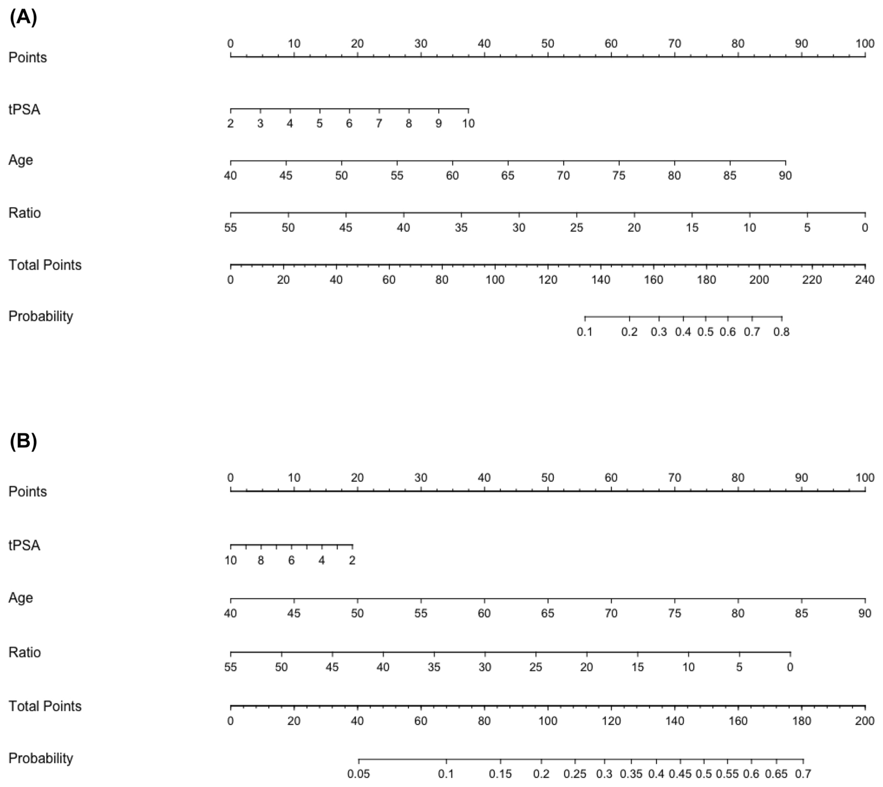
Figure 1. Nomograms used to estimate the probability of ISUP ≥ 3 (A) and ISUP < 3 (B) prostate cancer in patients with PIRADS 3 lesions (reproduced with permission from Ferraro et al. [17]).
Cancers 16 03084 g001
Figure 2. Box plot showing the difference in the estimated probability according to Nomogram B between patients with positive and negative biopsy results.
Cancers 16 03084 g002
Figure 3. Diagram showing the Receiver Operating Characteristic (ROC) curve analysis and the Area Under the Curve (AUC) value of Nomogram B.
Cancers 16 03084 g003
Table 1. Clinical and pathologic characteristics of the patients with PIRADS 3 lesions (N = 70).
VariablesTotal Number of Patients = 70
Age, years [median (range)] 66 (61–73)
PSA, ng/mL [median (IQR)] 5.50 (3.9–6.9)
PSA ratio, [median (IQR)]0.89 (0.52–1.3)
PSA density, ng/mL/mL [median (IQR)]0.1 (0.06–0.12)
Prostate volume, mL [median (IQR)]63 (49–100)
Dimension of the lesion, mm [median (IQR)]8 (6–10.5)
Location of the lesion, N [%]
- Apex23 (32.9%)
- Intermediate26 (37.1%)
- Base9 (12.9%)
Gland zone of the lesion
- Anterior13 (18.6%)
- Peripheric38 (54.3%)
- Transitional16 (22.9%)
Overall biopsy result
- Negative for PCa56 (80%)
- Positive for PCa14 (20%)
IQR (Interquartile range).
Table 2. Estimated probability according to Nomogram A (ISUP ≥ 3) and Nomogram B (ISUP < 3).
VariablesProbability [%]
Nomogram A overall, % [IQR]5% (0–15%)
- Patients with positive biopsy8% (0–20%)
Nomogram B overall, % [IQR]33% (27.75–40%)
- Patients with positive biopsy37% (29.50–50.75%)
IQR (interquartile range).
Disclaimer/Publisher’s Note: The statements, opinions and data contained in all publications are solely those of the individual author(s) and contributor(s) and not of MDPI and/or the editor(s). MDPI and/or the editor(s) disclaim responsibility for any injury to people or property resulting from any ideas, methods, instructions or products referred to in the content.