Article

High M2-TAM Infiltration and STAT3/NF-κB Signaling Pathway as a Predictive Factor for Tumor Progression and Death in Cervical Cancer

by
George Alexandre Lira
1,2,3,4,
Fábio Medeiros de Azevedo
5,
Ingrid Gabrielle dos Santos Lins
4,
Isabelle de Lima Marques
2,
Giovanna Afonso Lira
4,
Christina Eich
3 and
Raimundo Fernandes de Araujo Junior
1,2,3,6,*
1
Cancer and Inflammation Research Laboratory, Department of Morphology, Federal University of Rio Grande do Norte Natal, Natal 59072-970, RN, Brazil
2
Postgraduate Program in Health Science, Federal University of Rio Grande do Norte, Natal 59072-970, RN, Brazil
3
Department of Radiology, Leiden University Medical Center, 2333 ZA Leiden, The Netherlands
4
League Against Cancer from Rio Grande do Norte, Advanced Oncology Center, Natal 59075-740, RN, Brazil
5
Pathology Department, Federal University of Rio Grande do Norte, Natal 59012-570, RN, Brazil
6
Postgraduate Program in Functional and Structural Biology, Federal University of Rio Grande do Norte, Natal 59072-970, RN, Brazil
*
Author to whom correspondence should be addressed.
Cancers 2024, 16(14), 2496; https://doi.org/10.3390/cancers16142496
Submission received: 5 June 2024 / Revised: 28 June 2024 / Accepted: 3 July 2024 / Published: 9 July 2024
(This article belongs to the Section Cancer Metastasis)

Abstract

:

Simple Summary

Once in an immunosuppressive tumoral microenvironment (TME), macrophages take over an essential role by activating signaling pathways such as STAT3 and NFkB to keep the immune suppression and tumor progression. However, this role in cancer cervical (CC) is not still clear. For that, we studied the interaction of tumor associated macrophages (TAM) and STAT3 and NFkB pathways with of several cancer hallmarks in 691 patients with CC, and more importantly, their impact on clinical outcome of these patients. Interestingly, a strong immunosuppressive loop was identified via TAM and STAT3 and NFkB pathways in TME which reverberated in a higher expression of tumor progression markers, and consequently in a poor clinical outcome.

Abstract

Introduction: The tumor microenvironment (TME) plays a crucial role in the progression, invasion, and metastasis of cervical carcinoma (CC). Tumor-associated macrophages (TAMs) are significant components of the CC TME, but studies on their correlation with CC progression are still controversial. This study aimed to investigate the relationship between TAM infiltration, the STAT3/NF-κB signaling pathway, and Overall Survival (OS) in CC patients. Methods: In a retrospective study, 691 CC patients who had received a definitive histopathologic diagnosis of CC scored by the FIGO staging system and not undergone preoperative treatment were selected from a database. The effect of TAM infiltration on tumor progression biomarkers using Tissue Microarray (TMA) and immunohistochemistry was evaluated. Furthermore, the impact of the expression of these biomarkers and clinical–pathological parameters on recurrence-free (RF) and OS using Kaplan–Meier and multivariable Cox regression methods was also analyzed. Results: High stromal CD163 + 204 + TAMs density and via STAT3 and NF-κB pathways was relevant to the expression of E-cadherin, Vimentin, MMP9, VEGFα, Bcl-2, Ki-67, CD25, MIF, FOXP3, and IL-17 (all p < 0.0001). In addition, elevated TNM staging IV had a strong association correlation with STAT3 and NF-κB pathways (p < 0.0001), CD25 (p < 0.001), VEGFα (p < 0.001), MIF (p < 0.0001), and Ki-67 (p < 0.0001). On the other hand, overall and recurrence survival was shown to be strongly influenced by the expression of SNAIL (HR = 1.52), E-cadherin (HR = 1.78), and Ki-67 (HR = 1.44). Conclusion: M2-TAM and via STAT3/NF-κB pathways had a strong effect on CC tumor progression which reverberated in the severity of clinicopathological findings, becoming an important factor of poor prognosis.

1. Introduction

Cervical cancer is an important public health problem with a worldwide impact, especially in underdeveloped countries, with high incidence and mortality rates [1,2,3]. Patients with cervical cancer have a rate of 5-year relative survival of 91% when the cancer is diagnosed at earlier stages, dropping to 60% when diagnosed after it spreads to nearby tissues, organs, or regional lymph nodes [4].
Prognostic factors for cervical cancer include race, age, stage, grade, histologic type, tumor volume, lymph node involvement, performance status, and the treatment received [5]. However, new studies are needed on protein biomarkers for cervical cancer prognosis that can guide the treatment. In this line, immunological indicators have been considered important prognostic factors for cervical cancer [6]. Tumor-associated macrophages (TAMs) comprise the largest number of immune cells and the more important part of the tumor microenvironment (TME), acting as immune and tumor regulators [7]. In this context, they have been identified as prognostic factors and potential targets for immunotherapy [7,8,9].
Within tumors by interacting with tumor cells, macrophages play a key role in regulating the immune response, promoting the tumor escape to immunosurveillance [10]. When they shift towards the CD163 + 204 + M2-TAM phenotype through the activation of the signal transducer activator (STAT3) pathway caused by an immunosuppressive cytokine loop, they promote immunosuppression, drug resistance, tumor progression, and metastasis [11,12]. Tumors release immunosuppressive cytokines that stimulate the switch to M2-TAMs via STAT3 in the TME, leading to worse survival in solid tumors [13,14]. Some evidence points out that M2-TAM infiltration has been associated with more advanced FIGO stage and lymph node metastasis [15,16], as well as with higher expression of signal transducer activator (STAT3) in various solid tumors, including breast, prostate, colorectal, melanoma, and brain cancer [17,18]. Reports describe that IL-10, Programmed Death-Ligand 1 (PD-L1), and Transforming Growth Factor beta (TGFβ) promote the STAT3 and Factor Nuclear kappa B (NF-κB) activation which are critical to macrophage polarization [19,20,21]. Furthermore, the STAT3 pathway stimulates specific transcription factors in tumor cells that support the EMT process, resistance to apoptosis, invasion, proliferation, and immunosuppression [17,19,21].
Although there is biological evidence, the clinical impact of M2-TAM’s pro-tumor activity and via STAT3 and NF-κB signaling pathways on the overall survival of cervical cancer patients has not been clarified. Therefore, we conducted a study to validate the association of M2-TAM and via STAT/NF-κB pathways with biomarkers of tumor progression and survival in cervical cancer.

2. Material and Methods

2.1. Tissue Samples and Data Collection

This research is a retrospective and longitudinal study of 1390 women diagnosed with cervical cancer from 2003 to 2017 registered at the archives of the Advanced Oncology Center at the League Against Cancer from Rio Grande do Norte (LRNCC), Natal, Brazil (Supplementary Materials). After the exclusion of 699 patients (Figure 1), the inclusion criteria adopted to select 691 cases was as follows: (I) patients who underwent surgery with definitive histopathologic diagnosis of cervical cancer, (II) scored by TNM and the International Federation of Gynecology and Obstetrics (FIGO) staging system of cervical cancer [22], and (III) with enough surgical material to perform the tissue microarray (TMA). Furthermore, sociodemographic and clinicopathological data were also collected (Supplementary Materials). The Institutional Review Board (IRB) from LRNCC (CAE 2077017-2644266-2761333-3071847, Natal, Brazil) approved this study. The ethics committee permitted the research, including the collection of information from medical records, as well as paraffin block samples from 691 patients.

2.2. Tissue Microarray (TMA) Block Construction and Staining by Hematoxylin and Eosin (HE) and Immunohistochemistry (IHC)

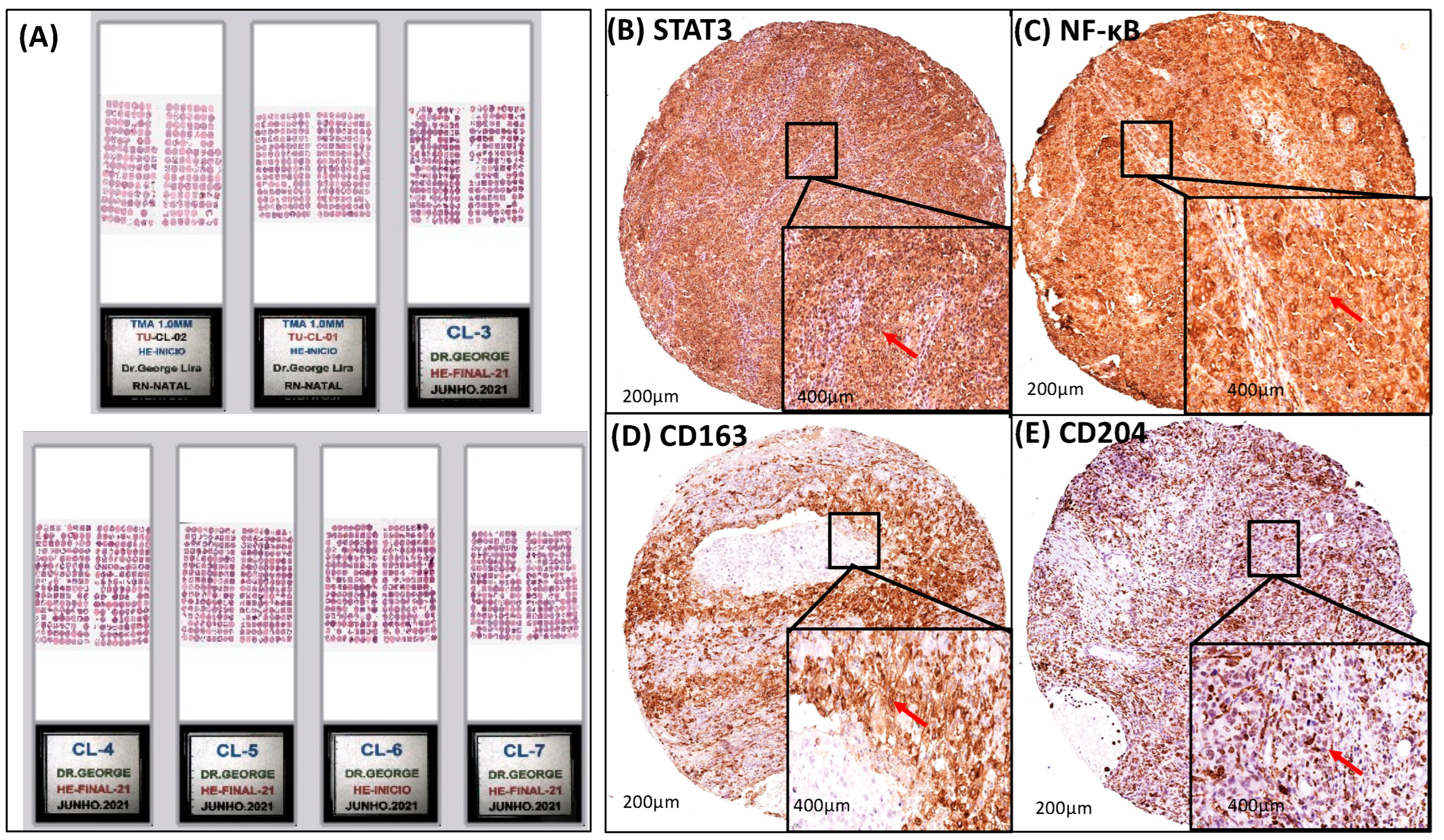
The Tissue Microarray blocks were constructed as described by Araujo Jr et al., 2015 [23]. Briefly, all hematoxylin and eosin slides were re-examined by a second experienced pathologist, and two morphologic representative fields of invasive cervical cancer were chosen and encircled with a marker pen. Two samples from each neoplasm, obtained from biopsies and/or surgical specimens, with one millimeter in diameter each, were taken from the donor blocks and transferred to the recipient block, using the Manual Tissue Microarrayer device I (Beecher Instruments, Silver Spring, MD, USA). Three-micrometer cuts of the microarray block were made using the Paraffin Tape-transfer System (Instrumedics, Saint Louis, MO, USA). One of the sections was stained with H&E to confirm the presence of tumor by light microscopy. Immunohistochemical reactions were performed on two different slides to increase the reliability of the results. The histological sections were placed on 8% salinized glass slides for the immunohistochemical method (Figure 2).

Immunohistochemistry (IHC)

Immunohistochemistry of TMA slides was performed as described by Araújo Jr et al., 2016 [24]. Using a microtome, 4 µm thin tissue slices were cut from tumors and transferred to gelatine-coated slides. The paraffin-embedded tumor tissue sections were incubated overnight at 4 °C with anti-CD25 to M2-TAM, anti-CD163 to M2-TAM, anti-IL-10, anti-TGF β, anti-VEGF α, anti-IL-17, anti-NFκB, anti-PDL-1, anti-E-Cadherin, anti-BCL-2, anti-MMP-9, anti-Ki-67, anti-Vimentin, anti-CD204, anti-SNAIL, anti-MIF, anti-STAT3, and anti-FOXP3 (Table S1). Slices were rinsed in PBS and treated with a secondary antibody conjugated to streptavidin/Haptoglobin-Related Protein (HRP) (Biocare Medical, Concord, CA, USA). Immunoreactivity to various proteins was visualized using a colorimetric-based detection kit (TrekAvidin-HRP Label + Kit from Biocare Medical, Pacheco, CA, USA) according to the manufacturer’s procedure. High-resolution images of the IHC staining were obtained from scanning an Aperio AT2 slide scanner (Aperio, Vista, CA, USA) at 200× and 400× magnification. The digital images were captured using a high-power objective (200× and 400×) for ImageScope software (12.4.6).

2.3. Quantitative Evaluation of Immunostaining

According to the staining intensity, it was considered positive, cells stained in brown in the nucleus, and/or cytoplasm, and/or membrane (Table S2). The expression of markers was considered according to the numerical result of the software image analysis algorithms (SP263) [25,26] (Supplementary Materials). In this line, based on the numerical result of the parameter in pixel, a median between the color intensity and the number of cells with positive staining was obtained [27,28,29] (Supplementary Materials). A hotspot was subjectively defined as the area with the highest density of Ki-67-positive tumor cells. To classify, we categorized Ki-67 into distinct low- and high-proliferating tumors, including up to 15% and greater than 15%, respectively [30] (Supplementary Materials).

2.4. Follow-Up

Overall survival (OS) was calculated as the length of time from the date of diagnosis or the start of treatment for a disease (a total of 691 patients), such as cancer, that patients diagnosed with the disease are still alive—a total of 434 alive and 257 deaths [31]. Recurrence-free survival (RFS) was calculated as the measure of time from random assignment to cancer recurrence or death from any cause [32] (Supplementary Materials).

2.5. Statistical Analysis

Statistical association between categorical variables was assessed using either the Chi-squared test or Fisher’s exact test. Locoregional relapse was defined as the occurrence of any local or regional recurrence and/or death resulting from the tumor. Cox regression analysis was used to perform univariate analyses for overall survival and recurrence-free survival. Hazard ratios with 95% CI were presented. The statistical analysis was conducted using SPSS software version 26 for Windows (SPSS for Windows, 12 Inc., Chicago, IL, USA).
Kaplan–Meier curves for overall survival and recurrence-free survival were generated within the R statistical environment version 4.3.0 using the univariate log rank (Mantel–Cox) test. A p-value threshold of 0.05 was considered statistically significant.

3. Results

3.1. Clinicopathologic Features of Patients

The characteristics of 691 patients are summarized in Table 1 and Table S2. The mean age was 52.5 years with 40.2% aged between 40 and 60. FIGO and TNM had more patients in stage I, 56.9% and 53.5%, respectively; 28.1% relapsed, and 37.2% died within 5 years (Table 1). Of those 468 patients indicated for surgical treatment, 41.10% had a tumor size larger than 4 cm, 43.4% presented angiolymphatic invasion, 10.5% with neural invasion, and 51.7% showed deeper stromal invasion. Lymph node metastasis was found in 26.7% and a positive resection margin in 9.6%. The most frequent pathological stage found was stage I in 58.8% (Table S3).

3.2. Analysis of Survival Rate, Clinicopathological Features, and Age

Univariate logistic Cox regression analysis was used to investigate the impact of age, TNM, and FIGO staging on death rates. Patients had hazard ratios of 3.67 (95% CI 1.98 to 6.80), 8.89 (95% CI 5.31 to 14.88), and 10.95 (95% CI 5.31 to 14.88) for older age, FIGO stage IV, and TNM stage, respectively. A significant statistical correlation was found between survival and age (years, <40: p < 0.0001; >40 and <60: p < 0.05; >60 and <80: p < 0.0001 and >80: p < 0.0001), FIGO staging (all categories, p < 0.0001), and TNM staging (all categories, p < 0.0001) (Table 2).
Furthermore, survival had significant statistical correlation with hemoglobin level (g/dL, until >8–12: p < 0.0001;), leukocytes count (leukocytosis: p < 0.0001), thrombocytosis (p < 0.0001), tumor size (>4 cm: p < 0.0001), positive HPV (p < 0.0001), neural and angiolymphatic invasion (p < 0.05); depth of stromal invasion (p < 0.0001), lymph node metastasis (p < 0.0001), number of resected iliac pelvic and retroperitoneal lymph nodes (6–10: p < 0.05 and >3: p < 0.05, respectively), TNM pathological Staging (IV: p < 0.0001), and chemotherapy (p < 0.01) (Table S4).

3.3. A Strong Association Interaction between CD204+ and CD163+ M2-TAM with STAT3 and NF-κB Signaling Pathways

The association correlation between the expression of CD204+ and CD163+ M2-TAM with STAT3/NF-κB signaling pathways in the tumor microenvironment (TME) was studied in 691 patients with cervical cancer (Table 3). Importantly, the correlation was strongly significant to STAT3 and NF-κB transcription factors (all p < 0.0001), showing that the higher expression of STAT3 was related to higher expression of NF-κB, CD163, and CD204 (Table 3). Cytoplasmic staining of NF-κB transcription factor and CD163 were identified in 65.8% and 65.6% of 691 patients with cervical cancer, respectively (Table 3 and Figure 2). On the other hand, STAT3 transcription factor and CD204 showed cytoplasmic/nuclear staining present in 85.4% and 85.6% of cases (Table 3 and Figure 2).

3.4. Transcription Factors (STAT3 and NF-κB) and M2-TAM (CD204 and CD163) Modulated EMT (Vimentin, E-Cadherin, and SNAIL) and Invasion (MMP9)

Protein expression of Vimentin (VIM), E-Cadherin (E-cad), MMP9, and SNAIL was correlated with STAT3, NF-κB, CD163, and CD204. Higher expression of E-Cadherin, MMP9, Vimentin (all p < 0.0001), and SNAIL (p < 0.001) significantly correlated with higher expression of STAT3 (Table 4). Higher expression of MMP9 (p < 0.0001), Vimentin (p < 0.0001), and E-cadherin (p < 0.05), but not SNAIL (p < 0.05), significantly correlated with higher expression of NF-κB (Table 4). Higher expression of CD163 was crucial to the high expression of MMP9 (p < 0.0001), Vimentin (p < 0.0001), E-cadherin (p < 0.0001), but not SNAIL (p > 0.05). Higher expression of STAT3, NF-κB, CD204, and CD163 had a strong correlation with higher expression of CD204 (all p < 0.0001 and SNAIL p < 0.05) (Table 5).
The cytoplasmic/membrane staining of Vimentin and E-cadherin was seen in 84.1% and 65.6% of the cases, respectively. The cytoplasmic/nuclear staining of MMP9 was observed in 89.4%, and the nuclear staining of SNAIL was found in 55% of 691 patients (Table 4 and Table 5, and Figure 3).

3.5. Transcription Factors (STAT3 and NF-κB) and M2-TAM (CD204 and CD163) Upregulated the Immunosuppression in the Cervical Cancer TME

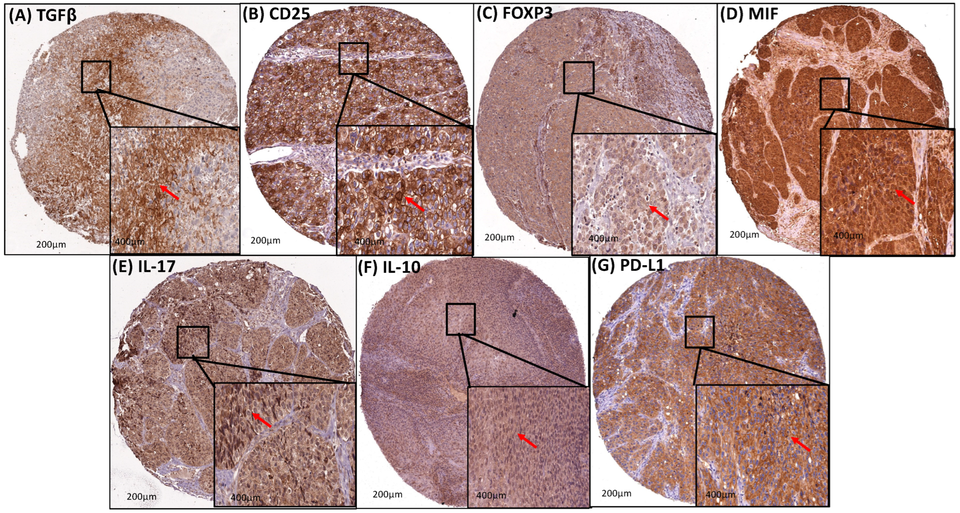
The staining of TGFβ, PD-L1, IL-10, FOXP3, IL-17, and MIF was cytoplasmic and present in 25%, 9.4%, 56.2%, 7.2%, 76.3%, 86.1%, and 85.2%, respectively, of the cervical cancer cases (Table 6 and Table 7, and Figure 4). Furthermore, these immunosuppressive markers were correlated with the expression of STAT3, NF-κB, CD163, and CD204. On the other hand, it was statistically significant with the higher expression of CD163 (p < 0.05) and CD204 (p < 0.05). Higher expression of CD25, FOXP3, IL-17, and MIF was strongly associated with the higher expression of STAT3, NF-κB, CD163, and CD204 (all p < 0.0001). However, the higher expression of IL-10 was associated with the expression of STAT3 (p < 0.001), CD163 (p < 0.05), and CD204 (p < 0.05) but not with NF-κB. Similarly, the association between the higher expression of PD-L1 was statistically significant with the higher expression of STAT3 (p < 0.001) and CD204 (p < 0.007) but not with NF-κB and CD163 (Table 6 and Table 7, and Figure 4).

3.6. Transcription Factors (STAT3 and NF-κB) and M2-TAM (CD204 and CD163) Had a Strong Association Correlation with Apoptosis, Angiogenesis, and Proliferation

The cytoplasmic/nuclear staining of Bcl-2 and the cytoplasmic staining of VEGFα were present in 77.4% and 78.4%, respectively (Table 8 and Table 9, and Figure 3). On the other hand, the nuclear staining of Ki-67 was found in 66.3% (Table 8 and Table 9, and Figure 3). There was a strong association between the higher expression of VEGFα and the expression of STAT3, NF-κB, CD163, and CD204 (all p < 0.0001). Likewise, the higher expression of Bcl-2 was statistically significant with higher expression of STAT3, NF-κB, CD163, and CD204 (all p < 0.0001 and NF-κB p < 0.05). And finally, the expression of Ki-67 was only statistically significant with NF-κB expression (p < 0.001) (Table 8 and Table 9).

3.7. Assessment of Overall Survival and Recurrence-Free Survival of Patients with Cervical Cancer Based on Protein Expression of STAT3, NF-κB, CD163, and CD204

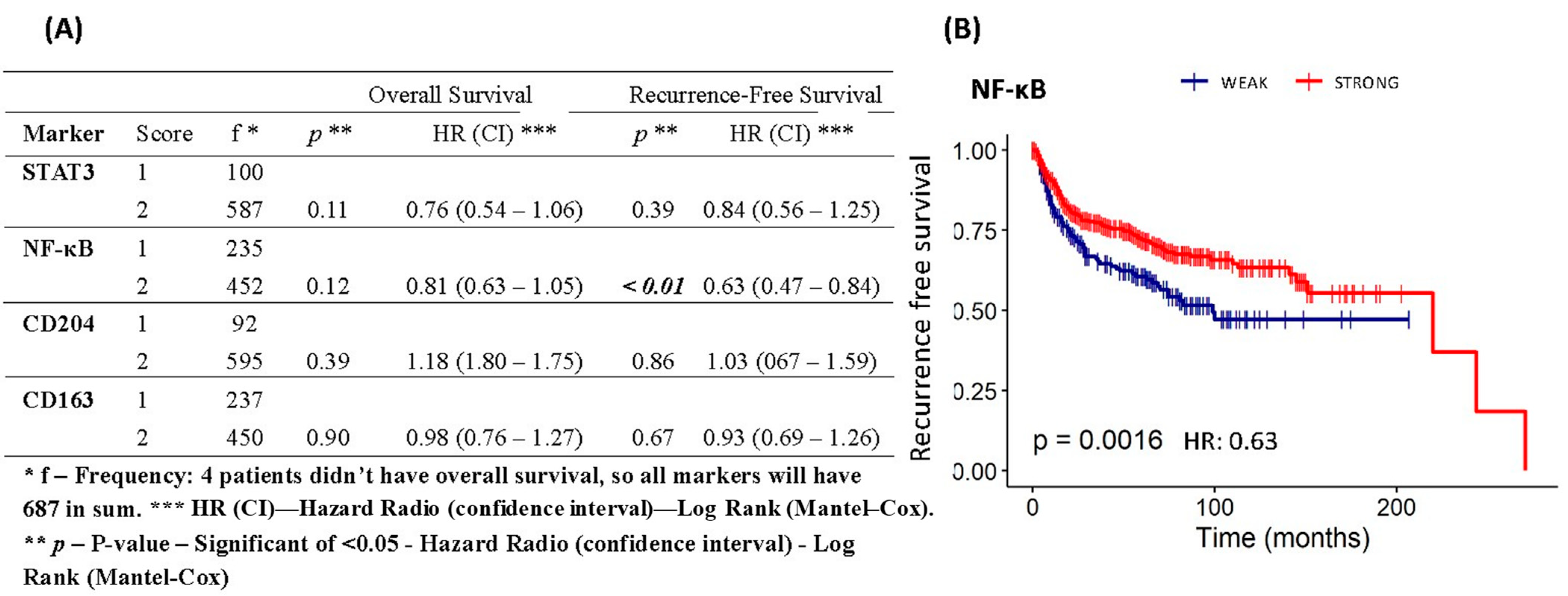
Based on Kaplan–Meier analysis, the STAT3, CD163, and CD204 expression had no significant correlation with the overall survival rate and recurrence-free rate, except the NF-κB expression (p < 0.01) that had a significant correlation with a recurrence-free survival rate. The patients who showed a higher NF-κB expression had a significantly more favorable outcome in recurrence-free survival than those with a lower expression (Figure 5).

3.8. Assessment of Overall Survival and Recurrence-Free Survival Evaluations of Patients with Cervical Cancer Based on Protein Expression of EMT, Invasion, Immunosuppression, Resistance to Apoptosis, Angiogenesis, and Proliferation

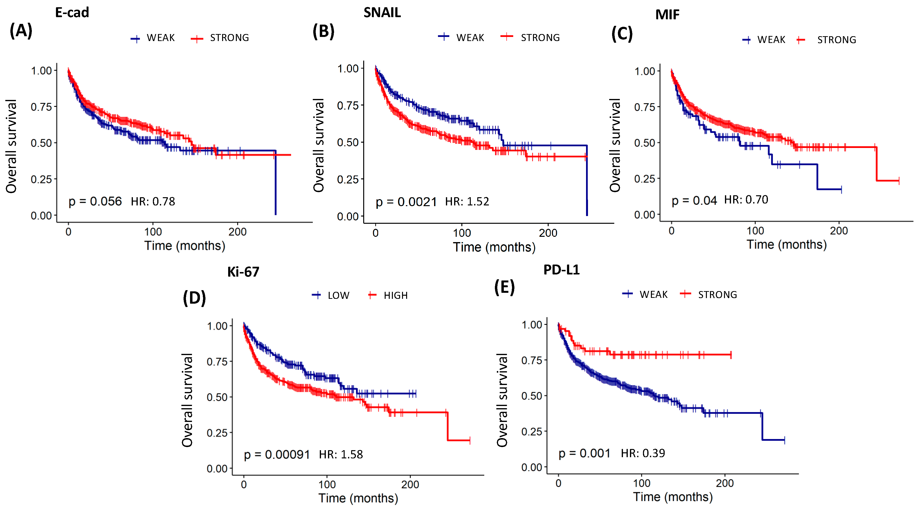
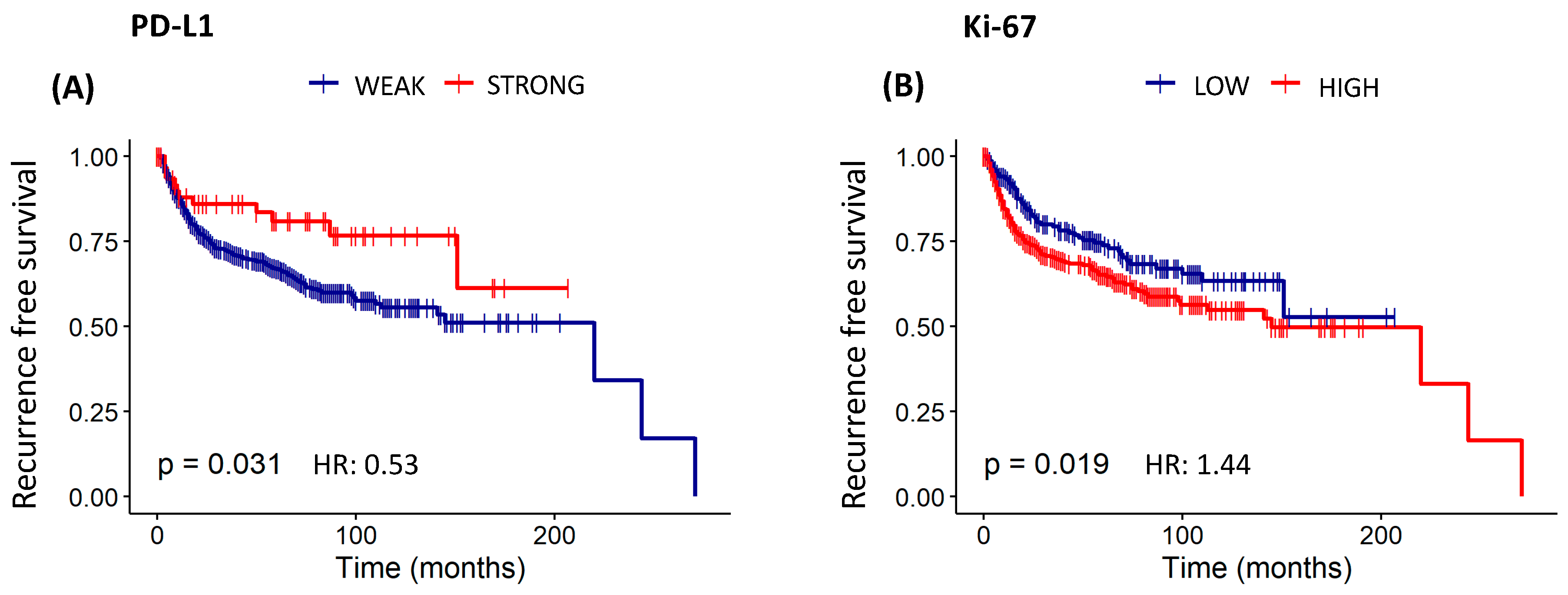
The study found statistically significant overall survival curves for E-cad (p < 0.05), SNAIL (p < 0.01), PD-L1 (p < 0.01), MIF (p < 0.05), and Ki-67 (p < 0.001) (Table 10 and Figure 6). Patients with a higher expression of E-cad (HR: 0.78) and MIF (HR: 0.70), as well as a lower expression of PD-L1 (HR: 0.39), had a significantly more favorable outcome when overall survival was analyzed. On the other hand, patients who showed a higher expression of SNAIL (HR: 1.52) and Ki-67 (HR: 1.58) had a significantly less favorable outcome in overall survival. Likewise, statistically significant recurrence-free survival curves to PD-L1 and Ki-67 (both p < 0.05) were found, pointing out that patients with a lower expression of PD-L1 (HR: 0.53) had a higher disease-free chance. On the other hand, those who had a higher expression of Ki-67 (HR: 1.44) showed a higher recurrence risk (Table 10 and Figure 7).

3.9. Association Correlation between Transcription Factors, EMT, Invasion, Immunosuppression, Apoptosis Resistance, Angiogenesis, and Proliferation with Clinical TNM Staging

Based on Fisher’s exact test analysis, the protein expression of STAT3 and NF-κB (both p < 0.01) had a significant correlation with TNM staging (Table 11). Furthermore, protein expression of VIM, E-cad, SNAIL, CD25, MIF, PD-L1, VEGFα, and Ki-67 had statistically high significance with TNM staging (p < 0.001, p < 0.0001, p < 0.0001, p < 0.001, p < 0.0001, p < 0.001, p < 0.001, and p < 0.0001, respectively) (Table S5).

3.10. Prognostic Significance of Patients by TNM Clinic Staging

In this study, the relationship between the TNM staging and death had a statistically significant correlation when overall survival and recurrence-free survival curves were studied using the Kaplan–Meier analysis (all p < 0.0001). In this context, patients with a higher stage (III and IV) had a significantly less favorable outcome in overall survival (III-HR: 4.43; IV-HR: 10.90) and recurrence-free survival (III-HR: 3.48; IV-HR: 10.82) (Table 12, and Figure 8).

3.11. Overall Survival of Patients with Cervical Cancer Based on Lifestyle Database, Laboratory Analysis, Treatment, and Clinicopathological Characteristics

Additionally, this study assessed the impact of clinicopathological variables on the overall survival of patients with cervical cancer. Table 13 summarizes the variables that increased the death risk. Interestingly, leukocytosis, thrombocytosis, therapy, neural invasion, compromised margin, and lymph node metastasis were independent prognostic factors for OS, enhancing the hazard of death (HR: 1.36–2.98). On the other hand, deeper stromal invasion, tumor size higher than 4 cm, and TNM pathological stage showed to be important factors to increase the hazard of death at 3.43, 4.32, and 7.58, respectively (Table 13 and Table S4).

4. Discussion

In this study, we found that a high degree of M2-TAM infiltration and the activation of STAT3/NF-κB signaling in TME of cervical cancer was associated with the upregulation of expression of protein factors related to immunosuppression, EMT, invasion, resistance to apoptosis, angiogenesis, and proliferation, which resonated in adverse clinical and pathological prognostic parameters. The interaction of M2-TAM and via STAT3/NF-κB with the tumor cells in TME creates a hostile environment for the immune cells but a favorable one for tumor progression [19,21]. In this context, we hypothesized that M2-TAMs and via STAT3/NF-κB crosstalk can modulate important hallmarks of tumor progression, negatively affecting the survival of 691 patients with cervical cancer.
The development of tumor progression by M2-TAM involves a complex interaction of signals, including stroma cells through cytokines, and chemokines, which stimulates the crosstalk of the STAT-3 pathway with NF-κB [33,34]. In TME, the crosstalk of STAT3 and NF-κB pathways promotes an important role in modulating cancer-associated inflammation-promoting TAM polarization towards M2, as well as stimulating resistance to apoptosis, EMT, invasion, and proliferation in tumor cells [35,36,37]. As clinical relevance, the increased expression of STAT3 and NF-κB has been associated with higher clinical TNM staging in solid tumors, which was confirmed in our study [38,39]. Furthermore, we found a strong association correlation between +CD163 + CD204M2-TAM with STAT3 and NF-κB signaling pathways, as well as with factors linked to tumor growth, dissemination, and immunosuppression [40,41,42,43,44].
EMT type III, EMT associated with metastasis, is an important event in the evolution of cancer that is induced by a set of signaling molecules, such as STAT3 and NF-κB, and signaling cell (M2-TAM) [20,45]. Collectively, they stimulate multiple signaling pathways, thereby activating a small set of transcription factors (TFs), for example, SNAIL, or master regulators of EMT. These TFs act to suppress the expression of epithelial markers such as E-cadherin, as well as activation of mesenchymal markers such as N-cadherin, Vimentin, matrix metalloproteinases, favoring tumor infiltration, dissemination, and metastasis associated with increased resistance to apoptosis (bcl-2) contributing to the poor prognosis of patients [45,46].
Here, the expression of E-cadherin, Vimentin, and SNAIL had a relevant correlation with the overall survival of 691 patients with cervical cancer. The loss of the cell–cell junction by lower expression of E-cadherin changes the cytoskeleton of epithelial cells to a mesenchymal phenotype, leading to higher expression of Vimentin and SNAIL, a transcription factor of EMT process [47,48,49]. These EMT biomarkers are remarkable factors of worse prognosis [50,51,52]. Furthermore, M2-TAM regulates the deposition or degradation of ECM components by secreting matrix metallopeptidase (MMPs), serine, and cathepsins, which contributes to the tumor dissemination [53,54,55,56]. Our results showed that CD163 + CD204 + M2-TAM via STAT3/NF-κB signaling pathways was strongly responsible for increasing protein factors of EMT and invasion (MMP9), reverberating in a worse prognosis scenario in patients with cervical cancer.
Resistance to apoptosis, proliferation, and angiogenesis are cancer hallmarks that contribute to the tumor growth and dissemination predicting evolution of clinical outcomes [57,58,59]. There is strong evidence that the interaction between cancer-specific Bcl-2, STAT3/NF-κB signaling and the recruitment of M2-TAM promote a pro-tumor microenvironment [21,60]. Likewise, M2-TAMs produce pro-angiogenic factors, such as VEGFα, to improve angiogenesis in TME and promote the tumor expansion [61]. In addition, the nuclear Ki-67 protein is generally expressed only in proliferating cells that are from G1 phase to mitosis, and its expression rapidly decreases immediately after mitosis [62]. For that, the immunostaining for Ki-67 expression is the gold standard, and a cutoff level of 10–14% positive staining is used to judge high risk of prognosis [63]. Our results showed a strong association correlation of CD163 + CD204 + M2-TAM and via STAT3/NF-κB signaling pathways with the higher expression of Bcl-2 and VEGFα, as well as with elevated index of Ki-67 (>15% positive staining). And more interestingly, the higher Ki-67 proliferation index found in our study was associated to higher clinical TNM stage (III and IV), disease recurrence, and hazard of death in patients with cervical cancer, which indicated a strong corroboration with reports in the literature [64,65,66].
In metastasis, NF-κB cooperates with STAT3 in promoting cancer progression, conferring resistance to apoptosis and plasticity. Furthermore, NF-κB and STAT3 interact and cooperate in regulating the interaction of malignant cells and the tumor microenvironment, especially immune cells such as TAMs that release cell-stimulating growth factors and cytokines, including IL-6, IL-10, and TNF-α [67,68,69].
In the TME, M2-TAMs exert an immunosuppressive function that acts on the positive regulation of the STAT3 pathway and the negative regulation of NF-κB in cells of the immune system [19,67,70,71,72]. In this line, macrophage migration inhibitory factor (MIF) has been implicated to upregulate STAT3 pathway in monocyte/macrophage towards M2-TAM [73]. Regulatory T cells (Treg cells) have been recognized as a core component of immunosuppression by suppressing cytotoxic T cells in many tumors [74]. FOXP3, a member of the Forkhead/winged-helix family of transcriptional regulators, converts naive T cells (CD4 + CD25-T cells) into suppressive inducible Foxp3+ Treg (CD4 + CD25 + FOXP3 + T cells) when regulated by TGF-β, IL-17, IL-10, and PD-L1 [75,76,77]. A high expression of CD25, a surface receptor of Treg cells, and FOXP3, as well as a low level of CD8+ T cells in the TME are associated with unfavorable prognosis [16,19,78]. Interestingly, our results showed a strong association correlation between CD163 + CD204 + M2TAM, via STAT3/NF-κB pathways and the expression of CD25, FOXP3, MIF, and IL-17 in 56.2%, 76.3%, 85.2%, and 86.1%, respectively, of 691 patients with cervical cancer. This expression profile, especially for CD25 and MIF, reverberated in a higher clinical TNM staging (III and IV) and consequently in an increased hazard of death and disease recurrence. On the other hand, the weak expression of TGFβ, IL-10, and PD-L1 in 75%, 92.8%, and 90.6% of patients did not have any association correlation with CD163 + 204 + M2-TAM, via STAT3 and NF-κB pathways or with clinical–pathological parameters. These weak labeling results may limit our analyses into the immunosuppressive role of these signaling pathways for the outcome of death. There are also interactions with alternative pathways that will act on tumor progression. Some reports describe that the tumor immune escape can be driven by other immunosuppressive cytokine loops, including CCL22, CCL17, and CCL18 that are also secreted by macrophage for recruiting Treg cells and modulate the immune response during the tumorigenic process [79,80].
An immunosuppressive molecular scenario reverberates often in an unfavorable clinical–pathological profile, on treatment failure and death. Some studies point out that older women, particularly those over age 70 years, independent of other factors such as comorbidities, histology, and stage of disease, show significantly decreased survival indexes [81,82,83]. In our study, patients of advanced age, 60–70 years and >80 years, presented a higher hazard of death (2.33 and 3.67, respectively). Pathological type, tumor size, FIGO stage, and lymph node metastasis are parameters related to recurrence and overall survival [84]. However, the criteria of lymph node metastasis seen in pathological TNM staging is considered an independent prognostic factor for overall survival in advanced disease, being used to guide postoperative adjuvant therapy [85]. In this line, our results showed that tumor size > 4 cm, FIGO staging IV, lymph node metastasis, and TNM pathological staging IV showed a strong correlation with overall survival (HR: 2.81–8.89). Furthermore, other parameters investigated, such as leukocytosis, thrombocytosis, depth of stromal invasion, and compromised margin, were relevant factors for death with HR ranging from 2.46 to 3.43 [86,87]. Paradoxically, overactivity of the immune system may allow the tumor evasion from the immune response by the tumor using specific immune cells known for their immunosuppressive activity [88]. In this context and as described previously, an elevated expression of Treg cells in TME and leukocytosis were observed in patients with locally advanced cervical cancer seen in cases of the higher clinical–pathological TNM staging (III and IV), reverberating in an elevated risk of death. Therefore, our study strongly reported that tumor progression is associated with the high expression of M2-TAM and STAT3/NF-κB pathways, leading to lower survival in cervical cancer. On the other hand, a weak association of analyzed immunosuppressive cytokines with the tumor progression and survival was a limitation of this study.

5. Conclusions

M2-TAM and STAT3/NF-κB pathways upregulated the expression of immunosuppressive components, leading to a stimulatory loop in EMT, invasion, angiogenesis, apoptosis resistance, and proliferation, which negatively reverberated on the clinic parameters, including TNM stage, disease recurrence, and death in 691 patients with cervical cancer. These findings showed that protein biomarkers related to the interaction of M2-TAM and cancer cells in TME have a strong predictive value in cervical cancer. Therefore, these results can act as new protein biomarkers that will contribute to new lines of oncological treatment.

Supplementary Materials

The following supporting information can be downloaded at: https://www.mdpi.com/article/10.3390/cancers16142496/s1, Table S1: Specification, clone, dilution, antigenic recovery, and incubation time of the primary antibodies; Table S2: Positivity pattern of antibody cell expression; Table S3: Distribution of patients by lifestyle database, laboratory analysis, clinical stage, treatment, and clinicopathological; Table S4: Cox-regression for overall survival to lifestyle, laboratory analysis, of clinical stage, clinicopathological characteristic and treatment; Table S5: Immunoexpression pattern of VIM, E-cad, MMP9, SNAIL, TGFβ, CD25, FOXP3, MIF, IL-17, IL-10, PD-L1, Bcl-2, VEGFα, and Ki-67 and correlation of association with TNM.

Author Contributions

R.F.d.A.J. and G.A.L. (George Alexandre Lira) designed the experiments. G.A.L. (George Alexandre Lira), I.d.L.M., I.G.d.S.L. and G.A.L. (George Alexandre Lira) collected human samples, built the TMA slides, and performed the immunohistochemistry. R.F.d.A.J., C.E., F.M.d.A. and G.A.L. (Giovanna Afonso Lira) analyzed the data. R.F.d.A.J. and G.A.L. (George Alexandre Lira) participated in the coordination of the study. R.F.d.A.J. and G.A.L. (George Alexandre Lira) drafted the manuscript. All authors have read and agreed to the published version of the manuscript.

Funding

We acknowledge support by postdoctoral fellowship from Raimundo Fernandes de Araujo Junior by CNPQ 401007/2022-7 (July 2022), doctoral fellowship from George Alexandre Lira by CNPQ 200998/2022-6 (June 2022), financial support by CNPq (grant number 301877/2019-0, March 2019)) and CAPES: Finance Code 001 (July 2017) We acknowledge support from Liga Norte-Riograndense contra o Cancer, Brazil, by allowing the collection of samples and data from medical records.

Institutional Review Board Statement

The study related to biopsies obtained from 691 patients undergoing surgical resection of cervical cancer was approved by the institutional committee (Nos. 2077017-2644266-2761333-3071847) on 23 May 2017.

Informed Consent Statement

Not applicable.

Data Availability Statement

The data that support the findings of this study are available from the corresponding author upon reasonable request.

Conflicts of Interest

The authors declare that they have no known competing financial interests or personal relationships that could have appeared to influence the work reported in this paper.

References

  1. Arbyn, M.; Weiderpass, E.; Bruni, L.; de Sanjosé, S.; Saraiya, M.; Ferlay, J.; Bray, F. Estimates of incidence and mortality of cervical cancer in 2018: A worldwide analysis. Lancet Glob. Health 2020, 8, e191–e203. [Google Scholar] [CrossRef] [PubMed]
  2. Sung, H.; Ferlay, J.; Siegel, R.L.; Laversanne, M.; Soerjomataram, I.; Jemal, A.; Bray, F.; Bsc, M.F.B.; Me, J.F.; Soerjomataram, M.I.; et al. Global Cancer Statistics 2020: GLOBOCAN Estimates of Incidence and Mortality Worldwide for 36 Cancers in 185 Countries. CA A Cancer J. Clin. 2021, 71, 209–249. [Google Scholar] [CrossRef]
  3. Santos, M.d.O.; Lima, F.C.d.S.d.; Martins, L.F.L.; Oliveira, J.F.P.; Almeida, L.M.d.; Cancela, M.d.C. Estimativa de Incidência de Câncer no Brasil, 2023–2025. Rev. Bras. Cancerol. 2023, 69, e213700. [Google Scholar] [CrossRef]
  4. Lei, J.; Ploner, A.; Elfström, K.M.; Wang, J.; Roth, A.; Fang, F.; Sundström, K.; Dillner, J.; Sparén, P. HPV Vaccination and the Risk of Invasive Cervical Cancer. N. Engl. J. Med. 2020, 383, 1340–1348. [Google Scholar] [CrossRef] [PubMed]
  5. Cohen, P.A.; Jhingran, A.; Oaknin, A.; Denny, L. Cervical cancer. Lancet 2019, 393, 169–182. [Google Scholar] [CrossRef] [PubMed]
  6. Chen, C.-C.; Wang, L.; Lin, J.-C.; Jan, J.-S. The prognostic factors for locally advanced cervical cancer patients treated by intensity-modulated radiation therapy with concurrent chemotherapy. J. Formos. Med. Assoc. 2015, 114, 231–237. [Google Scholar] [CrossRef]
  7. Gadducci, A.; Cosio, S. Pharmacological Treatment of Patients with Metastatic, Recurrent or Persistent Cervical Cancer Not Amenable by Surgery or Radiotherapy: State of Art and Perspectives of Clinical Research. Cancers 2020, 12, 2678. [Google Scholar] [CrossRef] [PubMed]
  8. Chanmee, T.; Ontong, P.; Konno, K.; Itano, N. Tumor-Associated Macrophages as Major Players in the Tumor Microenvironment. Cancers 2014, 6, 1670–1690. [Google Scholar] [CrossRef] [PubMed]
  9. Xiang, X.; Wang, J.; Lu, D.; Xu, X. Targeting tumor-associated macrophages to synergize tumor immunotherapy. Signal Transduct. Target. Ther. 2021, 6, 75. [Google Scholar] [CrossRef]
  10. Noy, R.; Pollard, J.W. Tumor-associated macrophages: From mechanisms to therapy. Immunity 2014, 41, 49–61. [Google Scholar] [CrossRef]
  11. Zhang, Y.; Cheng, S.; Zhang, M.; Zhen, L.; Pang, D.; Zhang, Q.; Li, Z. High-Infiltration of Tumor-Associated Macrophages Predicts Unfavorable Clinical Outcome for Node-Negative Breast Cancer. PLoS ONE 2013, 8, e76147. [Google Scholar] [CrossRef] [PubMed]
  12. Poh, A.R.; Ernst, M. Targeting Macrophages in Cancer: From Bench to Bedside. Front. Oncol. 2018, 8, 49. [Google Scholar] [CrossRef] [PubMed]
  13. Xue, Y.; Tong, L.; Liu, F.L.; Liu, A.; Zeng, S.; Xiong, Q.; Yang, Z.; He, X.; Sun, Y.; Xu, C. Tumor-infiltrating M2 macrophages driven by specific genomic alterations are associated with prognosis in bladder cancer. Oncol. Rep. 2019, 42, 581–594. [Google Scholar] [CrossRef] [PubMed]
  14. Kumar, A.T.; Knops, A.; Swendseid, B.; Martinez-Outschoom, U.; Harshyne, L.; Philp, N.; Rodeck, U.; Luginbuhl, A.; Cognetti, D.; Johnson, J.; et al. Prognostic Significance of Tumor-Associated Macrophage Content in Head and Neck Squamous Cell Carcinoma: A Meta-Analysis. Front. Oncol. 2019, 9, 656. [Google Scholar] [CrossRef]
  15. Guo, F.; Kong, W.; Zhao, G.; Cheng, Z.Z.; Ai, L.; Lv, J.; Feng, Y.C.; Ma, X.M. The correlation between tumor-associated macrophage infiltration and progression in cervical carcinoma. Biosci. Rep. 2021, 41. [Google Scholar] [CrossRef] [PubMed]
  16. Horta, B.; Pereira, T.; Medeiros, R.; Cerqueira, F. Cervical Cancer Outcome and Tumor-Associated Macrophages: Research Evidence. Immuno 2022, 2, 460–468. [Google Scholar] [CrossRef]
  17. Kanda, N.; Seno, H.; Konda, Y.; Marusawa, H.; Kanai, M.; Nakajima, T.; Kawashima, T.; Nanakin, A.; Sawabu, T.; Uenoyama, Y.; et al. STAT3 is constitutively activated and supports cell survival in association with survivin expression in gastric cancer cells. Oncogene 2004, 23, 4921–4929. [Google Scholar] [CrossRef]
  18. Scholz, A.; Heinze, S.; Detjen, K.M.; Peters, M.; Welzel, M.; Hauff, P.; Schirner, M.; Wiedenmann, B.; Rosewicz, S. Activated signal transducer and activator of transcription 3 (STAT3) supports the malignant phenotype of human pancreatic cancer. Gastroenterology 2003, 125, 891–905. [Google Scholar] [CrossRef]
  19. Júnior, R.F.d.A.; A Lira, G.; Schomann, T.; Cavalcante, R.S.; Vilar, N.F.; de Paula, R.C.M.; Gomes, R.F.; Chung, C.K.; Jorquera-Cordero, C.; Vepris, O.; et al. Retinoic acid-loaded PLGA nanocarriers targeting cell cholesterol potentialize the antitumour effect of PD-L1 antibody by preventing epithelial-mesenchymal transition mediated by M2-TAM in colorectal cancer. Transl. Oncol. 2023, 31, 101647. [Google Scholar] [CrossRef]
  20. Junior, R.F.d.A.; Eich, C.; Jorquera, C.; Schomann, T.; Baldazzi, F.; Chan, A.B.; Cruz, L.J. Ceramide and palmitic acid inhibit macrophage-mediated epithelial–mesenchymal transition in colorectal cancer. Mol. Cell. Biochem. 2020, 468, 153–168. [Google Scholar] [CrossRef]
  21. Cavalcante, R.S.; Ishikawa, U.; Silva, E.S.; Silva-Júnior, A.A.; Araújo, A.A.; Cruz, L.J.; Chan, A.B.; de Araújo Júnior, R.F. STAT3/NF-κB signalling disruption in M2 tumour-associated macrophages is a major target of PLGA nanocarriers/PD-L1 antibody immunomodulatory therapy in breast cancer. Br. J. Pharmacol. 2021, 178, 2284–2304. [Google Scholar] [CrossRef] [PubMed]
  22. Hideki, T.; Muneaki, S.; Mitsuya, I.; Nobuo, Y. TNM classification of gynaecological malignant tumours, eighth edition: Changes between the seventh and eighth editions. Jpn. J. Clin. Oncol. 2019, 49, 311–320. [Google Scholar]
  23. Araújo RFJr Lira, G.A.; Vilaça, J.A.; Guedes, H.G.; Leitão, M.C.; Lucena, H.F.; Ramos, C.C. Prognostic and diagnostic implications of MMP-2, MMP-9, and VEGF-α expressions in colorectal cancer. Pathol. Res. Pract. 2015, 211, 71–77. [Google Scholar] [CrossRef] [PubMed]
  24. Araújo Júnior, R.F.; Garcia, V.B.; Leitão, R.F.; Brito, G.A.; Miguel Ede, C.; Guedes, P.M.; de Araújo, A.A. Carvedilol Improves Inflammatory Response, Oxidative Stress and Fibrosis in the Alcohol-Induced Liver Injury in Rats by Regulating Kuppfer Cells and Hepatic Stellate Cells. PLoS ONE 2016, 11, e0148868. [Google Scholar] [CrossRef] [PubMed]
  25. Harada, K.; Dong, X.; Estrella, J.S.; Correa, A.M.; Xu, Y.; Hofstetter, W.L.; Sudo, K.; Onodera, H.; Suzuki, K.; Suzuki, A.; et al. Tumor-associated macrophage infiltration is highly associated with PD-L1 expression in gastric adenocarcinoma. Gastric Cancer Off. J. Int. Gastric Cancer Assoc. Jpn. Gastric Cancer Assoc. 2018, 21, 31–40. [Google Scholar] [CrossRef] [PubMed]
  26. Staniszewski, W. Virtual microscopy, data management and image analysis in Aperio ScanScope system. Folia Histochem. Cytobiol. 2010, 47, 699–701. [Google Scholar] [CrossRef]
  27. Fasanella, S.; Leonardi, E.; Cantaloni, C.; Eccher, C.; Bazzanella, I.; Aldovini, D.; Bragantini, E.; Morelli, L.; Cuorvo, L.; Ferro, A.; et al. Proliferative activity in human breast cancer: Ki-67 automated evaluation and the influence of different Ki-67 equivalent antibodies. Diagn. Pathol. 2011, 6, S7. [Google Scholar] [CrossRef] [PubMed]
  28. García-Rojo, M.; Sánchez, J.R.; de la Santa, E.; Durán, E.; Ruiz, J.L.; Silva, A.; Rubio, F.J.; Rodríguez, A.M.; Meléndez, B.; González, L.; et al. Automated image analysis in the study of lymphocyte subpopulation in eosinophilic oesophagitis. Diagn. Pathol. 2014, 9, S7. [Google Scholar] [CrossRef] [PubMed]
  29. Barricelli, B.R.; Casiraghi, E.; Gliozzo, J.; Huber, V.; Leone, B.E.; Rizzi, A.; Vergani, B. ki67 nuclei detection and ki67-index estimation: A novel automatic approach based on human vision modeling. BMC Bioinform. 2019, 20, 733. [Google Scholar] [CrossRef]
  30. Lashen, A.; Toss, M.S.; Green, A.R.; Mongan, N.P.; Rakha, E. Ki67 assessment in invasive luminal breast cancer: A comparative study between different scoring methods. Histopathology 2022, 81, 786–798. [Google Scholar] [CrossRef]
  31. Sakakibara, A.; Matsui, K.; Katayama, T.; Higuchi, T.; Terakawa, K.; Konishi, I. Age-related survival disparity in stage IB and IIB cervical cancer patients: Survival disparity in cervical cancer. J. Obstet. Gynaecol. Res. 2019, 45, 686–694. [Google Scholar] [CrossRef] [PubMed]
  32. Shin, W.; Park, S.-Y.; Seo, S.-S.; Lim, M.C.; Kim, J.-Y.; Kang, S. Predicting the risk of the distant recurrence of cervical cancer after concurrent chemoradiation: A validation study of the Korean Gynecologic Oncologic Group (KGOG)-1024 model. Gynecol. Oncol. 2021, 164, 62–67. [Google Scholar] [CrossRef] [PubMed]
  33. Quail, D.F.; Joyce, J.A. Microenvironmental regulation of tumor progression and metastasis. Nat. Med. 2013, 19, 1423–1437. [Google Scholar] [CrossRef] [PubMed]
  34. McAllister, S.S.; Weinberg, R.A. The tumour-induced systemic environment as a critical regulator of cancer progression and metastasis. Nature 2014, 16, 717–727. [Google Scholar] [CrossRef] [PubMed]
  35. Huang, R.; Wang, S.; Wang, N.; Zheng, Y.; Zhou, J.; Yang, B.; Wang, X.; Zhang, J.; Guo, L.; Wang, S.; et al. CCL5 derived from tumor-associated macrophages promotes prostate cancer stem cells and metastasis via activating β-catenin/STAT3 signaling. Cell Death Dis. 2020, 11, 234. [Google Scholar] [CrossRef] [PubMed]
  36. Kusaba, T.; Nakayama, T.; Yamazumi, K.; Yakata, Y.; Yoshizaki, A.; Inoue, K.; Nagayasu, T.; Sekine, I. Activation of STAT3 is a marker of poor prognosis in human colorectal cancer. Oncol. Rep. 2006, 15, 1445–1451. [Google Scholar] [CrossRef] [PubMed]
  37. Wen, W.; Lowe, G.; Roberts, C.M.; Finlay, J.; Han, E.S.; Glackin, C.A.; Dellinger, T.H. Pterostilbene Suppresses Ovarian Cancer Growth via Induction of Apoptosis and Blockade of Cell Cycle Progression Involving Inhibition of the STAT3 Pathway. Int. J. Mol. Sci. 2018, 19, 1983. [Google Scholar] [CrossRef] [PubMed]
  38. Shi, S.; Ma, H.-Y.; Zhang, Z.-G. Clinicopathological and prognostic value of STAT3/p-STAT3 in cervical cancer: A meta and bioinformatics analysis. Pathol. Res. Pract. 2021, 227, 153624. [Google Scholar] [CrossRef]
  39. Wu, P.; Wu, D.; Zhao, L.; Huang, L.; Shen, G.; Huang, J.; Chai, Y. Prognostic role of STAT3 in solid tumors: A systematic review and meta-analysis. Oncotarget 2016, 7, 19863–19883. [Google Scholar] [CrossRef]
  40. Naugler, W.E.; Karin, M. NF-κB and cancer—Identifying targets and mechanisms. Curr. Opin. Genet. Dev. 2008, 18, 19–26. [Google Scholar] [CrossRef]
  41. El-Arabey, A.A.; Abdalla, M.; Abd-Allah, A.R. Ovarian cancer immunotherapy of NF-κB may have a dark side. Hum. Cell Off. J. Hum. Cell Res. Soc. 2021, 34, 1019–1020. [Google Scholar] [CrossRef] [PubMed]
  42. Van Antwerp, D.J.; Martin, S.J.; Kafri, T.; Green, D.R.; Verma, I.M. Suppression of TNF-alpha-induced apoptosis by NF-kappaB. Science 1996, 274, 787–789. [Google Scholar] [CrossRef] [PubMed]
  43. Valeta-Magara, A.; Gadi, A.; Volta, V.; Walters, B.; Arju, R.; Giashuddin, S.; Zhong, H.; Schneider, R.J. Inflammatory Breast Cancer Promotes Development of M2 Tumor-Associated Macrophages and Cancer Mesenchymal Cells through a Complex Chemokine Network. Cancer Res. 2019, 79, 3360–3371. [Google Scholar] [CrossRef] [PubMed]
  44. Mancino, A.; Lawrence, T. Nuclear Factor-κB and Tumor-Associated Macrophages. Clin. Cancer Res. 2010, 16, 784–789. [Google Scholar] [CrossRef] [PubMed]
  45. Debnath, P.; Huirem, R.S.; Dutta, P.; Palchaudhuri, S. Epithelial–mesenchymal transition and its transcription factors. Biosci. Rep. 2021, 42. [Google Scholar] [CrossRef]
  46. Qureshi, R.; Arora, H.; Rizvi, M. EMT in cervical cancer: Its role in tumour progression and response to therapy. Cancer Lett. 2015, 356, 321–331. [Google Scholar] [CrossRef] [PubMed]
  47. Rodríguez-Sastre, M.A.; González-Maya, L.; Delgado, R.; Lizano, M.; Tsubaki, G.; Mohar, A.; García-Carrancá, A. Abnormal distribution of E-cadherin and β-catenin in different histologic types of cancer of the uterine cervix. Gynecol. Oncol. 2005, 97, 330–336. [Google Scholar] [CrossRef] [PubMed]
  48. E Koay, M.H.; Crook, M.; Stewart, C.J.R. Cyclin D1, E-cadherin and beta-catenin expression in FIGO Stage IA cervical squamous carcinoma: Diagnostic value and evidence for epithelial-mesenchymal transition. Histopathology 2012, 61, 1125–1133. [Google Scholar] [CrossRef] [PubMed]
  49. Mendonsa, A.M.; Na, T.-Y.; Gumbiner, B.M. E-cadherin in contact inhibition and cancer. Oncogene 2018, 37, 4769–4780. [Google Scholar] [CrossRef]
  50. Zhao, W.; Zhou, Y.; Xu, H.; Cheng, Y.; Kong, B. Snail family proteins in cervical squamous carcinoma: Expression and significance. Clin. Investig. Med. 2013, 36, E223–E233. [Google Scholar] [CrossRef]
  51. Kaufhold, S.; Bonavida, B. Central role of Snail1 in the regulation of EMT and resistance in cancer: A target for therapeutic intervention. J. Exp. Clin. Cancer Res. 2014, 33, 62. [Google Scholar] [CrossRef] [PubMed]
  52. Tian, Y.; Qi, P.; Niu, Q.; Hu, X. Combined Snail and E-cadherin Predicts Overall Survival of Cervical Carcinoma Patients: Comparison Among Various Epithelial-Mesenchymal Transition Proteins. Front. Mol. Biosci. 2020, 7, 22. [Google Scholar] [CrossRef] [PubMed]
  53. Lamouille, S.; Xu, J.; Derynck, R. Molecular mechanisms of epithelial–mesenchymal transition. Nat. Rev. Mol. Cell Biol. 2014, 15, 178–196. [Google Scholar] [CrossRef] [PubMed]
  54. Li, Y.; Wu, T.; Zhang, B.; Yao, Y.; Yin, G. Matrix metalloproteinase-9 is a prognostic marker for patients with cervical cancer. Med Oncol. 2012, 29, 3394–3399. [Google Scholar] [CrossRef] [PubMed]
  55. Martins, J.M.A.; Rabelo-Santos, S.H.; Westin, M.C.D.A.; Zeferino, L.C. Tumoral and stromal expression of MMP-2, MMP-9, MMP-14, TIMP-1, TIMP-2, and VEGF-A in cervical cancer patient survival: A competing risk analysis. BMC Cancer 2020, 20, 660. [Google Scholar]
  56. Ruffell, B.; Affara, N.I.; Coussens, L.M. Differential macrophage programming in the tumor microenvironment. Trends Immunol. 2012, 33, 119–126. [Google Scholar] [CrossRef] [PubMed]
  57. Neophytou, C.M.; Trougakos, I.P.; Erin, N.; Papageorgis, P. Apoptosis Deregulation and the Development of Cancer Multi-Drug Resistance. Cancers 2021, 13, 4363. [Google Scholar] [CrossRef] [PubMed]
  58. Li, L.T.; Jiang, G.; Chen, Q.; Zheng, J.N. Ki67 is a promising molecular target in the diagnosis of cancer (Review). Mol. Med. Rep. 2015, 11, 1566–1572. [Google Scholar] [CrossRef] [PubMed]
  59. Jiang, X.; Wang, J.; Deng, X.; Xiong, F.; Zhang, S.; Gong, Z.; Li, X.; Cao, K.; Deng, H.; He, Y.; et al. The role of microenvironment in tumor angiogenesis. J. Exp. Clin. Cancer Res. 2020, 39, 204. [Google Scholar] [CrossRef]
  60. Fathi, N.; Rashidi, G.; Khodadadi, A.; Shahi, S.; Sharifi, S. STAT3 and apoptosis challenges in cancer. Int. J. Biol. Macromol. 2018, 117, 993–1001. [Google Scholar] [CrossRef]
  61. Wang, Y.; Chang, T.; Wu, T.; Xu, W.; Dou, G.; Wang, Y.; Guo, C. M2 macrophages promote vasculogenesis during retinal neovascularization by regulating bone marrow-derived cells via SDF-1/VEGF. Cell Tissue Res. 2020, 380, 469–486. [Google Scholar] [CrossRef] [PubMed]
  62. Yang, C.; Zhang, J.; Ding, M.; Xu, K.; Li, L.; Mao, L.; Zheng, J. Ki67 targeted strategies for cancer therapy. Clin. Transl. Oncol. 2017, 20, 570–575. [Google Scholar] [CrossRef] [PubMed]
  63. Ibrahim, T.; Farolfi, A.; Scarpi, E.; Mercatali, L.; Medri, L.; Ricci, M.; Nanni, O.; Serra, L.; Amadori, D. Hormonal Receptor, Human Epidermal Growth Factor Receptor-2, and Ki67 Discordance between Primary Breast Cancer and Paired Metastases: Clinical Impact. Oncology 2012, 84, 150–157. [Google Scholar] [CrossRef] [PubMed]
  64. Li, C.; Zheng, M.; Zheng, X.; Fang, X.; Dong, J.; Wang, C.; Wang, T. Predictive Ki-67 Proliferation Index of Cervical Squamous Cell Carcinoma Based on IVIM-DWI Combined with Texture Features. Contrast Media Mol. Imaging 2021, 2021, 8873065. [Google Scholar] [CrossRef] [PubMed]
  65. Shi, Q.; Xu, L.; Yang, R.; Meng, Y.; Qiu, L. Ki-67 and P16 proteins in cervical cancer and precancerous lesions of young women and the diagnostic value for cervical cancer and precancerous lesions. Oncol. Lett. 2019, 18, 1351–1355. [Google Scholar] [CrossRef] [PubMed]
  66. Tu, Y.; Jiang, P.; Zhang, J.; Jiang, S.; Yi, Q.; Yuan, R. The positive threshold of the immunohistochemical parameter Ki67 for predicting the recurrence of cervical cancer. Int. J. Gynecol. Obstet. 2021, 158, 330–337. [Google Scholar] [CrossRef] [PubMed]
  67. Hensler, M.; Kasikova, L.; Fiser, K.; Rakova, J.; Skapa, P.; Laco, J.; Lanickova, T.; Pecen, L.; Truxova, I.; Vosahlikova, S.; et al. M2-like macrophages dictate clinically relevant immunosuppression in metastatic ovarian cancer. J. Immunother. Cancer 2020, 8, e000979. [Google Scholar] [CrossRef] [PubMed]
  68. Chen, Y.; Song, Y.; Du, W.; Gong, L.; Chang, H.; Zou, Z. Tumor-associated macrophages: An accomplice in solid tumor progression. J. Biomed. Sci. 2019, 26, 78. [Google Scholar] [CrossRef]
  69. Zhang, H.; Li, R.; Cao, Y.; Gu, Y.; Lin, C.; Liu, X.; Lv, K.; He, X.; Fang, H.; Jin, K.; et al. Poor Clinical Outcomes and Immunoevasive Contexture in Intratumoral IL-10-Producing Macrophages Enriched Gastric Cancer Patients. Ann. Surg. 2020, 275, e626–e635. [Google Scholar] [CrossRef]
  70. Fu, X.-L.; Duan, W.; Su, C.-Y.; Mao, F.-Y.; Lv, Y.-P.; Teng, Y.-S.; Yu, P.-W.; Zhuang, Y.; Zhao, Y.-L. Interleukin 6 induces M2 macrophage differentiation by STAT3 activation that correlates with gastric cancer progression. Cancer Immunol. Immunother. 2017, 66, 1597–1608. [Google Scholar] [CrossRef]
  71. Hagemann, T.; Lawrence, T.; McNeish, I.; Charles, K.A.; Kulbe, H.; Thompson, R.G.; Robinson, S.C.; Balkwill, F.R. “Re-educating” tumor-associated macrophages by targeting NF-kappa B. J. Exp. Med. 2008, 205, 1261–1268. [Google Scholar] [CrossRef] [PubMed]
  72. Wang, Y.; Shen, Y.; Wang, S.; Shen, Q.; Zhou, X. The role of STAT3 in leading the crosstalk between human cancers and the immune system. Cancer Lett. 2018, 415, 117–128. [Google Scholar] [CrossRef]
  73. Munir, M.T.; Kay, M.K.; Kang, M.H.; Rahman, M.; Al-Harrasi, A.; Choudhury, M.; Moustaid-Moussa, N.; Hussain, F.; Rahman, S.M. Tumor-Associated Macrophages as Multifaceted Regulators of Breast Tumor Growth. Int. J. Mol. Sci. 2021, 22, 6526. [Google Scholar] [CrossRef] [PubMed]
  74. Nizar, S.; Meyer, B.; Galustian, C.; Kumar, D.; Dalgleish, A. T regulatory cells, the evolution of targeted immunotherapy. Biochim. Biophys. Acta Rev. Cancer 2010, 1806, 7–17. [Google Scholar] [CrossRef] [PubMed]
  75. Murai, M.; Turovskaya, O.; Kim, G.; Madan, R.; Karp, C.L.; Cheroutre, H.; Kronenberg, M. Interleukin 10 acts on regulatory T cells to maintain expression of the transcription factor Foxp3 and suppressive function in mice with colitis. Nat. Immunol. 2009, 10, 1178–1184. [Google Scholar] [CrossRef] [PubMed]
  76. Yamagiwa, S.; Gray, J.D.; Hashimoto, S.; Horwitz, D.A. A role for TGF-beta in the generation and expansion of CD4(+)CD25(+) regulatory T cells from human peripheral blood. J. Immunol. 2001, 166, 7282–7289. [Google Scholar] [CrossRef] [PubMed]
  77. Que, Y.; Xiao, W.; Guan, Y.-X.; Liang, Y.; Yan, S.-M.; Chen, H.-Y.; Li, Q.-Q.; Xu, B.-S.; Zhou, Z.-W.; Zhang, X. PD-L1 Expression Is Associated with FOXP3+ Regulatory T-Cell Infiltration of Soft Tissue Sarcoma and Poor Patient Prognosis. J. Cancer 2017, 8, 2018–2025. [Google Scholar] [CrossRef] [PubMed]
  78. Fridman, W.H.; Pagès, F.; Sautès-Fridman, C.; Galon, J. The immune contexture in human tumours: Impact on clinical outcome. Nat. Rev. Cancer 2012, 12, 298–306. [Google Scholar] [CrossRef] [PubMed]
  79. Mantovani, A. From phagocyte diversity and activation to probiotics: Back to Metchnikoff. Eur. J. Immunol. 2008, 38, 3269–3273. [Google Scholar] [CrossRef]
  80. Curiel, T.J.; Coukos, G.; Zou, L.; Alvarez, X.; Cheng, P.; Mottram, P.; Evdemon-Hogan, M.; Conejo-Garcia, J.R.; Zhang, L.; Burow, M.; et al. Specific recruitment of regulatory T cells in ovarian carcinoma fosters immune privilege and predicts reduced survival. Nat. Med. 2004, 10, 942–949. [Google Scholar] [CrossRef]
  81. Venkatesulu, B.P.; Mallick, S.; Rath, G.K. Patterns of care of cervical cancer in the elderly: A qualitative literature review. J. Geriatr. Oncol. 2017, 8, 108–116. [Google Scholar] [CrossRef] [PubMed]
  82. Moore, K.N.; Java, J.J.; Slaughter, K.N.; Rose, P.G.; Lanciano, R.; DiSilvestro, P.A.; Thigpen, J.T.; Lee, Y.-C.; Tewari, K.S.; Chino, J.; et al. Is age a prognostic biomarker for survival among women with locally advanced cervical cancer treated with chemoradiation? An NRG Oncology/Gynecologic Oncology Group ancillary data analysis. Gynecol. Oncol. 2016, 143, 294–301. [Google Scholar] [CrossRef] [PubMed]
  83. Quinn, B.A.; Deng, X.; Colton, A.; Bandyopadhyay, D.; Carter, J.S.; Fields, E.C. Increasing age predicts poor cervical cancer prognosis with subsequent effect on treatment and overall survival. Brachytherapy 2018, 18, 29–37. [Google Scholar] [CrossRef] [PubMed]
  84. Miyahara, S.; Tsuji, K.; Shimada, M.; Shibuya, Y.; Shigeta, S.; Nagai, T.; Umezawa, R.; Tokunaga, H.; Jingu, K.; Yaegashi, N. The Impact of Histological Subtype on Survival Outcome of Patients with Stage IIB-IVA Cervical Cancer Who Received Definitive Radiotherapy. Tohoku J. Exp. Med. 2021, 255, 303–313. [Google Scholar] [CrossRef] [PubMed]
  85. Gien, L.; Covens, A. Lymph node assessment in cervical cancer: Prognostic and therapeutic implications. J. Surg. Oncol. 2008, 99, 242–247. [Google Scholar] [CrossRef] [PubMed]
  86. Eifel, P.J.; Jhingran, A.; Levenback, C.F.; Tucker, S. Predictive Value of a Proposed Subclassification of Stages I and II Cervical Cancer Based on Clinical Tumor Diameter. Int. J. Gynecol. Cancer 2009, 19, 2–7. [Google Scholar] [CrossRef]
  87. Liu, Y.-M.; Ni, L.-Q.; Wang, S.-S.; Lv, Q.-L.; Chen, W.-J.; Ying, S.-P. Outcome and prognostic factors in cervical cancer patients treated with surgery and concurrent chemoradiotherapy: A retrospective study. World J. Surg. Oncol. 2018, 16, 18. [Google Scholar] [CrossRef]
  88. Madeddu, C.; Sanna, E.; Nemolato, S.; Mulas, O.; Oppi, S.; Scartozzi, M.; La Nasa, G.; Maccio, A. Pathogenic and Prognostic Roles of Paraneoplastic Leukocytosis in Cervical Cancer: Can Genomic-Based Targeted Therapies Have a Role? A Literature Review and an Emblematic Case Report. Diagnostics 2022, 12, 1910. [Google Scholar] [CrossRef]
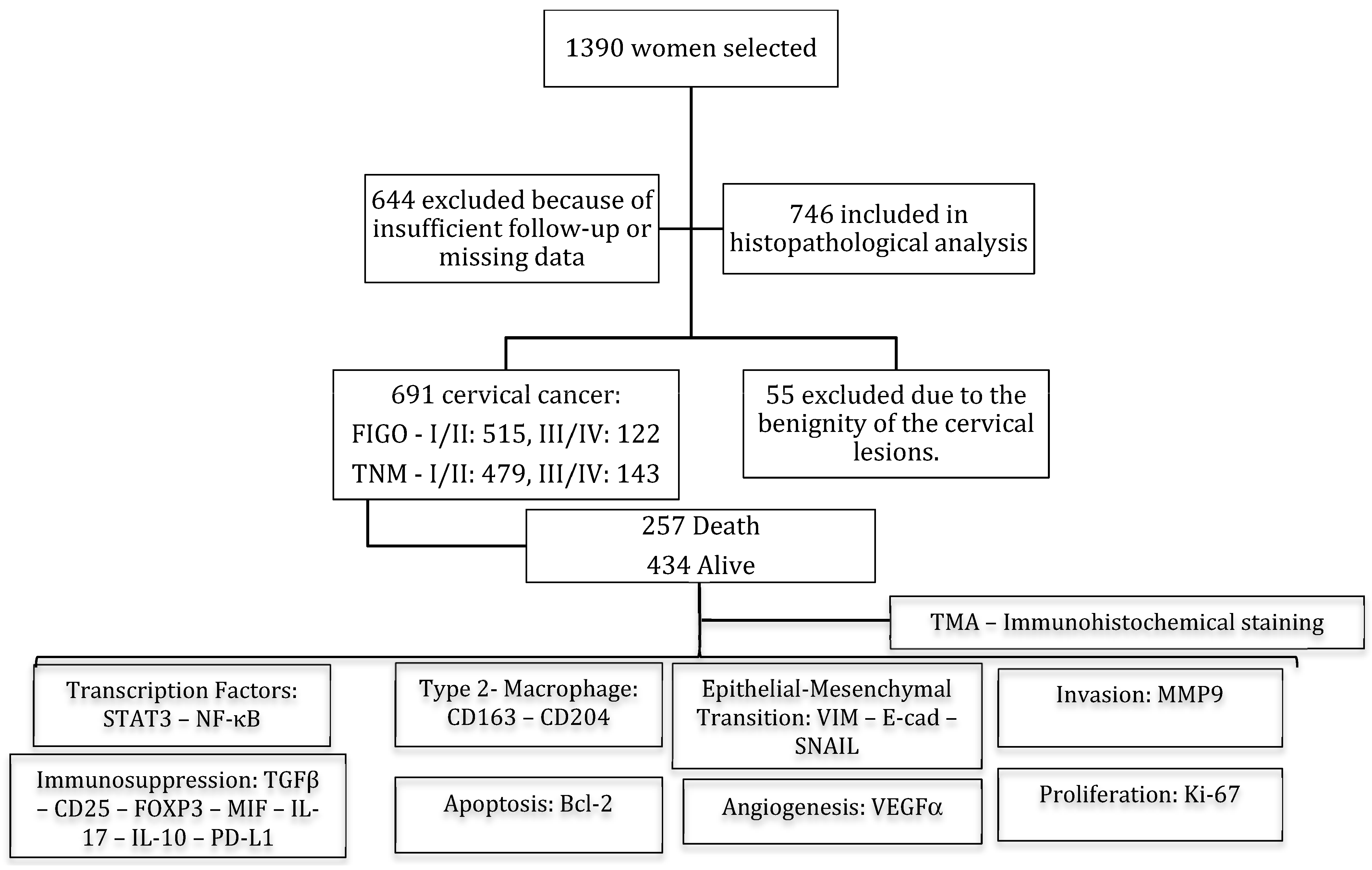
Figure 1. Flowchart of the women included in the study (2003–2017). FIGO/TNM predicts the outcome and determines the type of treatment that will be given. FIGO/TNM: stage I—confined to the organ of origin; stage II—invasion of surrounding organs or tissue; stage III—spread to distant lymph nodes or tissue within the pelvis; stage IV—distant metastasis. Tissue microarray (TMA) immunohistochemistry staining was evaluated for transcription factors (STAT3 and NF-κB), Type 2 Macrophage (CD163-CD204), Epithelial–Mesenchymal Transition (VIM, E-cad and SNAIL), Invasion (MMP9), Immunosuppression (TGFβ, CD25, FOXP3, MIF, IL-17, IL-10, and PD-L1), Apoptosis (Bcl-2), Angiogenesis (VEGFα), and Proliferation (Ki-67).
Cancers 16 02496 g001
Figure 2. Intracellular signaling modulation in TME by STAT3 and NF-κB pathways via CD163/CD204 M2-TAM. Modulation of intracellular signaling in the TME through the STAT3 and NF-κB pathways and immunostaining of M2-TAM through the biomarkers CD163 and CD204. (A) Tissue Microarray (TMA) slides in hematoxylin and eosin (H&E) with duplicates of the 746 patients performed in 2021, (B) brownish-brown diffuse staining with strong positive cytoplasmic/nuclear staining for anti-STAT3 (red arrow), (C) specific brownish-brown staining with strong positive cytoplasmic staining for anti-NF-κB (red arrow), (D) specific moderately diffuse brownish-brown staining with strong positive cytoplasmic staining for anti-CD163 (red arrow), and (E) moderately diffuse brownish-brown staining with strong positive cytoplasmic/nuclear staining for anti-CD204 (red arrow) in primary cervical cancer. The immunohistological profile presents images at 200× magnification; 400× magnification is shown in the lower right corner of each image. Arrow: the tumor cell with intracellular staining (cytoplasmic and/or nuclear) depending on each immunohistochemical marker.
Cancers 16 02496 g002
Figure 3. Protein expression profiles of cervical tumors to evaluate EMT, Invasion, Apoptosis, Angiogenesis, and Cell Proliferation. (A) diffuse brownish-brown coloration with strong cytoplasmic/membrane staining for anti-VIM (red narrow), (B) diffuse brownish-brown coloration with strong cytoplasmic/membrane for anti-E-cad (red narrow), (C) diffuse brownish-brown coloration with strong cytoplasmic staining/nuclear staining for anti-MMP9 (red narrow), (D) diffusely brownish-brown staining with strong nuclear staining for anti-SNAIL (red narrow), (E) diffusely brownish-brown staining with strong cytoplasmic/nuclear staining for anti-Bcl-2 (red narrow), (F) diffusely brownish-brown staining with strong cytoplasmic staining for anti-VEGFα (red narrow), and (G) restricted brownish-brown staining with some strong nuclear staining for anti-Ki-67 (red narrow) on Tissue Microarray (TMA) with primary cervical cancer. The immunohistological profile presents images at 200× magnification; 400× magnification is shown in the lower right corner of each image.
Cancers 16 02496 g003
Figure 4. Protein expression profiles of cervical tumors to assess immunosuppression. Strong diffuse brownish-brown cytoplasmic staining for anti-TGFβ (A), anti-CD25 (B), anti-FOXP3 (C), anti-MIF (D), anti-IL-17 (E), anti-IL-10 (F), and anti-PD-L1 (G) in TMA with primary cervical cancer. Red arrow: brownish-brown cytoplasmic staining.
Cancers 16 02496 g004
Figure 5. Survival analysis of the risk model by expression of STAT3, NF-κB, CD204, and CD163 proteins in 691 patients with cervical cancer. (A) Comparison of the predictive power of the multiple risk model using the results of Cox regression in overall survival and recurrence-free survival, significance is seen only for NF-κB. (B) Kaplan–Meier recurrence-free survival curve estimated by NF-κB staining of patients with cervical cancer after treatment with worse prognosis for weak staining (log-rank χ2 = 0.63; p = 0.0016).
Cancers 16 02496 g005
Figure 6. Survival analysis of the risk model by E-cad, SNAIL, MIF, Ki-67, and PD-L1 protein expression in 691 patients with cervical cancer. Kaplan–Meier Overall Survival Curve estimated by staining of (A) E-cad (log-rank χ2 = 0.78; p = 0.056), (B) SNAIL (log-rank χ2 = 1.52; p = 0.0021), (C) MIF (log-rank χ2 = 0.70; p = 0.04), (D) Ki-67 (log-rank χ2 = 1.58; p = 0.00091), and (E) PD-L1 (log-rank χ2 = 0.39; p = 0.001).
Cancers 16 02496 g006
Figure 7. Survival analysis of the risk model by PD-L1 and Ki-67 protein expression in 691 patients with cervical cancer. Kaplan–Meier Recurrence-Free Survival Curve estimated by staining of (A) PD-L1 (log-rank χ2 = 0.53; p = 0.031), and (B) Ki-67 (log-rank χ2 = 1.44; p = 0.019).
Cancers 16 02496 g007
Figure 8. Survival analysis of the risk model by Clinical TNM Staging in 691 patients with cervical cancer. Kaplan–Meier Overall Survival Curve estimated by TNM (I-IV) (A) and Kaplan–Meier Recurrence-Free Survival Curve estimated by TNM (I-IV) (B).
Cancers 16 02496 g008
Table 1. Distribution of 691 patients with cervical cancer by age, clinical stage, recurrence, and survival.
Clinicopathological FeaturesCategoryFrequencyPercentage
Age (Years)≤4018426.6%
>40 and ≤6027840.2%
>60 and ≤8020129.1%
>80273.9%
Not available10.1%
Total Number691100.0%
FIGO StagingI39356.9%
II12217.7%
III10114.6%
IV213.0%
Not available547.8%
Total Number691100.0%
TNM StagingI37053.5%
II10915.8%
III10615.3%
IV375.4%
Not available6910.0%
Total Number691100.0%
RecurrenceNo46066.6%
Yes19428.1%
Not available375.4%
Total Number691100.0%
DeathNo41760.3%
Yes25737.2%
Not available172.5%
Total Number691100.0%
Table 2. Cox regression for overall survival to clinical staging.
Clinicopathological FeaturesCategoryFreq *Significance **Hazard Ratio of Exp(B) ***
Age (Years)<=40182<0.0001
>40 and <=60277<0.051.50 (1.06–2.12)
>60 and <=80200<0.00012.33 (1.63–3.31)
>8027<0.00013.67 (1.98–6.80)
FIGO StagingI391<0.0001
SystemII122<0.00012.44 (1.74–3.41)
III100<0.00015.68 (4.15–7.79)
IV20<0.00018.89 (5.31–14.88)
TNM Staging SystemI369<0.0001
II109<0.00012.29 (1.60–3.28)
III105<0.00014.47 (3.23–6.17)
IV36<0.000110.95 (7.22–16.62)
* Frequency; ** Significant (p < 0.05); *** Log Rank (Mantel–Cox).
Table 3. Protein expression of STAT3, NF-κB, CD163, and CD204 in primary cervical tumors and the association correlation between them.
Marker STAT3 NF-κB
Immunoexpression12 12
Weak
n [%] = 101 [14.6]
Strong
n [%] = 590 [85.4]
p *Weak
n [%] = 236 [34.2]
Strong
n [%] = 455 [65.8]
p *
NF-κB1Weak n [%] = 236 [34.2]62 [9.0]174 [25.2]<0.0001- -
2Strong n [%] = 455 [65.8]39 [5.6]416 [60.2]
CD1631Weak n [%] = 238 [34.4]68 [9.8]170 [24.6]<0.0001118 [17.1]120 [17.4]<0.0001
2Strong n [%] = 453 [65.6]33 [4.8]420 [60.8] 118 [17.1]335 [48.5]
CD2041Weak n [%] = 93 [13.5]51 [7.4]42 [6.1]<0.000148 [6.9]45 [6.5]<0.0001
2Strong n [%] = 598 [86.5]50 [7.2]548 [79.3] 188 [27.2]410 [59.3]
Fisher’s exact test (* Significant p < 0.05); Abbreviations: STAT3 = Signal Transducer and Activator of Transcription 3; NF-κB = Nuclear Factor-kappa B and M2-TAM = Tumor-associated Macrophages Type 2.
Table 4. Protein expression of vimentin, e-cadherin, matrix metalloproteinase 9, SNAIL, STAT3, and NF-κB in primary cervical tumors and the association correlation between them.
Marker STAT3 NF-κB
Immunoexpression12 12
Weak
n [%] =
101 [14.6]
Strong
n [%] =
590 [85.4]
p *Weak
n [%] =
236 [34.2]
Strong
n [%] =
455 [65.8]
p *
VIM1Weak n [%] = 110 [15.9]58 [8.4]52 [7.5]<0.000168 [9.8]42 [6.1]<0.0001
2Strong n [%] = 581 [84.1]43 [6.2]538 [77.9] 168 [24.3]413 [59.8]
E-cad1Weak n [%] = 311 [45.0]79 [11.4]232 [33.6]<0.0001122 [17.7]189 [27.4]<0.05
2Strong n [%] = 380 [55.0]22 [3.2]358 [51.8] 114 [16.5]266 [38.5]
MMP91Weak n [%] = 73 [10.6]47 [6.8]26 [3.8]<0.000144 [6.4]29 [4.2]<0.0001
2Strong n [%] = 618 [89.4]54 [7.8]564 [81.6] 192 [27.8]426 [61.6]
SNAIL1Weak n [%] = 238 [34.4]47 [6.8]191 [27.6]<0.0178 [11.3]160 [23.2]0.61
2Strong n [%] = 453 [65.6]54 [7.8]399 [57.7] 158 [22.9]295 [42.7]
* Fisher’s exact test (Significant p < 0.05); Abbreviations: STAT3 = Signal Transducer and Activator of Transcription 3; NF-κB = Nuclear Factor-kappa B; VIM = Vimentin; E-cad = E-cadherin; and MMP9 = Matrix Metalloproteinase-9.
Table 5. Protein expression of vimentin, E-cadherin, matrix metalloproteinase 9, and SNAIL in primary cervical tumors and the association correlation with M2-TAM (CD163 and CD204).
Marker CD163 CD204
Immunoexpression12 12
Weak
n [%] =
238 [34.4]
Strong
n [%] = 453 [65.6]
p *Weak
n [%] = 93 [13.5]
Strong
n [%] =
598 [86.5]
p *
VIM1Weak n [%] = 110 [15.9]77 [11.1]33 [4.8]<0.000156 [8.1]54 [7.8]<0.0001
2Strong n [%] = 581 [84.1]161 [23.3]420 [60.8] 37 [5.4]544 [78.7]
E-cad1Weak n [%] = 311 [45.0]131 [19.0]180 [26.0]<0.000168 [9.8]243 [35.2]<0.0001
2Strong n [%] = 380 [55.0]107 [15.5]273 [39.5] 25 [3.6]355 [51.4]
MMP91Weak n [%] = 73 [10.6]53 [7.7]20 [2.9]<0.000144 [6.4]29 [4.2]<0.0001
2Strong n [%] = 618 [89.4]185 [26.8]433 [62.7] 49 [7.1]569 [82.3]
SNAIL1Weak n [%] = 238 [34.4]92 [13.3]146 [21.1]0.0941 [5.9]197 [28.5]<0.05
2Strong n [%] = 453 [65.6]146 [21.1]307 [44.4] 52 [7.5]401 [8.0]
* Fisher’s exact test (Significant p < 0.05); Abbreviations: VIM = Vimentin; E-cad = E-cadherin; MMP9 = Matrix Metalloproteinase 9; and M2-TAM = Tumor-associated Macrophages Type 2.
Table 6. Protein expression of TGFβ, CD25, FOXP3, MIF, IL-17, IL-10, and PD-L1 in primary cervical tumors and the association correlation with STAT3 and NF-κB.
Marker STAT3 NF-κB
Immunoexpression12 12
Weak
n [%] = 101 [14.6]
Strong
n [%] =
590 [85.4]
p *Weak
n [%] = 236 [34.2]
Strong
n [%] = 455 [65.8]
p *
TGFβ1Weak n [%] = 518 [75.0]80 [11.6]438 [63.4]0.25180 [26.0]338 [48.9]0.58
2Strong n [%] = 173 [25.0]21 [3.0]152 [22.0] 56 [8.1]117 [16.9]
CD251Weak n [%] = 303 [43.8]74 [10.7]229 [33.1]<0.0001137 [19.8]166 [24.0]<0.0001
2Strong n [%] = 388 [56.2]27 [3.9]361 [52.2] 99 [14.3]289 [41.8]
FOXP31Weak n [%] = 164 [23.7]66 [9.6]98 [14.2]<0.000172 [10.4]92 [13.3]<0.0001
2Strong n [%] = 527 [76.3]35 [5.1]492 [71.2] 164 [23.7]363 [52.5]
MIF1Weak n [%] = 102 [14.8]60 [8.7]42 [6.1]<0.000151 [7.4]51 [7.4]<0.0001
2Strong n [%] = 589 [85.2]41 [5.9]548 [79.3] 185 [26.8]404 [58.5]
IL-171Weak n [%] = 96 [13.9]56 [8.1]40 [5.8]<0.000157 [8.2]39 [5.6]<0.0001
2Strong n [%] = 595 [86.1]45 [6.5]550 [79.6] 179 [25.9]416 [60.2]
IL-101Weak n [%] = 641 [92.8]101 [14.6]540 [78.1]<0.001220 [31.8]421 [60.9]0.87
2Strong n [%] = 50 [7.2]0 [0.0]50 [7.2] 16 [2.3]34 [4.9]
PD-L11Weak n [%] = 626 [90.6]100 [14.5]526 [76.1]<0.001210 [30.4]416 [60.2]0.33
2Strong n [%] = 65 [9.4]1 [0.1]64 [9.3] 26 [3.8]39 [5.6]
* Fisher’s exact test (Significant p < 0.05); Abbreviations: TGFβ = Transforming growth factor β; CD25 = α-chain of IL-2 receptor; IL-10 = Interleukin 10; IL-17 = Interleukin 17; PD-L1 = Programmed cell death-ligand 1; FOXP3 = Forkhead box protein P3; MIF = macrophage migration inhibitory factor.
Table 7. Protein expression of TGFβ, CD25, FOXP3, MIF, IL-17, IL-10, and PD-L1 in primary cervical tumors and the association correlation with M2-TAM (CD163 and CD204).
Marker CD163 CD204
Immunoexpression12 12
Weak
n [%] =
238 [34.4]
Strong
n [%] = 453 [65.6]
p *Weak
n [%] = 93 [13.5]
Strong
n [%] =
598 [86.5]
p *
TGFβ1Weak n [%] = 518 [75.0]191 [27.6]327 [47.3]<0.0580 [11.6]438 [63.4]<0.01
2Strong n [%] = 173 [25.0]47 [6.8]126 [18.2] 13 [1.9]160 [23.2]
CD251Weak n [%] = 303 [43.8]137 [19.8]166 [24.0]<0.000169 [10.0]234 [33.9]<0.0001
2Strong n [%] = 388 [56.2]101 [14.6]287 [41.5] 24 [3.5]364 [52.7]
FOXP31Weak n [%] = 164 [23.7]85 [12.3]79 [11.4]<0.000164 [9.3]100 [14.5]<0.0001
2Strong n [%] = 527 [76.3]153 [22.1]374 [54.1] 29 [4.2]498 [72.1]
MIF1Weak n [%] = 102 [14.8]59 [8.5]43 [6.2]<0.000144 [6.4]58 [8.4]<0.0001
2Strong n [%] = 589 [85.2]179 [25.9]410 [59.3] 49 [7.1]540 [78.1]
IL-171Weak n [%] = 96 [13.9]69 [10.0]27 [3.9]<0.000155 [8.0]41 [5.9]<0.0001
2Strong n [%] = 595 [86.1]169 [24.5]426 [61.6] 38 [5.5]557 [80.6]
IL-101Weak n [%] = 641 [92.8]228 [33.0]413 [59.8]<0.0591 [13.2]550 [79.6]<0.05
2Strong n [%] = 50 [7.2]10 [1.4]40 [5.8] 2 [0.3]48 [6.9]
PD-L11Weak n [%] = 626 [90.6]223 [32.3]403 [58.3]0.05491 [13.2]535 [77.4]<0.01
2Strong n [%] = 65 [9.4]15 [2.2]50 [7.2] 2 [0.3]63 [9.1]
* Fisher’s exact test (Significant p < 0.05); Abbreviations: TGFβ = Transforming growth factor β; CD25 = α-chain of IL-2 receptor; IL-10 = Interleukin 10; IL-17 = Interleukin 17; PD-L1 = Programmed cell death-ligand 1; FOXP3 = Forkhead box protein P3; MIF = macrophage migration inhibitory.
Table 8. Protein expression of Bcl-2, VEGFα, and Ki-67 and the association correlation with STAT3 and NF-κB.
Marker STAT3 NF-κB
Immunoexpression12 12
Weak
n [%] = 101 [14.6]
Strong
n [%] = 590 [85.4]
p *Weak
n [%] = 236 [34.2]
Strong
n [%] = 455 [65.8]
p *
Bcl-21Weak n [%] = 156 [22.6]51 [7.4]105 [15.2]<0.000167 [9.7]89 [12.9]<0.05
2Strong n [%] = 535 [77.4]50 [7.2]485 [70.2] 169 [24.7]366 [53.0]
VEGFα1Weak n [%] = 149 [21.6]71 [10.3]78 [11.3]<0.000180 [11.6]69 [10.0]<0.0001
2Strong n [%] = 542 [78.4]30 [4.3]512 [74.1] 156 [22.6]386 [55.9]
Ki-671Low n [%] = 223 [33.7]39 [5.6]194 [28.1]0.2560 [8.7]173 [25.0]<0.01
2High n [%] = 458 [66.3]62 [9.0]396 [57.3] 176 [25.5]282 [40.8]
* Fisher’s exact test (significant p < 0.05); Abbreviations: VEGFα = vascular endothelial growth factor alfa; Bcl-2 = B-cell lymphoma 2; Ki-67 = marker of proliferation.
Table 9. Protein expression of Bcl2, VEGFα, and Ki-67 and the association correlation with M2-TAM (CD163 and CD204).
MarkerImmunoexpressionCD163 CD204
12 12
Weak
n [%] =
238 [34.4]
Strong
n [%] = 453 [65.6]
p *Weak
n [%] = 93 [13.5]
Strong
n [%] =
598 [86.5]
p *
Bcl-21Weak n [%] = 156 [22.6]77 [11.1]79 [11.4]<0.000146 [6.7]110 [15.9]<0.0001
2Strong n [%] = 535 [77.4]161 [23.3]374 [54.1] 47 [6.8]488 [70.6]
VEGFα1Weak n [%] = 149 [21.6]88 [12.7]61 [8.8]<0.000162 [9.0]87 [12.6]<0.0001
2Strong n [%] = 542 [78.4]150 [21.7]392 [56.7] 31 [4.5]511 [74.0]
Ki-671Low n [%] = 223 [33.7]85 [12.3]148 [21.4]0.4435 [5.1]198 [28.4]0.41
2High n [%] = 458 [66.3]62 [9.0]396 [57.3] 176 [25.5]282 [40.8]
* Fisher’s exact test (significant p < 0.05); Abbreviations: VEGFα = vascular endothelial growth factor alfa; Bcl-2 = B-cell lymphoma 2; Ki-67 = marker of proliferation.
Table 10. Results of univariable Cox regression model analysis on protein expression of EMT, immunosuppression, resistance to apoptosis, angiogenesis, and proliferation.
MarkerScoref *Overall SurvivalRecurrence-Free Survival
p **HR (CI) ***p **HR (CI) ***
VIM1109
25780.280.83 (0.60–1.16)0.140.75 (0.52–1.09)
E-cad1309
2378<0.050.78 (0.61–1.00)0.720.95 (0.71–1.26)
MMP9172
26150.451.19 (0.75–1.88)0.351.29 (0.74–2.22)
SNAIL1237
2450<0.011.52 (1.16–2.00)0.521.10 (1.81–1.48)
TGFß1515
21720.071.27 (0.97–1.67)0.411.14 (0.82–1.56)
CD251302
23850.400.90 (0.70–1.15)0.430.89 (0.66–1.19)
FOXP31163
25240.820.96 (0.72–1.29)0.960.99 (0.70–1.39)
MIF1101
2586<0.050.70 (0.50–0.98)0.250.79 (0.53–1.18)
IL-17195
25920.690.92 (0.64–1.34)0.460.85 (0.56–1.30)
IL-101637
2500.391.21 (0.77–1.89)0.210.65 (0.33–1.28)
PD-L11623
2640.0010.39 (0.22–0.70)<0.050.53 (0.29–0.95)
Bcl-21154
25330.060.76 (0.58–1.01)0.360.85 (0.56–1.30)
VEGFα1148
25390.070.76 (0.57–1.02)0.060.72 (0.51–1.10)
Ki-671232
2455<0.0011.58 (1.20–2.09)<0.051.44 (1.05–1.97)
* f—Frequency: 4 patients did not attain overall survival, so all markers will have 687 in sum; ** pp-value—Significant of <0.05; *** HR (CI)—Hazard Radio (confidence interval)—Log Rank (Mantel–Cox).
Table 11. Protein Expression of Transcription Factors (STAT3 and NF-κB) and Immunosuppression by M2-TAM (CD204 and CD163) in primary cervical tumors, and the association correlation with TNM.
MarkerScoreTNM
IIIIIIIVTotalp *
STAT3142 (6.8%)14 (2.3%)27 (4.3%)6 (1.0%)89 (14.3%)
2328 (52.7%)95 (15.3%)79 (12.7%)31 (5.0%)533 (85.7%)<0.01
NF-κB1109 (17.5%)44 (7.1%)48 (7.7%)13 (2.1%)214 (34.4%)
2261 (42.0%)65 (10.5%)58 (9.3%)24 (3.9%)408 (65.6%)<0.01
CD204149 (7.9%)10 (1.6%)21 (3.4%)5 (0.8%)85 (13.7%)
2321 (51.6%)99 (15.9%)85 (13.7%)32 (5.1%)537 (86.3%)0.15
CD1631128 (20.6%)33 (5.3%)39 (6.3%)12 (1.9%)212 (34.1%)
2242 (38.9%)76 (12.2%)67 (10.8%)25 (4.0%)410 (65.9%)0.77
* Fisher’s exact test (significant p < 0.05); TNM data are missing in 96 patients. In this analysis, 622 of them were categorized.
Table 12. Cox regression for overall survival and Recurrence-Free Survival to TNM.
Overall SurvivalRecurrence-Free Survival
Stagef *p ***HR ****f **p ***HR ****
I369 368
II109<0.00012.27 (1.59–3.26)109<0.00012.14 (1.44–3.17)
III105<0.00014.43 (3.20–6.14)104<0.00013.48 (2.39–5.06)
IV36<0.000110.90 (7.17–16.57)36<0.000110.82 (6.36–18.41)
* f—Frequency: 69 patients did not have TNM, so all categorized of 622 in sum, and 4 patients did not have overall survival, so all markers will have 617 in sum; ** 74 patients not available to recurrence, so all categorized of 617 in sum; *** Significant (p < 0.05); **** HR (CI)—Hazard Ratio (confidence interval)—Log Rank (Mantel–Cox).
Table 13. Multivariate Cox regression for overall survival.
VariablesCharacteristicsFreq *p **Hazard Ratio of Exp(B) ***
Hemoglobin Count (g/dL)until 829<0.00011.0
Leukocytes Count (leukocytes/mm3)Leukocytosis101<0.00012.55 (1.89–3.44)
Platelets Count (platelets/mm3)Thrombocytosis50<0.00012.46 (1.69–3.58)
Tumor Size ****>4192<0.00014.32 (1.98–9.41)
Invasion ****Yes2010.051.71 (1.12–2.62)
Neural Invasion ****Yes480.052.06 (1.14–3.71)
Depth of stromal invasion ****Total243<0.00013.43 (1.94–6.06)
Compromised margin ****Yes45<0.00012.98 (1.88–4.74)
Lymph node metastasis ****Positive125<0.00012.81 (1.92–4.11)
TNM Pathological Stage ****IV34<0.00017.58 (4.50–12.75)
External Radiation TherapyYes393<0.00011.68 (1.28–2.21)
ChemotherapyYes294<0.011.362 (1.06–1.74)
* Freq: Frequency; ** p Significant (p < 0.05); *** Log Rank (Mantel–Cox); **** The assessment was based on the 468 patients with cervical cancer who underwent surgery.
Disclaimer/Publisher’s Note: The statements, opinions and data contained in all publications are solely those of the individual author(s) and contributor(s) and not of MDPI and/or the editor(s). MDPI and/or the editor(s) disclaim responsibility for any injury to people or property resulting from any ideas, methods, instructions or products referred to in the content.