Systematic Review

Somatic, Genetic and Epigenetic Changes in Nephrogenic Rests and Their Role in the Transformation to Wilms Tumors, a Systematic Review

by
Tessa Bánki
1,
Jarno Drost
1,2,
Marry M. van den Heuvel-Eibrink
1,
Annelies M. C. Mavinkurve-Groothuis
1 and
Ronald R. de Krijger
1,3,*
1
Princess Máxima Center for Pediatric Oncology, 3584 CS Utrecht, The Netherlands
2
Oncode Institute, 3584 CS Utrecht, The Netherlands
3
Department of Pathology, University Medical Center Utrecht, 3584 CX Utrecht, The Netherlands
*
Author to whom correspondence should be addressed.
Cancers 2023, 15(5), 1363; https://doi.org/10.3390/cancers15051363
Submission received: 8 December 2022 / Revised: 13 February 2023 / Accepted: 17 February 2023 / Published: 21 February 2023
(This article belongs to the Section Pediatric Oncology)

Abstract

:

Simple Summary

We reviewed all studies investigating molecular changes in nephrogenic rests (NR), the presumed precursor lesions of Wilms tumors (WT) being the most frequent malignant childhood renal tumors, between 1990 and 2022. Only 23 studies were found, reporting 119 pairs of NR and corresponding WT, which may allow the detection of early genetic changes that play a role in tumorigenesis. Two genes, WT1 and WTX, and two chromosomal regions, 11p13 where WT1 is located, and 11p15 harboring the IGF-2 gene, were found to be mutated or show loss of imprinting, respectively, in both nephrogenic rests and WT, suggesting that these could be relevant early genetic events.

Abstract

Objective: To review somatic genetic changes in nephrogenic rests (NR), which are considered to be precursor lesions of Wilms tumors (WT). Methods: This systematic review is written according to the PRISMA statement. PubMed and EMBASE were systematically searched for articles in the English language studying somatic genetic changes in NR between 1990 and 2022. Results: Twenty-three studies were included in this review, describing 221 NR of which 119 were pairs of NR and WT. Single gene studies showed mutations in WT1 and WTX, but not CTNNB1 to occur in both NR and WT. Studies investigating chromosomal changes showed loss of heterozygosity of 11p13 and 11p15 to occur in both NR and WT, but loss of 7p and 16q occurred in WT only. Methylome-based studies found differential methylation patterns between NR, WT, and normal kidney (NK). Conclusions: Over a 30-year time frame, few studies have addressed genetic changes in NR, likely hampered by technical and practical limitations. A limited number of genes and chromosomal regions have been implicated in the early pathogenesis of WT, exemplified by their occurrence in NR, including WT1, WTX, and genes located at 11p15. Further studies of NR and corresponding WT are urgently needed.

1. Introduction

Nephrogenic rests (NR) are foci of aberrant embryonal tissue in the kidney, which are still present after 36 weeks of gestation [1,2]. They represent non-obligate precursor lesions of WT and may regress over time or progress to WT due to as yet unknown molecular factors [1,2]. The presence of multifocal or diffuse NR is referred to as nephroblastomatosis [3,4]. NR have been described as either perilobar NR (PLNR) or intralobular NR (ILNR), based on the topographic site in the kidney. Both types can also occur in the same kidney as well as in both kidneys of the same individual. PLNR are located at the periphery of the renal cortex and have been shown to consist mainly of blastemal or immature epithelial components (Figure 1A) [1,5]. ILNR are located within the lobes and are mostly composed of a combination of cystic epithelial elements surrounded by moderately cellular stromal elements (Figure 1B) [1,5,6]. In addition, heterologous components, including fat, may be present. ILNR are considered to arise prior to PLNR, and are usually not as well demarcated as PLNR [1,5,6]. Histologically, according to Beckwith’s criteria [3], NR can be subdivided into different histological subtypes such as dormant rests (especially blastemal cells with minimal proliferation), sclerosing rests (minimal blastemal components and presence of stromal maturation), hyperplastic rests (proliferative signs manifested by blastemal and epithelial cells and increased size), neoplastic rests (usually a discrete spherical nodule), and obsolete rests ((sclerotic) stromal and epithelial components, and minimal blastemal). Any histological subtype of NR has been shown to be able to transition to another NR subtype [1,4].
In infants, microscopic NR are identified in about 1% of autopsies, the majority of which will either remain stable or go into regression [3,7]. The occurrence of NR is partly determined by ethnic and demographic factors [6]. In White American children, 20% reveal WT-associated PLNR compared to 7% in Asian American children, and 2% in Japanese children. ILNR, on the other hand, are more common in Asian American (33%) and Japanese children (25%) relative to the White American children (17%) [8]. As ILNR seem to arise from abnormalities earlier in development, they also occur at a younger age in children, namely at a median age of 23 months, whereas PLNR are usually not discovered until a median age of 36 months [5]. PLNR occur mainly in females and ILNR are more often seen in males [6]. The age at diagnosis also differs between ethnic groups. NR are generally diagnosed earlier in Asian American patients (median age at diagnosis 31 months) than in White American patients (median age at diagnosis 39 months) [8].
NR have been described to lead to the development of WT in a subset of cases [3,5]. WT are the most commonly found malignant renal tumor in children [9]. WT are morphologically heterogeneous embryonic tumors, including epithelial, blastemal, stromal, and sometimes rhabdomyomatous elements [10]. Tumors can be classified in different histological types, which currently grossly determines risk stratification after neoadjuvant chemotherapy treatment [11]. The distinction between NR, mainly PLNR, and WT can be extremely challenging, both radiologically and pathologically, especially if a biopsy is submitted for evaluation. In fact, no single radiological or histological criterion, including size and shape of the lesion, can distinguish NR from WT and a 25% misdiagnosis rate has been reported for radiological assessment (see Figure 1C,D) [12,13]. PLNR are associated with epithelial, blastemal, or mixed type WT and stromal or heterologous elements are limited or absent. In contrast, there is an association with ILNR and stromal type WT and heterologous components are frequent [3]. Most WT occur sporadically and are unilateral, but they can also be familial and bilateral [6]. According to the International Society of Paediatric Oncology Renal Tumour Study Group (SIOP-RTSG), kidneys with unilateral WT contain NR in 40% of the cases (25% PLNR, 9% ILNR, 5% both PLNR and ILNR and 1% nephroblastomatosis). Bilateral WT are significantly more frequently associated with NR, as NR have been reported in 94% of the stage V cases [14].
Bilateral WT occur significantly more often in syndromes in the context of which WT recurrently occur [11]. It is beyond the scope of this review to discuss these syndromes, except for their association with NR. Examples are Denys–Drash and WAGR syndrome, which are associated with the presence of ILNR. Patients with these syndromes have a risk of about 30% and 95%, respectively, to develop WT [6]. Patients with these syndromes have a risk of up to 95% and 50%, respectively, to develop WT [15,16]. PLNR occur more frequently in patients with overgrowth syndromes such as Beckwith–Wiedemann syndrome (BWS) [6]. Some of the abovementioned syndromes have a known genetic driver, such as the occurrence of germline WT1 mutations in Denys–Drash and WAGR syndrome [11,17]. In addition, numerous recurrent somatic genetic abnormalities, both gene mutations and copy number variations (CNV) at specific chromosomal locations, as well as hypermethylated regions have been found in non-syndromic, sporadic WTs. These include mutations in WT1, CTNNB1, MYCN, TP53, AMER1, FBXW7, GPC3, MLLT1, DIS3L2, DICER1, DROSHA, DGCR8, SIX1 and SIX2, SMARCA4, ARID1A and chromosomal aberrations such as gain of chromosome arm 1q, and loss of 16q and 1p [11,18].
Although knowledge on tumor-driving changes in WT is increasing, the genetic changes underpinning the development of NR have not been extensively studied. Notably, most NR regress and only some develop into WT [7]. Identifying molecular changes underpinning the transformation of NR to WT may aid the understanding of WT pathogenesis and guide the development of targeted therapies. Therefore, we performed a systematic review of the literature in which molecular analysis was completed in NR or in a combination of NR and WT. This systematic review presents the current knowledge of somatic molecular changes in NR.

2. Materials and Methods

2.1. Search Strategy and Eligibility Criteria

This review was written according to the Preferred Reporting Items for Systematic Reviews and Meta-Analyses (PRISMA) statement [19,20] and the protocol was not registered. PubMed, EMBASE and Cochrane were systematically searched for all the available literature on molecular changes in NR in the English language, published from 1990 to 2022. The full search strategy is provided in Table S1. Articles were included if somatic molecular changes in NR were studied, including case reports in which specific analysis for NR was performed. Studies without molecular analysis in NR, non-English literature, animal studies or germline mutation analyses were excluded. Three authors (T.B., A.M., R.K.) assessed all articles independently based on the in- and exclusion criteria. After a comparison and the consensus of the authors on potentially relevant articles, the remaining publications were screened for eligibility criteria, based on their full text. Controversies were resolved by consensus.

2.2. Quality Assessment

The “Standards for Reporting Diagnostic Accuracy 2015” (STARD 2015) checklist [20] was used to assess the quality of the included studies individually and is provided in Supplementary Table S2a. The included case reports were assessed using the case report guidelines (CARE checklist 2013), the checklist is presented in Supplementary Table S2b [21]. The included studies were further assessed with the Oxford Centre for Evidence-Based Medicine Levels of Evidence Classification rubric [22], independently by the three authors (T.B., A.M., R.K.), for methodologic quality. In case of disagreement, it was solved by reaching consensus.

2.3. Data Extraction

From the included articles, the number of NR, WT, WT-NR pairs analyzed, mean age at time of diagnosis of WT, sex, histological type of WT and NR and whether a WT was bilateral or hereditary, were obtained. Furthermore, from each study, the genes or chromosomal regions investigated with associated molecular techniques were extracted.

3. Results

3.1. Search Strategy and Eligibility Criteria

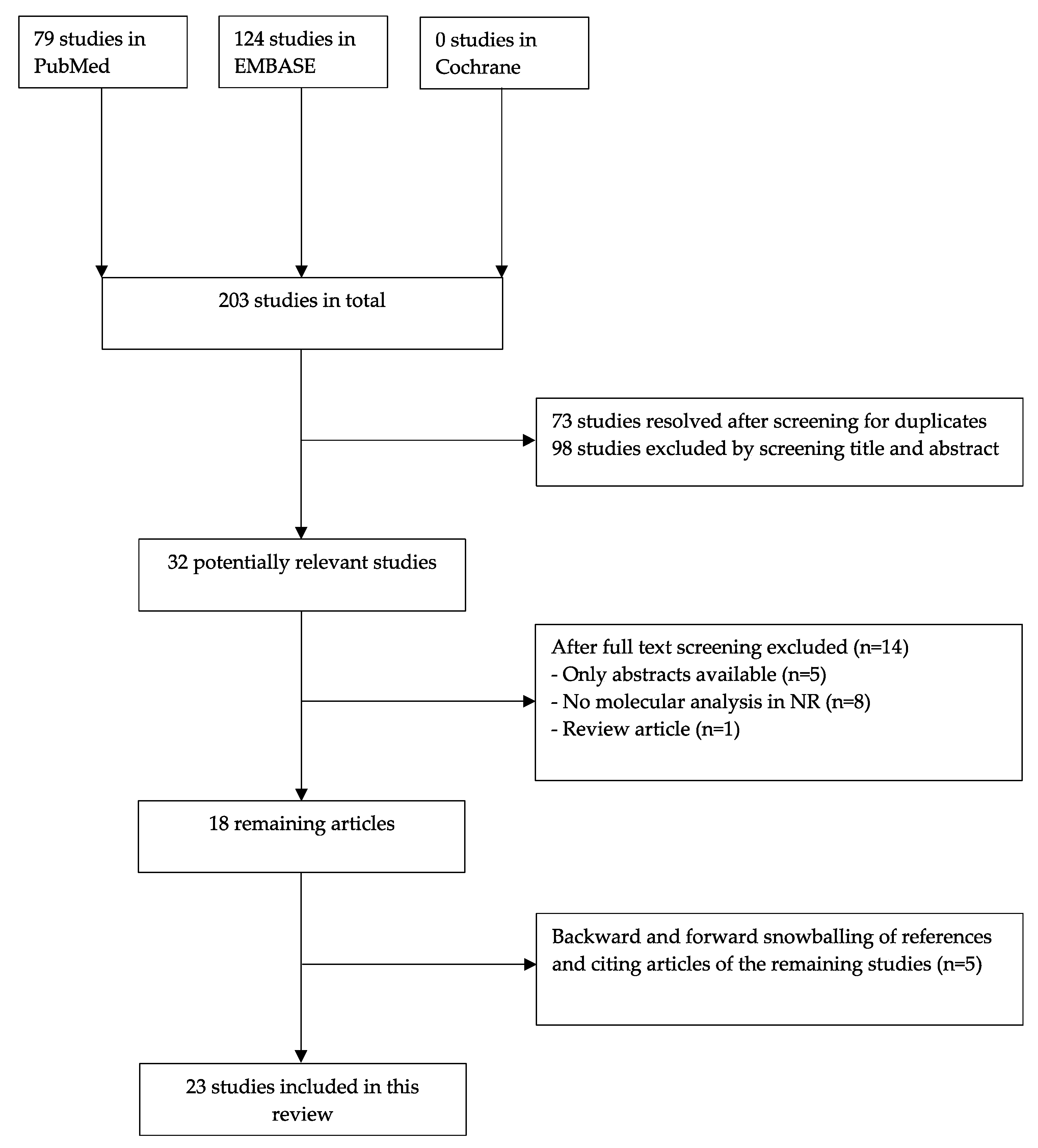
Our search in databases PubMed, EMBASE, and Cochrane generated a total of 203 articles. Seventy-three were duplicate articles that were found with more than one search engine and were therefore removed. The 130 remaining articles were screened for title and abstract after which 98 articles were excluded because of the in 2.1 mentioned criteria (Figure 2). We performed full-text screening in 32 potentially relevant studies, critically examining the in- and exclusion criteria. Consequently, 14 more articles were excluded, of which five were abstracts only and nine others were excluded since they did not meet the inclusion criteria. The remaining 18 articles were included in this systematic review. There were five additional articles identified by forward and backward snowballing, resulting in a total of 23 articles. In Figure 2, a flowchart of the search and selection process is presented based on the PRISMA scheme.

3.2. Quality Assessment

Quality assessment was completed for all 23 included articles. An overview of the quality assessment of 18 included articles, using the STARD checklist, is provided in Supplementary Table S3a (including the STARD Checklist in Table S2a). The remaining five case reports, using the CARE checklist, are presented in Supplementary Table S3b. All included articles could be classified as Oxford level 2 to 4, as shown in Table 1.

3.3. Characteristics of the Included Studies

Patient characteristics of the 23 included studies are presented in Table 1. In those studies, 221 NR were analyzed, of which 53 were ILNR, 136 were PLNR, 11 were nephroblastomatosis and 21 were not further specified. A total of 119 WT-NR pairs were analyzed with different methods including methylation analysis, sequencing analysis, LOH analysis, expression analysis and in situ hybridization. There were 28 WT-NR pairs reported with concomitant bilateral WT, whereas this was not explicitly reported in the other cases. Sex and age were described in only a few studies. In the eight studies describing sexual gender, there were 42 females and 30 males. The age at diagnosis for WT was variable ranging from 11 months to 144 months and for NR from 10 months to 192 months.

3.4. Chromosomal Changes

Table 2 presents the somatic changes that were found in NR, including the gene or chromosomal region that was involved and the detection method that was used. We identified seven studies in which LOH analysis was used. LOH of 11p15 was found in 13/64 investigated NR (20%), and the same loss was confirmed in the corresponding WT (n = 13) [31,38]. Three studies demonstrated LOH of 11p13, which revealed that this was present in 4/28 WT-NR pairs (14%) all representing ILNR. There was a single WT-PLNR pair that showed LOH of 11p13 in the WT only [35,38,40]. Hoban et al. described a case in which the LOH of chromosome 11 (both 11p13 and 11p15) was observed, where in fact all other maternal chromosomal loci were lost in the NR and WT in a Beckwith–Wiedemann patient [42]. Three studies involved LOH of 16q showing loss in 13/53 WTs (25%). In two studies, loss of 16q was not found in their associated NR [32,38], but Austruy et al. detected loss of 16q in one case of nephroblastomatosis [41]. LOH of 7p was investigated in two pairs of NR-WT, but LOH of 7p was only present in WT, not in the corresponding NR [32,37].
Furthermore, genome-wide comparative genome hybridization (CGH) was performed in two other studies. Vuononvirta et al. investigated 50 PLNR and divided them into three subgroups. The first group showed no copy number changes at all (44%). The second group contained eight cases in which whole chromosome changes were observed (16%). The remaining group, involving 20 cases, presented multiple partial chromosomal gains or losses (40%). In 76%, the NR contained part of the copy number changes seen in the corresponding WT [31]. Steenman et al. also performed whole genome analysis by CGH. This revealed losses of 1p, 7p, 4q and gains of 1q and 12q in nephroblastomatosis and their associated WT. Loss of 11p was found in nephroblastomatosis only. Changes only present in WT were loss of 9p and gain of 8, 10q and 18. Loss of 16q was detected in one case of WT and in one case of nephroblastomatosis adjacent to WT [40].
MdZin et al. found loss of chromosome 22 and showed that the frequency of chromosome 22 loss depended on NR morphology. Dormant, involuted and sclerosing PLNR presented monosomy 22 in 30%, whereas the hyperplastic and adenomatous PLNR showed monosomy 22 in 50%, increasing to a rate of 60–80% in WT [28].

3.5. Structural Aberrations in Candidate Gene Studies

While LOH analysis allows for detecting larger chromosomal regions without exactly identifying the responsible gene(s), candidate gene analysis by targeted sequencing might highlight abnormalities in putative oncogenes and tumor suppressor genes. Seven studies investigated single nucleotide changes in NR compared to WT. Four studies found mutations in the WT1 gene and showed that these can be present in both NR and WT [35,38,43,45]. Using Sanger sequencing analysis, Fukuzawa et al. showed that CTNNB1 mutations occurred only in WT (n = 6) and not in paired NR (n = 6) [35]. Another gene that may play a role in WT tumorigenesis, WTX, presented mutations in both NR (n = 1) and WT (n = 4) [29]. A KRAS gene mutation was found in one case of WT and adjacent NR in the setting of mosaicism [23,24]. In this same patient Slack et. al. [24] also described a somatic FBXW7 mutation in two WT but not in another WT nodule or the associated NR.
Two studies reported an absence of mutations of specific candidate genes. In the first study by Wegert et al., EGFR-internal tandem duplications (ITD) and BRAF-internal deletions (ID) were investigated, involving 208 WTs and 12 NR, but no changes in any of these genes were found [26]. In the second study, where PTEN was investigated, no abnormalities were found in the WT and NR included in the study by Grill et al. [30].

3.6. Epigenetic Studies

Epigenetic changes were described in ten studies, and six of them focused on DNA methylation changes. The different platforms used for methylation profiling in these studies included bisulfate sequencing [34], combined bisulfate and restriction analysis (COBRA) [32,33], pyrosequencing [31], and Illumina BeadChip microarray [25,27]. Charlton et al. [27], using the latter technique, performed a longitudinal study to find differences in DNA methylation to compare NR to NK as well as to WT. For this purpose, differentially methylated regions (DMR) were compared and shown to differ between WT, NR and NK [46]. Hypermethylation was seen in 55% of 629 differentially methylated regions (DMR) in NR as compared to NK [27]. When NR were compared to WT, in paired analysis, two subgroups of WT could be distinguished. In one group, NR and WT grossly harbored the same epigenetic profiles. Yet, the other group of WT showed hypervariability in the methylation profiles in comparison to NR, suggesting that there is a shift in methylation during the development from NK to NR and/or WT [27].
Vuononvirta et al. [31] used pyrosequencing to analyze H19. Hypermethylation of this gene is associated with LOI of IGF-2, which was found in 23/33 (70%) cases and hypermethylation of H19 in 37/40 (93%) of the PLNR cases. Brown et al. analyzed H19 in relation to LOI at 11p13 and 11p15. They found H19 hypermethylation in two WT-NR pairs using COBRA [32]. LOI at 11p13 and 11p15 was found in both WT-NR pairs. LOI of 11p13 leads to the decreased methylation of WT1 antisense regulatory region (ARR), which results in LOI of the noncoding antisense RNA WT1-AS and the alternate coding WT1 transcript AWT1. LOI of 11p15 is responsible for IGF-2 overexpression, another imprinted gene on chromosome 11 [32]. Hypomethylation of WT1 ARR in WT was also studied by Hancock et al. in two WT-NR pairs using bisulfate sequencing, revealing the lowest methylation levels in WT, the highest in NK, and NR showing methylation percentages between fetal kidney (FK) and NK. They also looked at the expression of AWT1 and WT1-AS, and found biallelic expression of AWT1 and WT1-AS in NR and WT, where monoallelic expression was found in the NK [34].
Cui et al. studied RNA expression of the H19 gene in association with IGF-2 expression, using in situ hybridization (ISH) in WT, NR and associated renal medulla. H19 was not expressed in WT and NR, but was present in normal renal medulla [39]. Yun et al. also investigated IGF-2 by in situ hybridization and Northern blotting in NR and WT. Both studies displayed comparable patterns in NR and WT, but there was a variable and heterogeneous level of expression. IGF-2 expression was frequently associated with blastema [39,44]. LOI of IGF-2 was studied by Ravenel et al. and was present in a WT–NR pair with two PLNR [36].
Coorens et al. used Illumina BeadChip microarray to demonstrate the presence of H19 hypermethylation in NK with clonal expansions (58%), while this hypermethylation was not found in NK without clones. One WT–NR pair was included, in which the WT and NR emerged from a similar ancestral clone at different time points, which is indicative of an association between clones, NR and WT all showing H19 hypermethylation [25].
Finally, Chilukamarri et al. investigated the GLIPR1/RTVP-1 gene. Hypomethylation of this gene was shown in 21 out of 24 WTs. There were two associated NR analyzed, which also showed hypomethylation of this gene [33].

4. Discussion

We have presented an overview of all molecular studies on nephrogenic rests between 1990 and 2022. As a result of this long-time frame, a wide range of techniques were used to examine chromosomal regions, copy number variations, individual genes and epigenetic changes in NR. A total of 23 studies were found showing loss of chromosomal arms 11p13 and 11p15, 1p, 4q and 11p, and gains in 1q, 7q and 12q, as well as mutations in WT1, WTX and KRAS to occur in both NR and WT, suggesting these are early events (Table 3). Mutations in CTNNB1 and FBXW7 and LOH of 16q and 7p are only present in WT, but not the associated NR, therefore likely representing late events (Table 3). Furthermore, differential methylation levels display a relationship between WT, NR, and NK.
Summary of early molecular events that occur in both NR and WT and late molecular events that occur in WT only. Late events are therefore likely not involved in the progression from NR to WT.
Little is known about the molecular pathogenesis of WT, including the mechanisms that affect transition from NR to WT, although NR have been recognized as precursor lesions [3]. Studying molecular changes in pairs of NR and WT might shed light on the timing of such events. If the same alteration occurs in NR and the associated WT, this can be considered as an early event. Indeed, LOI and LOH at 11p13 and 11p15 were found in both NR and WT, representing the chromosomal regions where the WT1 and the IGF2 genes are located, respectively [31,32,35,38,40]. No such gene correlations are known for the other chromosomal arms that were recurrently lost (1p, 4q, 7p and 11p), or gained (1q, 7q and 12q) [40]. MdZin et al. found an increasing frequency of monosomy of chromosome 22 from sclerotic/dormant PLNR to hyperplastic/adenomatous PLNR and then finally toward WT, suggesting that loss of chromosome 22 is an early event and that tumor suppressor genes on this chromosome might be involved in WT tumorigenesis [28]. Mutations in WT1 [35,38,43,45] and WTX [29] can also be considered early events, as they were found in both NR and WT. WT1 was first investigated in WT in 1991 [45,47], and WTX was first described in association with WT in 2007 (Figure 3) [48].
Genetic changes that are found in WT only, and not in NR, are likely to be late events and therefore not involved in the transition from NR to WT. For instance, LOH of 16q appears to be a late event [32,38,40,41]. However, in one case, loss of 16q was also found in the associated nephroblastomatosis [41]. Loss of 7p appears to occur later in development as well [32,37], although Steenman et al. found LOH of 7p in a case of nephroblastomatosis [40]. Furthermore, CTNNB1 mutations, which were first described in WT in 1999 (Figure 3), are considered late events in Wilms tumorigenesis [29,35]. No PTEN, EGFR and BRAF mutations or rearrangements were found in both WT and NR, implying that these genes do not seem to contribute to the transition of NR to WT [26,30].
It should be noted that, due to the long time interval, there was large variation between studies with regard to the number and type of polymorphic markers used for LOH or LOI analyses, which may have had an effect on the detection of molecular abnormalities. Likewise, for the candidate gene studies, not all genes have been investigated completely or information on the extent of screening is lacking. In one study, all exons of CTNNB1 were sequenced [35], while Park et al. [43] examined exons 2–10 for detecting mutations in WT1. In studies describing WT1, two studies used primers for only two exons to detect mutations in WT1, not mentioning if all other exons of WT1 were sequenced [35,38]. In one other study that examined both CTNNB1 and WTX, it was not reported which exons were sequenced [29]. Furthermore, many of the genes that have been shown to be involved in WT tumorigenesis, such as SIX1, SIX2, and DROSHA, have not been systematically analyzed in NR [49]. Thus, comprehensive analyses should be performed using WES or WGS to examine all genes and to prevent important genes from not being detected.
Regarding epigenetic changes, there appears to be a correlation between the methylation and expression patterns of NR as compared to WT. The similar or increased methylation of WT and NR with respect to NK suggests that this might play a role in tumorigenesis [27]. There was increased methylation of H19 in WT and NR, and also in the clonal expansions of NK, which suggests a transformation from NK to WT in the levels of methylation of H19. It is remarkable that the WT–NR pair included in this study arose from the same clone and that they are therefore phylogenetically related [25]. Together this may indicate that the clonal beds of NK are “primed” to become a WT. LOI at 11p15 and 11p13 and LOI of IGF-2 were detected in WT and NR, and thus seem to be early events [32,36]. The presence of LOI of 11p13 correlates with the decreased methylation of WT1 ARR, suggesting that imprinting defects at 11p13 may be involved in tumorigenesis [32,34]. H19 and IGF-2 are two parentally imprinted genes on 11p15, with opposite regulatory mechanisms: if H19 is silenced, IGF-2 on the other hand is upregulated [50,51]. This explains why there was expression of IGF-2 in both NR and WT, while H19 expression was not detected [39]. Together with ISH studies showing variable IGF-2 expression patterns in NR and WT, these data suggest a role for IGF-2 as an early driver of WT development [39,44]. Likewise, hypomethylation of GLIPR1/RTVP1 was found in WT and NR, suggesting that this specific change might also contribute to WT development [33].
As previously described, NR are present in approximately 40% of unilateral WT and in almost all cases of bilateral WTs [14]. Nephroblastomatosis is more frequently present in bilateral WT [52]. This suggests that not every WT might arise from NR. Conversely, not every NR develops into a WT either. WT and NR both develop from the same embryonic tissue, and both morphologically reflect embryonal renal tissue. Normally, nephrogenesis stops at 34 weeks of gestation and any remaining nephrogenic tissue is considered NR. However, little is known about the underlying factors that lead to such persistence. ILNR are frequent in tumors with stromal histology that are typically associated with aberrations in WT1 on chromosome locus 11p13. PLNR occur more often in blastemal and/or epithelial type WT and are associated with alterations in IGF-2 on 11p15 [18]. These associations have been confirmed in several included studies where a distinction was made between PLNR and ILNR. WT1 mutations and LOH of 11p13 are found in ILNR [35,38,43], and LOI of IGF-2 in PLNR [36]. However, LOH of 11p15 can also be found in ILNR [38]. Interestingly, there is a known association between WT1 and CTNNB1 mutations in WT [53,54]. However, it is notable that CTNNB1 mutations occur as late events and are only present in WT [35], while WT1 mutations can occur in NR and WT [35,38,43,45]. Even though NR are precursors of WT, there are only few studies which focus on and describe the molecular pathogenesis of NR. In this review, all studies between 1990 and 2020 which analyzed molecular changes in NR and used molecular techniques were included, resulting in only 23 relevant articles, i.e., less than 1 per year over the investigated time frame. We chose 1990 as a starting point (Figure 3), as it coincides with the first histopathologic description of NR and with the wider applicability of molecular methods in biomedical research, but even until 2000 few molecular studies were performed. In addition, most techniques required frozen lesional tissue, while frequently only formalin-fixed paraffin embedded (FFPE) material was available, limiting the possibilities of molecular analyses, at least in previous decades. Finally, it is virtually impossible to distinguish NR macroscopically, apart from patients with nephroblastomatosis, requiring expert microscopic detection on FFPE material with its inherent limitations.
Throughout this review, we have applied the histological definition of NR and nephroblastomatosis over the radiological definition, which is not 100% superimposable with the histological term of NR. The supplied histological material was used as a reference point. The diagnosis of NR and WT can only be made after histological confirmation. On CT imaging, NR and nephroblastomatosis may show distinct features from WT, mainly with regard to the shape and size of the lesion [13]. However, up to 25% of the NR and WT are still misdiagnosed, with half of nodules radiologically diagnosed as WT, turning out be NR after histological confirmation [12]. It is outside the scope of this review to further discuss the potential a newer imaging technology, such as diffusion-weighted imaging (DWI) MRI for the above distinction.
As presented in Table 1, the number of WT and NR examined varied greatly from study to study. Some genes have been studied on less than five tumors. Therefore, firm conclusions cannot be drawn from these studies. To do so in the future, larger-scale studies of WT and corresponding NR would be needed to assign certain genes a significant role in the development of NR to WT. In addition, mutations such as DROSHA and SIX1 and SIX2, which are known driver mutations in WT, have not been examined in NR. These genes, as can be seen in Figure 3, have been discovered from 2010 onwards. Many studies described in this review were completed at a time when these candidate genes were not yet identified and have therefore not been investigated in NR.
Until 2000, mRNA in situ hybridization (ISH), sequencing analysis, LOH analysis and CGH analysis were used to analyze NR (Figure 3). From 2006 methylation studies emerged in which the difference in methylation patterns between NR and WT was investigated [25,27,31,32,33,34]. Several methods that were used in studies presented in this review may currently be considered outdated with the advent of new molecular methods, including whole exome and whole genome sequencing, RNA sequencing and also spatial transcriptomics, which allows tissue context while analyzing gene expression profiles. These techniques may allow faster progress and seem particularly relevant for NR analysis, which still relies on morphological identification. It is important to consider tests for the early detection of genetic, epigenetic and somatic changes in NR that may become clinically relevant to the patient before NR develop into a WT. At the moment, not all genetic predispositions have been explored. Therefore, all children with WT or nephroblastomatosis should be referred to a clinical geneticist and advised to undergo whole exome or whole genome sequencing, as there is a high risk (up to 33%) of genetic predisposition, as was recently shown in a Dutch cohort [55]. This certainly applies to children with bilateral WT, in whom an 80% chance of genetic predisposition has recently been revealed [56].

5. Conclusions

In conclusion, over the past 30 years, several studies have looked at NR with or without associated WT. As a result, much has become known about the genetic changes in NR. LOH of 11p13 and 11p15, expression of IGF-2 and mutations in WT1 and WTX appear to play a role in the early tumorigenesis of WT. LOH of 16q and 7p and mutations in CTNNB1 seem to occur later in development. Methylation patterns of NR in comparison to WT appear to be similar. Due to rapid advances in (genome-wide) molecular techniques, the increased possibilities to use FFPE material and the availability of histologically confirmed frozen material, genetic changes in NR and corresponding WT may be investigated in larger series and might unravel early steps in the progression of WT tumorigenesis.

Supplementary Materials

The following supporting information can be downloaded at: https://www.mdpi.com/article/10.3390/cancers15051363/s1, Table S1: Search strategy PubMed and EMBASE, Table S2a: “Standards for Reporting Diagnostic Accuracy 2015” (STARD 2015) checklist, Table S2b: Case report guidelines (CARE checklist 2013), Table S3a: Quality assessment of 18 out of 23 included articles based on the STARD 2015 checklist, Table S3b: Quality assessment of 5 out of 23 included articles based on the CARE 2013 checklist.

Author Contributions

Conceptualization, T.B., J.D., A.M.C.M.-G. and R.R.d.K.; methodology, T.B., J.D., A.M.C.M.-G. and R.R.d.K.; writing—original draft preparation, T.B., A.M.C.M.-G. and R.R.d.K.; writing—review and editing, T.B., J.D., A.M.C.M.-G., M.M.v.d.H.-E. and R.R.d.K. All authors have read and agreed to the published version of the manuscript.

Funding

This research received no external funding.

Institutional Review Board Statement

Not applicable.

Informed Consent Statement

Not applicable.

Data Availability Statement

No new data were created in this study. Data sharing is not applicable to this article.

Conflicts of Interest

The authors declare no conflict of interest.

References

  1. Hennigar, R.A.; O’Shea, P.A.; Grattan-Smith, J.D. Clinicopathologic Features of Nephrogenic Rests and Nephroblastomatosis. Adv. Anat. Pathol. 2001, 8, 276–289. [Google Scholar] [CrossRef]
  2. Popov, S.D.; Sebire, N.J.; Vujanic, G.M. Wilms’ Tumour—Histology and Differential Diagnosis. In Wilms Tumor; Codon Publications: Singapore, 2016. [Google Scholar]
  3. Beckwith, J.B.; Kiviat, N.B.; Bonadio, J.F. Nephrogenic Rests, Nephroblastomatosis, and the Pathogenesis of Wilms’ Tumor. Pediatr. Pathol. 1990, 10, 1–36. [Google Scholar] [CrossRef]
  4. Lee Tsün, H.; Holman, R.L. Bilateral Nephroblastomatosis in a Premature Infant. J. Pathol. Bacteriol. 1961, 82, 249–255. [Google Scholar] [CrossRef]
  5. Beckwith, J.B. Nephrogenic Rests and the Pathogenesis of Wilms Tumor: Developmental and Clinical Considerations. Am. J. Med. Genet. 1998, 79, 268–273. [Google Scholar] [CrossRef]
  6. Fukuzawa, R.; Reeve, A.E. Molecular Pathology and Epidemiology of Nephrogenic Rests and Wilms Tumors. J. Pediatr. Hematol. Oncol. 2007, 29, 589–594. [Google Scholar] [CrossRef] [PubMed]
  7. Caiulo, V.A.; Latini, G.; Cataldi, L.; De Felice, C. Nephrogenic Rests. J. Pediatr. Hematol. Oncol. 2007, 29, 361–363. [Google Scholar] [CrossRef]
  8. Fukuzawa, R.; Breslow, N.E.; Morison, I.M.; Dwyer, P.; Kusafuka, T.; Kobayashi, Y.; Becroft, D.M.; Beckwith, J.B.; Perlman, E.J.; Reeve, A.E. Epigenetic Differences between Wilms’ Tumours in White and East-Asian Children. Lancet 2004, 363, 446–451. [Google Scholar] [CrossRef]
  9. Bozlu, G.; Cıtak, E.C. Evaluation of Renal Tumors in Children. Türk. Urol. Derg. Turk. J. Urol. 2018, 44, 268. [Google Scholar] [CrossRef]
  10. Vujanić, G.M.; Gessler, M.; Ooms, A.H.A.G.; Collini, P.; Coulomb-l’Hermine, A.; D’Hooghe, E.; de Krijger, R.R.; Perotti, D.; Pritchard-Jones, K.; Vokuhl, C.; et al. The UMBRELLA SIOP–RTSG 2016 Wilms Tumour Pathology and Molecular Biology Protocol. Nat. Rev. Urol. 2018, 15, 693–701. [Google Scholar] [CrossRef] [Green Version]
  11. SIOP-RTSG. Umbrella Protocol 2016 Version 1.8; SIOP-RTSG: Saarbrucken, Germany, 2017. [Google Scholar]
  12. Dzhuma, K.; Ducou Le Pointe, H.; Coulomb, A.; Tabone, M.; Bergeron, C.; Audry, G.; Irtan, S. Wilms Tumors and Their Precursors: Radiological Diagnosis versus Histology. Pediatr. Blood Cancer 2020, 67, e2841. [Google Scholar] [CrossRef]
  13. Sandberg, J.K.; Chi, Y.-Y.; Smith, E.A.; Servaes, S.; Hoffer, F.A.; Mullen, E.A.; Perlman, E.J.; Tornwall, B.; Ehrlich, P.F.; Geller, J.I.; et al. Imaging Characteristics of Nephrogenic Rests Versus Small Wilms Tumors: A Report From the Children’s Oncology Group Study AREN03B2. Am. J. Roentgenol. 2020, 214, 987–994. [Google Scholar] [CrossRef]
  14. Vujanić, G.M.; Apps, J.R.; Moroz, V.; Ceroni, F.; Williams, R.D.; Sebire, N.J.; Pritchard-Jones, K. Nephrogenic Rests in Wilms Tumors Treated with Preoperative Chemotherapy: The UK SIOP Wilms Tumor 2001 Trial Experience. Pediatr. Blood Cancer 2017, 64, e26547. [Google Scholar] [CrossRef]
  15. Liu, E.K.; Suson, K.D. Syndromic Wilms Tumor: A Review of Predisposing Conditions, Surveillance and Treatment. Transl. Androl. Urol. 2020, 9, 2370–2381. [Google Scholar] [CrossRef]
  16. Kalapurakal, J.A.; Dome, J.S.; Perlman, E.J.; Malogolowkin, M.; Haase, G.M.; Grundy, P.; Coppes, M.J. Management of Wilms’ Tumour: Current Practice and Future Goals. Lancet Oncol. 2004, 5, 37–46. [Google Scholar] [CrossRef] [PubMed]
  17. Al-Hussain, T.; Ali, A.; Akhtar, M. Wilms Tumor. Adv. Anat. Pathol. 2014, 21, 166–173. [Google Scholar] [CrossRef]
  18. Treger, T.D.; Chowdhury, T.; Pritchard-Jones, K.; Behjati, S. The Genetic Changes of Wilms Tumour. Nat. Rev. Nephrol. 2019, 15, 240–251. [Google Scholar] [CrossRef] [PubMed]
  19. Moher, D.; Liberati, A.; Tetzlaff, J.; Altman, D.G. Preferred Reporting Items for Systematic Reviews and Meta-Analyses: The PRISMA Statement. PLoS Med. 2009, 6, e1000097. [Google Scholar] [CrossRef] [Green Version]
  20. Bossuyt, P.M.; Reitsma, J.B.; Bruns, D.E.; Gatsonis, C.A.; Glasziou, P.P.; Irwig, L.; Lijmer, J.G.; Moher, D.; Rennie, D.; de Vet, H.C.W.; et al. STARD 2015: An Updated List of Essential Items for Reporting Diagnostic Accuracy Studies. Clin. Chem. 2015, 61, 1446–1452. [Google Scholar] [CrossRef] [PubMed] [Green Version]
  21. Riley, D.S.; Barber, M.S.; Kienle, G.S.; Aronson, J.K.; von Schoen-Angerer, T.; Tugwell, P.; Kiene, H.; Helfand, M.; Altman, D.G.; Sox, H.; et al. CARE Guidelines for Case Reports: Explanation and Elaboration Document. J. Clin. Epidemiol. 2017, 89, 218–235. [Google Scholar] [CrossRef]
  22. Howick, J.; Chalmers, I.G.P.; Glasziou, P.; Greenhalgh, T.; Heneghan, C.; Liberati, A.; Moschetti, I.; Phillips, B.; Thornton, H.; Goddard, O.; et al. The Oxford Levels of Evidence 2; Oxford Centar Evidence-Based Medicine: Oxford, UK, 2011. [Google Scholar]
  23. Chang, C.A.; Perrier, R.; Kurek, K.C.; Estrada-Veras, J.; Lehman, A.; Yip, S.; Hendson, G.; Diamond, C.; Pinchot, J.W.; Tran, J.M.; et al. Novel Findings and Expansion of Phenotype in a Mosaic RASopathy Caused by Somatic KRAS Variants. Am. J. Med. Genet. Part A 2021, 185, 2829–2845. [Google Scholar] [CrossRef]
  24. Slack, J.C.; Bründler, M.-A.; Chang, C.A.; Perrier, R.; Lafay-Cousin, L.; Kurek, K.C. Bilateral Nephroblastic Tumors and a Complex Renal Vascular Anomaly in a Patient With a Mosaic RASopathy: Novel Histopathologic Features and Molecular Insights. Pediatr. Dev. Pathol. 2021, 24, 235–240. [Google Scholar] [CrossRef] [PubMed]
  25. Coorens, T.H.H.; Treger, T.D.; Al-Saadi, R.; Moore, L.; Tran, M.G.B.; Mitchell, T.J.; Tugnait, S.; Thevanesan, C.; Young, M.D.; Oliver, T.R.W.; et al. Embryonal Precursors of Wilms Tumor. Science 2019, 366, 1247–1251. [Google Scholar] [CrossRef] [PubMed]
  26. Wegert, J.; Vokuhl, C.; Collord, G.; Del Castillo Velasco-Herrera, M.; Farndon, S.J.; Guzzo, C.; Jorgensen, M.; Anderson, J.; Slater, O.; Duncan, C.; et al. Recurrent Intragenic Rearrangements of EGFR and BRAF in Soft Tissue Tumors of Infants. Nat. Commun. 2018, 9, 2378. [Google Scholar] [CrossRef] [PubMed] [Green Version]
  27. Charlton, J.; Williams, R.D.; Sebire, N.J.; Popov, S.; Vujanic, G.; Chagtai, T.; Alcaide-German, M.; Morris, T.; Butcher, L.M.; Guilhamon, P.; et al. Comparative Methylome Analysis Identifies New Tumour Subtypes and Biomarkers for Transformation of Nephrogenic Rests into Wilms Tumour. Genome Med. 2015, 7, 11. [Google Scholar] [CrossRef] [Green Version]
  28. MdZIN, R.; Phillips, M.; Edwards, C.; Murch, A.; Charles, A. Perilobar Nephrogenic Rests and Chromosome 22. Pediatr. Dev. Pathol. 2011, 14, 485–492. [Google Scholar] [CrossRef] [PubMed]
  29. Fukuzawa, R.; Holman, S.K.; Chow, C.W.; Savarirayan, R.; Reeve, A.E.; Robertson, S.P. WTX Mutations Can Occur Both Early and Late in the Pathogenesis of Wilms Tumour. J. Med. Genet. 2010, 47, 791–794. [Google Scholar] [CrossRef]
  30. Grill, C.; Guelly, C.; Ebner, B.; Leuschner, I.; Hauser-Kronberger, C.; Hoefler, G.; Guertl, B. Loss of PTEN/MMAC1 Activity Is a Rare and Late Event in the Pathogenesis of Nephroblastomas. Hum. Pathol. 2010, 41, 1172–1177. [Google Scholar] [CrossRef]
  31. Vuononvirta, R.; Sebire, N.J.; Dallosso, A.R.; Reis-Filho, J.S.; Williams, R.D.; Mackay, A.; Fenwick, K.; Grigoriadis, A.; Ashworth, A.; Pritchard-Jones, K.; et al. Perilobar Nephrogenic Rests Are Nonobligate Molecular Genetic Precursor Lesions of Insulin-Like Growth Factor-II-Associated Wilms Tumors. Clin. Cancer Res. 2008, 14, 7635–7644. [Google Scholar] [CrossRef] [Green Version]
  32. Brown, K.W.; Power, F.; Moore, B.; Charles, A.K.; Malik, K.T.A. Frequency and Timing of Loss of Imprinting at 11p13 and 11p15 in Wilms’ Tumor Development. Mol. Cancer Res. 2008, 6, 1114–1123. [Google Scholar] [CrossRef] [Green Version]
  33. Chilukamarri, L.; Hancock, A.L.; Malik, S.; Zabkiewicz, J.; Baker, J.A.; Greenhough, A.; Dallosso, A.R.; Huang, T.H.-M.; Royer-Pokora, B.; Brown, K.W.; et al. Hypomethylation and Aberrant Expression of the Glioma Pathogenesis-Related 1 Gene in Wilms Tumors. Neoplasia 2007, 9, 970–978. [Google Scholar] [CrossRef] [Green Version]
  34. Hancock, A.L.; Brown, K.W.; Moorwood, K.; Moon, H.; Holmgren, C.; Mardikar, S.H.; Dallosso, A.R.; Klenova, E.; Loukinov, D.; Ohlsson, R.; et al. A CTCF-Binding Silencer Regulates the Imprinted Genes AWT1 and WT1-AS and Exhibits Sequential Epigenetic Defects during Wilms’ Tumourigenesis. Hum. Mol. Genet. 2007, 16, 343–354. [Google Scholar] [CrossRef] [PubMed] [Green Version]
  35. Fukuzawa, R.; Heathcott, R.W.; More, H.E.; Reeve, A.E. Sequential WT1 and CTNNB1 Mutations and Alterations of -Catenin Localisation in Intralobar Nephrogenic Rests and Associated Wilms Tumours: Two Case Studies. J. Clin. Pathol. 2006, 60, 1013–1016. [Google Scholar] [CrossRef] [Green Version]
  36. Ravenel, J.D.; Broman, K.W.; Perlman, E.J.; Niemitz, E.L.; Jayawardena, T.M.; Bell, D.W.; Haber, D.A.; Uejima, H.; Feinberg, A.P. Loss of Imprinting of Insulin-Like Growth Factor-II (IGF2) Gene in Distinguishing Specific Biologic Subtypes of Wilms Tumor. J. Natl. Cancer Inst. 2001, 93, 1698–1703. [Google Scholar] [CrossRef] [PubMed] [Green Version]
  37. Powlesland, R.M.; Charles, A.K.; Malik, K.T.A.; Reynolds, P.A.; Pires, S.; Boavida, M.; Brown, K.W. Loss of Heterozygosity at 7p in Wilms’ Tumour Development. Br. J. Cancer 2000, 82, 323–329. [Google Scholar] [CrossRef] [Green Version]
  38. Charles, A.K.; Brown, K.W.; Berry, P.J. Microdissecting the Genetic Events in Nephrogenic Rests and Wilms’ Tumor Development. Am. J. Pathol. 1998, 153, 991–1000. [Google Scholar] [CrossRef]
  39. Cui, H.; Hedborg, F.; He, L.; Nordenskjöld, A.; Sandstedt, B.; Pfeifer-Ohlsson, S.; Ohlsson, R. Inactivation of H19, an Imprinted and Putative Tumor Repressor Gene, Is a Preneoplastic Event during Wilms’ Tumorigenesis. Cancer Res. 1997, 57, 4469–4473. [Google Scholar]
  40. Steenman, M.; Redeker, B.; De Meulemeester, M.; Wiesmeijer, K.; Voute, P.A.; Westerveld, A.; Slater, R.; Mannens, M. Comparative Genomic Hybridization Analysis of Wilms Tumors. Cytogenet. Genome Res. 1997, 77, 296–303. [Google Scholar] [CrossRef] [PubMed]
  41. Austruy, E.; Candon, S.; Henry, I.; Gyapay, G.; Tournade, M.-F.; Mannens, M.; Callen, D.; Junien, C.; Jeanpierre, C. Characterization of Regions of Chromosomes 12 and 16 Involved in Nephroblastoma Tumorigenesis. Genes Chromosom. Cancer 1995, 14, 285–294. [Google Scholar] [CrossRef]
  42. Hoban, P.; Heighway, J.; White, G.M.; Baker, B.; Gardner, J.; Birch, J.; Morris-Jones, P.; Kelsey, A. Genome-Wide Loss of Maternal Alleles in a Nephrogenic Rest and Wilms’ Tumour from a BWS Patient. Hum. Genet. 1995, 95, 651–656. [Google Scholar] [CrossRef]
  43. Park, S.; Bernard, A.; Bove, K.E.; Sens, D.A.; Hazen-Martin, D.J.; Garvin, A.J.; Haber, D.A. Inactivation of WT1 in Nephrogenic Rests, Genetic Precursors to Wilms’ Tumour. Nat. Genet. 1993, 5, 363–367. [Google Scholar] [CrossRef]
  44. Yun, K.; Molenaar, A.J.; Fiedler, A.M.; Mark, A.J.; Eccles, M.R.; Becroft, D.M.; Reeve, A.E. Insulin-like Growth Factor II Messenger Ribonucleic Acid Expression in Wilms Tumor, Nephrogenic Rest, and Kidney. Lab. Invest. 1993, 69, 603–615. [Google Scholar]
  45. Pritchard-Jones, K.; Fleming, S. Cell Types Expressing the Wilms’ Tumour Gene (WT1) in Wilms’ Tumours: Implications for Tumour Histogenesis. Oncogene 1991, 6, 2211–2220. [Google Scholar] [PubMed]
  46. Zhang, Y.; Liu, H.; Lv, J.; Xiao, X.; Zhu, J.; Liu, X.; Su, J.; Li, X.; Wu, Q.; Wang, F.; et al. QDMR: A Quantitative Method for Identification of Differentially Methylated Regions by Entropy. Nucleic Acids Res. 2011, 39, e58. [Google Scholar] [CrossRef] [PubMed] [Green Version]
  47. Cowell, J.K.; Wadey, R.B.; Haber, D.A.; Call, K.M.; Housman, D.E.; Pritchard, J. Structural Rearrangements of the WT1 Gene in Wilms’ Tumour Cells. Oncogene 1991, 6, 595–599. [Google Scholar]
  48. Rivera, M.N.; Kim, W.J.; Wells, J.; Driscoll, D.R.; Brannigan, B.W.; Han, M.; Kim, J.C.; Feinberg, A.P.; Gerald, W.L.; Vargas, S.O.; et al. An X Chromosome Gene, WTX, Is Commonly Inactivated in Wilms Tumor. Science 2007, 315, 642–645. [Google Scholar] [CrossRef] [PubMed] [Green Version]
  49. Gadd, S.; Huff, V.; Walz, A.L.; Ooms, A.H.A.G.; Armstrong, A.E.; Gerhard, D.S.; Smith, M.A.; Auvil, J.M.G.; Meerzaman, D.; Chen, Q.-R.; et al. A Children’s Oncology Group and TARGET Initiative Exploring the Genetic Landscape of Wilms Tumor. Nat. Genet. 2017, 49, 1487–1494. [Google Scholar] [CrossRef] [PubMed] [Green Version]
  50. Chung, W. Chromosome 11p15.5 Regional Imprinting: Comparative Analysis of KIP2 and H19 in Human Tissues and Wilms’ Tumors. Hum. Mol. Genet. 1996, 5, 1101–1108. [Google Scholar] [CrossRef] [Green Version]
  51. Bergman, D.; Halje, M.; Nordin, M.; Engström, W. Insulin-Like Growth Factor 2 in Development and Disease: A Mini-Review. Gerontology 2013, 59, 240–249. [Google Scholar] [CrossRef]
  52. Charlton, J.; Irtan, S.; Bergeron, C.; Pritchard-Jones, K. Bilateral Wilms Tumour: A Review of Clinical and Molecular Features. Expert Rev. Mol. Med. 2017, 19, e8. [Google Scholar] [CrossRef]
  53. Royer-Pokora, B.; Weirich, A.; Schumacher, V.; Uschkereit, C.; Beier, M.; Leuschner, I.; Graf, N.; Autschbach, F.; Schneider, D.; von Harrach, M. Clinical Relevance of Mutations in the Wilms Tumor Suppressor 1 Gene WT1 and the Cadherin-Associated Protein Β1 Gene CTNNB1 for Patients with Wilms Tumors. Cancer 2008, 113, 1080–1089. [Google Scholar] [CrossRef]
  54. Duhme, C.; Busch, M.; Heine, E.; de Torres, C.; Mora, J.; Royer-Pokora, B. WT1-Mutant Wilms Tumor Progression Is Associated With Diverting Clonal Mutations of CTNNB1. J. Pediatr. Hematol. Oncol. 2021, 43, e180–e183. [Google Scholar] [CrossRef] [PubMed]
  55. Hol, J.A.; Kuiper, R.P.; van Dijk, F.; Waanders, E.; van Peer, S.E.; Koudijs, M.J.; Bladergroen, R.; van Reijmersdal, S.V.; Morgado, L.M.; Bliek, J.; et al. Prevalence of (Epi)Genetic Predisposing Factors in a 5-Year Unselected National Wilms Tumor Cohort: A Comprehensive Clinical and Genomic Characterization. J. Clin. Oncol. 2022, 40, 1892–1902. [Google Scholar] [CrossRef] [PubMed]
  56. van Peer, S.E.; Hol, J.A.; van der Steeg, A.F.W.; van Grotel, M.; Tytgat, G.A.M.; Mavinkurve-Groothuis, A.M.C.; Janssens, G.O.R.; Littooij, A.S.; de Krijger, R.R.; Jongmans, M.C.J.; et al. Bilateral Renal Tumors in Children: The First 5 Years’ Experience of National Centralization in The Netherlands and a Narrative Review of the Literature. J. Clin. Med. 2021, 10, 5558. [Google Scholar] [CrossRef] [PubMed]
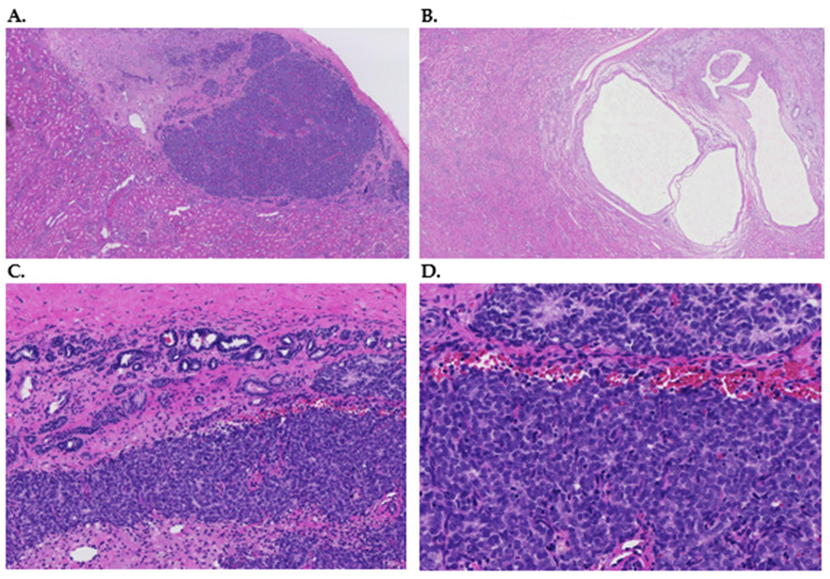
Figure 1. (A) Hematoxylin and eosin-stained slide with an overview of a hyperplastic perilobar nephrogenic rest, composed of epithelial structures without cytonuclear atypia of the composing cells. Magnification 2×. (B) Hematoxylin and eosin-stained slide with overview of an intralobar nephrogenic rest, composed of cystic structures with some intervening stroma. Both types of rests are surrounded by normal renal parenchyma. Magnification 2×. (C) Hematoxylin and eosin-stained section of a 9 mm lesion of the left kidney of 2-year-old male patient for which nephron-sparing surgery was performed. The patient underwent pre-operative chemotherapy according to the UMBRELLA protocol and also had a lesion of the contralateral kidney that was simultaneously removed. The image shows fibrous tissue of the kidney capsule at the top. Below there are glandular structures compatible with epithelial elements of a perilobar nephrogenic rest, as well as a blastemal component with limited nuclear atypia. Magnification 10×. (D) Hematoxylin and eosin-stained section of the same lesion as (C). This lesion was eventually classified as compatible with a perilobar nephrogenic rest after the careful review of multiple expert pathologists. Please note the bland nuclear morphology of the epithelium and blastema in this close-up image. Magnification 20×.
Cancers 15 01363 g001
Figure 2. Flowchart of the search and selection process.
Cancers 15 01363 g002
Figure 3. History of identified aberrations in WT and introduction of molecular methods. The above timeline shows selected WT-associated genes and there first description in relation to WT. The lower part indicates the year of the first description of the selected molecular methods. LOH, loss of heterozygosity; CGH, comparative genomic hybridization; RNA, ribonucleic acid; WES, whole exome sequencing; WGS, whole genome sequencing.
Cancers 15 01363 g003
Table 1. Characteristics of reviewed NR cases.
StudyOxford LevelNR (n)NR TypeWT (n)WT TypeWT-NR PairsBilateral WTSexAge at
Diagnosis
Chang et al., 2021 * [23]4Multiple
1
PLNR
NBL
1
-
epithelial type
-
1
-
Yes
-
M
F
21 months
36 months
Slack et al., 2021 [24]4MultiplePLNR1epithelial type1YesM21 months
Coorens et al., 2019 [25]41ND23ND1No NDND
Wegert et al., 2018 [26]212ND208NDNDNDNDND
Charlton et al., 2015 [27]22217 PLNR, 5 ILNR36ND20NDNDND
MdZin et al., 2011 [28]49
1
9 PLNR
1 PLNR
2
1
blastemal type
mixed type
1
1
Yes
No
M
M
42 months
48 months
Fukuzawa et al., 2010 [29]444 ILNR43 stromal type, 1 blastemal type4ND3F, 1MND
Grill et al., 2010 [30]22618 PLNR, 8 ILNR2214 mixed type, 2 stromal type, 4 blastemal type and 1 regressive type, 1 not known22NDNDND
Vuononvirta et al., 2008 [31]25050 PLNR25ND2517/50 NRNDND
Brown et al., 2008 ** [32]4MultiplePLNR51ND22; 2 WT-NR pairsNDND
Chilukamarri et al., 2007 [33]42ND245 stromal type, 1 epithelial type, 4 blastemal type, 8 mixed type, 6 not known 25; 2 WT-NR pairs
8M, 16F (WT-NR pairs 2F)WT 36 ^ months
Hancock et al., 2007 [34]2MultipleND2mixed type22 WT-NR pairsNDND
Fukuzawa et al., 2006 [35]433 ILNR2stromal type 2NDNDND
Ravenel et al., 2001 [36] 422 PLNR60ND1NDNDND
Powlesland et al., 1999 [37]22PLNR73 mixed type, 1 stromal type, 3 blastemal type22, 1 WT-NR pair 4 M, 3F (WT-NR pair M)WT 43 months ^
Charles et al., 1998 [38]24222 PLNR, 17 ILNR, 3 both types139NDND9; 2 WT-NR pairsNDNR 49 months ^ PLNR 54 ^, ILNR 44 ^
Cui et al., 1997 [39]287 PLNR, 1 ILNR14ND7NDNDND
Steenman et al., 1997 [40]2 77 NBL46ND62, no WT-NR pairsNDND
Austruy et al., 1995 [41]22NBL 28ND23, no WT-NR pairsNDND
Hoban et al., 1995 [42]4MultiplePLNR, ILNR1ND1NDF11 months
Park et al., 1993 [43]4Multiple1 ILNR, multiple PLNR19blastemal type 2ND2F (WT-NR pair)11 months and 48–84/144 months
Yun et al., 1993 [44]21513 ILNR, 2 PLNR 316 blastemal type, 5 stromal dominant, 20 mixed type15ND14M, 17F (WT-NR pairs 8M, 7F)WT 43 months ^
Pritchard-Jones et al., 1991 [45]2MultipleNBL and not known3218 mixed type, 4 epithelial type and 10 not known NDNDNDND
NR, nephrogenic rests; WT, Wilms tumors; n, population size expressed as number of NR or WT; ND, no (complete/clear) data; PLNR, perilobar nephrogenic rests; ILNR, intralobular nephrogenic rests; NBL, nephroblastomatosis; ^, mean; *, one case already reported by Slack et al.; **, two cases already reported by Hancock et al.
Table 2. Genetic changes in NR.
StudyGene/Chromosomal RegionMethodResultsConclusion
Chang et al., 2021 [23] KRAS, FBXW7NGS/WGSAssociation between nephroblastomatosis and KRAS. Possible association between KRAS and bilateral WT and between mosaic KRAS and NBL.
Slack et al., 2021 [24]KRAS, FBXW7NGSMosaic KRAS has similar frequencies in WT and adjacent NR. Bilateral WT, but not two adjacent NR, contained the FBXW7 mutation. Similar KRAS allele frequencies in WT and NR. FBXW7 mutation seems to be a late event in WT tumorigenesis
Coorens et al., 2019 [25]Genome wide WGS, WES
Methylation analysis
Clonal nephrogenesis in 14/23 (61%) WTs (4/4 bilateral). WT and NR from same patient arose at different times from the same ancestral clone.
H19 hypermethylation in 7/12 NK with clonal nephrogenesis but not in NK without clonal nephrogenesis.
There is an association between VAF of embryonal clonal expansions, H19 hypermethylation and development of WT.
Wegert et al., 2018 [26]EGFR, BRAFWGSNo mutations in both NR (n = 12) and WT (n = 208)No EGFR, BRAF mutations in NR and WT
Charlton et al., 2015 [27]Genome wideComprehensive methylome analysisNR vs. NK: 629 DMR, 55% showed hypermethylation.
NR vs. WT: 2 subgroups WT, one group showed the same epigenetics as NR and one group presented increased methylation variability.
Methylation profiles vary significantly between NK, NRs and WTs and alterations in the methylome lead to NR formation and transformation to WTs.
MdZin et al., 2011 [28]Chromosome 22FISH and microsatellite analysisDormant, involuted and sclerosing NR displayed monosomy 22 in 30%, hyperplastic and adenomatous NR in 50%, and 60–80% in nuclei of WT.More common loss of chromosome 22 in the development of PLNR (from dormant to hyperplastic) to WT.
Fukuzawa et al., 2010 [29]WTX, CTNNB1Sequencing analysis, MLPA/microsatellite analysisCTNNB1 mutation: n = 0 in NR, n = 4 in WT. WTX mutation: n = 1 in NR, n = 4 in WT.WTX can occur as an early event, or in later stages of development, CTNNB1 is a late event.
Grill et al., 2010 [30]PTENLOH-analysis, sequencing analysisNone of the WT and none of the ILNR/PLNR showed LOH.PTEN does not play a role in tumorigenesis. Downregulation does not cause WNT-pathway activation.
Vuononvirta et al., 2008 [31]Genome wideaCGH
LOH-analysis Methylation analysis
PLNR 3 groups: no copy number changes (44%); single, whole chromosome changes (16%); multiple gains or losses (40%). In 76% NR changes correspond to WT.
11p15 LOH in 10/39 (26%), in NR and tumor (n = 9). H19 hypermethylation in 37/40 (93%) PLNR.
PLNR are non-obligate precursors of WT.
Brown et al., 2008 [32]LOI 11p13/15, LOH 16q, 7pLOH-analysis

Methylation analysis
LOI 11p13 and 11p15; LOH 7p/16q not in NR, but in WT.

H19 DMR analysis increased in NR and WT (n = 2). Reduced methylation of WT1 ARR in NR and WT.
LOI at 11p13 and 11p15 are early events in NR, and occur prior to LOH at 7p or 16q. H19 DMR methylation was increased in NR and WT.
Chilukamarri et al., 2007 [33]GLIPR1/RTVP-1Methylation analysisHypomethylation WT (21/24) and NR (n = 2)Hypomethylation of GLIPR1/RTVP1 may play a role in WT tumorigenesis.
Hancock et al., 2007 [34] WT1Methylation and expression analysisWT showed hypomethylation of WT1 ARR on both alleles. WT1 methylation differs between FK, NR and WT. Imprinting defects at 11p13 contribute to WT tumorigenesis.
Fukuzawa et al., 2006 [35]CTNNB1, WT1

11p13
Sequencing analysis

LOH-analysis
No CTNNB1 mutations in NR, n = 2 in WT. WT1 mutation: n = 3 in NR and n = 2 in associated WT.
11p13 LOH in ILNR and tumor (n = 1)
Mutations in the CTNNB1 occur in the later stages of WT tumorigenesis. Mutations of WT1 are early events in ILNR.
Ravenel et al., 2001 [36]IGF-2Expression analysis LOI of IGF-2 in 2 PLNR and associated WT. LOI of IGF-2 seems to be an early event in the development of WT.
Powlesland et al., 1999 [37]7pLOH-analysis LOH of 7p in 7/77 WT, one associated NR has no LOH of 7p. LOH of 7p seems to be a late event in WT tumorigenesis.
Charles et al., 1998 [38]WT1
11p15, 11p13, 16q
Sequencing analysis
LOH-analysis
Two pairs of ILNR and WT showed WT1 mutation.
LOH at 11p15 in 3/25 (12%, all ILNR). LOH at 11p13 in 3/26 (12%, 2 in ILNR, in case with PLNR only in tumor). Loss of 16q in 4/23, only in tumors.
LOH at 11p13 and 11p15 is seen in ILNR and WT. PLNR showed no LOH 11p events occur early in WT development. Genetic changes at 16q are a late event.
Cui et al., 1997 [39]H19, IGF-2ISHIGF-2 expression present in NR, WT and kidney medulla. H19 is not expressed in NR and WT in contrast to renal medulla. Pattern of IGF-2 expression differs in NR and WT. Association between expression of IGF-2 and H19, but H19 inactivation also without effect on IGF-2 expression status. Loss of H19 expression possibly involved in blastemal overgrowth.
Steenman et al., 1997 [40]Genome wideCGHLosses in 1p, 4q, 7p and gains in 7q, 1q and 12q can occur in both tumor and NBL, even as LOH at 1p and 11p13; loss of 11 only in NBL; loss of 9p, 16q and gain of 8, 10q and 18 are only seen in WT. Two specific 1p regions involved in WT etiology.
Austruy et al., 1995 [41]16q LOH analysisLOH of 16q in 7/28 WT and in one out of two associated NBL. LOH of 16q can also occur as an early event Wilms tumorigenesis.
Hoban et al., 1995 [42]Genome wideLOCH analysis LOCH present on chromosome 11 (11p13/p15), but also on all other chromosomes.Loss of all maternal loci, including #11, suggests to be an early genetic event.
Park et al., 1993 [43]WT1Sequencing analysisWT1 mutations in both cases in NR and WT (n = 2), both somatic.WT1 inactivation seems to be an early genetic event.
Yun et al., 1993 [44] IGF-2mRNA ISHIGF-2 hybridization patterns of NR equal to WTs. IGF-2 expression in NR variable. IGF-2 transcripts more frequent in tumors with blastema.Occasional NR also displayed different IGF-2 expression, suggesting NR could be precursor lesions of WT.
Pritchard-Jones et al., 1991 [45]WT1mRNA ISHNBL have high levels of expression, similar to WT.WT1 contributes to WT tumorigenesis.
NGS, next generation sequencing; WGS, whole genome sequencing; WES, whole exome sequencing; NK, normal kidney; VAF, variable allele frequency; DMR, differently methylated regions; FISH, fluorescent in situ hybridization; MLPA, multiplex ligation dependent probe amplification; LOH, loss of heterozygosity; aCGH, array comparative genomic hybridization; LOI, loss of imprinting; WT1 ARR, WT1 antisense regulatory region; FK, foetal kidney; ISH, in situ hybridization. CGH, comparative genomic hybridization; LOCH, loss of constitutional heterozygosity.
Table 3. Early and late events.
Early Events Late Events
  • LOI and LOH of 11p13
  • LOI and LOH of 11p15
  • Loss of 1p, 4q and, 11p
  • Gains in 1q, 7q and 12q
  • Loss of chromosome 22
  • Mutations in WT1, WTX and KRAS
  • LOH of 16q
  • LOH of 7p
  • Mutations in CTNNB1 and FBXW7
Disclaimer/Publisher’s Note: The statements, opinions and data contained in all publications are solely those of the individual author(s) and contributor(s) and not of MDPI and/or the editor(s). MDPI and/or the editor(s) disclaim responsibility for any injury to people or property resulting from any ideas, methods, instructions or products referred to in the content.