Article

Predictive Factors of Response to Immunotherapy in Lymphomas: A Multicentre Clinical Data Warehouse Study (PRONOSTIM)

by
Marion Detroit
1,
Mathis Collier
2,
Nathanaël Beeker
2,
Lise Willems
3,
Justine Decroocq
3,
Bénédicte Deau-Fischer
3,
Marguerite Vignon
3,
Rudy Birsen
3,
Frederique Moufle
4,
Clément Leclaire
4,
Elisabeth Balladur
4,
Paul Deschamps
5,
Adrien Chauchet
6,
Rui Batista
7,
Samuel Limat
8,9,
Jean-Marc Treluyer
2,10,
Laure Ricard
11,
Nicolas Stocker
11,
Olivier Hermine
12,
Sylvain Choquet
13,
Véronique Morel
13,
Carole Metz
1,
Didier Bouscary
3,
Marie Kroemer
8,9 and
Jérémie Zerbit
14,*
add Show full author list remove Hide full author list
1
Pharmacy Department, Pitié-Salpêtrière Hospital, Greater Paris University Hospitals (AP-HP), Sorbonne University, 75013 Paris, France
2
Clinical Research Unit, Cochin Hospital, AP-HP, Centre Paris-Cité University, 75014 Paris, France
3
Hematology Department, Cochin Hospital, AP-HP, Centre Paris-Cité University, 75014 Paris, France
4
Adult Department, Hospital at Home, AP-HP, Centre Paris-Cité University, 75014 Paris, France
5
Hematology Oncology Department, André Mignot Hospital, 78157 Le Chesnay, France
6
Hematology Department, University Hospital of Besançon, 25000 Besançon, France
7
Pharmacy Department, Cochin Hospital, AP-HP, Centre Paris-Cité University, 75014 Paris, France
8
Pharmacy Department, University Hospital of Besançon, 25000 Besançon, France
9
French National Institute of Health and Medical Research (INSERM), Etablissement Français du Sang Bourgogne Franche-Comte (EFS BFC), UMR1098, RIGHT, University of Bourgogne Franche-Comté, 25000 Besançon, France
10
Regional Pharmacovigilance Center, Pharmacology Department, Cochin Hospital, AP-HP, Centre Paris-Cité University, 75014 Paris, France
11
Hematology Department, Saint Antoine Hospital, AP-HP, INSERM UMRs 938, Sorbonne University, 75012 Paris, France
12
Hematology Department, Necker Hospital, AP-HP, Centre Paris-Cité University, 75015 Paris, France
13
Hematology Department, Pitié-Salpêtrière Hospital, AP-HP, Sorbonne University, 75013 Paris, France
14
Cancer Treatment Unit, Pharmacy Department, Hospital at Home, AP-HP, Centre Paris-Cité University, 75014 Paris, France
*
Author to whom correspondence should be addressed.
Cancers 2023, 15(16), 4028; https://doi.org/10.3390/cancers15164028
Submission received: 10 July 2023 / Revised: 4 August 2023 / Accepted: 7 August 2023 / Published: 9 August 2023
(This article belongs to the Section Cancer Informatics and Big Data)

Abstract

:

Simple Summary

Immunotherapy is increasingly used in lymphoma strategy. Risk-adapted therapeutical management and set-up scores to -stratify the most vulnerable patients by risk are becoming major concerns. With the continuing upward trend of real-world data usage in addition to clinical trial data, it is possible to test the feasibility of using data from clinical data warehouses (CDWs) to identify new predictive factors for response or toxicity to immunotherapy. Based on a large set of biological and clinical factors, our results confirm already known predictors factors of CAR T (chimeric antigen receptor T) cells: age, elevated lactate dehydrogenase, and C-Reactive Protein at the time of infusion. Additionally male gender, low hemoglobin, and hypo- or hyperkalemia are demonstrated to be predictive factors for progression after CAR T cell therapy. Thus, the attractiveness of CDW for generating data by building ever larger cohorts is proven, enabling significant results to be obtained in line with those previously described in the literature.

Abstract

Immunotherapy (IT) is a major therapeutic strategy for lymphoma, significantly improving patient prognosis. IT remains ineffective for a significant number of patients, however, and exposes them to specific toxicities. The identification predictive factors around efficacy and toxicity would allow better targeting of patients with a higher ratio of benefit to risk. PRONOSTIM is a multicenter and retrospective study using the Clinical Data Warehouse (CDW) of the Greater Paris University Hospitals network. Adult patients with Hodgkin lymphoma or diffuse large-cell B lymphoma treated with immune checkpoint inhibitors or CAR T (Chimeric antigen receptor T) cells between 2017 and 2022 were included. Analysis of covariates influencing progression-free survival (PFS) or the occurrence of grade ≥3 toxicity was performed. In total, 249 patients were included. From this study, already known predictors for response or toxicity of CAR T cells such as age, elevated lactate dehydrogenase, and elevated C-Reactive Protein at the time of infusion were confirmed. In addition, male gender, low hemoglobin, and hypo- or hyperkalemia were demonstrated to be potential predictive factors for progression after CAR T cell therapy. These findings prove the attractiveness of CDW in generating real-world data, and show its essential contribution to identifying new predictors for decision support before starting IT.

1. Introduction

Over the past few years, the management of malignant lymphoma (ML) has been largely modified through the development of immunotherapy [1,2,3,4,5,6,7]. Impressive results obtained with CAR T (Chimeric antigen receptor T) cells in third line treatment or beyond have led to the initiation of clinical trials assessing their efficacy earlier in the therapeutic strategy. Recently, results from the ZUMA-7 and TRANSFORM trials have demonstrated their efficacy in second-line treatment for large B cell lymphoma [8,9].
Despite their efficacy, treatments with ICI (immune checkpoint inhibitor) and CAR T cells are associated with substantial toxicities. The main toxicities are immune-related adverse events (irAE) underlying their mechanism of action, cytokine release syndrome (CRS), and immune effector cell-associated neurotoxicity syndrome (ICANS) [10,11,12]. However, the efficacy of immunotherapies varies widely, with complete response rates in the order of 40–50% in clinical trials, demonstrating that less than half of patients with ML can benefit from them [7,13,14,15]. Therefore, the establishment of predictive factors for immunotherapy in the ML context in order to maximise therapeutic benefit and reduce toxicity is a challenging topic.
A few studies have aimed to assess predictors of ICI efficacy and toxicity in patients with ML [16,17,18,19,20,21,22,23]. In contrast, most studies have sought to identify factors associated with the clinical efficacy and/or toxicity of CAR T cells. A low C-reactive protein (CRP) level on the day of CAR T cell infusion, associated with high absolute lymphocyte count at apheresis or low peak ferritin, was predictive of response to axicabtagene ciloleucel (axi-cel) according to Jacobson et al. [24]. Another study demonstrated that extra nodal infiltration of two or more sites and high lymphoma burden were risk factors for early progression after CAR T cell therapy when considering lactate dehydrogenase (LDH) level and total metabolic tumour volume [25]. Other studies, including the JULIET trial, have demonstrated that an elevated LDH level at baseline is associated with poorer overall survival (OS) [14,26,27]. Low tumour burden at baseline and low systemic inflammation, on the contrary, were found to be associated with durable axi-cel response [28]. Interestingly, beyond biological factors, the DESCAR-T registry demonstrated that the type of CAR T cells used was significantly associated with both progression-free survival (PFS) and OS [29]. Other studies have focused on the predictive factors of toxicities. A high EASIX (Endothelial Activation and Stress Index) score has been found to predict severe CRS and ICANS after CAR T cell therapy, setting up a relationship between LDH, creatinine, and platelet levels [30]. Additionally, in combination with ferritin and CRP value, the baseline EASIX score was associated with the onset of CRS and ICANS after axi-cel infusion [31]. The CAR-HEMATOTOX score was set up to stratify risk for hematological toxicity, infectious complications, and poor survival outcomes prior to CAR T cells infusion, and might be useful for risk-adapted management of these toxicities [32,33]. Other factors that might predict CAR T cell efficacy and/or toxicity are currently under investigation, such as the composition of the intestinal microbiome, tumour necrosis factor-alpha, acute kidney injury, high sensitivity troponin, and brain natriuretic peptide [34,35,36,37].
In the context of the establishment of massive healthcare databases, this study aimed to determine whether a clinical data warehouse or repository might be useful for predicting the toxicity and efficacy of immunotherapies used in ML.

2. Materials and Methods

2.1. Study Design and Population

We conducted a real-world retrospective multicentre study (PRONOSTIM, NCT05450367). The study population included all consecutive patients with relapsed and refractory (R/R) diffuse large B cell lymphoma (DLBCL) and Hodgkin lymphoma (HL) treated with authorized anti-CD19 CAR T cell therapy and ICI (anti-Programmed cell Death protein 1 (anti-PD-1)) in 21 Greater Paris University Hospitals (AP-HP) from August 2017 to May 2022. Exclusion criteria were as follows: patients under 18 years old at the time of infusion, patients who opposed the collection of their personal data, and patients who received both CAR T cell and ICI therapy. Real-world data were retrospectively collected from medical records using the AP-HP clinical data warehouse (CDW). The study received approval from the Institutional Review Board (CES-22-03_PRONOSTIM, IRB00011591) of the scientific and ethical committee of the AP-HP. All subjects included in this study were informed about the reuse of their data for research, and subjects who objected to the reuse of their data were excluded from this study in accordance with French legislation. All procedures related to this work adhered to the ethical standards of the relevant national and institutional committees on human experimentation and with the Helsinki Declaration of 1975, as revised in 2008.

2.2. Data Collection

The AP-HP CDW contains available data on all inpatient visits for HL or DLBCL to 21 AP-HP hospitals, as follows: age, sex, body mass index (BMI), diagnosis and date, performance status, Ann Arbor stage, prognostic value (International Prognostic Score for HL [38] and International Prognostic Index for DLBCL [39]), comorbidities, number and type of prior lines of treatment before CAR T cells or ICI infusion, laboratory tests and microbiological results, occurrence, and AE grade. The sociodemographic and biological data used in this study were structured data obtained via automatic CDW extraction. Likewise, where applicable, the date of death (from any cause) was extracted from the Systems Medicalization Program. Comorbidities were extracted from International Classification of Diseases codes used during previous hospitalizations and before the ML diagnosis date of each patient, if available. ML characteristics, previous treatment details, outcomes, and toxicities (presence/absence and grade) were obtained by semi-automatic extraction through keyword searches or manual extraction by rereading medical reports. To determine covariates influencing PFS, data were extracted as close as possible to the date of the first infusion. Likewise, to determine those influencing the occurrence of grade ≥3 toxicity, data were extracted within 30 days of starting treatment.

2.3. Endpoints

The primary endpoint was treatment response, categorized as follows: (1) complete response (disappearance of disease signs); (2) partial response (disappearance of at least 50% of disease signs); (3) stable disease (no decrease or increase of disease signs); and (4) progression (increased signs of disease). Secondary endpoints were cumulative incidence of grade III or more AE, OS, and PFS. AE were graded according to the National Cancer Institute Common Terminology Criteria for Adverse Events (CTCAE) version 5.0 [40]. CRS and ICANS were graded according to the consensus criteria from the American Society for Transplantation and Cellular Therapies [12].
PFS was measured from the date of first immunotherapy infusion to the date of death from any cause, disease relapse or progression, or the date of data censoring. OS was calculated from the date of first immunotherapy infusion until the date of death from any cause or the date of data censoring.

2.4. Statistical Analysis

Quantitative variables were described with means, medians, standard deviation (SD), interquartile range (IQR), and extreme values. Qualitative variables were described with numbers and proportions. OS and PFS were estimated using the Kaplan–Meier method.
A Cox proportional-hazards model with a stepwise selection procedure to select predictive covariates was used to assess the influence of said covariates on PFS as stratified by hematological malignancies (HL vs. DLBCL). DLBCL anti-PD-1 treated patients were excluded from this analysis due to the small sample size. To examine the association between predictive covariates and the occurrence of toxicity grade ≥ 3 within 30 days of the treatment infusion, a logistic regression model was used. This model was stratified according to the type of treatment (CAR T cells vs. anti-PD-1). The toxicities considered were hematological, infectious, digestive, liver, kidney, cardiac, pulmonary, cutaneous, and endocrine for anti-PD-1, with CRS and ICANS additionally considered for CAR T cells.
In order to reduce the amount of missing data, several measures were taken: missing BMI, WBC (White Blood Cells), and hemoglobin values were automatically retrieved from hospitalization and consultation reports when available and plausible, while other variables with missing values were imputed by the MICE algorithm (Multiple Imputation by Chained Equations) with twenty imputations of ten iterations each. The variables to be imputed were as follows: number of previous treatments, BMI, disease stage, PS (performance status), ASAT (aspartate aminotransferase), ALAT (alanine aminotransferase), bicarbonates, bilirubin, creatinine, CRP, GGT (Gamma-glutamyl transferase), hemoglobin, LDH, WBC, alkaline phosphatases, potassium, sodium, and urea, which were imputed based on biological variables, demographics, and comorbidities.
Data management was performed using Python 3.7 (Python software Foundation, Amsterdam, The Netherlands) and quantitative analyses were carried out with R 4.0.0 (R Foundation for Statistical Computing, Vienna, Austria), using the survival package version 3.3 for Cox regression. We followed the recommendations of The STrengthening the Reporting of OBservational Studies in Epidemiology (STROBE) Initiative. A detailed statistical analysis plan was developed and carried out before the data were frozen.

3. Results

3.1. Demographic and Clinical Characteristics of Patients

We retrospectively enrolled 249 lymphoma patients from 21 centres, of whom 117 were treated by anti-PD-1 and 132 by CAR T cells (Figure 1). Seventy-three (29.3%) patients had HL and received anti-PD-1 therapy. One hundred and seventy-six patients (70.7%) had DLBCL; of these, 132 received CAR T cell therapy and the 44 received anti-PD-1 therapy (Table 1).
Among the HL cohort, the patients’ median age at time to treat (TT) was equal to 54.0 years (interquartile range [IQR], 34.0–71.0). Almost two thirds of the patients were men (47/73, 64.4%). Most patients had a PS equal to 0 or 1 (n = 41; 69.5%) and a disseminated disease (stage III/IV, n = 64; 87.6%). IPS was high in 65.4% (n = 17) of patients. Before the first anti-PD-1 infusion, patients received a median of two (IQR, 1–3) prior lines of treatment.
Among the DLBCL cohort treated with anti-PD-1, the patients’ median age at TT was equal to 57.0 years (IQR, 38.5–74.2) and the sex ratio was equal to 1. Twenty-four patients presented a PS < 2 (77.4%) and thirty-nine (88.6%) a disease stage ≥ III. IPI was low in 83.3% (n = 15) of patients. Before the first anti-PD-1 infusion, patients received a median of 1.5 (IQR, 1–3) prior lines of treatment.
Among the DLBCL cohort treated by CAR T cells, median age at TT was equal to 63.0 years (IQR, 55.0–69.2). Almost two thirds of the patients were men (82/132, 62.1%). Most patients had a PS equal to 0 or 1 (n = 106, 84.1%) and presented a disseminated disease (n = 123, 93.2%). IPI was low in 71.1% (n = 69) of patients. Before infusion, patients received a median of two (IQR, 2–3) prior lines treatment and 76.5% (n = 101) of patients received bridging therapy, including chemotherapy regimens, anti-CD20 containing regimens, or both. Because of lymphoma progression, 5.9% (6/101) received more than one line of bridging therapy.

3.2. Efficacy Outcomes

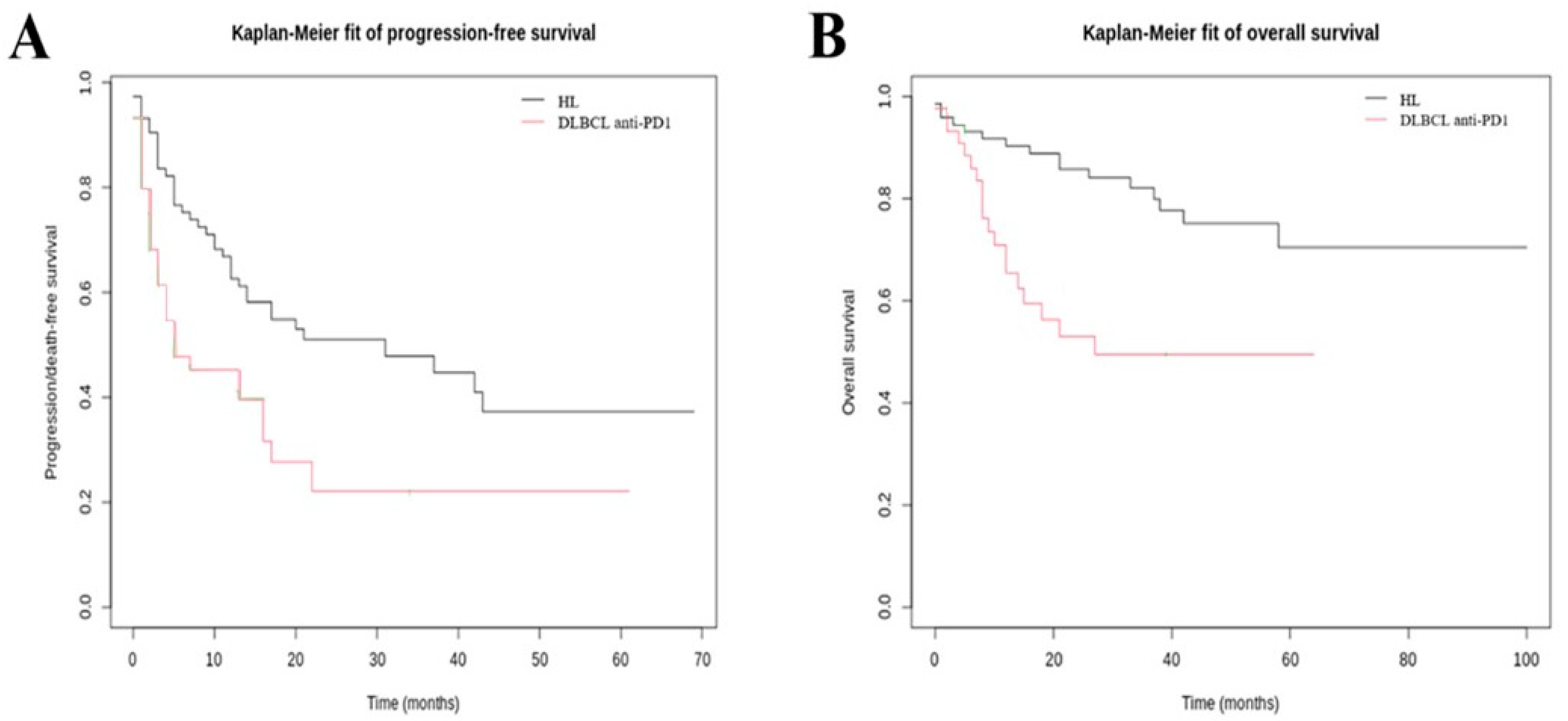
The best overall response rate (ORR) of HL patients was 51.4%; thirty patients (41.7%) had a complete response, six patients (8.3%) had a partial response, and one patient (1.4%) had a stable disease. Following anti-PD-1 therapy, the median PFS was 16.5 months (IQR, 5.9–29.5) (Figure 2A). Median OS was not reached (Figure 2B).
Among the 44 DLBCL anti-PD-1 treated patients, the best ORR was 34.1%; thirteen patients (29.5%) had a complete response, one patient (2.3%) had a partial response, and one patient (2.3%) had a stable disease. Following anti-PD-1 therapy, the median PFS was 5.3 months (IQR, 2.0–15.5) (Figure 2A). The median OS was 13.3 months (IQR, 7.6–38.6) (Figure 2B).
The best ORR of DLBCL CAR T cell-treated patients was 44.3%; 52 patients (39.7%) had a complete response, five patients (3.8%) had a partial response, and one patient (0.8%) had a stable disease. Following CAR T cells, the median PFS was 3.8 months (IQR, 1.6–11.1) (Figure 3A). The median OS from infusion was 15.5 months (IQR, 9.2–26.4) (Figure 3B). The PFS was significantly improved after axi-cel infusion compared to after tisagenlecleucel (tisa-cel) infusion (p = 0.008), with a median PFS equal to 5.1 months (IQR, 2.7–14) versus 3.1 months (IQR, 1.0–8.4) (Figure 3C). The median OS was 17.2 months (IQR, 10.2–28.0) and 14.8 months (IQR, 9.1–24.9) for axi-cel and tisa-cel, respectively (Figure 3D). The OS was significantly improved after axi-cel infusion compared to after tisa-cel (p = 0.05).

3.3. Response Prediction

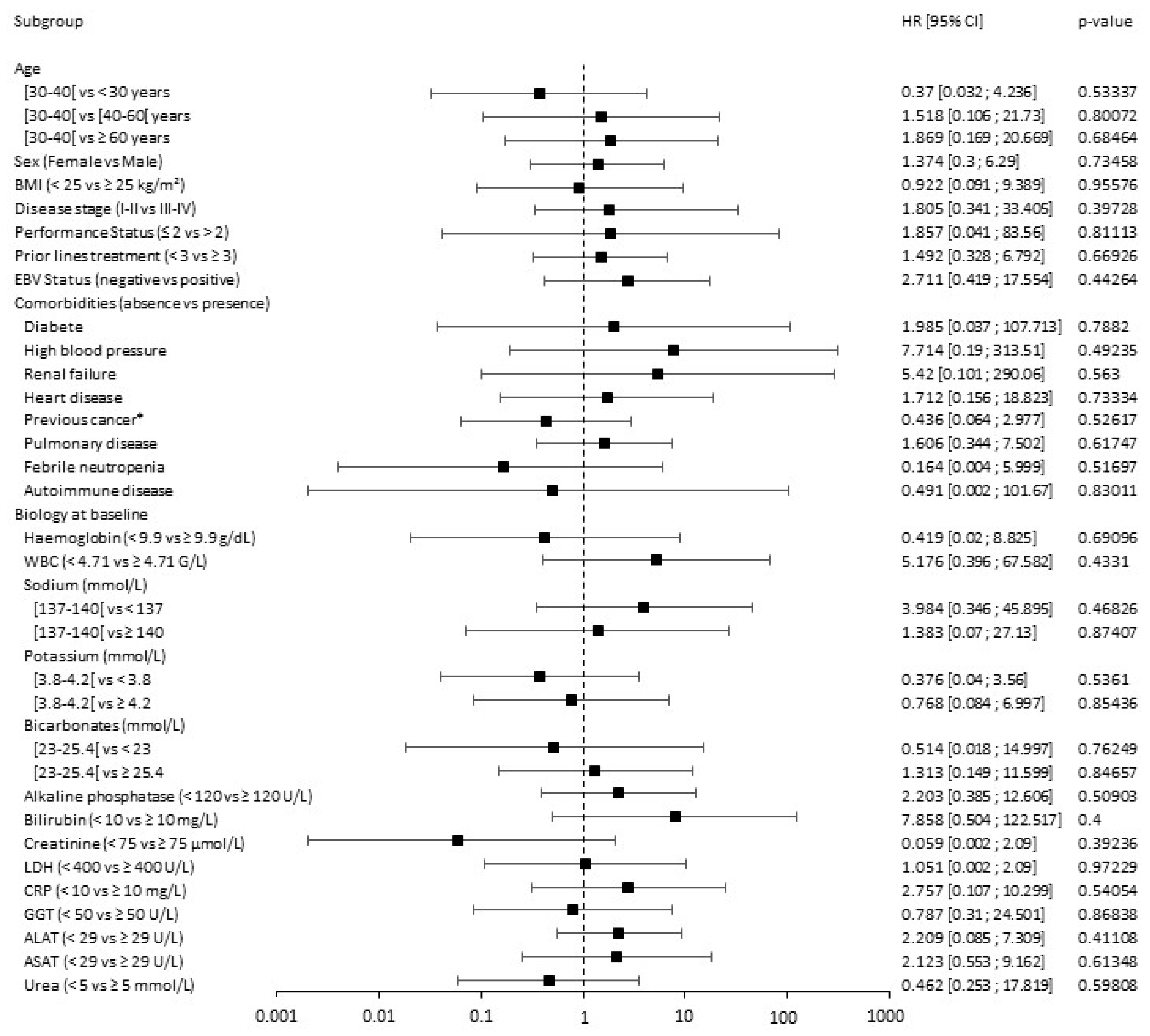
Univariate analysis of the HL patient cohort is presented in Figure 4. None of age, gender, PS, disease stage, or Epstein–Barr infection influenced PFS in HL patients treated by anti-PD-1 therapy. Moreover, among all clinical assessments considered, none significantly impacted treatment response as measured by PFS.
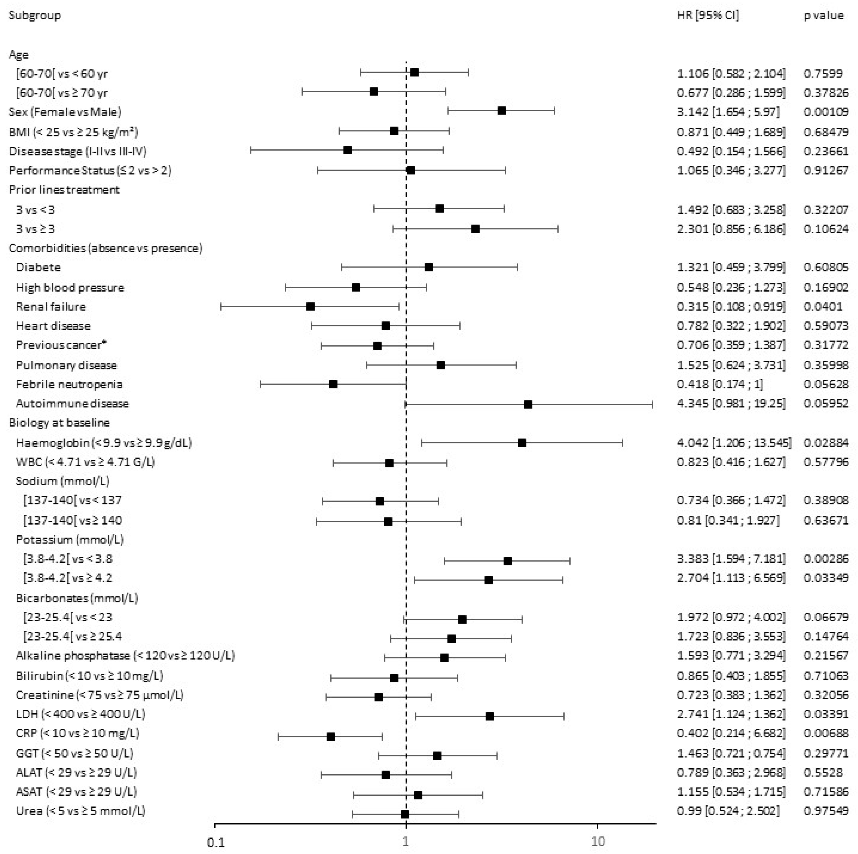
The univariate analysis of DLBCL CAR T cell-treated patients is summarized in Figure 5.
Among demographic parameters, male sex was significantly associated with disease progression (HR = 3.142, 95% CI: 1.654–5.97; p = 0.00109). Interestingly, age (p = 0.7599 for age < 60 and p = 0.37826 for age ≥ 70 years old), disease stage (p = 0.23661), and PS (p = 0.91267) were not predictive factors for PFS. Additionally, early CAR T cell treatment (i.e., < 3 prior lines treatment) compared to later treatment (≥ 3 prior lines treatment) did not impact PFS (p = 0.32207 and p = 0.10624, respectively). Among comorbidities, there was a trend towards a negative association between autoimmune disorders and PFS (p = 0.05952). Among biological factors, LDH ≥ 400 U/L was significantly associated with poorer PFS (HR = 2.741, 95% CI = 1.124–6.682; p = 0.03391). Additionally, a rate of haemoglobin strictly inferior to 9.9 g/dL (p = 0.02884) and extreme potassium concentration at baseline (i.e., <3.8 and ≥4.2 mmol/L, respectively) were significantly associated with PFS (p = 0.00286 and p = 0.03349, respectively).

3.4. Adverse Events

The most common anti-PD-1-related AEs (grade superior or equal to 3) for HL and DLBCL patients were blood disorders (78.1% and 59.1%, respectively), hepatic disorders (34.2% and 25.0%, respectively) and kidney disorders (21.9% and 18.2%, respectively) (Table 2). When focusing on patients treated with CAR T cells within 30 days after treatment infusion, 118 out of 132 patients (89.4%) experienced a grade 3 or 4 AE. The most common drug-related AE of grade ≥ 3 were CRS (n = 15, 11.4%), ICANS (n = 15, 11.4%) and blood disorders (n = 40, 30.3%). Overall, 103 patients had a CRS of any grade and 47 patients had an ICANS of any grade (Table 2).

3.5. Toxicity Prediction

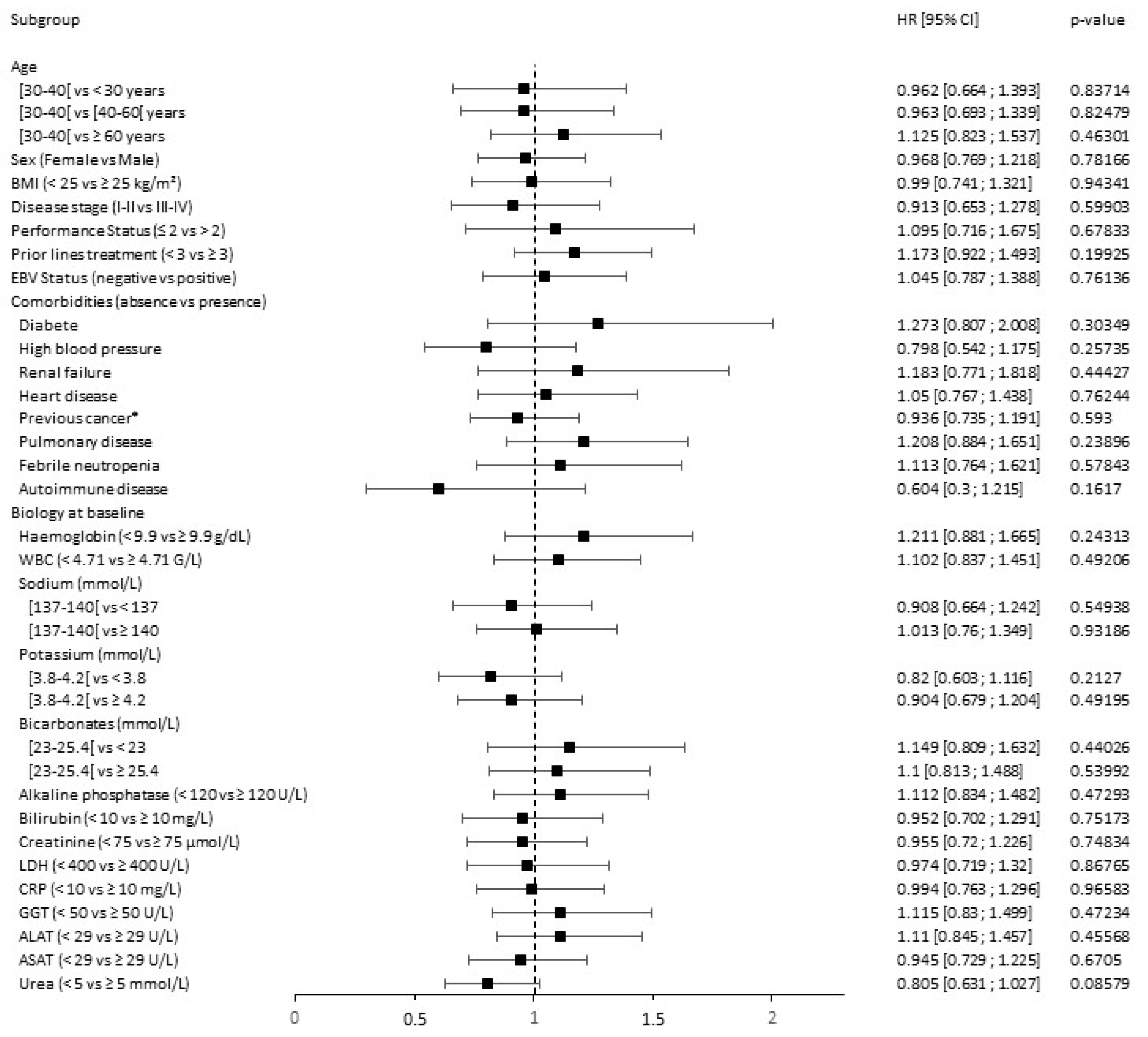
Neither demographic factors, treatment history, or clinical biomarkers influenced the occurrence of toxicity induced by anti-PD-1 (Figure 6).
Toxicities induced by CAR T cells were not influenced by demographic characteristics, PS, disease stage, treatment history, or comorbidities (Figure 7).
Nevertheless, age ≥ 70 seemed to be negatively associated with CAR T cell tolerance (HZ = 1.208, p = 0.08645). Among biological values at baseline, only an elevated CRP portended poorer tolerance to CAR T cells. Indeed, with a hazard ratio equal to 1.301 (95% CI: 1.098–1.542), a CRP level ≥ 10 mg/L was significantly associated with increase in toxicity occurrence (p = 0.00317).

4. Discussion

A total of 249 patients with ML treated by anti-PD-1 or CAR T cell therapy were analyzed in this real-world study. Considering survival and toxicity data obtained outside of trials, these findings are consistent with the previous literature. Known predictive response or toxicity factors of CAR T cells such as age, elevated LDH, and CRP at the time of infusion were confirmed by the PRONOSTIM study. Additionally, new factors were highlighted, including gender, hemoglobin, and kalemia.
The median PFS from HL patients treated with anti-PD-1 echoed the ORR of the CHECKMATE 205 and KEYNOTE 087 studies, in which the median PFS was 14.7 and 13.7 and the ORR was 69.0% and 51.4%, respectively [6,7]. Regarding toxicity, the most common drug-related AEs were blood, hepatic, and kidney disorders.
To the best of our knowledge, most of the published data identifying predictive factors of anti-PD-1 efficacy and toxicity refer to patients with solid tumours, and are not relevant for extrapolation to ML [22,41,42,43,44,45,46]. In our study, the influence of demographic factors, clinical and biological characteristics at baseline, the number of prior lines of treatment, and comorbidities were all considered. We showed the absence of significant influence of age or sex on both PFS and occurrence of adverse events in anti-PD-1 treatment. Sex-based benefit following treatment with anti-PD-1 remains a contentious issue. Although safety profiles and survival of men and women are comparable, differences can be driven by other factors such as age, hormonal status, the influence of oestrogens in women, delay in diagnosis in men, and the greater overall longevity of women [47,48,49,50]. Regarding age, Nishijima et al. and Baldini et al. showed a significant survival benefit with anti-PD-1 in younger patients and in older patients with an age limit of 65–70 years, as well as a more frequent irAE in older subjects [51,52]. None of these studies looked at HL patients. BMI ≥ 25 kg/m2 and obesity were not associated with survival or tolerance outcomes; these results are consistent with previous results [16]. Univariate analysis of the above factors did not allow us to identify more significant predictors of survival and toxicity to ICI.
In terms of OS and PFS, our results are consistent with survival rates from the JULIET and ZUMA-1 trials and from the French DESCAR-T registry [1,2,29]. Regarding toxicity, most common adverse events following CAR T cell infusion were CRS and ICANS, occurring in 78.0% (11.4% of grade ≥ 3) and 35.6% (12.1% of grade ≥ 3) of patients, respectively. In the pivotal ZUMA-1 and JULIET studies, grade ≥ 3 CRS was 13.0% for axi-cel and 22.0% for tisa-cel. Similarly, grade ≥ 3 ICANS was 28.0% and 12.0%, respectively. The better safety profile in our real-world study can be explained by better experience over time and new management strategies around CRS and ICANS.
The identification of predictive factors associated with the efficacy and safety of CAR T cell therapy in the DLBCL population is a hot topic. Interestingly, in our study males benefitted less than females from CAR T cells in terms of PFS. To the best of our knowledge this association has not yet been demonstrated, although a previous study highlighted that females were at greater risk of severe toxicity after CAR T cell infusion [53]. As previously demonstrated, no age-related differences in efficacy or toxicity were observed, suggesting that age alone should not limit use of CAR T cells [54,55]. Regarding comorbidities, we tried to use the ICD-10 classification to identify predictive factors. Surprisingly we observed an unexpected positive impact of renal failure on PFS. Outlier results associated with a few patients indicate the existence of a random selection bias and that potential unmeasured confounders may undermine the validity of our conclusions. As previously published, no association between obesity and toxicity or response was identified [56]. Potential markers of greater burden of more aggressive disease have been evaluated in previous studies. Analysis suggests that patients with elevated pre-infusion LDH have poorer PFS [25,57]. LDH greater than 400 U/L levels before CAR T cell infusion was associated with maximal predictive significance for PFS, confirming the cut-off defined by Rabinovich et al. [27]. Literature data remain contentious about the level of LDH and the severity of ICANS or CRS [30,58]. In our study, we did not highlight any correlation between LDH levels and ICANS or CRS severity, in accordance with the results obtained by Pennisi et al. [30]. High CRP levels at baseline have previously been reported in patients with severe CRS or ICANS [30,31]. Our results suggest that a CRP dosage superior or equal to 10 mg/L initially portends poorer tolerance. In another study, CRP levels obtained on the day of CAR T cell infusion strictly inferior to 30 mg/L were correlated with improved duration of response (p = 0.003), PFS (p < 0.001) and OS (p < 0.001), while an increased day 0 and peak CRP were associated with grade ≥ 3 ICANS, though not with CRS [59]. As recently described, moderate toxicity manifesting as grade 2 CRS may be associated with favourable clinical outcomes [60]. Thus, the occurrence of CRS and ICANS was analysed jointly in this study.

5. Conclusions

In this study, we used data from a clinical data warehouse to identify predictors of immunotherapy response in lymphomas. Based on a large set of biological and clinical factors, our results confirm already known predictors and demonstrate male gender, low hemoglobin, and hypo- or hyperkalemia as predictive factors for progression after CAR T cell therapy. These risk factors should be considered together with others already highlighted rather than independently, as evidenced by the emergence of numerous predictive scores. The associations between predictors should be used as an enhanced risk stratification algorithm for decision support before starting treatment, and their usefulness should be confirmed in prospective cohorts. In this context, the use of the clinical data warehouse shows an essential contribution to identifying new predictive characteristics not previously evidenced by clinical trials or more typical real-world studies.

Author Contributions

Conceptualization, M.D., M.C., N.B., M.K. and J.Z.; Data curation, M.D. and J.Z.; Formal analysis, M.D., M.C., N.B., M.K. and J.Z.; Funding acquisition, J.Z.; Investigation, M.D. and M.C.; Methodology, M.D., M.C., N.B., M.K. and J.Z.; Project administration, J.Z.; Resources, J.Z.; Software, M.C. and N.B.; Supervision, M.K. and J.Z.; Validation, M.D., L.W., J.D., B.D.-F., M.V., R.B. (Rudy Birsen), F.M., C.L., E.B., P.D., A.C., R.B. (Rui Batista), S.L., J.-M.T., L.R., N.S., O.H., S.C., V.M., C.M., D.B., M.K. and J.Z.; Visualization, M.D.; Writing—original draft, M.D.; Writing—review and editing, M.K. and J.Z. All authors have read and agreed to the published version of the manuscript.

Funding

This research received no external funding.

Institutional Review Board Statement

This study was conducted in accordance with the Declaration of Helsinki and approved by the Institutional Review Board (or Ethics Committee) of the scientific and ethical committee of the AP-HP (CES-22-03_PRONOSTIM, IRB00011591) on 8 February 2022.

Informed Consent Statement

All subjects included in this study were informed about the reuse of their data for research, and subjects who objected to the reuse of their data were excluded from this study in accordance with French legislation.

Data Availability Statement

The datasets generated and/or analyzed during the current study are available from the corresponding author on reasonable request.

Acknowledgments

We are grateful to all principal investigators of the PRONOSTIM study. The authors thank the Institutional Review Board of the scientific and ethical committee of the Greater Paris University Hospitals.

Conflicts of Interest

S.C. served on the scientific advisory board for Gilead/Kite, Novartis. The other authors report no conflict of interest relevant to this manuscript.

References

  1. Neelapu, S.S.; Locke, F.L.; Bartlett, N.L.; Lekakis, L.J.; Miklos, D.B.; Jacobson, C.A.; Braunschweig, I.; Oluwole, O.O.; Siddiqi, T.; Lin, Y.; et al. Axicabtagene Ciloleucel CAR T-Cell Therapy in Refractory Large B-Cell Lymphoma. N. Engl. J. Med. 2017, 377, 2531–2544. [Google Scholar] [CrossRef]
  2. Schuster, S.J.; Bishop, M.R.; Tam, C.S.; Waller, E.K.; Borchmann, P.; McGuirk, J.P.; Jäger, U.; Jaglowski, S.; Andreadis, C.; Westin, J.R.; et al. Tisagenlecleucel in Adult Relapsed or Refractory Diffuse Large B-Cell Lymphoma. N. Engl. J. Med. 2019, 380, 45–56. [Google Scholar] [CrossRef] [PubMed]
  3. Fowler, N.H.; Dickinson, M.; Dreyling, M.; Martinez-Lopez, J.; Kolstad, A.; Butler, J.; Ghosh, M.; Popplewell, L.; Chavez, J.C.; Bachy, E.; et al. Tisagenlecleucel in Adult Relapsed or Refractory Follicular Lymphoma: The Phase 2 ELARA Trial. Nat. Med. 2022, 28, 325–332. [Google Scholar] [CrossRef] [PubMed]
  4. Wang, M.; Munoz, J.; Goy, A.; Locke, F.L.; Jacobson, C.A.; Hill, B.T.; Timmerman, J.M.; Holmes, H.; Jaglowski, S.; Flinn, I.W.; et al. KTE-X19 CAR T-Cell Therapy in Relapsed or Refractory Mantle-Cell Lymphoma. N. Engl. J. Med. 2020, 382, 1331–1342. [Google Scholar] [CrossRef] [PubMed]
  5. Jacobson, C.A.; Chavez, J.C.; Sehgal, A.R.; William, B.M.; Munoz, J.; Salles, G.; Munshi, P.N.; Casulo, C.; Maloney, D.G.; de Vos, S.; et al. Axicabtagene Ciloleucel in Relapsed or Refractory Indolent Non-Hodgkin Lymphoma (ZUMA-5): A Single-Arm, Multicentre, Phase 2 Trial. Lancet Oncol. 2022, 23, 91–103. [Google Scholar] [CrossRef] [PubMed]
  6. Chen, R.; Zinzani, P.L.; Fanale, M.A.; Armand, P.; Johnson, N.A.; Brice, P.; Radford, J.; Ribrag, V.; Molin, D.; Vassilakopoulos, T.P.; et al. Phase II Study of the Efficacy and Safety of Pembrolizumab for Relapsed/Refractory Classic Hodgkin Lymphoma. J. Clin. Oncol. 2017, 35, 2125–2132. [Google Scholar] [CrossRef]
  7. Armand, P.; Engert, A.; Younes, A.; Fanale, M.; Santoro, A.; Zinzani, P.L.; Timmerman, J.M.; Collins, G.P.; Ramchandren, R.; Cohen, J.B.; et al. Nivolumab for Relapsed/Refractory Classic Hodgkin Lymphoma After Failure of Autologous Hematopoietic Cell Transplantation: Extended Follow-Up of the Multicohort Single-Arm Phase II CheckMate 205 Trial. J. Clin. Oncol. 2018, 36, 1428–1439. [Google Scholar] [CrossRef]
  8. Axicabtagene Ciloleucel as Second-Line Therapy for Large B-Cell Lymphoma|NEJM. Available online: https://www.nejm.org/doi/full/10.1056/NEJMoa2116133 (accessed on 16 January 2023).
  9. Kamdar, M.; Solomon, S.R.; Arnason, J.; Johnston, P.B.; Glass, B.; Bachanova, V.; Ibrahimi, S.; Mielke, S.; Mutsaers, P.; Hernandez-Ilizaliturri, F.; et al. Lisocabtagene Maraleucel versus Standard of Care with Salvage Chemotherapy Followed by Autologous Stem Cell Transplantation as Second-Line Treatment in Patients with Relapsed or Refractory Large B-Cell Lymphoma (TRANSFORM): Results from an Interim Analysis of an Open-Label, Randomised, Phase 3 Trial. Lancet 2022, 399, 2294–2308. [Google Scholar] [CrossRef]
  10. Thompson, J.A.; Schneider, B.J.; Brahmer, J.; Achufusi, A.; Armand, P.; Berkenstock, M.K.; Bhatia, S.; Budde, L.E.; Chokshi, S.; Davies, M.; et al. Management of Immunotherapy-Related Toxicities, Version 1.2022, NCCN Clinical Practice Guidelines in Oncology. J. Natl. Compr. Cancer Netw. 2022, 20, 387–405. [Google Scholar] [CrossRef]
  11. Santomasso, B.D.; Nastoupil, L.J.; Adkins, S.; Lacchetti, C.; Schneider, B.J.; Anadkat, M.; Atkins, M.B.; Brassil, K.J.; Caterino, J.M.; Chau, I.; et al. Management of Immune-Related Adverse Events in Patients Treated with Chimeric Antigen Receptor T-Cell Therapy: ASCO Guideline. J. Clin. Oncol. 2021, 39, 3978–3992. [Google Scholar] [CrossRef]
  12. Lee, D.W.; Santomasso, B.D.; Locke, F.L.; Ghobadi, A.; Turtle, C.J.; Brudno, J.N.; Maus, M.V.; Park, J.H.; Mead, E.; Pavletic, S.; et al. ASTCT Consensus Grading for Cytokine Release Syndrome and Neurologic Toxicity Associated with Immune Effector Cells. Biol. Blood Marrow Transplant. 2019, 25, 625–638. [Google Scholar] [CrossRef] [PubMed] [Green Version]
  13. Locke, F.L.; Ghobadi, A.; Jacobson, C.A.; Miklos, D.B.; Lekakis, L.J.; Oluwole, O.O.; Lin, Y.; Braunschweig, I.; Hill, B.T.; Timmerman, J.M.; et al. Long-Term Safety and Activity of Axicabtagene Ciloleucel in Refractory Large B-Cell Lymphoma (ZUMA-1): A Single-Arm, Multicentre, Phase 1-2 Trial. Lancet Oncol. 2019, 20, 31–42. [Google Scholar] [CrossRef] [PubMed]
  14. Schuster, S.J.; Tam, C.S.; Borchmann, P.; Worel, N.; McGuirk, J.P.; Holte, H.; Waller, E.K.; Jaglowski, S.; Bishop, M.R.; Damon, L.E.; et al. Long-Term Clinical Outcomes of Tisagenlecleucel in Patients with Relapsed or Refractory Aggressive B-Cell Lymphomas (JULIET): A Multicentre, Open-Label, Single-Arm, Phase 2 Study. Lancet Oncol. 2021, 22, 1403–1415. [Google Scholar] [CrossRef] [PubMed]
  15. Chen, R.; Zinzani, P.L.; Lee, H.J.; Armand, P.; Johnson, N.A.; Brice, P.; Radford, J.; Ribrag, V.; Molin, D.; Vassilakopoulos, T.P.; et al. Pembrolizumab in Relapsed or Refractory Hodgkin Lymphoma: 2-Year Follow-up of KEYNOTE-087. Blood 2019, 134, 1144–1153. [Google Scholar] [CrossRef] [Green Version]
  16. De Filippi, R.; Morabito, F.; Santoro, A.; Tripepi, G.; D’Alò, F.; Rigacci, L.; Ricci, F.; Morelli, E.; Zinzani, P.L.; Pinto, A. Body Mass Index Is Not Associated with Survival Outcomes and Immune-Related Adverse Events in Patients with Hodgkin Lymphoma Treated with the Immune Checkpoint Inhibitor Nivolumab. J. Transl. Med. 2021, 19, 489. [Google Scholar] [CrossRef]
  17. Fattizzo, B.; Cavallaro, F.; Folino, F.; Barcellini, W. Recent Insights into the Role of the Microbiome in Malignant and Benign Hematologic Diseases. Crit. Rev. Oncol. Hematol. 2021, 160, 103289. [Google Scholar] [CrossRef]
  18. Swaidani, S.; Roopkumar, J.; Jun-Shim, Y.; Charles, C.; Paul, S.; Kundu, S.; Funchain, P.; Rayman, P.; Pavicic, P.G.; Diaz-Montero, C.M.; et al. Biomarker Assessment of Venous Thromboembolism in Cancer Patients Receiving Checkpoint Blockade. Blood 2019, 134, 3648. [Google Scholar] [CrossRef]
  19. Colombo, A.; Hav, M.; Singh, M.; Xu, A.; Gamboa, A.; Lemos, T.; Gerdtsson, E.; Chen, D.; Houldsworth, J.; Shaknovich, R.; et al. Single-Cell Spatial Analysis of Tumor Immune Architecture in Diffuse Large B-Cell Lymphoma. Blood Adv. 2022, 6, 4675–4690. [Google Scholar] [CrossRef]
  20. Hatic, H.; Sampat, D.; Goyal, G. Immune Checkpoint Inhibitors in Lymphoma: Challenges and Opportunities. Ann. Transl. Med. 2021, 9, 1037. [Google Scholar] [CrossRef]
  21. Roemer, M.G.M.; Advani, R.H.; Ligon, A.H.; Natkunam, Y.; Redd, R.A.; Homer, H.; Connelly, C.F.; Sun, H.H.; Daadi, S.E.; Freeman, G.J.; et al. PD-L1 and PD-L2 Genetic Alterations Define Classical Hodgkin Lymphoma and Predict Outcome. J. Clin. Oncol. 2016, 34, 2690–2697. [Google Scholar] [CrossRef] [Green Version]
  22. Jeong, A.-R.; Ball, E.D.; Goodman, A.M. Predicting Responses to Checkpoint Inhibitors in Lymphoma: Are We Up to the Standards of Solid Tumors? Clin. Med. Insights Oncol. 2020, 14, 1179554920976366. [Google Scholar] [CrossRef]
  23. Chen, C.; Liu, S.; Jiang, X.; Huang, L.; Chen, F.; Wei, X.; Guo, H.; Shao, Y.; Li, Y.; Li, W. Tumor Mutation Burden Estimated by a 69-Gene-Panel Is Associated with Overall Survival in Patients with Diffuse Large B-Cell Lymphoma. Exp. Hematol. Oncol. 2021, 10, 20. [Google Scholar] [CrossRef]
  24. Jacobson, C.A.; Hunter, B.; Armand, P.; Kamihara, Y.; Ritz, J.; Rodig, S.J.; Wright, K.; Lipschitz, M.; Redd, R.A.; Maus, M.V.; et al. Axicabtagene Ciloleucel in the Real World: Outcomes and Predictors of Response, Resistance and Toxicity. Blood 2018, 132, 92. [Google Scholar] [CrossRef]
  25. Vercellino, L.; Di Blasi, R.; Kanoun, S.; Tessoulin, B.; Rossi, C.; D’Aveni-Piney, M.; Obéric, L.; Bodet-Milin, C.; Bories, P.; Olivier, P.; et al. Predictive Factors of Early Progression after CAR T-Cell Therapy in Relapsed/Refractory Diffuse Large B-Cell Lymphoma. Blood Adv. 2020, 4, 5607–5615. [Google Scholar] [CrossRef] [PubMed]
  26. Iacoboni, G.; Villacampa, G.; Martinez-Cibrian, N.; Bailén, R.; Lopez Corral, L.; Sanchez, J.M.; Guerreiro, M.; Caballero, A.C.; Mussetti, A.; Sancho, J.; et al. Real-world Evidence of Tisagenlecleucel for the Treatment of Relapsed or Refractory Large B-cell Lymphoma. Cancer Med. 2021, 10, 3214–3223. [Google Scholar] [CrossRef] [PubMed]
  27. Rabinovich, E.; Pradhan, K.; Sica, R.A.; Bachier-Rodriguez, L.; Mantzaris, I.; Kornblum, N.; Shastri, A.; Gritsman, K.; Goldfinger, M.; Verma, A.; et al. Elevated LDH Greater than 400 U/L Portends Poorer Overall Survival in Diffuse Large B-Cell Lymphoma Patients Treated with CD19 CAR-T Cell Therapy in a Real World Multi-Ethnic Cohort. Exp. Hematol. Oncol. 2021, 10, 55. [Google Scholar] [CrossRef] [PubMed]
  28. Locke, F.L.; Rossi, J.M.; Neelapu, S.S.; Jacobson, C.A.; Miklos, D.B.; Ghobadi, A.; Oluwole, O.O.; Reagan, P.M.; Lekakis, L.J.; Lin, Y.; et al. Tumor Burden, Inflammation, and Product Attributes Determine Outcomes of Axicabtagene Ciloleucel in Large B-Cell Lymphoma. Blood Adv. 2020, 4, 4898–4911. [Google Scholar] [CrossRef]
  29. Bachy, E.; Le Gouill, S.; Di Blasi, R.; Sesques, P.; Manson, G.; Cartron, G.; Beauvais, D.; Roulin, L.; Gros, F.X.; Rubio, M.T.; et al. A Real-World Comparison of Tisagenlecleucel and Axicabtagene Ciloleucel CAR T Cells in Relapsed or Refractory Diffuse Large B Cell Lymphoma. Nat. Med. 2022, 28, 2145–2154. [Google Scholar] [CrossRef]
  30. Pennisi, M.; Sanchez-Escamilla, M.; Flynn, J.R.; Shouval, R.; Alarcon Tomas, A.; Silverberg, M.L.; Batlevi, C.; Brentjens, R.J.; Dahi, P.B.; Devlin, S.M.; et al. Modified EASIX Predicts Severe Cytokine Release Syndrome and Neurotoxicity after Chimeric Antigen Receptor T Cells. Blood Adv. 2021, 5, 3397–3406. [Google Scholar] [CrossRef]
  31. Greenbaum, U.; Strati, P.; Saliba, R.M.; Torres, J.; Rondon, G.; Nieto, Y.; Hosing, C.; Srour, S.A.; Westin, J.; Fayad, L.E.; et al. CRP and Ferritin in Addition to the EASIX Score Predict CAR-T-Related Toxicity. Blood Adv. 2021, 5, 2799–2806. [Google Scholar] [CrossRef]
  32. Rejeski, K.; Perez, A.; Sesques, P.; Hoster, E.; Berger, C.; Jentzsch, L.; Mougiakakos, D.; Frölich, L.; Ackermann, J.; Bücklein, V.; et al. CAR-HEMATOTOX: A Model for CAR T-Cell-Related Hematologic Toxicity in Relapsed/Refractory Large B-Cell Lymphoma. Blood 2021, 138, 2499–2513. [Google Scholar] [CrossRef] [PubMed]
  33. Rejeski, K.; Perez, A.; Iacoboni, G.; Penack, O.; Bücklein, V.; Jentzsch, L.; Mougiakakos, D.; Johnson, G.; Arciola, B.; Carpio, C.; et al. The CAR-HEMATOTOX Risk-Stratifies Patients for Severe Infections and Disease Progression after CD19 CAR-T in R/R LBCL. J. Immunother. Cancer 2022, 10, e004475. [Google Scholar] [CrossRef] [PubMed]
  34. Boardman, A.P.; Gutgarts, V.; Flynn, J.R.; Devlin, S.M.; Tomas, A.A.; Fein, J.A.; Slingerland, J.; Parascondola, A.; Lin, R.J.; Scordo, M.; et al. Predictors and Implications of Renal Injury in Large Cell Lymphoma Patients Receiving CD19 CAR T Cell Therapy; ASH. Blood 2022, 140, 4671–4672. [Google Scholar] [CrossRef]
  35. Shah, H.; O’Conke, D.; Zhao, R.; Jalota, A.; Patel, M.S.; Winter, A.; Williams, L.S.; Khouri, J.; Jagadeesh, D.; Anwer, F.; et al. Assessment of Major Adverse Cardiac Events (MACE) and Arrhythmias in Patients with Large B-Cell Lymphoma Undergoing Anti-CD19 Chimeric Antigen Receptor T-Cell (CAR-T) Therapy: Impact of Baseline Cardiac Biomarkers; ASH. Blood 2022, 140, 4673–4675. [Google Scholar] [CrossRef]
  36. Smith, M.; Dai, A.; Ghilardi, G.; Amelsberg, K.V.; Devlin, S.M.; Pajarillo, R.; Slingerland, J.B.; Beghi, S.; Herrera, P.S.; Giardina, P.; et al. Gut Microbiome Correlates of Response and Toxicity Following Anti-CD19 CAR T Cell Therapy. Nat. Med. 2022, 28, 713–723. [Google Scholar] [CrossRef] [PubMed]
  37. Wang, Y.; Song, Z.; Geng, Y.; Gao, L.; Xu, L.; Tang, G.; Ni, X.; Chen, L.; Chen, J.; Wang, T.; et al. The Risk Factors and Early Predictive Model of Hematotoxicity after CD19 Chimeric Antigen Receptor T Cell Therapy. Front. Oncol. 2022, 12, 987965. [Google Scholar] [CrossRef]
  38. Hasenclever, D.; Diehl, V. A Prognostic Score for Advanced Hodgkin’s Disease. International Prognostic Factors Project on Advanced Hodgkin’s Disease. N. Engl. J. Med. 1998, 339, 1506–1514. [Google Scholar] [CrossRef]
  39. A Predictive Model for Aggressive Non-Hodgkin’s Lymphoma|NEJM. Available online: https://www.nejm.org/doi/full/10.1056/nejm199309303291402 (accessed on 5 January 2023).
  40. Common Terminology Criteria for Adverse Events (CTCAE). 2017. Available online: https://ctep.cancer.gov/protocoldevelopment/electronic_applications/ctc.htm (accessed on 5 January 2023).
  41. Baruch, E.N.; Youngster, I.; Ben-Betzalel, G.; Ortenberg, R.; Lahat, A.; Katz, L.; Adler, K.; Dick-Necula, D.; Raskin, S.; Bloch, N.; et al. Fecal Microbiota Transplant Promotes Response in Immunotherapy-Refractory Melanoma Patients. Science 2021, 371, 602–609. [Google Scholar] [CrossRef]
  42. Bigot, F.; Castanon, E.; Baldini, C.; Hollebecque, A.; Carmona, A.; Postel-Vinay, S.; Angevin, E.; Armand, J.-P.; Ribrag, V.; Aspeslagh, S.; et al. Prospective Validation of a Prognostic Score for Patients in Immunotherapy Phase I Trials: The Gustave Roussy Immune Score (GRIm-Score). Eur. J. Cancer 2017, 84, 212–218. [Google Scholar] [CrossRef]
  43. Aggen, D.H.; Ager, C.R.; Obradovic, A.Z.; Chowdhury, N.; Ghasemzadeh, A.; Mao, W.; Chaimowitz, M.G.; Lopez-Bujanda, Z.A.; Spina, C.S.; Hawley, J.E.; et al. Blocking IL1 Beta Promotes Tumor Regression and Remodeling of the Myeloid Compartment in a Renal Cell Carcinoma Model: Multidimensional Analyses. Clin. Cancer Res. 2021, 27, 608–621. [Google Scholar] [CrossRef]
  44. Angell, H.K.; Bruni, D.; Barrett, J.C.; Herbst, R.; Galon, J. The Immunoscore: Colon Cancer and Beyond. Clin. Cancer Res. 2020, 26, 332–339. [Google Scholar] [CrossRef] [PubMed] [Green Version]
  45. Ettinger, D.S.; Wood, D.E.; Aisner, D.L.; Akerley, W.; Bauman, J.R.; Bharat, A.; Bruno, D.S.; Chang, J.Y.; Chirieac, L.R.; D’Amico, T.A.; et al. NCCN Guidelines Insights: Non-Small Cell Lung Cancer, Version 2.2021. J. Natl. Compr. Cancer Netw. 2021, 19, 254–266. [Google Scholar] [CrossRef] [PubMed]
  46. Le, D.T.; Durham, J.N.; Smith, K.N.; Wang, H.; Bartlett, B.R.; Aulakh, L.K.; Lu, S.; Kemberling, H.; Wilt, C.; Luber, B.S.; et al. Mismatch Repair Deficiency Predicts Response of Solid Tumors to PD-1 Blockade. Science 2017, 357, 409–413. [Google Scholar] [CrossRef] [PubMed] [Green Version]
  47. Klein, S.L.; Flanagan, K.L. Sex Differences in Immune Responses. Nat. Rev. Immunol. 2016, 16, 626–638. [Google Scholar] [CrossRef] [PubMed]
  48. Conforti, F.; Pala, L.; Bagnardi, V.; De Pas, T.; Martinetti, M.; Viale, G.; Gelber, R.D.; Goldhirsch, A. Cancer Immunotherapy Efficacy and Patients’ Sex: A Systematic Review and Meta-Analysis. Lancet Oncol. 2018, 19, 737–746. [Google Scholar] [CrossRef]
  49. Duma, N.; Abdel-Ghani, A.; Yadav, S.; Hoversten, K.P.; Reed, C.T.; Sitek, A.N.; Enninga, E.A.L.; Paludo, J.; Aguilera, J.V.; Leventakos, K.; et al. Sex Differences in Tolerability to Anti-Programmed Cell Death Protein 1 Therapy in Patients with Metastatic Melanoma and Non-Small Cell Lung Cancer: Are We All Equal? Oncologist 2019, 24, e1148–e1155. [Google Scholar] [CrossRef] [Green Version]
  50. van der Kooij, M.K.; Dekkers, O.M.; Aarts, M.J.B.; van den Berkmortel, F.W.P.J.; Boers-Sonderen, M.J.; de Groot, J.W.B.; Hospers, G.A.P.; Piersma, D.; van Rijn, R.S.; Suijkerbuijk, K.P.M.; et al. Sex-Based Differences in Treatment with Immune Checkpoint Inhibition and Targeted Therapy for Advanced Melanoma: A Nationwide Cohort Study. Cancers 2021, 13, 4639. [Google Scholar] [CrossRef]
  51. Nishijima, T.F.; Muss, H.B.; Shachar, S.S.; Moschos, S.J. Comparison of Efficacy of Immune Checkpoint Inhibitors (ICIs) between Younger and Older Patients: A Systematic Review and Meta-Analysis. Cancer Treat. Rev. 2016, 45, 30–37. [Google Scholar] [CrossRef]
  52. Baldini, C.; Martin Romano, P.; Voisin, A.-L.; Danlos, F.-X.; Champiat, S.; Laghouati, S.; Kfoury, M.; Vincent, H.; Postel-Vinay, S.; Varga, A.; et al. Impact of Aging on Immune-Related Adverse Events Generated by Anti-Programmed Death (Ligand)PD-(L)1 Therapies. Eur. J. Cancer 2020, 129, 71–79. [Google Scholar] [CrossRef]
  53. Inc, M.G. Car T-Cells Associated Acute Toxicity in B-Cell Non-Hodgkin Lymphoma: Real-World Study from the Descar-T Registry by Dr. Pierre Sesques. Available online: https://library.ehaweb.org/eha/2022/eha2022-congress/357074/pierre.sesques.car.t-cells.associated.acute.toxicity.in.b-cell.non-hodgkin.html?f=listing%3D0%2Abrowseby%3D8%2Asortby%3D2%2Asearch%3Dcar-t (accessed on 30 September 2022).
  54. Neelapu, S.S.; Jacobson, C.A.; Oluwole, O.O.; Munoz, J.; Deol, A.; Miklos, D.B.; Bartlett, N.L.; Braunschweig, I.; Jiang, Y.; Kim, J.J.; et al. Outcomes of Older Patients in ZUMA-1, a Pivotal Study of Axicabtagene Ciloleucel in Refractory Large B-Cell Lymphoma. Blood 2020, 135, 2106–2109. [Google Scholar] [CrossRef]
  55. Ram, R.; Grisariu, S.; Shargian-Alon, L.; Amit, O.; Bar-On, Y.; Stepensky, P.; Yeshurun, M.; Avni, B.; Hagin, D.; Perry, C.; et al. Toxicity and Efficacy of Chimeric Antigen Receptor T-Cell Therapy in Patients with Diffuse Large B-Cell Lymphoma above the Age of 70 Years Compared to Younger Patients—A Matched Control Multicenter Cohort Study. Haematologica 2022, 107, 1111–1118. [Google Scholar] [CrossRef] [PubMed]
  56. Wudhikarn, K.; Bansal, R.; Khurana, A.; Hathcock, M.A.; Bennani, N.N.; Paludo, J.; Villasboas, J.C.; Wang, Y.; Johnston, P.B.; Ansell, S.M.; et al. The Impact of Obesity and Body Weight on the Outcome of Patients with Relapsed/Refractory Large B-Cell Lymphoma Treated with Axicabtagene Ciloleucel. Blood Cancer J. 2021, 11, 124. [Google Scholar] [CrossRef] [PubMed]
  57. Westin, J.R.; Tam, C.S.; Borchmann, P.; Jaeger, U.; McGuirk, J.P.; Holte, H.; Waller, E.K.; Jaglowski, S.; Bishop, M.R.; Andreadis, C.; et al. Correlative Analyses of Patient and Clinical Characteristics Associated with Efficacy in Tisagenlecleucel-Treated Relapsed/Refractory Diffuse Large B-Cell Lymphoma Patients in the Juliet Trial. Blood 2019, 134, 4103. [Google Scholar] [CrossRef]
  58. Karschnia, P.; Jordan, J.T.; Forst, D.A.; Arrillaga-Romany, I.C.; Batchelor, T.T.; Baehring, J.M.; Clement, N.F.; Gonzalez Castro, L.N.; Herlopian, A.; Maus, M.V.; et al. Clinical Presentation, Management, and Biomarkers of Neurotoxicity after Adoptive Immunotherapy with CAR T Cells. Blood 2019, 133, 2212–2221. [Google Scholar] [CrossRef]
  59. Jacobson, C.A.; Hunter, B.D.; Redd, R.; Rodig, S.J.; Chen, P.-H.; Wright, K.; Lipschitz, M.; Ritz, J.; Kamihara, Y.; Armand, P.; et al. Axicabtagene Ciloleucel in the Non-Trial Setting: Outcomes and Correlates of Response, Resistance, and Toxicity. J. Clin. Oncol. 2020, 38, 3095–3106. [Google Scholar] [CrossRef] [PubMed]
  60. Brammer, J.E.; Braunstein, Z.; Katapadi, A.; Porter, K.; Biersmith, M.; Guha, A.; Vasu, S.; Yildiz, V.O.; Smith, S.A.; Buck, B.; et al. Early Toxicity and Clinical Outcomes after Chimeric Antigen Receptor T-Cell (CAR-T) Therapy for Lymphoma. J. Immunother. Cancer 2021, 9, e002303. [Google Scholar] [CrossRef]
Figure 1. Flow chart of study design and patient inclusion. CAR T cells: chimeric antigen receptor T cells, DLBCL: diffuse large B-cell lymphoma, HL: Hodgkin lymphoma, PD-1: programmed cell death 1.
Cancers 15 04028 g001
Figure 2. Kaplan–Meier curves of anti-PD-1 treated patients. (A) Kaplan–Meier curve for PFS and (B) Kaplan–Meier curve for OS. DLBCL: diffuse large B-cell lymphoma; HL: Hodgkin lymphoma; OS: overall survival; PD-1: programmed cell death 1; PFS: progression-free survival.
Cancers 15 04028 g002
Figure 3. Kaplan–Meier curves of CAR T cell-treated patients. (A) Kaplan–Meier curve for PFS, regardless CAR T cells type; (B) Kaplan–Meier curve for OS irrespective of CAR T cell type; (C) Kaplan–Meier curve for PFS depending on CAR T cell type (tisagenlecleucel versus axicabtagen ciloleucel); and (D) Kaplan–Meier curve for OS depending on CAR T cell type (tisagenlecleucel versus axicabtagen ciloleucel). Axi-cel: axicabtagene ciloleucel; CAR T cells: chimeric antigen receptor T cells; DLBCL: diffuse large B-cell lymphoma; OS: overall survival; PFS: progression-free survival; tisa-cel: tisagenlecleucel.
Cancers 15 04028 g003
Figure 4. Forest plot of analyses of predictive factors for PFS of HL anti-PD-1 treated patients. ALAT: alanine aminotransferase; ASAT: aspartate aminotransferase; BMI: body mass index; CI: confidence interval; CRP: C-reactive protein; EBV: Epstein–Barr virus; GGT: gamma-glutamyl transferase; HR: hazard ratio; LDH: lactate dehydrogenase; WBC: white blood cells; *: other than hematological. Black points represent the value of the hazard ratio (HR) and segment the value of the 95% CI. The first category in parentheses is taken as the reference category for comparison and HR computation. HR < 1 represents a prognosis factor associated with a prolonged survival or a lower toxicity, while HR > 1 represents a prognosis factor associated with shorter survival or greater toxicity. A prognostic factor is statistically significant if the 95% CI does not contain 1. The Cox univariate model was used for calculating the HR and associated two-sided p value.
Cancers 15 04028 g004
Figure 5. Forest plot of analyses of predictive factors for PFS of DLBCL CAR T cell-treated patients ALAT: alanine aminotransferase; ASAT: aspartate aminotransferase; BMI: body mass index; CI: confidence interval; CRP: C-reactive protein; EBV: Epstein–Barr virus; GGT: gamma-glutamyl transferase; HR: hazard ratio; LDH: lactate dehydrogenase; WBC: white blood cells; *: other than hematological. Black points represent the value of the hazard ratio (HR) and segment the value of the 95% CI. The first category in parentheses is taken as the reference category for comparison and HR computation. HR < 1 represents a prognosis factor associated with a prolonged survival or a lower toxicity, while HR > 1 represents a prognosis factor associated with shorter survival or greater toxicity. A prognostic factor is statistically significant if the 95% CI does not contain 1. The Cox univariate model was used for calculating the HR and associated two-sided p value.
Cancers 15 04028 g005
Figure 6. Forest plot of analyses of predictive factors for toxicity of anti-PD-1 treated patients. ALAT: alanine aminotransferase; ASAT: aspartate aminotransferase; BMI: body mass index; CI: confidence interval; CRP: C-reactive protein; EBV: Epstein–Barr virus; GGT: gamma-glutamyl transferase; HR: hazard ratio; LDH: lactate dehydrogenase; WBC: white blood cells; *: other than hematological. Black points represent the value of the hazard ratio (HR) and segment the value of the 95% CI. The first category in parentheses is taken as the reference category for comparison and HR computation. HR < 1 represents a prognosis factor associated with a prolonged survival or a lower toxicity, while HR > 1 represents a prognosis factor associated with shorter survival or greater toxicity. A prognostic factor is statistically significant if the 95% CI does not contain 1. The Cox univariate model was used for calculating the HR and associated two-sided p value.
Cancers 15 04028 g006
Figure 7. Forest plot of analyses of predictive factors for toxicity of CAR T cell-treated patients. ALAT: alanine aminotransferase; ASAT: aspartate aminotransferase; BMI: body mass index; CI: confidence interval; CRP: C-reactive protein; EBV: Epstein–Barr virus; GGT: gamma-glutamyl transferase; HR: hazard ratio; LDH: lactate dehydrogenase; WBC: white blood cells; *: other than hematological. Black points represent the value of the hazard ratio (HR) and segment the value of the 95% CI. The first category in parentheses is taken as the reference category for comparison and HR computation. HR < 1 represents a prognosis factor associated with a prolonged survival or a lower toxicity, while HR > 1 represents a prognosis factor associated with shorter survival or greater toxicity. A prognostic factor is statistically significant if the 95% CI does not contain 1. The Cox univariate model was used for calculating the HR and associated two-sided p value.
Cancers 15 04028 g007
Table 1. Baseline demographic and clinical characteristics of patients.
Hodgkin Lymphoma CohortDLBCL Anti-PD-1 Treated CohortDLBCL CAR T Cells Treated Cohort
Characteristics n = 73n = 44n = 132
Sex
Malen (%)47 (64.4)22 (50.0)82 (62.1)
Femalen (%)26 (35.6)22 (50.0)50 (37.9)
Age at time of (1st) treatment infusion (years)Med [IQR]54.0 [34.0–71.0]57.0 [38.5–74.2]63.0 [55.0–69.2]
min; max18.0; 86.018.0; 84.025.0; 80.0
BMI (kg/m2)Med [IQR]23.5 [19.7–25.8]23.6 [20.3–26.4]24.5 [21.7–27.1]
min; max14.9; 34.713.3; 31.614.6; 39.5
Including obesity backgroundn (%)6 (8.2)1 (2.3)6 (4.5)
Comorbidities
Diabeten (%)3 (4.1)5 (11.4)11 (8.3)
High blood pressuren (%)13 (17.8)6 (13.6)31 (23.5)
Renal failuren (%)9 (12.3)4 (9.1)15 (11.4)
Heart diseasen (%)18 (24.7)7 (15.9)29 (22.0)
Previous cancer * n (%)18 (24.7)15 (34.1)36 (27.3)
Pulmonary diseasen (%)11 (15.1)7 (15.9)16 (12.1)
Febrile neutropenian (%)6 (8.2)6 (13.6)17 (12.9)
Autoimmune diseasen (%)2 (2.7)1 (2.3)5 (3.8)
EBV positive statusn (%)18 (24.7)6 (13.6)12 (9.1)
Stage at diagnosis
Local disease (I/II stages)n (%)9 (12.3)5 (11.4)9 (6.8)
Dissiminated disease (III/IV stages)n (%)64 (87.7)39 (88.6)123 (93.2)
PS at diagnosis
0–2n (%)53 (89.8)27 (87.1)118 (93.6)
3–4n (%)6 (10.2)4 (12.9)8 (6.4)
Missingn (%)14 (19.2)13 (29.5)6 (4.5)
Pronostic score at diagnosis
IPI
0–3n (%)-15 (83.3%)69 (71.1%)
≥4n (%)-3.0 (16.7%)28 (28.8%)
Missingn (%)-26 (59.1)35 (26.5)
IPS
0–3n (%)9 (34.6%)--
≥4n (%)17 (65.4%)--
Missingn (%)47 (64.4)--
Prior lines treatmentMed [IQR]2.0 [1.0–3.0]1.5 [1.0–3.0]2.0 [2.0–3.0]
min; max0.0; 7.01.0; 9.00.0; 6.0
Including autograftn (%)16 (21.9)6 (13.6)27 (20.5)
Last PFS (months)Med [IQR]5.0 [2.5–10.0]5.0 [2.2–10.8]3.0 [2.0–6.0]
min; max1.0; 79.01.0; 72.01.0; 53.0
Missingn (%)10 (13.7)2 (4.5)1 (0.8)
Biology at baseline
Potassium (mmol/L)Med [IQR]4.0 [3.8–4.3]4.1 [3.8–4.3]4.1 [3.7–4.3]
min; max3.0; 6.33.0; 5.12.4; 6.4
Missingn (%)1 (1.4)7 (15.9)1 (0.8)
Hemoglobin (g/dL)Med [IQR]11.4 [9.8–13.0]11.4 [9.7–12.8]10.5 [9.2–12.3]
min; max5.0; 16.97.7; 15.73.3; 15.5
Missingn (%)0 (0.0)3 (6.8)3 (2.3)
LDH (U/L)Med [IQR]266.0 [197.0–327.5]250.5 [202.5–337.5]334.5 [229.5–544.2]
min; max107.0; 2224.0156.0; 1109.0134.0; 4428.0
Missingn (%)2 (2.7)10 (22.7)4 (3.0)
BMI: body mass index; CAR T cells: chimeric antigen receptor T cells; DLBCL: diffuse large B-cell lymphoma; HL: Hodgkin lymphoma; IPI: international prognostic index; IPS: international prognostic score; IQR: interquartile range; LDH: lactate dehydrogenase; Med: median; PD-1: programmed cell death 1; PFS: progression-free survival; PS: performance status; * other than hematological.
Table 2. Safety analysis of infused patients.
Hodgkin Lymphoma CohortDLBCL Anti-PD-1 Treated CohortDLBCL CAR T Cells Treated Cohort
Adverse events n = 73n = 44n = 132
CRSn (%)--103 (78.0)
Including grade ≥3n (%)--15 (11.4)
ICANSn (%)--47 (35.6)
Including grade ≥3n (%)--16 (12.1)
Hematological disorders grade ≥3n (%)35 (47.9)16 (36.4)40 (30.3)
Hepatic disorders grade ≥3n (%)11 (15.1)4 (9.1)34 (25.8)
Kidney disorders grade ≥3n (%)5 (6.8)2 (4.5)30 (22.7)
Endocrine disorders grade ≥3n (%)7 (9.6)2 (4.5)1 (0.8)
Infections grade ≥3n (%)6.0 (8.2)0.0 (0.0)35.0 (26.5)
Cardiac disorders grade ≥3n (%)6.0 (8.2)0.0 (0.0)9.0 (6.8)
Skin disorders grade ≥3n (%)5 (6.8)1 (2.3)3 (2.3)
Neurological disorders grade ≥3n (%)4 (5.5)1 (2.3)1 (0.8)
Digestive disorders grade ≥3n (%)3 (4.1)3 (6.8)12 (9.1)
Respiratory and lung disorders grade ≥3n (%)3.0 (4.1)0.0 (0.0)1.0 (0.8)
CAR T cells: chimeric antigen receptor T cells; CRS: cytokine release syndrome; DLBCL: diffuse large B-cell lymphoma; ICANS: immune effector cell-associated neurotoxicity syndrome; PD-1: programmed cell death 1.
Disclaimer/Publisher’s Note: The statements, opinions and data contained in all publications are solely those of the individual author(s) and contributor(s) and not of MDPI and/or the editor(s). MDPI and/or the editor(s) disclaim responsibility for any injury to people or property resulting from any ideas, methods, instructions or products referred to in the content.