
Cell-Free Circulating (Tumor) DNA before Surgery as a Prognostic Factor in Non-Metastatic Colorectal Cancer: A Systematic Review

 ,
,  and
and
Simple Summary
Abstract
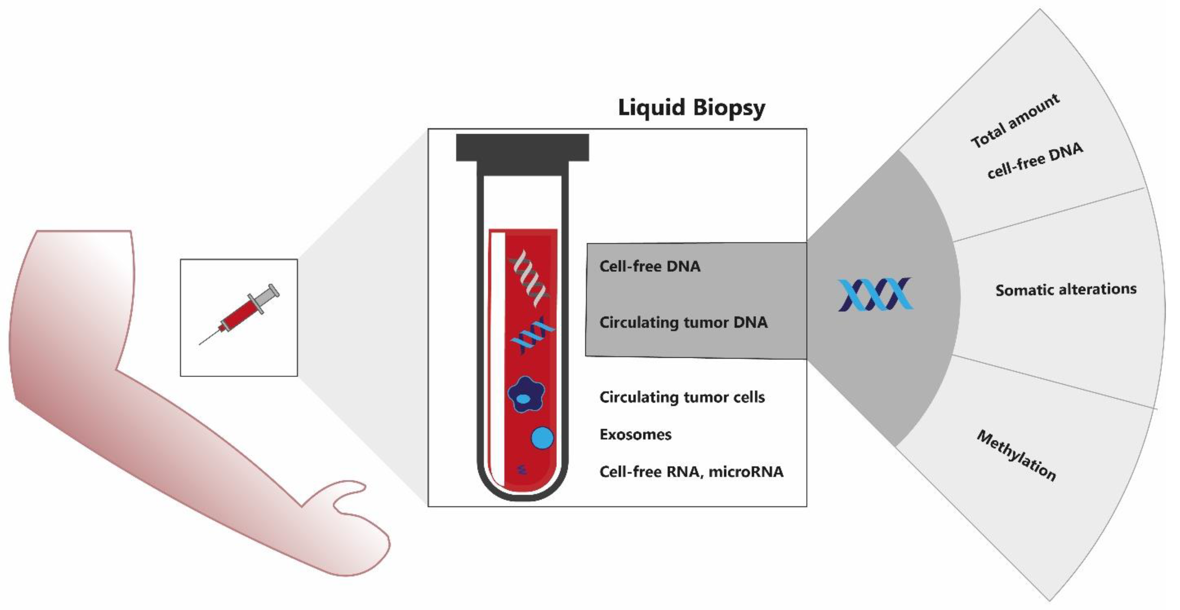
1. Introduction
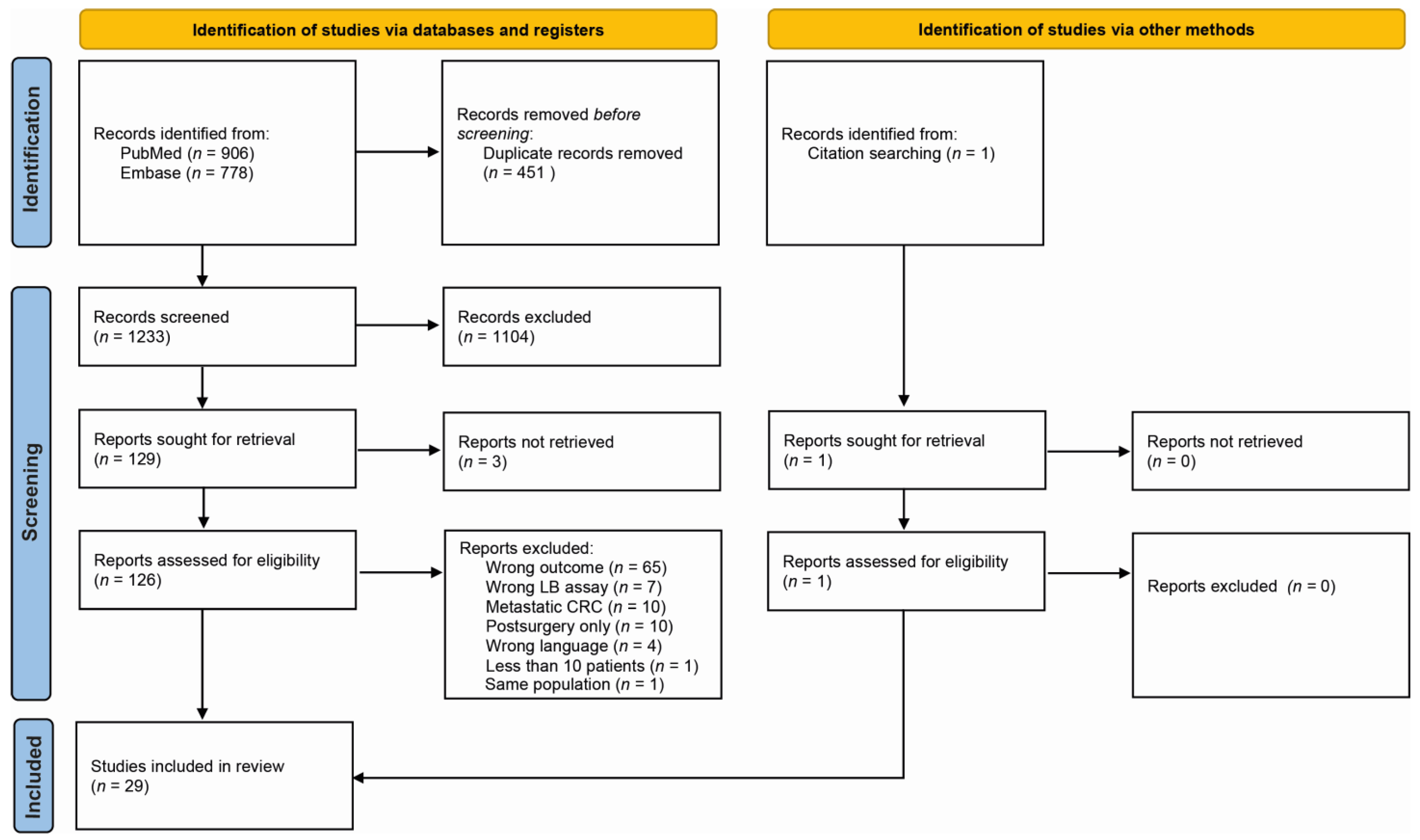
2. Materials and Methods
2.1. Study Design
2.2. Search Strategy
2.3. Inclusion and Exclusion Criteria
2.4. Evidence Synthesis
2.5. Quality of Evidence
3. Results
3.1. Cell-Free DNA
Summary of Included Studies
3.2. Somatic Alterations
Summary of Included Studies
3.3. Methylation
Summary of Included Studies
4. Discussion
5. Conclusions
Supplementary Materials
Author Contributions
Funding
Conflicts of Interest
References
- Elferink, M.A.G.; De Jong, K.P.; Klaase, J.M.; Siemerink, E.J.; De Wilt, J.H.W. Metachronous metastases from colorectal cancer: A population-based study in North-East Netherlands. Int. J. Color. Dis. 2015, 30, 205–212. [Google Scholar] [CrossRef] [PubMed]
- van Gestel, Y.R.; de Hingh, I.H.; van Herk-Sukel, M.P.; van Erning, F.N.; Beerepoot, L.V.; Wijsman, J.H.; Slooter, G.D.; Rutten, H.J.; Creemers, G.-J.M.; Lemmens, V.E. Patterns of metachronous metastases after curative treatment of colorectal cancer. Cancer Epidemiol. 2014, 38, 448–454. [Google Scholar] [CrossRef] [PubMed]
- Qaderi, S.M.; Galjart, B.; Verhoef, C.; Slooter, G.D.; Koopman, M.; Verhoeven, R.H.A.; de Wilt, J.H.W.; van Erning, F.N. Disease recurrence after colorectal cancer surgery in the modern era: A population-based study. Int. J. Color. Dis. 2021, 36, 2399–2410. [Google Scholar] [CrossRef] [PubMed]
- Braendengen, M.; Tveit, K.M.; Berglund, Å.; Birkemeyer, E.; Frykholm, G.; Påhlman, L.; Wiig, J.N.; Byström, P.; Bujko, K.; Glimelius, B. Randomized Phase III Study Comparing Preoperative Radiotherapy With Chemoradiotherapy in Nonresectable Rectal Cancer. J. Clin. Oncol. 2008, 26, 3687–3694. [Google Scholar] [CrossRef] [PubMed]
- Bosset, J.-F.; Collette, L.; Calais, G.; Mineur, L.; Maingon, P.; Radosevic-Jelic, L.; Daban, A.; Bardet, E.; Beny, A.; Ollier, J.-C. Chemotherapy with Preoperative Radiotherapy in Rectal Cancer. N. Engl. J. Med. 2006, 355, 1114–1123. [Google Scholar] [CrossRef] [PubMed]
- Bahadoer, R.R.; Dijkstra, E.A.; van Etten, B.; Marijnen, C.A.M.; Putter, H.; Kranenbarg, E.M.-K.; Roodvoets, A.G.H.; Nagtegaal, I.D.; Beets-Tan, R.G.H.; Blomqvist, L.K.; et al. Short-course radiotherapy followed by chemotherapy before total mesorectal excision (TME) versus preoperative chemoradiotherapy, TME, and optional adjuvant chemotherapy in locally advanced rectal cancer (RAPIDO): A randomised, open-label, phase 3 trial. Lancet Oncol. 2021, 22, 29–42. [Google Scholar] [CrossRef]
- Seymour, M.T.; Morton, D. on behalf of the International FOxTROT Trial Investigators FOxTROT: An international randomised controlled trial in 1052 patients (pts) evaluating neoadjuvant chemotherapy (NAC) for colon cancer. J. Clin. Oncol. 2019, 37, 3504. [Google Scholar] [CrossRef]
- Jung, F.; Lee, M.; Doshi, S.; Zhao, G.; Cheung, K.L.T.; Chesney, T.; Guidolin, K.; Englesakis, M.; Lukovic, J.; O’Kane, G.; et al. Neoadjuvant therapy versus direct to surgery for T4 colon cancer: Meta-analysis. Br. J. Surg. 2021, 109, 30–36. [Google Scholar] [CrossRef]
- Diehl, F.; Schmidt, K.; Choti, M.A.; Romans, K.; Goodman, S.; Li, M.; Thornton, K.; Agrawal, N.; Sokoll, L.; Szabo, S.A.; et al. Circulating mutant DNA to assess tumor dynamics. Nat. Med. 2008, 14, 985–990. [Google Scholar] [CrossRef]
- Heitzer, E.; Haque, I.S.; Roberts, C.E.S.; Speicher, M.R. Current and future perspectives of liquid biopsies in genomics-driven oncology. Nat. Rev. Genet. 2019, 20, 71–88. [Google Scholar] [CrossRef]
- Tie, J.; Wang, Y.; Tomasetti, C.; Li, L.; Springer, S.; Kinde, I.; Silliman, N.; Tacey, M.; Wong, H.-L.; Christie, M.; et al. Circulating tumor DNA analysis detects minimal residual disease and predicts recurrence in patients with stage II colon cancer. Sci. Transl. Med. 2016, 8, 346ra92. [Google Scholar] [CrossRef] [PubMed]
- Reinert, T.; Henriksen, T.V.; Christensen, E.; Sharma, S.; Salari, R.; Sethi, H.; Knudsen, M.; Nordentoft, I.K.; Wu, H.-T.; Tin, A.S.; et al. Analysis of Plasma Cell-Free DNA by Ultradeep Sequencing in Patients With Stages I to III Colorectal Cancer. JAMA Oncol. 2019, 5, 1124–1131. [Google Scholar] [CrossRef] [PubMed]
- Parikh, A.R.; Van Seventer, E.E.; Siravegna, G.; Hartwig, A.V.; Jaimovich, A.; He, Y.; Kanter, K.; Fish, M.G.; Fosbenner, K.D.; Miao, B.; et al. Minimal Residual Disease Detection using a Plasma-only Circulating Tumor DNA Assay in Patients with Colorectal Cancer. Clin. Cancer Res. 2021, 27, 5586–5594. [Google Scholar] [CrossRef] [PubMed]
- Henriksen, T.V.; Reinert, T.; Christensen, E.; Sethi, H.; Birkenkamp-Demtröder, K.; Gögenur, M.; Gögenur, I.; Zimmermann, B.G.; Dyrskjøt, L.; Andersen, C.L.; et al. The effect of surgical trauma on circulating free DNA levels in cancer patients—implications for studies of circulating tumor DNA. Mol. Oncol. 2020, 14, 1670–1679. [Google Scholar] [CrossRef]
- Bos, A.; van Erning, F.; van Gestel, Y.; Creemers, G.; Punt, C.; van Oijen, M.; Lemmens, V. Timing of adjuvant chemotherapy and its relation to survival among patients with stage III colon cancer. Eur. J. Cancer 2015, 51, 2553–2561. [Google Scholar] [CrossRef]
- Bettegowda, C.; Sausen, M.; Leary, R.J.; Kinde, I.; Wang, Y.; Agrawal, N.; Bartlett, B.R.; Wang, H.; Luber, B.; Alani, R.M.; et al. Detection of circulating tumor DNA in early- and late-stage human malignancies. Sci. Transl. Med. 2014, 6, 224. [Google Scholar] [CrossRef]
- Moher, D.; Liberati, A.; Tetzlaff, J.; Altman, D.G.; The PRISMA Group. Preferred Reporting Items for Systematic Reviews and Meta-Analyses: The PRISMA Statement. J. Clin. Epidemiol. 2009, 62, 1006–1012. [Google Scholar] [CrossRef]
- Schraa, S.; Van Rooijen, K.; Koopman, M.; Vink, G.; Fijneman, R. Cell-Free Circulating (Tumor) DNA before Surgery as a Prognostic Factor in Non-Metastatic Colorectal Cancer: A Systematic Review. Available online: https://doi.org/10.17605/OSF.IO/UJSQP (accessed on 11 April 2022). [CrossRef]
- Hayden, J.A.; Van Der Windt, D.A.; Cartwright, J.L.; Côté, P.; Bombardier, C. Assessing Bias in Studies of Prognostic Factors. Ann. Intern. Med. 2013, 158, 280–286. [Google Scholar] [CrossRef]
- Czeiger, D.; Shaked, G.; Sebbag, G.; Vakhrushev, A.; Flomboym, A.; Lior, Y.; Belochitski, O.; Ariad, S.; Douvdevani, A. Elevated Cell-Free DNA Measured by a Simple Assay Is Associated With Increased Rate of Colorectal Cancer Relapse. Am. J. Clin. Pathol. 2016, 145, 852–857. [Google Scholar] [CrossRef]
- Guadalajara, H.; Domínguez-Berzosa, C.; García-Arranz, M.; Herreros, M.D.; Pascual, I.; Sanz-Baro, R.; García-Olmo, D.C.; García-Olmo, D. The concentration of deoxyribonucleic acid in plasma from 73 patients with colorectal cancer and apparent clinical correlations. Cancer Detect. Prev. 2008, 32, 39–44. [Google Scholar] [CrossRef]
- Fleming, C.A.; O’Leary, D.P.; Wang, J.; Redmond, H.P. Association of Observed Perioperative Cell-Free DNA Dynamics With Early Recurrence in Patients With Colon Cancer. JAMA Surg. 2020, 155, 168–170. [Google Scholar] [CrossRef] [PubMed]
- Zhong, Y.; Zhou, Q.; Zhang, Y.; Zhou, S.; Zhang, G.; Jiang, C.; Zhang, Z.; Zhang, X.; Xu, J.; Jin, C.; et al. Cell-free DNA as a biomarker for colorectal cancer: A retrospective analysis in patients before and after surgery. Cell. Mol. Biol. 2020, 66, 135–141. [Google Scholar] [CrossRef] [PubMed]
- Wang, J.-Y.; Hsieh, J.-S.; Chang, M.-Y.; Huang, T.-J.; Chen, F.-M.; Cheng, T.-L.; Alexandersen, K.; Huang, Y.-S.; Tzou, W.-S.; Lin, S.-R. Molecular Detection of APC, K-ras, and p53 Mutations in the Serum of Colorectal Cancer Patients as Circulating Biomarkers. World J. Surg. 2004, 28, 721–726. [Google Scholar] [CrossRef] [PubMed]
- Lecomte, T.; Berger, A.; Zinzindohoué, F.; Micard, S.; Landi, B.; Blons, H.; Beaune, P.; Cugnenc, P.-H.; Laurent-Puig, P. Detection of free-circulating tumor-associated DNA in plasma of colorectal cancer patients and its association with prognosis. Int. J. Cancer 2002, 100, 542–548. [Google Scholar] [CrossRef] [PubMed]
- Shin, S.-J.; Chun, S.-M.; Kim, T.-I.; Kim, Y.J.; Choi, H.-J.; Jang, S.J.; Kim, J. Feasibility of multiplexed gene mutation detection in plasma samples of colorectal cancer patients by mass spectrometric genotyping. PLoS ONE 2017, 12, e0176340. [Google Scholar] [CrossRef] [PubMed]
- Reinert, T.; Schøler, L.V.; Thomsen, R.; Tobiasen, H.; Vang, S.; Nordentoft, I.; Lamy, P.; Kannerup, A.-S.; Mortensen, F.V.; Stribolt, K.; et al. Analysis of circulating tumour DNA to monitor disease burden following colorectal cancer surgery. Gut 2016, 65, 625–634. [Google Scholar] [CrossRef] [PubMed]
- Schøler, L.V.; Reinert, T.; Ørntoft, M.-B.W.; Kassentoft, C.G.; Árnadóttir, S.S.; Vang, S.; Nordentoft, I.K.; Knudsen, M.; Lamy, P.; Andreasen, D.; et al. Clinical Implications of Monitoring Circulating Tumor DNA in Patients with Colorectal Cancer. Clin. Cancer Res. 2017, 23, 5437–5445. [Google Scholar] [CrossRef]
- Thomsen, C.E.B.; Appelt, A.L.; Andersen, R.F.; Lindebjerg, J.; Jensen, L.H.; Jakobsen, A. The prognostic value of simultaneous tumor and serum RAS/RAF mutations in localized colon cancer. Cancer Med. 2017, 6, 928–936. [Google Scholar] [CrossRef]
- Tarazona, N.; Gimeno-Valiente, F.; Gambardella, V.; Zuniga, S.; Rentero-Garrido, P.; Huerta, M.; Roselló, S.; Martinez-Ciarpaglini, C.; Carbonell-Asins, J.A.; Carrasco, F.; et al. Targeted next-generation sequencing of circulating-tumor DNA for tracking minimal residual disease in localized colon cancer. Ann. Oncol. 2019, 30, 1804–1812. [Google Scholar] [CrossRef]
- Nakamura, Y.; Yokoyama, S.; Matsuda, K.; Tamura, K.; Mitani, Y.; Iwamoto, H.; Mizumoto, Y.; Murakami, D.; Kitahata, Y.; Yamaue, H. Preoperative detection of KRAS mutated circulating tumor DNA is an independent risk factor for recurrence in colorectal cancer. Sci. Rep. 2021, 11, 1–8. [Google Scholar] [CrossRef]
- Allegretti, M.; Cottone, G.; Carboni, F.; Cotroneo, E.; Casini, B.; Giordani, E.; Amoreo, C.A.; Buglioni, S.; Diodoro, M.; Pescarmona, E.; et al. Cross-sectional analysis of circulating tumor DNA in primary colorectal cancer at surgery and during post-surgery follow-up by liquid biopsy. J. Exp. Clin. Cancer Res. 2020, 39, 1–12. [Google Scholar] [CrossRef] [PubMed]
- Phallen, J.; Sausen, M.; Adleff, V.; Leal, A.; Hruban, C.; White, J.; Anagnostou, V.; Fiksel, J.; Cristiano, S.; Papp, E.; et al. Direct detection of early-stage cancers using circulating tumor DNA. Sci. Transl. Med. 2017, 9, 403. [Google Scholar] [CrossRef] [PubMed]
- Suzuki, T.; Suzuki, T.; Yoshimura, Y.; Yahata, M.; Yew, P.Y.; Nakamura, T.; Nakamura, Y.; Park, J.-H.; Matsuo, R. Detection of circulating tumor DNA in patients of operative colorectal and gastric cancers. Oncotarget 2020, 11, 3198–3207. [Google Scholar] [CrossRef] [PubMed]
- Matthaios, D.; Balgkouranidou, I.; Karayiannakis, A.; Bolanaki, H.; Xenidis, N.; Amarantidis, K.; Chelis, L.; Romanidis, K.; Chatzaki, A.; Lianidou, E.; et al. Methylation status of the APC and RASSF1A promoter in cell-free circulating DNA and its prognostic role in patients with colorectal cancer. Oncol. Lett. 2016, 12, 748–756. [Google Scholar] [CrossRef] [PubMed]
- Xue, G.; Lu, C.-J.; Pan, S.-J.; Zhang, Y.-L.; Miao, H.; Shan, S.; Zhu, X.-T.; Zhang, Y. DNA hypomethylation of CBS promoter induced by folate deficiency is a potential noninvasive circulating biomarker for colorectal adenocarcinomas. Oncotarget 2017, 8, 51387–51401. [Google Scholar] [CrossRef]
- Rasmussen, S.L.; Krarup, H.B.; Sunesen, K.G.; Johansen, M.B.; Stender, M.T.; Pedersen, I.S.; Madsen, P.H.; Thorlacius-Ussing, O. The prognostic efficacy of cell-free DNA hypermethylation in colorectal cancer. Oncotarget 2018, 9, 7010–7022. [Google Scholar] [CrossRef]
- Lin, P.-C.; Lin, J.-K.; Lin, C.-H.; Lin, H.-H.; Yang, S.-H.; Jiang, J.-K.; Chen, W.-S.; Chou, C.-C.; Tsai, S.-F.; Chang, S.-C. Clinical Relevance of Plasma DNA Methylation in Colorectal Cancer Patients Identified by Using a Genome-Wide High-Resolution Array. Ann. Surg. Oncol. 2014, 22, 1419–1427. [Google Scholar] [CrossRef]
- Wallner, M.; Herbst, A.; Behrens, A.; Crispin, A.; Stieber, P.; Göke, B.; Lamerz, R.; Kolligs, F.T. Methylation of Serum DNA Is an Independent Prognostic Marker in Colorectal Cancer. Clin. Cancer Res. 2006, 12, 7347–7352. [Google Scholar] [CrossRef]
- Herbst, A.; Wallner, M.; Rahmig, K.; Stieber, P.; Crispin, A.; Lamerz, R.; Kolligs, F.T. Methylation of helicase-like transcription factor in serum of patients with colorectal cancer is an independent predictor of disease recurrence. Eur. J. Gastroenterol. Hepatol. 2009, 21, 565–569. [Google Scholar] [CrossRef]
- Liu, Y.; Chew, M.H.; Tham, C.K.; Tang, C.L.; Ong, S.Y.; Zhao, Y. Methylation of serum SST gene is an independent prognostic marker in colorectal cancer. Am. J. Cancer Res. 2016, 6, 2098–2108. [Google Scholar]
- Bedin, C.; Enzo, M.V.; Del Bianco, P.; Pucciarelli, S.; Nitti, D.; Agostini, M. Diagnostic and prognostic role of cell-free DNA testing for colorectal cancer patients. Int. J. Cancer 2017, 140, 1888–1898. [Google Scholar] [CrossRef] [PubMed]
- Song, L.; Guo, S.; Wang, J.; Peng, X.; Jia, J.; Gong, Y.; Yang, B.; Xiao, W.; Dong, C.; Liu, H.; et al. The blood mSEPT9 is capable of assessing the surgical therapeutic effect and the prognosis of colorectal cancer. Biomark. Med. 2018, 12, 961–973. [Google Scholar] [CrossRef] [PubMed]
- Constâncio, V.; Nunes, S.; Moreira-Barbosa, C.; Freitas, R.; Oliveira, J.; Pousa, I.; Oliveira, J.; Soares, M.; Dias, C.G.; Dias, T.; et al. Early detection of the major male cancer types in blood-based liquid biopsies using a DNA methylation panel. Clin. Epigenetics 2019, 11, 1–18. [Google Scholar] [CrossRef] [PubMed]
- Arellano, M.L.; García-Arranz, M.; Ruiz, R.; Olivera, R.; Magallares, S.; Olmedillas-Lopez, S.; Valdes-Sanchez, T.; Guadalajara, H.; García-Olmo, D. A First Step to a Biomarker of Curative Surgery in Colorectal Cancer by Liquid Biopsy of Methylated Septin 9 Gene. Dis. Markers 2020, 2020, 1–5. [Google Scholar] [CrossRef] [PubMed]
- Jin, S.; Zhu, D.; Shao, F.; Chen, S.; Guo, Y.; Li, K.; Wang, Y.; Ding, R.; Gao, L.; Ma, W.; et al. Efficient detection and post-surgical monitoring of colon cancer with a multi-marker DNA methylation liquid biopsy. Proc. Natl. Acad. Sci. USA 2021, 118, 2017421118. [Google Scholar] [CrossRef]
- Luo, H.; Zhao, Q.; Wei, W.; Zheng, L.; Yi, S.; Li, G.; Wang, W.; Sheng, H.; Pu, H.; Mo, H.; et al. Circulating tumor DNA methylation profiles enable early diagnosis, prognosis prediction, and screening for colorectal cancer. Sci. Transl. Med. 2020, 12, 524. [Google Scholar] [CrossRef]
- Underhill, H.R.; Kitzman, J.O.; Hellwig, S.; Welker, N.C.; Daza, R.; Baker, D.N.; Gligorich, K.M.; Rostomily, R.C.; Bronner, M.P.; Shendure, J. Fragment Length of Circulating Tumor DNA. PLoS Genet. 2016, 12, e1006162. [Google Scholar] [CrossRef]
- Jiang, P.; Chan, C.W.M.; Chan, K.C.A.; Cheng, S.H.; Wong, J.; Wong, V.W.-S.; Wong, G.L.H.; Chan, S.L.; Mok, T.S.K.; Chan, H.L.Y.; et al. Lengthening and shortening of plasma DNA in hepatocellular carcinoma patients. Proc. Natl. Acad. Sci. USA 2015, 112, E1317–E1325. [Google Scholar] [CrossRef]
- Wu, T.-L.; Zhang, D.; Chia, J.-H.; Tsao, K.-C.; Sun, C.-F.; Wu, J.T. Cell-free DNA: Measurement in various carcinomas and establishment of normal reference range. Clin. Chim. Acta 2002, 321, 77–87. [Google Scholar] [CrossRef]
- Leon, S.A.; Shapiro, B.; Sklaroff, D.M.; Yaros, M.J. Free DNA in the serum of cancer patients and the effect of therapy. Cancer Res. 1977, 37, 646–650. [Google Scholar]
- Spindler, K.G.; Boysen, A.K.; Pallisgård, N.; Johansen, J.S.; Tabernero, J.; Sørensen, M.M.; Jensen, B.V.; Hansen, T.F.; Sefrioui, D.; Andersen, R.F.; et al. Cell-Free DNA in Metastatic Colorectal Cancer: A Systematic Review and Meta-Analysis. Oncologist 2017, 22, 1049–1055. [Google Scholar] [CrossRef] [PubMed]
- Wan, J.C.M.; Massie, C.; Garcia-Corbacho, J.; Mouliere, F.; Brenton, J.D.; Caldas, C.; Pacey, S.; Baird, R.; Rosenfeld, N. Liquid biopsies come of age: Towards implementation of circulating tumour DNA. Nat. Rev. Cancer 2017, 17, 223–238. [Google Scholar] [CrossRef] [PubMed]
- Raskov, H.; Søby, J.H.; Troelsen, J.T.; Bojesen, R.D.; Gögenur, I. Driver Gene Mutations and Epigenetics in Colorectal Cancer. Ann. Surg. 2020, 271, 75–85. [Google Scholar] [CrossRef] [PubMed]
- Taieb, J.; Le Malicot, K.; Shi, Q.; Lorca, F.P.; Bouché, O.; Tabernero, J.; Mini, E.; Goldberg, R.M.; Folprecht, G.; Van Laethem, J.L.; et al. Prognostic Value of BRAF and KRAS Mutations in MSI and MSS Stage III Colon Cancer. J. Natl. Cancer Inst. 2017, 109, 1–12. [Google Scholar] [CrossRef] [PubMed]
- Lee, J.-S.; Kim, M.; Seong, M.-W.; Kim, H.-S.; Lee, Y.K.; Kang, H.J. Plasma vs. serum in circulating tumor DNA measurement: Characterization by DNA fragment sizing and digital droplet polymerase chain reaction. Clin. Chem. Lab. Med. (CCLM) 2019, 58, 527–532. [Google Scholar] [CrossRef] [PubMed]
- De Cuba, E.M.V.; Snaebjornsson, P.; Heideman, D.A.M.; Van Grieken, N.C.T.; Bosch, L.J.W.; Fijneman, R.J.A.; Belt, E.; Bril, H.; Stockmann, H.B.A.C.; Hooijberg, E.; et al. Prognostic value of BRAF and KRAS mutation status in stage II and III microsatellite instable colon cancers. Int. J. Cancer 2016, 138, 1139–1145. [Google Scholar] [CrossRef]
- Feinberg, A.; Koldobskiy, A.P.F.M.A.; Göndör, A. Epigenetic modulators, modifiers and mediators in cancer aetiology and progression. Nat. Rev. Genet. 2016, 17, 284–299. [Google Scholar] [CrossRef]
- Liu, M.C.; Oxnard, G.R.; Klein, E.A.; Swanton, C.; Seiden, M.V. Sensitive and specific multi-cancer detection and localization using methylation signatures in cell-free DNA. Ann. Oncol. 2020, 31, 745–759. [Google Scholar] [CrossRef]
- Chaudhuri, A.A.; Chabon, J.J.; Lovejoy, A.F.; Newman, A.M.; Stehr, H.; Azad, T.D.; Khodadoust, M.S.; Esfahani, M.S.; Liu, C.L.; Zhou, L.; et al. Early Detection of Molecular Residual Disease in Localized Lung Cancer by Circulating Tumor DnA Profiling. Cancer Discov. 2017, 7, 1394–1403. [Google Scholar] [CrossRef]
- Chae, Y.K.; Oh, M. Detection of Minimal Residual Disease Using ctDNA in Lung Cancer: Current Evidence and Future Directions. J. Thorac. Oncol. 2019, 14, 16–24. [Google Scholar] [CrossRef]
- Cullinane, C.; Fleming, C.; Leary, D.P.O.; Hassan, F.; Kelly, L.; Sullivan, M.J.O.; Corrigan, M.A.; Redmond, H.P. Association of Circulating Tumor DNA With Disease-Free Survival in Breast Cancer A Systematic Review and Meta-analysis. JAMA Netw Open 2020, 3, e2026921. [Google Scholar] [CrossRef] [PubMed]
- Yang, C.; Zou, K.; Zheng, L.; Xiong, B. Prognostic and clinicopathological significance of circulating tumor cells detected by RT-PCR in non-metastatic colorectal cancer: A meta-analysis and systematic review. BMC Cancer 2017, 17, 725. [Google Scholar] [CrossRef] [PubMed]
- Abdalla, T.S.A.; Meiners, J.; Riethdorf, S.; König, A.; Melling, N.; Gorges, T.; Karstens, K.-F.; Izbicki, J.R.; Pantel, K.; Reeh, M. Prognostic value of preoperative circulating tumor cells counts in patients with UICC stage I-IV colorectal cancer. PLoS ONE 2021, 16, e0252897. [Google Scholar] [CrossRef] [PubMed]
- Riethdorf, S.; O’Flaherty, L.; Hille, C.; Pantel, K. Clinical applications of the CellSearch platform in cancer patients. Adv. Drug Deliv. Rev. 2018, 125, 102–121. [Google Scholar] [CrossRef]
- Sur, D.; Burz, C.; Sabarimurugan, S.; Irimie, A. Diagnostic and Prognostic Significance of MiR-150 in Colorectal Cancer: A Systematic Review and Meta-Analysis. J. Pers. Med. 2020, 10, 99. [Google Scholar] [CrossRef]
- Moody, L.; Dvoretskiy, S.; An, R.; Mantha, S.; Pan, Y.-X. The Efficacy of miR-20a as a Diagnostic and Prognostic Biomarker for Colorectal Cancer: A Systematic Review and Meta-Analysis. Cancers 2019, 11, 1111. [Google Scholar] [CrossRef]
- Foxtrot Collaborative Group Feasibility of preoperative chemotherapy for locally advanced, operable colon cancer: The pilot phase of a randomised controlled trial. Lancet Oncol. 2012, 13, 1152–1160. [CrossRef]
- Berg, I.V.D.; van de Weerd, S.; Roodhart, J.M.L.; Vink, G.R.; Braak, R.R.J.C.V.D.; Jimenez, C.R.; Elias, S.G.; van Vliet, D.; Koelink, M.; Hong, E.; et al. Improving clinical management of colon cancer through CONNECTION, a nation-wide colon cancer registry and stratification effort (CONNECTION II trial): Rationale and protocol of a single arm intervention study. BMC Cancer 2020, 20, 776. [Google Scholar] [CrossRef]
- Malmstrøm, M.L.; Brisling, S.; Klausen, T.W.; Săftoiu, A.; Perner, T.; Vilmann, P.; Gögenur, I. Staging with computed tomography of patients with colon cancer. Int. J. Color. Dis. 2017, 33, 9–17. [Google Scholar] [CrossRef]
- Wang, Y.; Li, L.; Cohen, J.D.; Kinde, I.; Ptak, J.; Popoli, M.; Schaefer, J.; Silliman, N.; Dobbyn, L.; Tie, J.; et al. Prognostic Potential of Circulating Tumor DNA Measurement in Postoperative Surveillance of Nonmetastatic Colorectal Cancer. JAMA Oncol. 2019, 5, 1118–1123. [Google Scholar] [CrossRef]
- Tie, J.; Cohen, J.D.; Wang, Y.; Christie, M.; Simons, K.; Lee, M.; Wong, R.; Kosmider, S.; Ananda, S.; McKendrick, J.; et al. Circulating Tumor DNA Analyses as Markers of Recurrence Risk and Benefit of Adjuvant Therapy for Stage III Colon Cancer. JAMA Oncol. 2019, 5, 1710–1717. [Google Scholar] [CrossRef]
- Mathios, D.; Johansen, J.S.; Cristiano, S.; Medina, J.E.; Phallen, J.; Larsen, K.R.; Bruhm, D.C.; Niknafs, N.; Ferreira, L.; Adleff, V.; et al. Detection and characterization of lung cancer using cell-free DNA fragmentomes. Nat. Commun. 2021, 12, 1–14. [Google Scholar] [CrossRef] [PubMed]
- Simera, I.; Moher, D.; Hoey, J.; Schulz, K.F.; Altman, D.G. The EQUATOR Network and reporting guidelines: Helping to achieve high standards in reporting health research studies. Maturitas 2009, 63, 4–6. [Google Scholar] [CrossRef]
- Schulz, K.F.; Altman, D.G.; Moher, D.; CONSORT Group. WITHDRAWN: CONSORT 2010 statement: Updated guidelines for reporting parallel group randomised trials. Int. J. Surg. 2010, 340, 726–732. [Google Scholar] [CrossRef]
- von Elm, E.; Altman, D.; Egger, M.; Pocock, S.; Gøtzsche, P.; Vandenbroucke, J.; Initiative, S. Faculty Opinions recommendation of The Strengthening the Reporting of Observational Studies in Epidemiology (Strobe) statement: Guidelines for reporting observational studies. Fac. Opin. Post Publ. Peer Rev. Biomed. Lit. 2008, 18, 1495–1499. [Google Scholar]
- Sauerbrei, W.; Taube, S.E.; McShane, L.M.; Cavenagh, M.M.; Altman, D.G. Reporting Recommendations for Tumor Marker Prognostic Studies (REMARK): An Abridged Explanation and Elaboration. J. Natl. Cancer Inst. 2018, 110, 803–811. [Google Scholar] [CrossRef]
- Wilkinson, M.D.; Dumontier, M.; Aalbersberg, I.J.; Appleton, G.; Axton, M.; Baak, A.; Blomberg, N.; Boiten, J.W.; da Silva Santos, L.B.; Bourne, P.E.; et al. The FAIR Guiding Principles for scientific data management and stewardship. Sci. Data 2016, 3, 160018. [Google Scholar] [CrossRef]
- Page, M.J.; McKenzie, J.E.; Bossuyt, P.M.; Boutron, I.; Hoffmann, T.C.; Mulrow, C.D.; Shamseer, L.; Tetzlaff, J.M.; Akl, E.A.; Brennan, S.E.; et al. The PRISMA 2020 statement: An updated guideline for reporting systematic reviews. BMJ 2021, 372, n71. [Google Scholar] [CrossRef]


| Author, Year | Biomarkers | Assay | n | Tumor Stage | % Detectable Presurgery | Correlation with Stage | Outcome |
|---|---|---|---|---|---|---|---|
| Cell-Free DNA | |||||||
| Czeiger et al., 2016 [20] | Cell-free DNA | SYBR Gold fluorometry | 38 | I: n = 5 | 100% detectable | Higher levels in stage IV | mHR for DFS = 6.03 (95% CI 1.87–19.41) |
| II: n = 20 | 49% above cutoff of 800 ng/mL | mHR for OS = 3.53 (95% CI 1.46–8.55) | |||||
| III: n = 7 | |||||||
| IV: n = 5 | |||||||
| Guadalajara et al., 2008 [21] | Cell-free DNA | Spectrophotometry (NanoDrop) | 73 | I: n = 17 | Not reported. | Higher levels in stage IV | No significant correlation between cfDNA concentration and development of metastases or mortality. Trend toward worse prognosis for patients with cfDNA concentration >60 ng/μL |
| II: n = 25 | |||||||
| III: n = 19 | |||||||
| IV: n = 11 | |||||||
| Benign: n = 1 | |||||||
| Fleming et al., 2020 [22] | Cell-free DNA | Spectrophotometry (NanoDrop) | 20 | I–II: n = 9 | Not reported | Not reported | Slightly higher cfDNA levels in patient with a recurrence compared to non-recurrence patients |
| III: n = 11 | |||||||
| Zhong et al., 2020 [23] | Cell-free DNA | qPCR | 60 | I–II: n = 26 | Not reported. | Yes | cfDNA concentration was an independent risk factor for PFS in both univariate and multivariate regression analysis |
| III–IV: n = 34 | |||||||
| Somatic alterations | |||||||
| Wang et al., 2004 [24] | APC, KRAS, TP53 | PCR-SSCP tumor-naive, serum | 104 | I: n = 7 II: n = 49 III: n = 39 IV: n = 9 | 0.46 | Non-significant trend | 75% vs. 9.5% recurrences (p < 0.001) |
| Lecomte et al., 2002 [25] | KRAS (codon 12, 13) Also: cfDNA, p16 methylation | PCR tumor-informed, plasma | 58 | I: n = 8 II: n = 21 III: n = 16 IV: n = 13 | cfDNA: 43% KRAS2: 45% p16: 68% Overall: 68% (stage I–III) | No | Significant worse RFS for ctDNA+ stage I–III patients: 2 y RFS of 66% (95% CI 36–84%) vs. 100%. mHR for OS in stage I–IV = 13 (95% CI 1.5–112). |
| Shin et al., 2017 [26] | KRAS | Sequenom MassARRAY + modified ultrahigh-sensitivity assay tumor-naive, plasma | 160 | I–II: n = 19 III: n = 35 IV: n = 106 | 17% in stage I–III | Correlation with heavier tumor burden | 89% vs. 78% recurrences in stage I–III patients. Lower PFS (17 vs. 21 months), but not significant |
| Reinert et al., 2016 [27] | Patient-specific somatic structural variants | dPCR tumor-informed, plasma | 11 | I: n = 1 II: n = 5 III: n = 2 IV: n = 3 | 0.73 | Non-significant trend | ctDNA+: 5/8 rec ctDNA−: 1/3 rec |
| Scholer et al., 2017 [28] | Patient-specific somatic structural variants and SNVs | dPCR tumor-informed, plasma | 27 | I: n = 5 II: n = 8 III: n = 8 IV: n = 6 | 0.74 | Yes | 8/10 ctDNA+ in stage I–III patients with relapse 6/11 ctDNA+ in stage I–III patients without relapse |
| Thomsen et al., 2017 [29] | RAS, BRAF | dPCR tumor-informed, serum | 294 | I: n = 40 II: n = 151 III: n = 103 | 0.42 | Yes | RAS: mHR for DFS = 2.18 (95% CI 1.26–3.77). mHR for OS = 2.30 (95% CI 1.27–4.15). BRAF and pMMR: mHR for DFS = 3.61 (95% CI 1.70–7.67). mHR for OS = 3.45 (95% CI 1.52–7.85). |
| Tarazona et al., 2019 [30] | 29 cancer-related genes | dPCR tumor-informed, plasma | 94 | I: n = 14 II: n = 41 III: n = 39 | 0.64 | Lower levels in stage I | No relation between ctDNA and outcome: uHR for DFS = 0.93 (95% CI: 0.33–2.69) |
| Nakamura et al., 2021 [31] | KRAS (codon 12, 13) | dPCR tumor-naive, plasma | 180 | I–III: n = 154 IV: n = 26 | 33% (30% in stage I–III) | Non-significant trend | Increased recurrence risk for ctDNA+ patients (27% vs. 3%). mHR for RFS = 2.18 (95% CI 1.02–4.61) |
| Reinert et al., 2019 [12] | Patient-specific mutations | Multiplex PCR-based NGS tumor-informed, plasma | 125 | I: n = 5 II: n = 39 III: n = 81 | 0.89 | Lower levels in stage I | No significant association between ctDNA and outcome |
| Allegretti et al., 2020 [32] | 15 cancer-related genes | Targeted NGS + dPCR tumor-naive, plasma | 39 | I: n = 9 II: n = 14 III: n = 11 NR: n = 5 | 0.44 | Weak, non-significant trend | 3/10 recurrences in follow-up patients. 3 recurrences: 100% ctDNA+ before surgery. 7 non-recurrences: 4/7 ctDNA+ before surgery |
| Phallen et al., 2017 [33] | 58 cancer-related genes | Targeted NGS tumor-naive, plasma | 42 | I: n = 8 II: n = 9 III: n = 10 IV: n = 15 | 0.83 | Lower levels in stage I | uHR for PFS/OS = 1.13 (95% CI 1.03–1.24) in stage I–III mHR for PFS = 36.3 (95% CI 2.8–471.1) in stage I–IV |
| Suzuki et al., 2020 [34] | 52 cancer-related genes | Targeted NGS tumor-naive, plasma | 154 | I: n = 29 II: n = 64 III: n = 50 IV: n = 11 | 0.73 | Non-significant trend | 4 recurrences in CRC patients with detectable ctDNA before surgery. MAF heat plot does not discriminate between recurrence and non-recurrence patients |
| Methylation | |||||||
| Matthaios et al., 2016 [35] | APC, RASSF1A methylation | PCR | 155 | I–III: n = 88 IV: n = 67 | APC: 33% RASSF1A: 25% | Yes | APC: OS 27 vs. 81 months RASSF1A: OS 46 vs. 71 months (p < 0.001) |
| Xue et al., 2017 [36] | Cystathionine-beta-synthase (CBS) hypomethylation | PCR | 95 | I: n = 10 II: n = 22 III: n = 38 IV: n = 15 | 0.64 | Yes | RR of RFP = 1.54 (95% CI 1.18–3.02) RR of OS = 1.35 (95% CI 1.09–2.41) |
| Rasmussen et al., 2018 [37] | 30 gene promotor regions | PCR | 193 | I–III: n = 159 IV: n = 34 | NR | Non-significant trend | Signification association between OS and >4 methylated regions RARB or RASSF1A: mHR for OS = 2.53 (95% CI 1.60–3.90) |
| Lin et al., 2015 [38] | TWIST1, FLI1, AGBL4 | qPCR (Sequenom MassArray) | 353 | I: n = 42 II: n = 140 III: n = 108 IV: n = 63 | ≥1 meth: 93% AGBL4: 65% FLI1: 66% TWIST1: 70% | No | No significant association between (number of) methylated genes and DFS |
| Wallner et al., 2006 [39] | TMEFF2, HLTF, hMLH1 | qPCR | 77 | I: n = 10 II: n = 24 III: n = 24 IV: n = 15 | HLTF: 22% HPP1: 12% hMLH1: 23% | Yes | TMEFF2 or HLTF: mRRD = 3.4 (95% CI 1.4–8.1) |
| Herbst et al., 2009 [40] | HLTF, TMEFF2 | qPCR | 106 | I: n = 13 II: n = 39 III: n = 54 | HLTF: 12% TMEFF2: 6% | No | HLTF: mRRR = 2.5 (95% CI 1.1–5.6). Significant worse RFS (p = 0.014). |
| Liu et al., 2016 [41] | SST, MAL, TAC1, SEPT9, EYA4, CRABP1, NELL1 | qPCR | 165 | I: n = 26 II: n = 62 III: n = 62 IV: n = 15 | 0.5 | NR | mSST: mHR for DFS = 2.60 (95% CI 1.37–4.94) mSST: mHR for CSD = 1.96 (95% CI 1.06–3.62) |
| Bedin et al., 2017 [42] | SFRP1, OSMR Also: total amount cfDNA | qPCR | 114 | I: n = 38 II: n = 29 III: n = 32 IV: n = 15 | 0.67 | Methylation: No cfDNA: Higher levels in stage III/IV | Methylation: no significant association with DFS/OS cfDNA: high level associated with poor prognosis. mHR for OS ALU83 = HR 2.71 (95%CI 1.22–6.02), mHR for OS ALU244 = 2.40 (95% CI 1.11–5.19). |
| Song et al., 2018 [43] | SEPT9 | qPCR | 120 | I: n = 14 II: n = 40 III: n = 45 IV: n = 21 | 0.87 | Yes | uHR for OS = HR 2.51 (95% CI 1.03–6.12) |
| Constâncio et al., 2019 [44] | APC, FOXA1, GSTP1, HOXD3, RARβ2, RASSF1A, SEPT9, SOX17 | qPCR | 100 | I–II: n = 39 III: n = 43 IV: n = 18 | SEPT9: 8% SOX17: 11% | Higher levels in stage IV | Significant association between RARβ2, SEPT9 and SOX17 and DSM |
| Leon Arellano et al., 2020 [45] | SEPT9 | qPCR | 10 | II: n = 4 III: n = 3 IV: n = 3 | 0.8 | NR | No significant association with recurrence ctDNA+: 1/5 recurrence ctDNA−: 0/2 recurrence |
| Jin et al., 2021 [46] | SEPT9 | qPCR | 82 | I: n = 5 II: n = 30 III: n = 40 IV: n = 7 | 0.89 | Higher levels for stage III and IV | No significant association with recurrence |
| Luo et al., 2020 [47] | Diagnostic score (cd-score) including 9 methylation markers | Targeted NGS + dPCR | 801 | I: n = 38 II: n = 139 III: n = 209 IV: n = 406 | 0.88 | Higher levels for stage III and IV | mHR for OS = 2.24 (SE 0.11) |
Publisher’s Note: MDPI stays neutral with regard to jurisdictional claims in published maps and institutional affiliations. |
© 2022 by the authors. Licensee MDPI, Basel, Switzerland. This article is an open access article distributed under the terms and conditions of the Creative Commons Attribution (CC BY) license (https://creativecommons.org/licenses/by/4.0/).
Share and Cite
Schraa, S.J.; van Rooijen, K.L.; Koopman, M.; Vink, G.R.; Fijneman, R.J.A. Cell-Free Circulating (Tumor) DNA before Surgery as a Prognostic Factor in Non-Metastatic Colorectal Cancer: A Systematic Review. Cancers 2022, 14, 2218. https://doi.org/10.3390/cancers14092218
Schraa SJ, van Rooijen KL, Koopman M, Vink GR, Fijneman RJA. Cell-Free Circulating (Tumor) DNA before Surgery as a Prognostic Factor in Non-Metastatic Colorectal Cancer: A Systematic Review. Cancers. 2022; 14(9):2218. https://doi.org/10.3390/cancers14092218
Chicago/Turabian StyleSchraa, Suzanna J., Karlijn L. van Rooijen, Miriam Koopman, Geraldine R. Vink, and Remond J. A. Fijneman. 2022. "Cell-Free Circulating (Tumor) DNA before Surgery as a Prognostic Factor in Non-Metastatic Colorectal Cancer: A Systematic Review" Cancers 14, no. 9: 2218. https://doi.org/10.3390/cancers14092218
APA StyleSchraa, S. J., van Rooijen, K. L., Koopman, M., Vink, G. R., & Fijneman, R. J. A. (2022). Cell-Free Circulating (Tumor) DNA before Surgery as a Prognostic Factor in Non-Metastatic Colorectal Cancer: A Systematic Review. Cancers, 14(9), 2218. https://doi.org/10.3390/cancers14092218