Clinical Significance of Tumor Markers for Advanced Thymic Carcinoma: A Retrospective Analysis from the NEJ023 Study
Abstract
Simple Summary
Abstract
1. Introduction
2. Materials and Methods
2.1. Patient Cohort
2.2. Data Analysis
2.3. Statistical Analysis
3. Results
3.1. Patient Characteristics
3.2. Tumor Markers
3.3. Univariate Analysis of the Relationship between the OS/PFS and Each Tumor Marker
3.4. Multivariate Analysis of the OS/PFS Including Factors and Tumor Markers
4. Discussion
5. Conclusions
Supplementary Materials
Author Contributions
Funding
Institutional Review Board Statement
Informed Consent Statement
Data Availability Statement
Acknowledgments
Conflicts of Interest
References
- de Jong, W.K.; Blaauwgeers, J.L.; Schaapveld, M.; Timens, W.; Klinkenberg, T.J.; Groen, H.J. Thymic epithelial tumours: A population-based study of the incidence, diagnostic procedures and therapy. Eur. J. Cancer 2008, 44, 123–130. [Google Scholar] [CrossRef]
- Engels, E.A.; Pfeiffer, R.M. Malignant thymoma in the United States: Demographic patterns in incidence and associations with subsequent malignancies. Int. J. Cancer 2003, 105, 546–551. [Google Scholar] [CrossRef]
- Kelly, R.J.; Petrini, I.; Rajan, A.; Wang, Y.; Giaccone, G. Thymic malignancies: From clinical management to targeted therapies. J. Clin. Oncol. 2011, 29, 4820–4827. [Google Scholar] [CrossRef] [PubMed]
- Kondo, K.; Monden, Y. Therapy for thymic epithelial tumors: A clinical study of 1320 patients from Japan. Ann. Thorac. Surg. 2003, 76, 878–884. [Google Scholar] [CrossRef]
- Weksler, B.; Dhupar, R.; Parikh, V.; Nason, K.S.; Pennathur, A.; Ferson, P.F. Thymic carcinoma: A multivariate analysis of factors predictive of survival in 290 patients. Ann. Thorac. Surg. 2013, 95, 299–303. [Google Scholar] [CrossRef] [PubMed]
- Lemma, G.L.; Lee, J.W.; Aisner, S.C.; Langer, C.J.; Tester, W.J.; Johnson, D.H.; Loehrer, P.J., Sr. Phase II study of carboplatin and paclitaxel in advanced thymoma and thymic carcinoma. J. Clin. Oncol. 2011, 29, 2060–2065. [Google Scholar] [CrossRef]
- Masaoka, A. Staging system of thymoma. J. Thorac. Oncol. 2010, 5, S304–S312. [Google Scholar] [CrossRef]
- Travis, W.D.; Brambilla, E.; Müller-Hermelink, H.K. Tumours of the thymus. In Pathology and Genetics of Tumors of the Lung, Pleura, Thymus and Heart, 3rd ed.; World Health Organization Classification of Tumors, IARC Press: Lyon, France, 2004; Volume 10, pp. 145–248. [Google Scholar]
- Litvak, A.M.; Woo, K.; Hayes, S.; Huang, J.; Rimner, A.; Sima, C.S.; Moreira, A.L.; Tsukazan, M.; Riely, G.J. Clinical characteristics and outcomes for patients with thymic carcinoma: Evaluation of Masaoka staging. J. Thorac. Oncol. 2014, 9, 1810–1815. [Google Scholar] [CrossRef] [PubMed]
- Hirai, F.; Yamanaka, T.; Taguchi, K.; Daga, H.; Ono, A.; Tanaka, K.; Kogure, Y.; Shimizu, J.; Kimura, T.; Fukuoka, J.; et al. A multicenter phase II study of carboplatin and paclitaxel for advanced thymic carcinoma: WJOG4207L. Ann. Oncol. 2015, 26, 363–368. [Google Scholar] [CrossRef]
- Hamaji, M.; Omasa, M.; Nakagawa, T.; Miyahara, S.; Suga, M.; Kawakami, K.; Aoyama, A.; Date, H. Survival outcomes of patients with high-grade and poorly differentiated thymic neuroendocrine carcinoma. Int. Cardiovasc. Thorac. Surg. 2020, 31, 98–101. [Google Scholar] [CrossRef]
- Miyata, R.; Hamaji, M.; Omasa, M.; Miyahara, S.; Aoyama, A.; Takahashi, Y.; Sumitomo, R.; Huang, C.L.; Hijiya, K.; Nakagawa, T.; et al. The treatment and survival of patients with postoperative recurrent thymic carcinoma and neuroendocrine carcinoma: A multicenter retrospective study. Surg. Today 2021, 51, 502–510. [Google Scholar] [CrossRef] [PubMed]
- Ettinger, D.S.W.; Wood, D.E.; Aisner, D.L.; Akerley, W.; Bauman, J.R.; Bharat, A.; Bruno, D.; Chang, J.Y.; Chirieac, L.R.; D’Amico, T.A. Thymomas and Thymic Carcinomas. In NCCN Guidelines; National Comprehensive Cancer Network: Version 1.2021; NCCN org; Available online: https://www.nccn.org/guidelines/category_1 (accessed on 22 December 2021).
- Agatsuma, T.; Koizumi, T.; Kanda, S.; Ito, M.; Urushihata, K.; Yamamoto, H.; Hanaoka, M.; Kubo, K. Combination chemotherapy with doxorubicin, vincristine, cyclophosphamide, and platinum compounds for advanced thymic carcinoma. J. Thorac. Oncol. 2011, 6, 2130–2134. [Google Scholar] [CrossRef] [PubMed]
- Koizumi, T.; Takabayashi, Y.; Yamagishi, S.; Tsushima, K.; Takamizawa, A.; Tsukadaira, A.; Yamamoto, H.; Yamazaki, Y.; Yamaguchi, S.; Fujimoto, K.; et al. Chemotherapy for advanced thymic carcinoma: Clinical response to cisplatin, doxorubicin, vincristine, and cyclophosphamide (ADOC chemotherapy). Am. J. Clin. Oncol. 2002, 25, 266–268. [Google Scholar] [CrossRef]
- Liu, J.M.; Wang, L.S.; Huang, M.H.; Hsu, W.H.; Yen, S.H.; Shiau, C.Y.; Li, A.F.; Tiu, C.M.; Tseng, S.W.; Huang, B.S. Topoisomerase 2alpha plays a pivotal role in the tumor biology of stage IV thymic neoplasia. Cancer 2007, 109, 502–509. [Google Scholar] [CrossRef]
- Hosaka, Y.; Tsuchida, M.; Toyabe, S.; Umezu, H.; Eimoto, T.; Hayashi, J. Masaoka stage and histologic grade predict prognosis in patients with thymic carcinoma. Ann. Thorac. Surg. 2010, 89, 912–917. [Google Scholar] [CrossRef]
- Ogawa, K.; Toita, T.; Uno, T.; Fuwa, N.; Kakinohana, Y.; Kamata, M.; Koja, K.; Kinjo, T.; Adachi, G.; Murayama, S. Treatment and prognosis of thymic carcinoma: A retrospective analysis of 40 cases. Cancer 2002, 94, 3115–3119. [Google Scholar] [CrossRef]
- Wu, J.; Wang, Z.; Jing, C.; Hu, Y.; Yang, B.; Hu, Y. The incidence and prognosis of thymic squamous cell carcinoma: A Surveillance, Epidemiology, and End Results Program population-based study. Medicine 2021, 100, e25331. [Google Scholar] [CrossRef]
- Ahmad, U.; Yao, X.; Detterbeck, F.; Huang, J.; Antonicelli, A.; Filosso, P.L.; Ruffini, E.; Travis, W.; Jones, D.R.; Zhan, Y.; et al. Thymic carcinoma outcomes and prognosis: Results of an international analysis. J. Thorac. Cardiovasc. Surg. 2015, 149, 95–101. [Google Scholar] [CrossRef] [PubMed]
- Yano, M.; Sasaki, H.; Yokoyama, T.; Yukiue, H.; Kawano, O.; Suzuki, S.; Fujii, Y. Thymic carcinoma: 30 cases at a single institution. J. Thorac. Oncol. 2008, 3, 265–269. [Google Scholar] [CrossRef]
- Lim, Y.J.; Song, C.; Kim, J.S. Improved survival with postoperative radiotherapy in thymic carcinoma: A propensity-matched analysis of Surveillance, Epidemiology, and End Results (SEER) database. Lung Cancer 2017, 108, 161–167. [Google Scholar] [CrossRef] [PubMed]
- Valdivia, D.; Cheufou, D.; Fels, B.; Puhlvers, S.; Mardanzai, K.; Zaatar, M.; Weinreich, G.; Taube, C.; Theegarten, D.; Stuschke, M.; et al. Potential Prognostic Value of Preoperative Leukocyte Count, Lactate Dehydrogenase and C-Reactive Protein in Thymic Epithelial Tumors. Pathol. Oncol. Res. 2021, 27, 629993. [Google Scholar] [CrossRef] [PubMed]
- Grunnet, M.; Sorensen, J.B. Carcinoembryonic antigen (CEA) as tumor marker in lung cancer. Lung Cancer 2012, 76, 138–143. [Google Scholar] [CrossRef] [PubMed]
- Tomita, M.; Shimizu, T.; Ayabe, T.; Yonei, A.; Onitsuka, T. Prognostic significance of tumour marker index based on preoperative CEA and CYFRA 21-1 in non-small cell lung cancer. Anticancer Res. 2010, 30, 3099–3102. [Google Scholar] [PubMed]
- Barlesi, F.; Gimenez, C.; Torre, J.P.; Doddoli, C.; Mancini, J.; Greillier, L.; Roux, F.; Kleisbauer, J.P. Prognostic value of combination of Cyfra 21-1, CEA and NSE in patients with advanced non-small cell lung cancer. Res. Med. 2004, 98, 357–362. [Google Scholar] [CrossRef] [PubMed]
- Molina, R.; Filella, X.; Augé, J.M.; Fuentes, R.; Bover, I.; Rifa, J.; Moreno, V.; Canals, E.; Viñolas, N.; Marquez, A.; et al. Tumor markers (CEA, CA 125, CYFRA 21-1, SCC and NSE) in patients with non-small cell lung cancer as an aid in histological diagnosis and prognosis. Comparison with the main clinical and pathological prognostic factors. Tumour Biol. 2003, 24, 209–218. [Google Scholar] [CrossRef]
- Pujol, J.L.; Boher, J.M.; Grenier, J.; Quantin, X. Cyfra 21-1, neuron specific enolase and prognosis of non-small cell lung cancer: Prospective study in 621 patients. Lung Cancer 2001, 31, 221–231. [Google Scholar] [CrossRef]
- Nisman, B.; Biran, H.; Ramu, N.; Heching, N.; Barak, V.; Peretz, T. The diagnostic and prognostic value of ProGRP in lung cancer. Anticancer Res. 2009, 29, 4827–4832. [Google Scholar]
- Jørgensen, L.G.; Hansen, H.H.; Cooper, E.H. Neuron specific enolase, carcinoembryonic antigen and lactate dehydrogenase as indicators of disease activity in small cell lung cancer. Eur. J. Cancer Clin. Oncol. 1989, 25, 123–128. [Google Scholar] [CrossRef]
- Ko, R.; Shukuya, T.; Okuma, Y.; Tateishi, K.; Imai, H.; Iwasawa, S.; Miyauchi, E.; Fujiwara, A.; Sugiyama, T.; Azuma, K.; et al. Prognostic Factors and Efficacy of First-Line Chemotherapy in Patients with Advanced Thymic Carcinoma: A Retrospective Analysis of 286 Patients from NEJ023 Study. Oncologist 2018, 23, 1210–1217. [Google Scholar] [CrossRef]
- Detterbeck, F.C.; Nicholson, A.G.; Kondo, K.; Van Schil, P.; Moran, C. The Masaoka-Koga stage classification for thymic malignancies: Clarification and definition of terms. J. Thorac. Oncol. 2011, 6, S1710–S1716. [Google Scholar] [CrossRef]
- Mori, Y.; Hoshi, H.; Jinnouchi, S.; Harada, K.; Onoe, K.; Nagamachi, S.; Ozawa, M.; Watanabe, K.; Tawara, Y. Clinical evaluation of serum neuron-specific enolase levels in patients with lung cancer. Radioisotopes 1987, 36, 78–80. [Google Scholar] [CrossRef] [PubMed]
- Lee, D.S.; Kim, S.J.; Kang, J.H.; Hong, S.H.; Jeon, E.K.; Kim, Y.K.; Yoo Ie, R.; Park, J.G.; Jang, H.S.; Lee, H.C.; et al. Serum Carcinoembryonic Antigen Levels and the Risk of Whole-body Metastatic Potential in Advanced Non-small Cell Lung Cancer. J. Cancer 2014, 5, 663–669. [Google Scholar] [CrossRef] [PubMed]
- Ran, C.; Sun, J.; Qu, Y.; Long, N. Clinical value of MRI, serum SCCA, and CA125 levels in the diagnosis of lymph node metastasis and para-uterine infiltration in cervical cancer. World J. Surg. Oncol. 2021, 19, 343. [Google Scholar] [CrossRef]
- Lee, Q.; Yu, X.; Yu, W. The value of PIVKA-II versus AFP for the diagnosis and detection of postoperative changes in hepatocellular carcinoma. J. Int. Med. 2021, 4, 77–81. [Google Scholar] [CrossRef]
- Oremek, G.M.; Sauer-Eppel, H.; Bruzdziak, T.H. Value of tumour and inflammatory markers in lung cancer. Anticancer Res. 2007, 27, 1911–1915. [Google Scholar]
- Eisenhauer, E.A.; Therasse, P.; Bogaerts, J.; Schwartz, L.H.; Sargent, D.; Ford, R.; Dancey, J.; Arbuck, S.; Gwyther, S.; Mooney, M.; et al. New response evaluation criteria in solid tumours: Revised RECIST guideline (version 1.1). Eur. J. Cancer 2009, 45, 228–247. [Google Scholar] [CrossRef] [PubMed]
- Mimae, T.; Tsuta, K.; Kondo, T.; Nitta, H.; Grogan, T.M.; Okada, M.; Asamura, H.; Tsuda, H. Protein expression and gene copy number changes of receptor tyrosine kinase in thymomas and thymic carcinomas. Ann. Oncol. 2012, 23, 3129–3137. [Google Scholar] [CrossRef]
- Thomas, A.; Chen, Y.; Berman, A.; Schrump, D.S.; Giaccone, G.; Pastan, I.; Venzon, D.J.; Liewehr, D.J.; Steinberg, S.M.; Miettinen, M.; et al. Expression of mesothelin in thymic carcinoma and its potential therapeutic significance. Lung Cancer 2016, 101, 104–110. [Google Scholar] [CrossRef]
- Aesif, S.W.; Aubry, M.C.; Yi, E.S.; Kloft-Nelson, S.M.; Jenkins, S.M.; Spears, G.M.; Greipp, P.T.; Sukov, W.R.; Roden, A.C. Loss of p16(INK4A) Expression and Homozygous CDKN2A Deletion Are Associated with Worse Outcome and Younger Age in Thymic Carcinomas. J. Thorac. Oncol. 2017, 12, 860–871. [Google Scholar] [CrossRef]
- Koh, H.M.; Jang, B.G.; Lee, H.J.; Hyun, C.L. Prognostic and clinicopathological roles of programmed death-ligand 1 (PD-L1) expression in thymic epithelial tumors: A meta-analysis. Thorac. Cancer 2020, 11, 3086–3098. [Google Scholar] [CrossRef]
- Tiseo, M.; Damato, A.; Longo, L.; Barbieri, F.; Bertolini, F.; Stefani, A.; Migaldi, M.; Gnetti, L.; Camisa, R.; Bordi, P.; et al. Analysis of a panel of druggable gene mutations and of ALK and PD-L1 expression in a series of thymic epithelial tumors (TETs). Lung Cancer 2017, 104, 24–30. [Google Scholar] [CrossRef] [PubMed]
- Yokoyama, S.; Miyoshi, H.; Nakashima, K.; Shimono, J.; Hashiguchi, T.; Mitsuoka, M.; Takamori, S.; Akagi, Y.; Ohshima, K. Prognostic Value of Programmed Death Ligand 1 and Programmed Death 1 Expression in Thymic Carcinoma. Clin. Cancer Res. 2016, 22, 4727–4734. [Google Scholar] [CrossRef] [PubMed]
- Sakane, T.; Sakamoto, Y.; Masaki, A.; Murase, T.; Okuda, K.; Nakanishi, R.; Inagaki, H. Mutation Profile of Thymic Carcinoma and Thymic Neuroendocrine Tumor by Targeted Next-generation Sequencing. Clin. Lung Cancer 2021, 22, 92–99.e4. [Google Scholar] [CrossRef] [PubMed]
- Terzi, N.; Yilmaz, I.; Batur, S.; Yegen, G.; Yol, C.; Arikan, E.; Oz, A. C-KIT mutation in thymic carcinomas. Pol. J. Pathol. 2020, 71, 120–126. [Google Scholar] [CrossRef]
- Conforti, F.; Zhang, X.; Rao, G.; De Pas, T.; Yonemori, Y.; Rodriguez, J.A.; McCutcheon, J.N.; Rahhal, R.; Alberobello, A.T.; Wang, Y.; et al. Therapeutic Effects of XPO1 Inhibition in Thymic Epithelial Tumors. Cancer Res. 2017, 77, 5614–5627. [Google Scholar] [CrossRef]
- Yang, X.; Yu, L.; Li, F.; Yu, T.; Zhang, Y.; Liu, H. Successful treatment of thymic carcinoma with dermatomyositis and interstitial pneumonia: A case report. Thorac. Cancer 2019, 10, 2031–2034. [Google Scholar] [CrossRef]
- Takahashi, F.; Tsuta, K.; Nagaoka, T.; Miyamoto, H.; Saito, Y.; Amano, H.; Uchida, K.; Morio, Y.; Shimizu, K.; Sasaki, S.; et al. Successful resection of dermatomyositis associated with thymic carcinoma: Report of a case. Surg. Today 2008, 38, 245–248. [Google Scholar] [CrossRef] [PubMed]
- Baudin, E.; Gigliotti, A.; Ducreux, M.; Ropers, J.; Comoy, E.; Sabourin, J.C.; Bidart, J.M.; Cailleux, A.F.; Bonacci, R.; Ruffié, P.; et al. Neuron-specific enolase and chromogranin A as markers of neuroendocrine tumours. Br. J. Cancer 1998, 78, 1102–1107. [Google Scholar] [CrossRef]
- Modlin, I.M.; Oberg, K.; Taylor, A.; Drozdov, I.; Bodei, L.; Kidd, M. Neuroendocrine tumor biomarkers: Current status and perspectives. Neuroendocrinology 2014, 100, 265–277. [Google Scholar] [CrossRef]
- Yan, P.; Han, Y.; Tong, A.; Liu, J.; Wang, X.; Liu, C. Prognostic value of neuron-specific enolase in patients with advanced and metastatic non-neuroendocrine non-small cell lung cancer. Biosci. Rep. 2021, 41, BSR20210866. [Google Scholar] [CrossRef]
- Ferrigno, D.; Buccheri, G.; Giordano, C. Neuron-specific enolase is an effective tumour marker in non-small cell lung cancer (NSCLC). Lung Cancer 2003, 41, 311–320. [Google Scholar] [CrossRef]
- Li, S.; Cao, L.; Wang, X.; Wang, F.; Wang, L.; Jiang, R. Neuron-Specific Enolase Is an Independent Prognostic Factor in Resected Lung Adenocarcinoma Patients with Anaplastic Lymphoma Kinase Gene Rearrangements. Med. Sci. Monit. 2019, 25, 675–690. [Google Scholar] [CrossRef] [PubMed]
- Xu, S.; Li, X.; Zhang, H.; Zu, L.; Yang, L.; Shi, T.; Zhu, S.; Lei, X.; Song, Z.; Chen, J. Frequent Genetic Alterations and Their Clinical Significance in Patients With Thymic Epithelial Tumors. Front. Oncol. 2021, 11, 667148. [Google Scholar] [CrossRef] [PubMed]
- Moreira, A.L.; Won, H.H.; McMillan, R.; Huang, J.; Riely, G.J.; Ladanyi, M.; Berger, M.F. Massively parallel sequencing identifies recurrent mutations in TP53 in thymic carcinoma associated with poor prognosis. J. Thorac. Oncol. 2015, 10, 373–380. [Google Scholar] [CrossRef] [PubMed]
- Tateyama, H.; Eimoto, T.; Tada, T.; Mizuno, T.; Inagaki, H.; Hata, A.; Sasaki, M.; Masaoka, A. p53 Protein Expression and p53 Gene Mutation in Thymic Epithelial Tumors: An Immunohistochemical and DNA Sequencing Study. Am. J. Clin. Pathol. 1995, 104, 375–381. [Google Scholar] [CrossRef]
- Kaira, K.; Endo, M.; Abe, M.; Nakagawa, K.; Ohde, Y.; Okumura, T.; Takahashi, T.; Murakami, H.; Tsuya, A.; Nakamura, Y.; et al. Biologic correlation of 2-[18F]-fluoro-2-deoxy-D-glucose uptake on positron emission tomography in thymic epithelial tumors. J. Clin. Oncol. 2010, 28, 3746–3753. [Google Scholar] [CrossRef]
- Zhou, X.; Hao, Q.; Lu, H. Mutant p53 in cancer therapy-the barrier or the path. J. Mol. Cell Biol. 2019, 11, 293–305. [Google Scholar] [CrossRef]
- Xu, F.Z.; Zhang, Y.B. Correlation analysis between serum neuron-specific enolase and the detection of gene mutations in lung adenocarcinoma. J. Thorac. Dis. 2021, 13, 552–561. [Google Scholar] [CrossRef]
- Giaccone, G.; Kim, C.; Thompson, J.; McGuire, C.; Kallakury, B.; Chahine, J.J.; Manning, M.; Mogg, R.; Blumenschein, W.M.; Tan, M.T.; et al. Pembrolizumab in patients with thymic carcinoma: A single-arm, single-centre, phase 2 study. Lancet Oncol. 2018, 19, 347–355. [Google Scholar] [CrossRef]
- Cho, J.; Kim, H.S.; Ku, B.M.; Choi, Y.L.; Cristescu, R.; Han, J.; Sun, J.M.; Lee, S.H.; Ahn, J.S.; Park, K.; et al. Pembrolizumab for Patients With Refractory or Relapsed Thymic Epithelial Tumor: An Open-Label Phase II Trial. J. Clin. Oncol. 2019, 37, 2162–2170. [Google Scholar] [CrossRef]
- Katsuya, Y.; Horinouchi, H.; Seto, T.; Umemura, S.; Hosomi, Y.; Satouchi, M.; Nishio, M.; Kozuki, T.; Hida, T.; Sukigara, T.; et al. Single-arm, multicentre, phase II trial of nivolumab for unresectable or recurrent thymic carcinoma: PRIMER study. Eur. J. Cancer 2019, 113, 78–86. [Google Scholar] [CrossRef] [PubMed]
- Assoun, S.; Theou-Anton, N.; Nguenang, M.; Cazes, A.; Danel, C.; Abbar, B.; Pluvy, J.; Gounant, V.; Khalil, A.; Namour, C.; et al. Association of TP53 mutations with response and longer survival under immune checkpoint inhibitors in advanced non-small-cell lung cancer. Lung Cancer 2019, 132, 65–71. [Google Scholar] [CrossRef] [PubMed]
- Zucali, P.A.; Di Tommaso, L.; Petrini, I.; Battista, S.; Lee, H.S.; Merino, M.; Lorenzi, E.; Voulaz, E.; De Vincenzo, F.; Simonelli, M.; et al. Reproducibility of the WHO classification of thymomas: Practical implications. Lung Cancer 2013, 79, 236–241. [Google Scholar] [CrossRef] [PubMed][Green Version]


| Characteristics | n (%) |
|---|---|
| Age, median(range), y | 61 (13–84) |
| Sex, male/female | 137/54 (71.7/28.3) |
| Eastern Cooperative Oncology Group performance status | |
| 0–1 | 248 (86.7) |
| 2–3 | 146 (10.8) |
| Unknown | 7 (2.4) |
| Histology | |
| Squamous cell carcinoma | 190 (66.4) |
| Undifferentiated carcinoma | 30 (10.5) |
| Poorly differentiated neuroendocrine carcinoma | 29 (10.1) |
| Well-differentiated neuroendocrine carcinoma | 8 (2.8) |
| Adenocarcinoma | 4 (1.4) |
| Mucoepidermoid carcinoma | 2 (0.7) |
| Sarcomatoid carcinoma | 2 (0.7) |
| Papillary adenocarcinoma | 1 (0.5) |
| Lymphoepithelioma-like carcinoma | 1 (0.5) |
| Basaloid carcinoma | 1 (0.5) |
| Staging | |
| Masaoka–Koga staging | |
| Stage III or postoperative recurrence | 69 (24.1) |
| Stage IVa | 75 (26.2) |
| Stage IVb | 144 (50.3) |
| World Health Organization TNM staging | |
| Stage III or postoperative recurrence | 64 (22.4) |
| Stage IV | 222 (77.6) |
| First-line chemotherapy regimens | |
| Platinum-based doublets | 178 (62.2) |
| Other multidrug chemotherapies | 98 (34.3) |
| Single agents | 10 (3.5) |
| Number of tumor marker types assessed | |
| 6 | 41 (14.3) |
| 5 | 65 (22.7) |
| 4 | 56 (19.6) |
| 3 | 56 (19.6) |
| 2 | 38 (13.3) |
| 1 | 14 (4.9) |
| 0 | 16 (5.6) |
| Tumor Marker | n (%) | Median (Range) |
|---|---|---|
| All patients (n = 286) | ||
| CEA level (normal range ≤ 5.0 ng/mL) | 237 (82.9) | 2.1 (0.2–182.8) ng/mL |
| CYFRA level (normal range ≤ 3.5 ng/mL) | 209 (73.1) | 3.8 (0.4–150) ng/mL |
| SCC antigen level (normal range ≤ 1.5 ng/mL) | 190 (66.4) | 0.95 (0.2–70) ng/mL |
| ProGRP level (normal range ≤ 80 pg/mL) | 166 (58.0) | 27.6 (4.0–1890) pg/mL |
| NSE level (normal range ≤ 10 ng/mL) | 147 (51.4) | 11.8 (1.9–231.4) ng/mL |
| AFP level (normal range ≤ 10 ng/mL) | 107 (37.4) | 3.2 (1.0–40) ng/mL |
| Patients with SCC (n = 190) | ||
| CEA level | 167 (87.9) | 2.0 (0.2–83.5) ng/mL |
| CYFRA level | 149 (78.4) | 4.4 (0.5–150) ng/mL |
| SCC antigen level | 136 (71.6) | 1.0 (0.2–70) ng/mL |
| ProGRP level | 103 (54.2) | 28.9 (4.0–97.6) pg/mL |
| NSE level | 93 (48.9) | 10.4 (1.9–113.2) ng/mL |
| AFP level | 76 (40.0) | 3.05 (1.0–26.3) ng/mL |
| Tumor Marker | OS | PFS | |||||
|---|---|---|---|---|---|---|---|
| n (%) | Median (95% CI) | HR (95% CI) * | p Value † | Median (95% CI) | HR (95% CI) * | p Value † | |
| CEA level (n = 237) | |||||||
| Normal | 202 (85.2) | 31.2 (25.9–37.1) | 1 | 8.5 (7.1–9.8) | 1 | ||
| Elevated | 35 (14.8) | 25.6 (11.3–40.8) | 1.57 (0.99–2.51) | 0.054 | 6.2 (4.3–9.2) | 1.42 (0.85–2.37) | 0.172 |
| CYFRA level (n = 209) | |||||||
| Normal | 100 (47.8) | 34.9 (26.3–48.2) | 1 | 8.5 (7.0–11.7) | 1 | ||
| Elevated | 109 (52.1) | 24.3 (18.4–31.0) | 1.31 (0.93–1.85) | 0.123 | 8.4 (6.0–9.2) | 0.96 (0.67–1.36) | 0.814 |
| SCC antigen level (n = 190) | |||||||
| Normal | 145 (76.3) | 33.9 (25.7–45.4) | 1 | 8.1 (7.0–9.0) | 1 | ||
| Elevated | 45 (23.7) | 27.2 (16.3–39.4) | 1.31 (0.87–1.99) | 0.194 | 9.6 (7.8–12.8) | 0.84 (0.55–1.28) | 0.491 |
| ProGRP level (n = 166) | |||||||
| Normal | 157 (94.6) | 26.5 (18.4–35.5) | 1 | 7.4 (6.2–8.8) | |||
| Elevated | 9 (5.4) | 27.8 (3.7–82.3) | 0.94 (0.41–2.15) | 0.885 | 14.1 (3.7–27.8) | 0.82 (0.33–2.02) | 0.662 |
| NSE level (n = 147) | |||||||
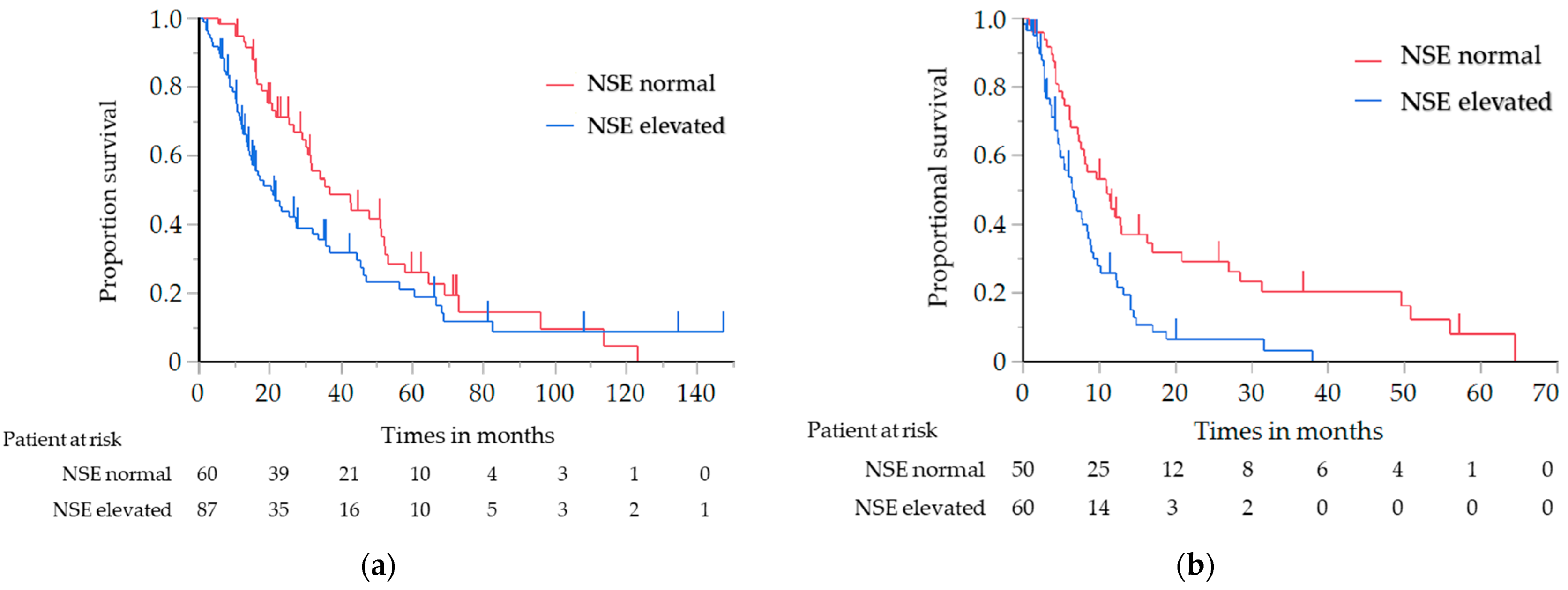
| Normal | 60 (40.8) | 36.8 (29.9–51.9) | 11.0 (7.3–16.2) | 1 | |||
| Elevated | 87 (59.2) | 20.3 (13.9–31.9) | 1.55 (1.04–2.31) | 0.029 | 6.4 (4.6–8.6) | 2.04 (1.31–3.18) | 0.001 |
| AFP level (n = 107) | |||||||
| Normal | 101 (94.4) | 28.9 (18.1–45.4) | 1 | 8.0 (5.8–9.0) | 1 | ||
| Elevated | 6 (5.6) | 33.4 (15.7–45.4) | 1.04 (0.33–3.34) | 0.945 | 4.8 (1.6–NE) | 2.60 (0.91–7.440) | 0.065 |
| Tumor Marker | OS | PFS | |||||
|---|---|---|---|---|---|---|---|
| n (%) | Median (95% CI) | HR (95% CI) * | p value † | Median (95% CI) | HR (95% CI) * | p Value † | |
| CEA level (n = 167) | |||||||
| Normal | 147 (88.0) | 31.9 (26.3–42.7) | 1 | 8.5 (7.3–9.9) | 1 | ||
| Elevated | 20 (12.0) | 28.9 (10.3–42.4) | 1.52 (0.83–2.80) | 0.175 | 6.2 (3.7–10.1) | 1.32 (0.70–2.48) | 0.387 |
| CYFRA level (n = 149) | |||||||
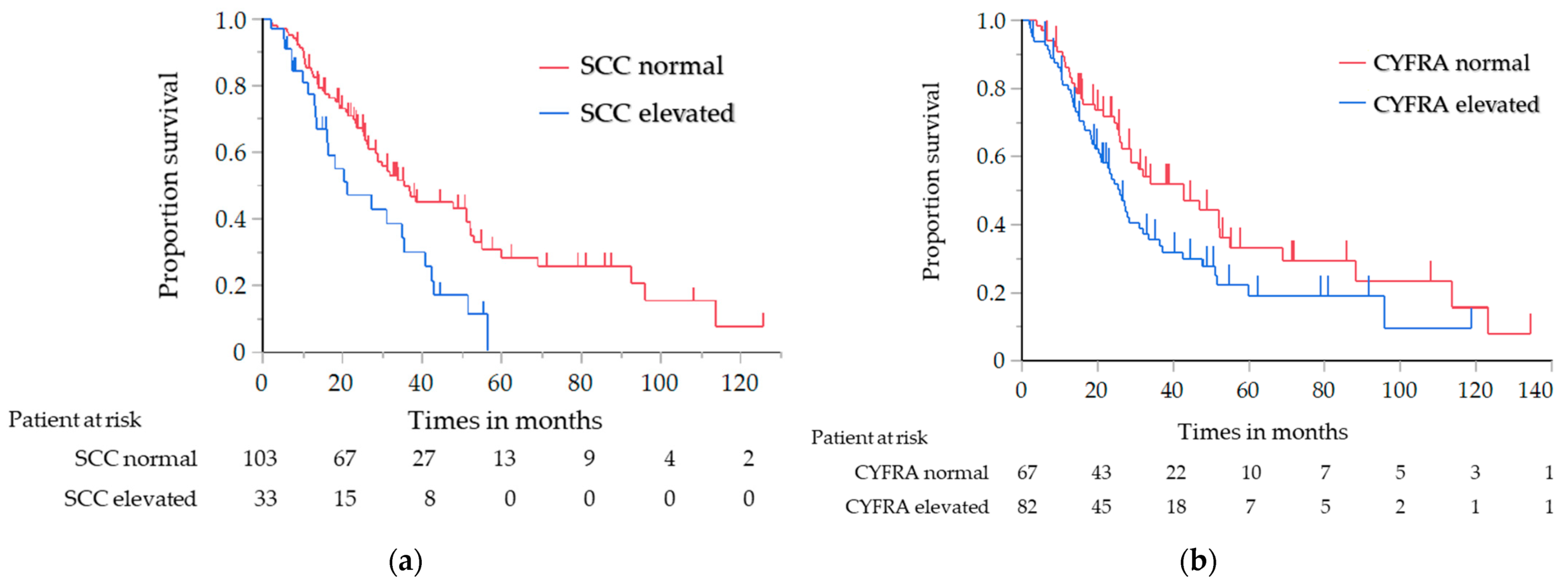
| Normal | 67(45.0) | 42.7 (26.3–52.2) | 1 | 9.2 (7.2–12.4) | 1 | ||
| Elevated | 82 (55.0) | 25.7 (13.4–35.4) | 1.52 (1.00–2.32) | 0.047 | 8.4 (5.8–9.8) | 1.05 (0.69–1.61) | 0.808 |
| SCC antigen level (n = 136) | |||||||
| Normal | 103 (75.7) | 35.5 (28.3–52.0) | 1 | 8.4 (7.2–10.9) | 1 | ||
| Elevated | 33 (24.3) | 21.1 (13.4–35.4) | 1.95 (1.20–3.16) | 0.006 | 8.6 (4.1–10.2) | 1.29 (0.79–2.10) | 0.301 |
| ProGRP level (n = 103) | |||||||
| Normal | 101(98.1) | 27.2 (20.8–37.1) | 1 | 7.6 (6.2–9.6) | 1 | ||
| Elevated | 2 (1.9) | 21.0 (14.2–27.8) | 1.95 (0.47–7.91) | 0.358 | 27.8 (NE–NE) | 0.55 (0.08–4.02) | 0.551 |
| NSE level (n = 93) | |||||||
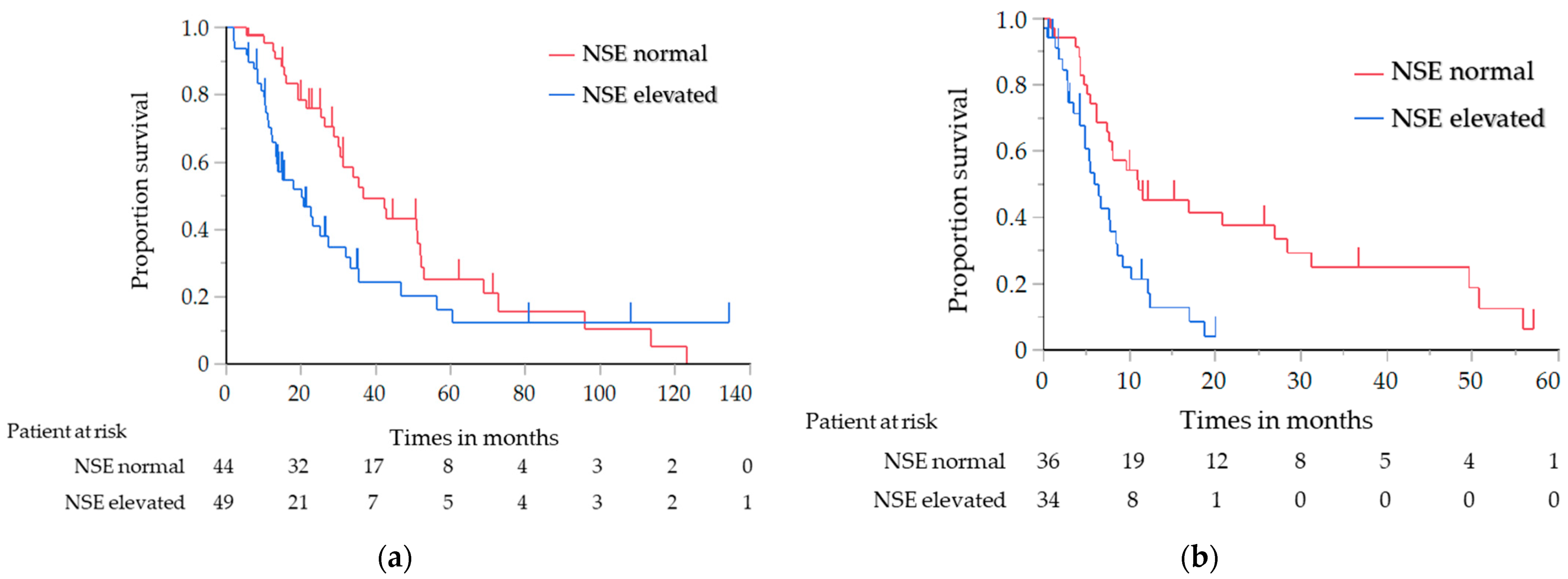
| Normal | 44 (47.3) | 36.8 (29.9–51.9) | 1 | 11.0 (7.3–28.4) | 1 | ||
| Elevated | 49 (52.7) | 20.3 (13.2–27.2) | 1.71 (1.05–2.80) | 0.030 | 5.9 (4.2–8.4) | 2.52 (1.40–4.54) | 0.001 |
| AFP level (n = 76) | |||||||
| Normal | 73 (96.1) | 28.9 (16.6–42.8) | 1 | 8.1 (5.8–9.2) | 1 | ||
| Elevated | 3 (3.9) | 45.4 (NE–NE) | 0.53 (0.07–3.88) | 0.527 | 4.8 (1.6–4.9) | 4.12 (1.20–14.18) | 0.015 |
| Category | N | Median (95% CI) (Months) | Univariate | Multivariate | ||
|---|---|---|---|---|---|---|
| HR (95% CI) | p Value | HR (95% CI) | p Value | |||
| NSE level | ||||||
| Normal | 60 | 36.8 (29.9–51.9) | 1 | 1 | ||
| Elevated | 87 | 20.3 (13.9–31.9) | 1.55 (1.04–2.31) | 0.030 | 1.67 (1.02–2.73) | 0.042 |
| Age (y) | ||||||
| <65 | 182 | 30.5 (23.2–36.7) | 1 | 1 | ||
| ≥65 | 104 | 31.2 (25.7–37.1) | 1.03 (0.76–1.39) | 0.853 | 0.74 (0.45–1.22) | 0.241 |
| Sex | ||||||
| Female | 84 | 21.3 (16.4–35.5) | 1 | 1 | ||
| Male | 202 | 31.9 (27.2–40.8) | 0.71 (0.53–0.97) | 0.031 | 0.84 (0.52–1.35) | 0.463 |
| Eastern Cooperative Oncology Group performance status | ||||||
| 0–1 | 248 | 32.0 (27.8–37.9) | 1 | 1 | ||
| 2–3 | 31 | 17.7 (11.3–21.3) | 1.75 (1.13–2.72) | 0.012 | 1.58 (0.83–3.00) | 0.162 |
| Histology | ||||||
| Squamous cell carcinoma | 190 | 31.9 (27.2–38.3) | 1 | 1 | ||
| Neuroendocrine carcinoma | 37 | 27.0 (16.3–45.0) | 1.35 (0.89–2.06) | 0.157 | 0.67 (0.37–1.20) | 0.174 |
| Others | 59 | 21.3 (14.8–35.9) | 1.32 (0.92–1.89) | 0.137 | 0.67 (0.36–1.20) | 0.176 |
| Masaoka stage | ||||||
| Recurrence/III | 67 | 36.5 (28.9–51.7) | 1 | 1 | ||
| IVa | 75 | 42.8 (28.2–52.9) | 0.82 (0.54–1.26) | 0.365 | 0.91 (0.20–4.16) | 0.907 |
| IVb | 144 | 21.3 (16.3–28.5) | 1.72 (1.19–2.48) | 0.004 | 2.16 (0.50–9.21) | 0.299 |
| (IVb vs. IVa) | 2.09 (1.47–2.99) | <0.001 | 2.36 (1.37–4.07) | 0.002 | ||
| World Health Organization TNM stage | ||||||
| Recurrence/III | 64 | 38.3 (30.5–51.9) | 1 | 1 | ||
| IV | 222 | 27.2 (23.2–33.9) | 1.37 (0.96–1.99) | 0.087 | 0.39 (0.09–1.75) | 0.281 |
| Volume-reduction surgery | ||||||
| Yes | 23 | 52.0 (28.5–123.2) | 1 | 1 | ||
| No | 263 | 28.9 (24.4–34.9) | 2.13 (1.19–3.84) | 0.012 | 1.50 (0.62–3.62) | 0.366 |
| Volume-reduction radiotherapy | ||||||
| Yes | 47 | 42.4 (32.0–52.2) | 1 | 1 | ||
| No | 240 | 27.2 (23.9–31.9) | 1.43 (0.96–2.21) | 0.093 | 1.47 (0.82–2.62) | 0.196 |
| First-line chemotherapy regimen | ||||||
| Platinum-based doublet | 178 | 30.7 (24.5–37.9) | 1 | |||
| Monotherapy | 10 | 54.9 (1.1–95.9) | 0.73 (0.34–1.58) | 0.431 | 0.50 (0.13–1.84) | 0.300 |
| Other multidrug regimens | 98 | 29.9 (23.2–37.1) | 0.94 (0.70–1.27) | 0.702 | 0.92 (0.58–1.47) | 0.735 |
Publisher’s Note: MDPI stays neutral with regard to jurisdictional claims in published maps and institutional affiliations. |
© 2022 by the authors. Licensee MDPI, Basel, Switzerland. This article is an open access article distributed under the terms and conditions of the Creative Commons Attribution (CC BY) license (https://creativecommons.org/licenses/by/4.0/).
Share and Cite
Mimori, T.; Shukuya, T.; Ko, R.; Okuma, Y.; Koizumi, T.; Imai, H.; Takiguchi, Y.; Miyauchi, E.; Kagamu, H.; Sugiyama, T.; et al. Clinical Significance of Tumor Markers for Advanced Thymic Carcinoma: A Retrospective Analysis from the NEJ023 Study. Cancers 2022, 14, 331. https://doi.org/10.3390/cancers14020331
Mimori T, Shukuya T, Ko R, Okuma Y, Koizumi T, Imai H, Takiguchi Y, Miyauchi E, Kagamu H, Sugiyama T, et al. Clinical Significance of Tumor Markers for Advanced Thymic Carcinoma: A Retrospective Analysis from the NEJ023 Study. Cancers. 2022; 14(2):331. https://doi.org/10.3390/cancers14020331
Chicago/Turabian StyleMimori, Tomoyasu, Takehito Shukuya, Ryo Ko, Yusuke Okuma, Tomonobu Koizumi, Hisao Imai, Yuichi Takiguchi, Eisaku Miyauchi, Hiroshi Kagamu, Tomohide Sugiyama, and et al. 2022. "Clinical Significance of Tumor Markers for Advanced Thymic Carcinoma: A Retrospective Analysis from the NEJ023 Study" Cancers 14, no. 2: 331. https://doi.org/10.3390/cancers14020331
APA StyleMimori, T., Shukuya, T., Ko, R., Okuma, Y., Koizumi, T., Imai, H., Takiguchi, Y., Miyauchi, E., Kagamu, H., Sugiyama, T., Azuma, K., Namba, Y., Yamasaki, M., Tanaka, H., Takashima, Y., Soda, S., Ishimoto, O., Koyama, N., Kobayashi, K., & Takahashi, K. (2022). Clinical Significance of Tumor Markers for Advanced Thymic Carcinoma: A Retrospective Analysis from the NEJ023 Study. Cancers, 14(2), 331. https://doi.org/10.3390/cancers14020331


