Review

Discovery and Validation of Clinically Relevant Long Non-Coding RNAs in Colorectal Cancer

by
Madison Snyder
1,2,
Susana Iraola-Guzmán
1,2,†,
Ester Saus
1,2 and
Toni Gabaldón
1,2,3,4,*
1
Barcelona Supercomputing Centre (BSC-CNS), Plaça Eusebi Güell, 1-3, 08034 Barcelona, Spain
2
Institute for Research in Biomedicine (IRB Barcelona), The Barcelona Institute of Science and Technology, Baldiri Reixac, 10, 08028 Barcelona, Spain
3
Catalan Institution for Research and Advanced Studies (ICREA), 08010 Barcelona, Spain
4
Centro de Investigación Biomédica En Red de Enfermedades Infecciosas (CIBERINFEC), 08028 Barcelona, Spain
*
Author to whom correspondence should be addressed.
Current address: Genomes for Life-GCAT lab Group, Institute for Health Science Research Germans Trias i Pujol (IGTP), 08916 Badalona, Spain.
Cancers 2022, 14(16), 3866; https://doi.org/10.3390/cancers14163866
Submission received: 12 July 2022 / Revised: 4 August 2022 / Accepted: 4 August 2022 / Published: 10 August 2022
(This article belongs to the Section Cancer Biomarkers)

Abstract

:

Simple Summary

Recent efforts in biomedical research have focused on the identification of molecular biomarkers to improve the diagnosis, prognosis and eventually treatment of the most common human diseases worldwide, including cancer. In this context, a large number of studies point to a pivotal role of long non-coding RNAs (lncRNAs) in the pathophysiology of carcinogenesis, suggesting diagnostic or therapeutic potential. However, for most of them, supporting evidence is scarce and often based on a single large-scale analysis. Here, focusing on colorectal cancer (CRC), we present an overview of the main approaches for discovering and validating lncRNA candidate molecules, and provide a curated list of the most promising lncRNAs associated with this malignancy.

Abstract

Colorectal cancer (CRC) is the third most prevalent cancer worldwide, with nearly two million newly diagnosed cases each year. The survival of patients with CRC greatly depends on the cancer stage at the time of diagnosis, with worse prognosis for more advanced cases. Consequently, considerable effort has been directed towards improving population screening programs for early diagnosis and identifying prognostic markers that can better inform treatment strategies. In recent years, long non-coding RNAs (lncRNAs) have been recognized as promising molecules, with diagnostic and prognostic potential in many cancers, including CRC. Although large-scale genome and transcriptome sequencing surveys have identified many lncRNAs that are altered in CRC, most of their roles in disease onset and progression remain poorly understood. Here, we critically review the variety of detection methods and types of supporting evidence for the involvement of lncRNAs in CRC. In addition, we provide a reference catalog that features the most clinically relevant lncRNAs in CRC. These lncRNAs were selected based on recent studies sorted by stringent criteria for both supporting experimental evidence and reproducibility.

1. Introduction

Cancer is among the most prominent life-threatening diseases in the world, with over 19 million newly diagnosed cases in 2020 alone [1]. Notably, colorectal cancer (CRC) is one of the most commonly diagnosed cancers, with nearly 2 million new cases each year (~10% of all new cancer cases). Moreover, CRC is the second leading cause of all cancer-related deaths, claiming almost 1 million lives in 2020 [1]. Although extremely deadly in the advanced stages, the development of CRC is gradual. Beginning from the pathological transformation of normal colonic epithelium to adenomatous polyp, CRC ultimately leads to invasive cancer [2]. CRC progression is generally categorized into five stages (0 to IV), depending on the extensiveness and clinical features [3]. The lethality of CRC is largely correlated to the stage of the disease at diagnosis. At its early stages (stages 0–II), CRC is very treatable, with 5-year survival rates as high as 90%. However, only 38% of CRC cases are diagnosed at an early, localized stage. By the later stages, the 5-year survival rate dramatically decreases to as low as 14% [4]. Hence, early diagnosis of CRC is key to saving lives.
While CRC has historically affected older populations, recent trends show an increase in cases in those under 50 years old. This has resulted in a decrease in the median age of patients with CRC from 72 to 66 years old. This is particularly concerning, as early onset CRC is often diagnosed at advanced (less treatable) stages, as compared to CRC in the traditional patient population [5]. Despite these negative trends, overall CRC mortality and incidence rates have consistently improved each year, reflecting the rise in preventative screenings, new testing, and targeted treatments [4].
Given the prominence and severity of this disease, there has been a large effort through recent research to better understand its causes, prognosis, and outcome. However, further research is still needed to improve prevention and treatment through more novel discoveries. This includes the identification of diagnostic and prognostic biomarkers. Compared to traditional diagnostic methods, such as colonoscopy or prognostic methods, including measuring tumor size and metastasis, the detection of biomarkers is less invasive. Unlike traditional tests, biomarker analysis can be carried out using urine, fecal, plasma, saliva or serum samples [6]. Thus, biomarkers have the potential to distinguish between benign and cancerous tumors (polyps vs. carcinomas) less invasively, while providing more accurate predictions of disease progression, likelihood of relapse, and even chance of onset [6].
Biomarkers have been a tremendous success at better informing diagnosis, prognosis, treatment, and preventative measures in other cancers [7,8,9,10]. One of the most famous examples, mutations in BRCA1/BRCA2 genes, have given patients the ability to reliably assess their risk of developing breast cancer in their lifetime, as well as their risk of relapse after a first bout of breast cancer. Additionally, mutations in BRCA1/BRCA2 genes can be used for informing patient care and treatment strategies after disease onset [7]. Similarly, the diagnostic lncRNA biomarker, PCA3, has been approved for clinical use in suspected cases of prostate cancer [11,12]. The ratio of PCA3, which encodes a prostate-specific RNA, and a prostate-specific antigen (PSA), is measured in urine samples and used to increase the specificity of the diagnosis [11,12,13]. In CRC, many different molecules have been identified as potential biomarkers, including lncRNAs. LncRNAs are RNA molecules longer than 200 bp that do not code for proteins. They have been broadly classified into sense, antisense, bidirectional, intronic and intergenic lncRNAs, depending on their relative position to protein-coding genes [14]. The vast majority of characterized lncRNAs are synthesized by Polymerase II, and subsequently spliced and 5′-capped. Additionally, some lncRNAs are also polyadenylated [15]. lncRNAs are poorly conserved, showing fewer exons, and generally have limited expression. Many lncRNAs are localized in the cell nucleus, where they exert regulatory functions by binding to DNA or DNA-associated proteins [16]. Other lncRNAs are transported to the cytosol, where they can interact with other cytosolic molecules. LncRNA mechanisms of action are generally classified into the following four main groups: chromatin regulation, gene regulation, scaffolding and condensation, and post-transcriptional regulation, as illustrated in Figure 1 [17]. LncRNA expression is generally limited by space and time, as it is often tissue or cell-type specific. Alterations in the pattern of expression of lncRNAs have been recurrently reported in cancer, where they can act as either oncogenes or tumor suppressors [18]. Overexpression and/or downregulation of lncRNAs in tumors is often associated with additional epigenetic alterations, such as DNA (de)methylation of promoters or enhancers [19,20,21]. It has been shown that differential expression of a subset of lncRNAs is associated with CRC heterogeneous features and also with functional pathways that mediate CRC, such as TGF-β and WNT pathways, immunity, epithelial-mesenchymal-transition (EMT), and angiogenesis [22]. The biomarker potential of lncRNAs has been increasingly studied in CRC in recent years [23]. However, most of these candidate lncRNAs currently lack proper experimental validation or characterization to be considered promising targets.
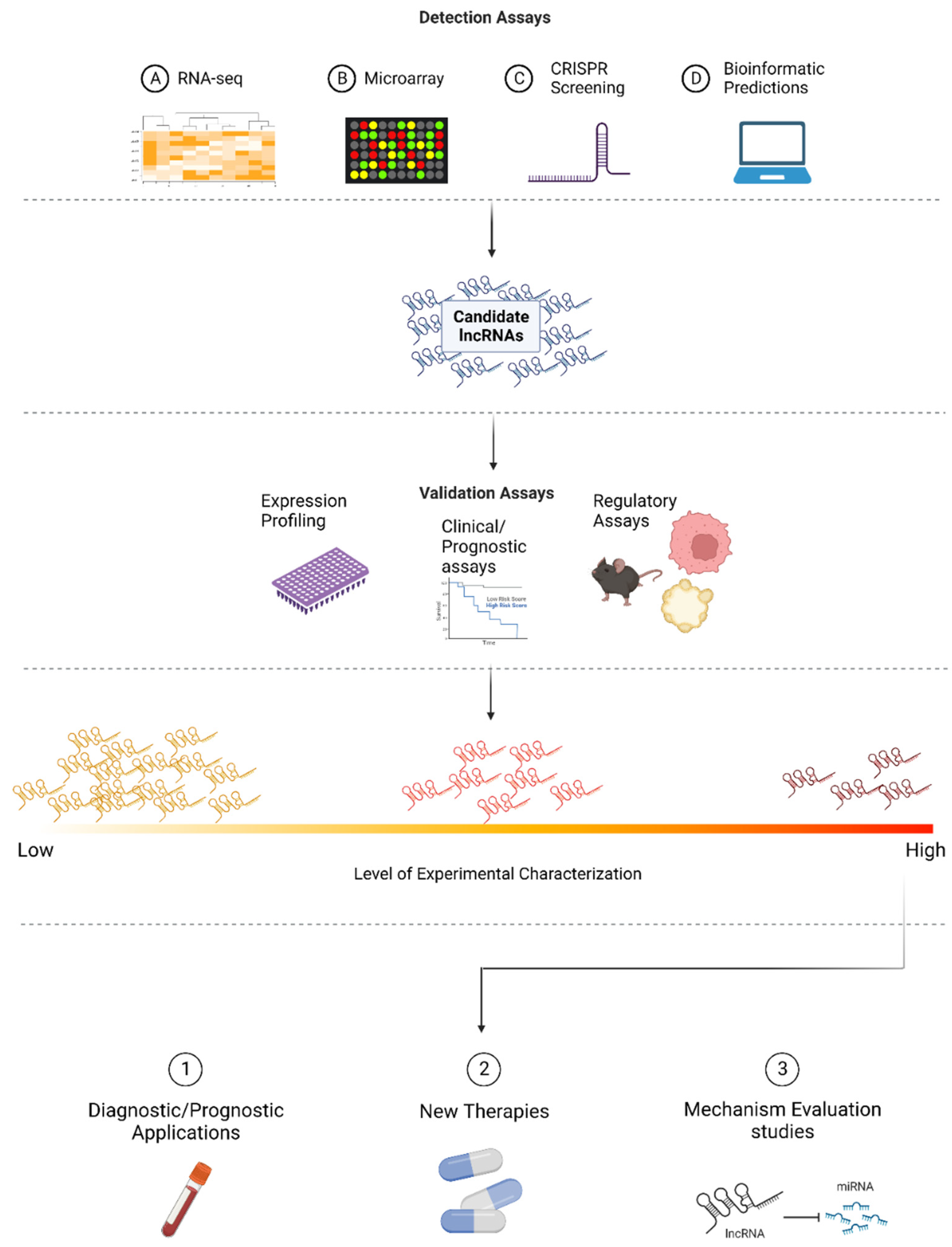
In this review, we outline the different modes of discovery for identifying lncRNAs as potential biomarkers in CRC and the advantages and disadvantages of each of these methods. We also discuss the common methods for evaluating the role and prognostic potential of previously identified lncRNAs. Based on recent studies, we provide a list of the most promising candidates based on study reproducibility and level of experimental characterization. Finally, we discuss the current use of lncRNAs as biomarkers in CRC and their potential as therapeutic targets in the future. Figure 2 provides an overview of the discovery and validation approaches for identifying clinically relevant lncRNAs, as described in detail in this review.

2. Approaches to Identify Relevant lncRNAs in CRC

Traditionally, approaches for determining the diagnosis and prognosis of CRC cases have been limited to non-molecular factors. In regard to diagnosis, colonoscopy and subsequent biopsy have been the gold standard in CRC screening [24]. Similarly, prognostic tools have been dominated by clinical and histological criteria including measurements of tumor size, tumor grade (stages described previously), and patient age among others [25]. However, recent research has started to move away from these approaches. Instead, focus has shifted towards identifying the prognostic and therapeutic potential of molecular biomarkers, including lncRNAs. LncRNAs comprise the majority of noncoding RNAs, many of which have unknown functions [14,26]. With the rise of next-generation sequencing (NGS) technologies and the subsequent ability to collect and analyze large volumes of data, many lncRNAs with prognostic and therapeutic potential in CRC have been identified. However, many of these candidates result from large-scale approaches that do not constitute conclusive proof, and therefore require further validation. Here, we provide a summary of the most used methods for identifying lncRNAs involved in CRC and discuss the advantages and disadvantages of each.

2.1. RNA Sequencing

With the increased accessibility of NGS, many researchers have begun to study transcriptional alterations in cancer through RNA sequencing (RNA-Seq). This technique allows researchers to reconstruct and quantify the expression of transcripts present in biological samples [27]. RNA-Seq studies that compare a CRC tumor and healthy tissue from the same patient can be used to uncover the differential expression and somatic mutations of various lncRNAs. These differentially expressed or mutated lncRNAs have the potential to be involved in the onset and progression of CRC, warranting further study.
Due to its untargeted approach, RNA-Seq uniquely allows for the discovery of novel lncRNAs. However, RNA-Seq does have some disadvantages. One significant challenge in detecting lncRNAs through RNA-Seq is their low relative abundance. Compared to protein coding genes, lncRNAs are extremely lowly expressed, constituting a minute fraction of the total RNA transcripts in a sample. To address this issue, target enrichment techniques utilizing probe-based strategies have been developed, enabling more effective lncRNA detection [28,29]. Given the large amounts of data that RNA-Seq analyses produce, there is also a risk of false positive detection of transcripts. This can be due to noisy expression or from transcripts that encode for proteins [27]. In fact, one study that investigated the reproducibility of differential expression results from identical replicates found that up to 8% of differentially expressed (DE) genes identified by RNA-Seq were false positives, even when using stringent identification parameters [30]. Maybe most concerningly, some studies have questioned the reproducibility of RNA-Seq results, as the resulting analyses are often dependent on the quality control, alignment, and quantification tools that are used in the analysis pipeline [31,32]. Moreover, the lack of normalization in analytical statistical methods often means that results are over dispersed and replicate dependent [32,33].

2.2. Microarrays

Microarrays are another genome-wide screening approach that can be used to identify lncRNAs involved in CRC. Microarrays are glass slides, lined with selected DNA oligonucleotide sequences in known locations that can hybridize specific lncRNAs (converted to cDNA) from a biological sample. Complementary base pairing between the sample and the oligonucleotide sequences on the chip produces light proportional to gene expression. Thus, hybridization allows for the detection of gene expression changes in a previously selected group of candidate lncRNAs in cancer cells [34,35]. Compared to RNA-Seq, microarrays are often less costly and require less complex and extensive bioinformatic analysis. However, they do require a preselection of lncRNAs that are suspected to have a biologically relevant function in CRC. Consequently, microarrays do not allow for the discovery of novel lncRNAs [27]. Recent work has also called into question the reliability and reproducibility of microarrays, due to unstable surface deposition chemistries [36]. A comparison of five different microarray data platforms revealed that there is poor concordance between systems in their output of results [37,38]. Additionally, poor sensitivity in detecting lowly expressed molecules, such as lncRNAs, limits detection efficiency [39].

2.3. CRISPR-Cas9 Screening

CRISPR-Cas9 screening for lncRNAs is one of the most optimizable modes of discovery for identifying candidate lncRNAs because of the many variables that can be altered when building a screen [40,41]. CRISPR-Cas9 screens can be designed with cells perturbed through loss of function techniques (inhibition or deletion) or gain of function techniques (activation). Screenings can also be specific to a number of lncRNA targets or to larger lncRNA pools. After selecting a perturbation method and the target lncRNAs, a single-guide RNA (sgRNA) library must be selected. NGS data of sgRNA counts are then used to identify lncRNAs associated with diseases such as CRC [42,43]. Compared to RNA-Seq or microarray analyses, there is no need to assume that differential expression implies function. Instead, CRISPR-Cas9 screening in a variety of cell lines can be used to identify lncRNAs through cancerous phenotypes, such as proliferation or drug resistance, independent of differential expression [42]. A major benefit of CRISPR-Cas9 screening is its customizability and effective targeting of lncRNAs [44]. However, statistical analyses of these screens are often hindered by the use of limited replicates [43]. Given the recent implementation of this technique, there is also a need to establish benchmarks to improve evaluation and reproducibility [42].

2.4. Bioinformatic Approaches

Several bioinformatic approaches that analyze genomic or transcriptomic data can be used to identify lncRNAs with prognostic or therapeutic potential in CRC. One such approach is the “detecting lncRNA cancer association” (DRACA) method, which was developed to evaluate potential molecular biomarkers by predicting lncRNA-cancer associations [45,46]. DRACA uses matrix factorization to consider interactions between lncRNAs, cancer prognosis, and other factors, such as miRNAs and genes, to predict lncRNA-cancer association. Using already available data on cancer prognosis, this approach results in both novel and biologically meaningful discoveries [46]. Likewise, the tool OncodriveFML was developed to identify known somatic mutations in genomic elements (such as lncRNAs) to predict those that have undergone positive selection during tumorigenesis. These lncRNAs have a high functional mutation bias and further role in CRC [47]. While innovative in its approach, OncodriveFML does have some limitations. OncodriveFML relies on the characterization of the functional impact of mutations in its calculations, meaning less characterized mutations in noncoding regions will not be counted. It also only predicts based on nucleotide substitutions, forgoing the identification of lncRNAs with insertions or deletions [47]. Another tool, the ExInAtor, also relies on mutational patterns of tumoral DNA, rather than changes in gene expression, to identify tumor driver lncRNAs [48]. This approach is especially advantageous for its specificity, rapid computation, and its ability to identify lncRNAs involved in tumorigenesis and not solely in upstream regulatory processes. However, ExInAtor does not evaluate the functional impact of mutations in lncRNAs, limiting the sensitivity of this approach and leaving room for many false negatives. Candidate lncRNAs identified by this approach also unexpectedly harbored many repeats and had lower GC content, indicating a possible bias of the tool [48]. An additional source is the RNA Atlas expanded with non-coding RNAs, which covers more than 3310 novel lncRNAs from RNAseq experiments that were performed in more than 300 human tissues and cell lines, including CRC [49].

3. Experimental Validation of Candidate lncRNAs

The discovery techniques outlined in the previous section are useful approaches for identifying lncRNAs of interest. However, given the existing biases and limitations of these approaches, further evaluation is needed to assess the true functional significance and clinical potential of these candidate lncRNAs. Here, we examine several common approaches used for this purpose.

3.1. Expression Profiling

As discussed above, one of the most common ways to identify lncRNAs with prognostic potential in CRC is through expression profiling in tumor and healthy tissues using RNA-Seq or microarrays. However, inconsistencies in the analysis of gene expression data can limit the reliability of these results. Real-time quantitative PCR (RT-qPCR) is the gold standard for verifying credible gene expression results [50]. In regard to lncRNAs in CRC, RT-qPCR can be used to amplify a target lncRNA in both tumoral and normal tissue. After each amplification cycle, the quantity of amplified target lncRNA is directly measured. Quantification assessments are then compared between tissue types to generate an expression profile [51]. While being a trusted validation method, many variables may jeopardize the reliable implementation of RT-qPCR. These include the quality of RNA, quantity and quality of reference genes, type of priming approach, and target abundance [52,53]. Although RT-qPCR is limited by technical and biological variability, it is often viewed as the benchmark technology in expression profiling, due to its sensitivity and specificity [50,51,52,53].
Another approach for validating expression profile data is through fluorescence in situ hybridization (FISH). FISH combines microscopy with the use of fluorescent probes that bind a target nucleic acid sequence to measure its presence and abundance within a particular tissue [54]. FISH can monitor RNA targets with high specificity and sensitivity and can accommodate different tissue types, including formalin-fixed paraffin embedded (FFPE) tissues, in which CRC tissues are often stored [55,56]. While a promising method, FISH is highly dependent on probe design, leaving room for variability in efficiency and accuracy of the approach [55]. An alternative methodology is RNAScope, which is a combined technique for the detection of target molecules of RNA in FFPE tissues [57]. In brief, multiple oligonucleotide-probes hybridize to target RNA molecules, then labeled (enzymatically or with fluorophores) amplifier molecules are hybridized to the probes, allowing detection by microscopy. Compared with previous in-situ hybridization techniques (ISH), RNAScope presents different advantages, such as increased sensitivity and specificity, and the possibility of multiple target detection. For this reason, it has been proposed as a valid alternative to RT-qPCR, for the characterization of biomarkers in cancer tissues [58,59,60,61]. More recently, the increasing interest in the localization and quantification of lncRNAs has stimulated the application of RNAScope to the detection of lncRNAs in FFPE tissue of patients with cancer [62,63]. The time-consuming set-up and the elevated cost are the main drawbacks of this methodology.

3.2. Clinical Significance

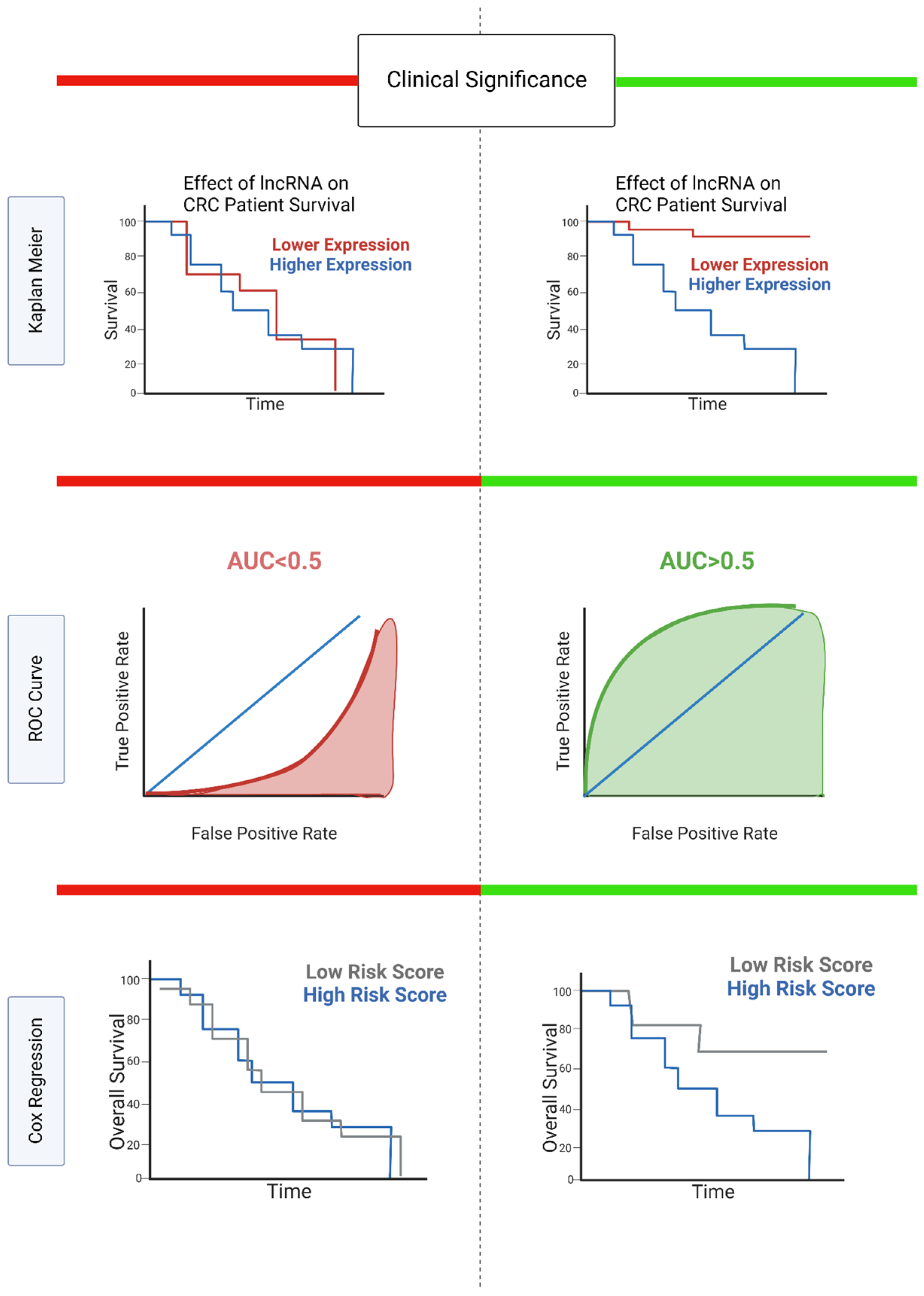
To evaluate the clinical significance of candidate lncRNAs, it is important to consider the clinicopathological, diagnostic, and prognostic value of these molecules. The correlation between candidate lncRNA expression and clinicopathological characteristics, such as age, sex, tumor size, tumor grade, and distant metastasis, is often analyzed to provide preliminary evidence of a lncRNA’s clinical relevance in CRC [64,65]. The most relevant analytical methods for defining clinical significance are summarized in Figure 3, and further explained here (Figure 3).
In terms of diagnosis, the receiver operating characteristics curve (ROC) is a trusted method to evaluate the sensitivity and specificity of biomarkers in CRC. ROC curves plot the true positive rate against the false positive rate for discriminating between CRC/non-CRC cases [66]. Consequently, ROC analysis can be used to determine the diagnostic relevance of a lncRNA biomarker, with the area under the ROC curve (AUC) as the quantifiable measurement of diagnostic accuracy. An ROC curve with an AUC of 0.5 describes a lncRNA biomarker with no discriminating ability between the CRC and non-CRC cases. AUCs above 0.5 provide some discriminatory ability, with an AUC of 1 representing a perfect biomarker [66].
Defining the prognostic value of lncRNAs is also essential to improve disease projections and patient care through more personalized treatment options. One common way to evaluate the prognostic potential of lncRNAs is through Cox regression analysis. Cox regression analysis is used to determine the relationship between predictors and the time that passes before an event occurs [67]. In this case, univariate Cox regression models are used to define the relationship between the predictor (lncRNA expression levels) and patient survival time. Univariate Cox regression between lncRNAs and overall survival has been carried out in multiple cancers, including colorectal cancer [68,69]. Multiple regression analysis can be used to analyze the significance between multiple variables, painting a more comprehensive picture of the prognostic potential of lncRNAs. This method can be more sensitive to comorbidities or covariates that potentially affect patient prognosis [70]. In this case, multiple lncRNA expression profiles, along with other variables, such as patient age, histology, or tumor grade, can be considered, giving a more advanced prognostic evaluation of the lncRNA signature.
Additionally, the Kaplan–Meier method is a powerful tool used to visualize survival differences in CRC. Kaplan–Meier plots show the overall survival rate of patients over a designated period of time. By separating patients into groups of high and low expression of a particular lncRNA, these plots can be used to visualize the effect of lncRNA expression on overall survival in CRC. Unlike Cox regression analyses, multiple variables cannot be considered in this survival analysis.
Given that a patient’s prognosis is a strong determinant of the therapeutic action that follows, prognostic and clinicopathological testing is incredibly important in defining the clinical significance of lncRNAs [69,71,72].

3.3. Regulatory Significance

In many cases, previously identified lncRNAs are predicted to have roles in tumorigenesis, migration, and invasion of cancer cells. To study the effect of aberrantly expressed lncRNAs on cancer development, researchers have investigated lncRNA-dependent progression and molecular networking of cancer cells through a variety of methods. In regard to cancer progression, dysregulating (silencing or overexpressing) a lncRNA of interest allows for the study of lncRNA-dependent cell proliferation, migration, invasion, and colony formation. The effect of an aberrantly expressed lncRNA on these essential cancer processes can be tested through apoptotic/CCK-8, wound healing, transwell, and cell formation assays, respectively [71]. These regulatory assays can be performed in several models of CRC, including CRC cell lines, colon organoids, and mouse models.
The regulatory significance of target lncRNAs is also highly dependent on their binding partners and network interactions. Proteins and mRNAs that are coexpressed with lncRNA targets can be predicted through bioinformatic softwares, including RPIseq, lncPRO, lncBASE, and Capsule-LPI, but also through techniques such as RNA-Seq [72,73,74,75]. The expression profiles of these predicted partners in CRC can then be examined through Western blots, dual luciferase assay, and RNA immunoprecipitation/RNA pull down [76,77]. A more extensive list of methods for the evaluation of lncRNA-dependent phenotypes is provided in Table 1.

4. Validated lncRNA Candidates in CRC

Countless studies have identified lncRNAs as candidate biomarkers in CRC. However, many of these studies fail to properly evaluate the role and diagnostic or prognostic potential of these lncRNAs. Carrying out experimental validation of candidate lncRNAs is important to elucidate which candidates are the most promising for future study. A recent study that surveyed a large dataset of lncRNAs found a total of 229 lncRNAs that were differentially expressed in CRC tumors, underscoring CRC heterogeneity. Among the well-established molecular features, the authors classified the tumors based on (i) microsatellite instability (MSI) caused by a deficient DNA mismatch repair (MMR), (ii) chromosome instability (CIN), (iii) the CpG island methylator phenotype (CIMP), and (iv) KRAS, BRAF, and TP53 gene mutations [22]. We surveyed recent studies to compile a list of candidate lncRNA biomarkers (listed in Table S1) that have been validated in CRC, using at least two of the following four methods: expression profiling, clinical or prognostic significance testing, regulatory significance testing (in vitro), and regulatory significance testing (in vivo). LncRNA candidates were primarily sourced using the cancer lncRNA census 2 (CLC2) [79]. Additionally, we included lncRNAs identified in our previous review on this topic, as well as from a previously published RNA-Seq dataset [23,29]. Testing information for each lncRNA was manually collected by reviewing articles on PubMed. This list includes 117 candidate transcripts, comprising both oncogenic and tumor suppressive lncRNAs and is organized in 4 categories by the extent of reproducibility and experimental characterization. To illustrate the nature of these candidates, we describe some of them here, focusing on two examples of lncRNAs with opposing contributions to CRC and with different levels of characterization, the lesser characterized LINC01296 and the extensively studied oncogene HOTAIR. Additionally, in Figure 1, we provide a comprehensive overview of the most frequently reported mechanism of action of candidate lncRNAs biomarkers, with strong supportive evidence in CRC (as listed in Table S1).

4.1. LINC01296

Long intergenic non-coding RNA 1296, also known as DUXAP9 (double homeobox A pseudogene 9), is a 68 kb long pseudogene located in 14q11.2. It is associated with cancer, including CRC, and has been recently proposed as a promising biomarker for CRC prognosis by several authors [80]. Information on this gene available at the Genecards website suggests that LINC01296 is poorly conserved (there are no described orthologs or paralogs), is located in a highly variable region, and is moderately expressed in only six human tissues, which are as follows: white blood cells, lymph nodes, heart, skeletal muscle, adipocyte and kidney [81]. Several studies have explored the expression of LINC01296 in CRC tissues, cell lines (SW480, SW620, LoVo, HT29, DLD1, HCT116, HCT-8, HCT-8/5-FU) and in vivo rodent models, and have shown overexpression of this lncRNA in tumoral samples compared with matching adjacent tissues [80,82,83]. Additionally, these same studies have shown a significant association with the overexpression of LINC01296 and tumor stage, lymph node metastasis, distant metastasis, as well as poor clinical prognosis and chemoresistance. In contrast, an earlier meta-analysis found a correlation between LINC01296 overexpression and better CRC prognosis [84]. Such contradictory results underscore the need for further studies using larger cohorts to elucidate LINC01296 implications in CRC. In vitro studies have shown the association of LINC01296 overexpression with cell proliferation, metastasis and chemoresistance to 5-fluorouracil (5-FU), supporting an oncogenic role [82]. Mechanistically, the LINC01296 transcript functions as an endogenous sponge of miR-26a to regulate mucin1 (MUC1) expression, catalyzed by GALNT3, which in turn modulates the activity of the PI3K/AKT pathway, and thereby the carcinogenesis process [82]. Proposed mechanisms of action include down-regulation of p15 for proliferation, and promotion of invasion by regulating the EMT process through the miR-141-3p/ZEB1-ZEB2 axis [80,83].

4.2. HOTAIR

The homeobox (HOX) transcript antisense intergenic RNA gene, first described in 2007 [85], is located at the HOXC locus (12q13.13) and encodes for a ~2.4 kb long transcript known as HOTAIR (http:/ensembl.org; accessed on 5 April 2022). HOTAIR is an oncogenic long intergenic non-coding transcript (lincRNA) that is highly expressed in CRC and multiple other tumor types [86]. HOTAIR shows conserved gene structure, high polymorphism (more than 6500 variant alleles have been described (http:/ensembl.org: accessed on 5 April 2022), and low sequence conservation [87]. HOTAIR was the first lncRNA described to act in trans [87], as a molecular scaffold, for the assembly of regulatory complexes, including PRC2 and LSD1 proteins, promoting epigenetic repression of the HOXD gene, through histone H3K27 trimethylation and histone H3K4me2 demethylation, respectively [85,88]. Other evidence showed a critical effect of HOTAIR activity on cell cycle progression and proliferation by regulating different molecules. Recently, novel mechanisms of action, including competitive endogenous RNA (ceRNA) and the microRNA sponge, have also been described [89] (see Figure 1 for further details). Of note, the overrepresentation of sponge and ceRNAs mechanisms of action as the most common for lncRNAs associated with CRC begs the question of whether this is due to study biases—i.e., recent studies looking for lncRNAs that target miRNA, etc.—or whether this is a recurrent mechanism in the context of the disease.
HOTAIR expression is positively correlated with the onset and progression of different types of cancer, such as breast, bladder, gastric and CRC, among others [90,91,92]. This overexpression correlates with poor overall survival rate, tumor stage, and metastasis [12]. In CRC, HOTAIR causes cell proliferation and metastasis by inducing EMT [93,94]. Several genetic variants have been associated with a high risk of CRC through increased HOTAIR overexpression [95,96,97]. In addition, HOTAIR transcripts have been detected in heterogeneous types of samples, including serum and other body fluids. HOTAIR expression has been shown to be high in CRC cell lines, tumor samples, and metastatic samples [94,98]. Interestingly, a recent study consistently found that downregulation of HOTAIR repressed the viability and metastasis of CRC cell lines in vitro, and suppressed the tumorigenesis, migration and invasion of CRC in vivo [94]. Importantly, another study has demonstrated that HOTAIR overexpression is associated with resistance to 5-FU-based chemotherapy. Both studies suggest a critical role of this molecule in CRC pathophysiology and a promising role as therapeutic target for CRC patients [98].
Overall, compelling evidence points to a potential role of HOTAIR as a biomarker for CRC prediction and diagnosis, due to its detection in bodily fluids [99,100], prognosis [101], and as a potential therapeutic target for the development of novel strategies to target its overexpression [94,98]. So far, a patent has been filed for HOTAIR in gastric cancer diagnosis [12], encouraging the development of novel applications.

4.3. Other Promising Candidates

Beyond LINC01296 and HOTAIR, countless other candidates have accumulated increasing evidence of a strong regulatory role in CRC. Some of the most well studied lncRNA candidates include the oncogenes NEAT1 and CCAT1 and the tumor suppressor genes GAS5 and MEG3 (Table 2). Notably, these lncRNA candidates often have additional, sometimes opposing, roles in other types of cancer. This emphasizes the need for tissue-specific studies to accurately evaluate the therapeutic potential of these lncRNAs. As lncRNA candidates have become more widely studied, several controversies over their role within a cancer specific context have ensued. Two of these controversies surround the lncRNAs MALAT1 and BANCR in CRC. Initially, these lncRNAs were thought to be overexpressed in CRC tissues, and therefore were proposed as oncogenic factors that promoted tumorigenesis [102]. In the case of BANCR, initial findings suggested that it induced EMT through a MEK signaling pathway, ultimately leading to an increase in the migratory abilities of cancer cells [102]. In the case of MALAT1, initial studies observed several mutations at the 3′ end of the MALAT1 transcript, elucidating its role in promoting cell migration and invasion in CRC [103]. While numerous studies have corroborated findings in support of the oncogenic role of BANCR and MALAT1, others have challenged these claims. In direct conflict with the initial studies, researchers using an in vivo model of BANCR overexpression in CRC found that the overexpression of BANCR inhibits tumor growth [104]. Conversely, another group using a similar in vivo model found that silencing of BANCR inhibits tumor growth, in line with the findings of initial studies [105]. Similarly, while MALAT1 has been extensively studied as an oncogenic lncRNA, a recent study suggests that downregulation of MALAT1 results in increased cancer cell migration and correlates with reduced patient survival [106]. These findings suggest that MALAT1 acts as a tumor suppressor, contrary to previous evidence [106,107]. These two examples illustrate the complexity of lncRNAs roles in cancer and underscore the need for further studies to clarify their roles and their potential as diagnostic or prognostic markers or as targets for new therapies.

5. Current Use of lncRNAs in Clinical Practice

LncRNAs are actively being explored as independent biomarkers or in combination with other lncRNAs or proteins for diagnosis and prognosis. The first lncRNA to be validated as a biomarker and used in clinical practice was PCA3 for the diagnosis of prostate cancer [11]. More recently, several clinical trials have investigated the association of lncRNAs with several diseases (for example, preeclampsia, NCt03903393; lung cancer, NCt03830619; acute ischemic stroke, NCt04175691) (Winkle et al., 2021). For CRC, there are two clinical trials underway at the time of writing this review [108]. The first explores the application of CCAT1 for early diagnosis of the disease (NCT04269746). The second examines the role of HOTTIP (in combination with the protein EIF4EBP1) as a biomarker for diagnosis and prognosis by measuring its abundance in serum and the presence of the single nucleotide polymorphism HOTTIP rs1859168 (NCT04729855). Both lncRNAs passed our selection criteria and are included in Table S1. Importantly, due to the efforts over the last decade, the list of lncRNA candidates for pre-clinical and clinical studies is growing, (see Table S1), which has resulted in more than 4000 patents for lncRNAs in CRC [109].
RNA-based drugs target the proteome, the transcriptome (including non-coding RNA), and the genome in an unprecedented way [110]. RNA therapeutics are specific and are able to target both common and rare or untargetable diseases in a personalized manner [111]. Nowadays, there is a growing interest in novel treatment strategies, with special focus on lncRNAs due to their advantageous characteristics, such as tissue specificity, regulatory roles, aberrant expression in cancer, and being detectable in serum samples. Additionally, at least three small molecules have been proven to prevent the action of two lncRNAs, GAS5 and MALAT1, related with diabetes and carcinogenesis, respectively [112]. Two clinical studies (NCT02508441; NCT03985072) have tested the effects of ASOs targeting mitochondrial lncRNA on solid tumors [113]. Overall, lncRNAs represent a promising source of novel tools in the RNA-therapeutics era that can be applied to a plethora of conditions, including CRC.

6. Conclusions

CRC continues to be one of the most common cancers worldwide, affecting millions of people each year. With an increasingly younger patient cohort and a high mortality rate, there is great need for the development of less invasive and more timely techniques for the diagnosis, prognosis and treatment of CRC. Previously, non-invasive diagnostic techniques and prognostic monitoring of CRC have been limited. However, recent studies have focused on novel tools and strategies for improving both our understanding of the disease and the perspective of CRC patients. In this review, we highlight the study of lncRNAs as potential biomarkers for the diagnosis, prognosis, and treatment of CRC, including an overview of the most commonly implemented methods for the detection and validation of these lncRNAs. While much of the research has focused on the discovery of these candidate biomarkers, it is paramount that lncRNA candidates are validated at the molecular, prognostic, and regulatory level. In this way, future research should focus on the most therapeutically actionable lncRNA targets. Although lncRNAs are not yet a common tool in CRC clinical practice, their growing use in other cancers is encouraging for the application of RNA therapeutics and personalized medicine in CRC. In the future, the continued evaluation of candidate biomarkers and subsequent clinical trials will undoubtedly aid in the transition to more personalized approaches for assessing CRC cases. By improving diagnosis, prognostic predictions, and targeted therapies, lncRNAs have the potential to alter treatment strategies and clinical outcomes for the better.

Supplementary Materials

The following supporting information can be downloaded at: https://www.mdpi.com/article/10.3390/cancers14163866/s1, Table S1: List of candidate lncRNAs and their experimental characterization.

Author Contributions

M.S. and T.G. defined the criteria and compiled the list of clinically relevant lncRNAs. M.S. wrote the first draft of the manuscript with contributions from E.S., S.I.-G. and T.G. All authors have read and agreed to the published version of the manuscript.

Funding

This research was funded by the Spanish Ministry of Science and Innovation with grant PGC2018-099921-B-I00, cofounded by European Regional Development Fund (ERDF); by the Catalan Research Agency (AGAUR) SGR423; by the European Union’s Horizon 2020 research and innovation programme (Grant ERC-2016-724173); by TRANSCOLONCAN COST action network (CA17118); by the Gordon and Betty Moore Foundation (Grant GBMF9742); by the “La Caixa” foundation (Grant LCF/PR/HR21/00737), and by the Instituto de Salud Carlos III (IMPACT grant IMP/00019) and CIBERINFEC (grant CB21/13/00061-ISCIII-SGEFI/ERDF). This research was made possible by the Fulbright U.S. Student Grant Program, sponsored by the U.S. Department of State, Bureau of Education and Cultural Affairs.

Acknowledgments

We would like to thank the other members of the Gabaldón group for their insightful discussions.

Conflicts of Interest

The authors declare no conflict of interest. The funders had no role in the design of the study; in the collection, analyses, or interpretation of data; in the writing of the manuscript; or in the decision to publish the results.

References

  1. Sung, H.; Ferlay, J.; Siegel, R.L.; Laversanne, M.; Soerjomataram, I.; Jemal, A.; Bray, F. Global Cancer Statistics 2020: GLOBOCAN Estimates of Incidence and Mortality Worldwide for 36 Cancers in 185 Countries. CA Cancer J. Clin. 2021, 71, 209–249. [Google Scholar] [CrossRef] [PubMed]
  2. Jass, J.R. Colorectal Cancer: A Multipathway Disease. Crit. Rev. Oncog. 2006, 12, 273–287. [Google Scholar] [CrossRef] [PubMed]
  3. Amin, M.B.; Edge, S.B.; Greene, F.L.; Byrd, D.R.; Brookland, R.K.; Washington, M.K.; Gershenwald, J.E.; Compton, C.C.; Hess, K.R.; Sullivan, D.C.; et al. AJCC Cancer Staging Manual; Springer International Publishing: New York, NY, USA, 2018; ISBN 9783319406176. [Google Scholar]
  4. Siegel, R.L.; Miller, K.D.; Goding Sauer, A.; Fedewa, S.A.; Butterly, L.F.; Anderson, J.C.; Cercek, A.; Smith, R.A.; Jemal, A. Colorectal Cancer Statistics, 2020. CA Cancer J. Clin. 2020, 70, 145–164. [Google Scholar] [CrossRef] [PubMed] [Green Version]
  5. Mauri, G.; Sartore-Bianchi, A.; Russo, A.-G.; Marsoni, S.; Bardelli, A.; Siena, S. Early-Onset Colorectal Cancer in Young Individuals. Mol. Oncol. 2019, 13, 109–131. [Google Scholar] [CrossRef] [Green Version]
  6. Chen, S.; Fang, Y.; Sun, L.; He, R.; He, B.; Zhang, S. Long Non-Coding RNA: A Potential Strategy for the Diagnosis and Treatment of Colorectal Cancer. Front. Oncol. 2021, 11, 762752. [Google Scholar] [CrossRef]
  7. Basu, N.N.; Ingham, S.; Hodson, J.; Lalloo, F.; Bulman, M.; Howell, A.; Evans, D.G. Risk of Contralateral Breast Cancer in BRCA1 and BRCA2 Mutation Carriers: A 30-Year Semi-Prospective Analysis. Fam. Cancer 2015, 14, 531–538. [Google Scholar] [CrossRef]
  8. Gonzalez, D.; Martinez, P.; Wade, R.; Hockley, S.; Oscier, D.; Matutes, E.; Dearden, C.E.; Richards, S.M.; Catovsky, D.; Morgan, G.J. Mutational Status of the TP53 Gene as a Predictor of Response and Survival in Patients with Chronic Lymphocytic Leukemia: Results from the LRF CLL4 Trial. J. Clin. Oncol. 2011, 29, 2223–2229. [Google Scholar] [CrossRef]
  9. Miller, B.E.; Tal-Singer, R.; Rennard, S.I.; Furtwaengler, A.; Leidy, N.; Lowings, M.; Martin, U.J.; Martin, T.R.; Merrill, D.D.; Snyder, J.; et al. Plasma Fibrinogen Qualification as a Drug Development Tool in Chronic Obstructive Pulmonary Disease. Perspective of the Chronic Obstructive Pulmonary Disease Biomarker Qualification Consortium. Am. J. Respir. Crit. Care Med. 2016, 193, 607–613. [Google Scholar] [CrossRef]
  10. Gordetsky, J.; Epstein, J. Grading of Prostatic Adenocarcinoma: Current State and Prognostic Implications. Diagn. Pathol. 2016, 11, 25. [Google Scholar] [CrossRef] [Green Version]
  11. Groskopf, J.; Aubin, S.M.J.; Deras, I.L.; Blase, A.; Bodrug, S.; Clark, C.; Brentano, S.; Mathis, J.; Pham, J.; Meyer, T.; et al. APTIMA PCA3 Molecular Urine Test: Development of a Method to Aid in the Diagnosis of Prostate Cancer. Clin. Chem. 2006, 52, 1089–1095. [Google Scholar] [CrossRef] [Green Version]
  12. Qian, Y.; Shi, L.; Luo, Z. Long Non-Coding RNAs in Cancer: Implications for Diagnosis, Prognosis, and Therapy. Front. Med. 2020, 7, 612393. [Google Scholar] [CrossRef] [PubMed]
  13. Xu, Y.-H.; Deng, J.-L.; Wang, G.; Zhu, Y.-S. Long Non-Coding RNAs in Prostate Cancer: Functional Roles and Clinical Implications. Cancer Lett. 2019, 464, 37–55. [Google Scholar] [CrossRef] [PubMed]
  14. Derrien, T.; Johnson, R.; Bussotti, G.; Tanzer, A.; Djebali, S.; Tilgner, H.; Guernec, G.; Martin, D.; Merkel, A.; Knowles, D.G.; et al. The GENCODE v7 Catalog of Human Long Noncoding RNAs: Analysis of Their Gene Structure, Evolution, and Expression. Genome Res. 2012, 22, 1775–1789. [Google Scholar] [CrossRef] [PubMed] [Green Version]
  15. Guttman, M.; Amit, I.; Garber, M.; French, C.; Lin, M.F.; Feldser, D.; Huarte, M.; Zuk, O.; Carey, B.W.; Cassady, J.P.; et al. Chromatin Signature Reveals over a Thousand Highly Conserved Large Non-Coding RNAs in Mammals. Nature 2009, 458, 223–227. [Google Scholar] [CrossRef] [PubMed]
  16. Statello, L.; Guo, C.-J.; Chen, L.-L.; Huarte, M. Gene Regulation by Long Non-Coding RNAs and Its Biological Functions. Nat. Rev. Mol. Cell Biol. 2021, 22, 96–118. [Google Scholar] [CrossRef]
  17. Poursheikhani, A.; Abbaszadegan, M.R.; Kerachian, M.A. Mechanisms of Long Non-Coding RNA Function in Colorectal Cancer Tumorigenesis. Asia Pac. J. Clin. Oncol. 2021, 17, 7–23. [Google Scholar] [CrossRef]
  18. Ashouri, A.; Sayin, V.I.; Van den Eynden, J.; Singh, S.X.; Papagiannakopoulos, T.; Larsson, E. Pan-Cancer Transcriptomic Analysis Associates Long Non-Coding RNAs with Key Mutational Driver Events. Nat. Commun. 2016, 7, 13197. [Google Scholar] [CrossRef] [Green Version]
  19. Wu, Z.; Liu, X.; Liu, L.; Deng, H.; Zhang, J.; Xu, Q.; Cen, B.; Ji, A. Regulation of lncRNA Expression. Cell. Mol. Biol. Lett. 2014, 19, 561–575. [Google Scholar] [CrossRef]
  20. Zhang, C.; Wang, X.; Li, X.; Zhao, N.; Wang, Y.; Han, X.; Ci, C.; Zhang, J.; Li, M.; Zhang, Y. The Landscape of DNA Methylation-Mediated Regulation of Long Non-Coding RNAs in Breast Cancer. Oncotarget 2017, 8, 51134–51150. [Google Scholar] [CrossRef] [Green Version]
  21. Song, P.; Wu, L.; Guan, W. Genome-Wide Identification and Characterization of DNA Methylation and Long Non-Coding RNA Expression in Gastric Cancer. Front. Genet. 2020, 11, 91. [Google Scholar] [CrossRef] [Green Version]
  22. James de Bony, E.; Bizet, M.; Van Grembergen, O.; Hassabi, B.; Calonne, E.; Putmans, P.; Bontempi, G.; Fuks, F. Comprehensive Identification of Long Noncoding RNAs in Colorectal Cancer. Oncotarget 2018, 9, 27605–27629. [Google Scholar] [CrossRef] [PubMed] [Green Version]
  23. Saus, E.; Brunet-Vega, A.; Iraola-Guzmán, S.; Pegueroles, C.; Gabaldón, T.; Pericay, C. Long Non-Coding RNAs As Potential Novel Prognostic Biomarkers in Colorectal Cancer. Front. Genet. 2016, 7, 54. [Google Scholar] [CrossRef] [PubMed] [Green Version]
  24. Vatandoost, N.; Ghanbari, J.; Mojaver, M.; Avan, A.; Ghayour-Mobarhan, M.; Nedaeinia, R.; Salehi, R. Early Detection of Colorectal Cancer: From Conventional Methods to Novel Biomarkers. J. Cancer Res. Clin. Oncol. 2016, 142, 341–351. [Google Scholar] [CrossRef] [PubMed]
  25. Kılıç, Ö.; Gültekin, Y.; Biri, İ.; Yılmaz, A.U. The Prognostic Factors on Survival Rates for Patients with Stage II-III Colon Cancer. Turk. J. Colorectal Dis. 2021, 32, 122–128. [Google Scholar] [CrossRef]
  26. Robinson, E.K.; Covarrubias, S.; Carpenter, S. The How and Why of lncRNA Function: An Innate Immune Perspective. Biochim. Biophys. Acta Gene Regul. Mech. 2020, 1863, 194419. [Google Scholar] [CrossRef]
  27. Lee, C.; Kikyo, N. Strategies to Identify Long Noncoding RNAs Involved in Gene Regulation. Cell Biosci. 2012, 2, 37. [Google Scholar] [CrossRef] [Green Version]
  28. Morlion, A.; Everaert, C.; Nuytens, J.; Hulstaert, E.; Vandesompele, J.; Mestdagh, P. Custom Long Non-Coding RNA Capture Enhances Detection Sensitivity in Different Human Sample Types. RNA Biol. 2021, 18, 215–222. [Google Scholar] [CrossRef]
  29. Iraola-Guzmán, S.; Brunet-Vega, A.; Pegueroles, C.; Saus, E.; Hovhannisyan, H.; Casalots, A.; Pericay, C.; Gabaldón, T. Target Enrichment Enables the Discovery of lncRNAs with Somatic Mutations or Altered Expression in Paraffin-Embedded Colorectal Cancer Samples. Cancers 2020, 12, 2844. [Google Scholar] [CrossRef]
  30. Li, S.; Łabaj, P.P.; Zumbo, P.; Sykacek, P.; Shi, W.; Shi, L.; Phan, J.; Wu, P.-Y.; Wang, M.; Wang, C.; et al. Detecting and Correcting Systematic Variation in Large-Scale RNA Sequencing Data. Nat. Biotechnol. 2014, 32, 888–895. [Google Scholar] [CrossRef] [Green Version]
  31. Simoneau, J.; Dumontier, S.; Gosselin, R.; Scott, M.S. Current RNA-Seq Methodology Reporting Limits Reproducibility. Brief. Bioinform. 2021, 22, 140–145. [Google Scholar] [CrossRef] [Green Version]
  32. Conesa, A.; Madrigal, P.; Tarazona, S.; Gomez-Cabrero, D.; Cervera, A.; McPherson, A.; Szcześniak, M.W.; Gaffney, D.J.; Elo, L.L.; Zhang, X.; et al. A Survey of Best Practices for RNA-Seq Data Analysis. Genome Biol. 2016, 17, 13. [Google Scholar] [CrossRef] [PubMed] [Green Version]
  33. Gu, X. Statistical Detection of Differentially Expressed Genes Based on RNA-Seq: From Biological to Phylogenetic Replicates. Brief. Bioinform. 2016, 17, 243–248. [Google Scholar] [CrossRef] [PubMed]
  34. Toolabi, N.; Daliri, F.S.; Mokhlesi, A.; Talkhabi, M. Identification of Key Regulators Associated with Colon Cancer Prognosis and Pathogenesis. J. Cell Commun. Signal. 2022, 16, 115–127. [Google Scholar] [CrossRef]
  35. Gao, C.; Zhao, D.; Zhao, Q.; Dong, D.; Mu, L.; Zhao, X.; Guo, M.; Xu, A.; Fang, L.; Liu, Q.; et al. Microarray Profiling and Co-Expression Network Analysis of lncRNAs and mRNAs in Ovarian Cancer. Cell Death Discov. 2019, 5, 93. [Google Scholar] [CrossRef] [PubMed] [Green Version]
  36. Brittain, W.J.; Brandsetter, T.; Prucker, O.; Rühe, J. The Surface Science of Microarray Generation-A Critical Inventory. ACS Appl. Mater. Interfaces 2019, 11, 39397–39409. [Google Scholar] [CrossRef]
  37. Camarillo, C.; Swerdel, M.; Hart, R.P. Comparison of Microarray and Quantitative Real-Time PCR Methods for Measuring MicroRNA Levels in MSC Cultures. Methods Mol. Biol. 2011, 698, 419–429. [Google Scholar]
  38. Sato, F.; Tsuchiya, S.; Terasawa, K.; Tsujimoto, G. Intra-Platform Repeatability and Inter-Platform Comparability of microRNA Microarray Technology. PLoS ONE 2009, 4, e5540. [Google Scholar] [CrossRef] [Green Version]
  39. Wang, Y.; Barbacioru, C.; Hyland, F.; Xiao, W.; Hunkapiller, K.L.; Blake, J.; Chan, F.; Gonzalez, C.; Zhang, L.; Samaha, R.R. Large Scale Real-Time PCR Validation on Gene Expression Measurements from Two Commercial Long-Oligonucleotide Microarrays. BMC Genomics 2006, 7, 59. [Google Scholar] [CrossRef] [Green Version]
  40. Esposito, R.; Bosch, N.; Lanzós, A.; Polidori, T.; Pulido-Quetglas, C.; Johnson, R. Hacking the Cancer Genome: Profiling Therapeutically Actionable Long Non-Coding RNAs Using CRISPR-Cas9 Screening. Cancer Cell 2019, 35, 545–557. [Google Scholar] [CrossRef] [Green Version]
  41. Pulido-Quetglas, C.; Johnson, R. Designing Libraries for Pooled CRISPR Functional Screens of Long Noncoding RNAs. Mamm. Genome 2021. [Google Scholar] [CrossRef]
  42. Liu, S.J.; Horlbeck, M.A.; Cho, S.W.; Birk, H.S.; Malatesta, M.; He, D.; Attenello, F.J.; Villalta, J.E.; Cho, M.Y.; Chen, Y.; et al. CRISPRi-Based Genome-Scale Identification of Functional Long Noncoding RNA Loci in Human Cells. Science 2017, 355, aah7111. [Google Scholar] [CrossRef] [PubMed] [Green Version]
  43. Li, W.; Xu, H.; Xiao, T.; Cong, L.; Love, M.I.; Zhang, F.; Irizarry, R.A.; Liu, J.S.; Brown, M.; Liu, X.S. MAGeCK Enables Robust Identification of Essential Genes from Genome-Scale CRISPR/Cas9 Knockout Screens. Genome Biol. 2014, 15, 554. [Google Scholar] [CrossRef] [PubMed]
  44. Aparicio-Prat, E.; Arnan, C.; Sala, I.; Bosch, N.; Guigó, R.; Johnson, R. DECKO: Single-Oligo, Dual-CRISPR Deletion of Genomic Elements Including Long Non-Coding RNAs. BMC Genomics 2015, 16, 846. [Google Scholar] [CrossRef] [PubMed] [Green Version]
  45. Xi, G.; Ziyu, X.; Yiting, L.; Zonghang, L.; Lifeng, Z. Construction of Competing Endogenous RNA Network and Identification of Novel Molecular Biomarkers in Colon Cancer: A Bioinformatic Analysis. Medicine 2021, 100, e25369. [Google Scholar] [CrossRef] [PubMed]
  46. Yan, H.; Chai, H.; Zhao, H. Detecting lncRNA-Cancer Associations by Combining miRNAs, Genes, and Prognosis With Matrix Factorization. Front. Genet. 2021, 12, 639872. [Google Scholar] [CrossRef] [PubMed]
  47. Mularoni, L.; Sabarinathan, R.; Deu-Pons, J.; Gonzalez-Perez, A.; López-Bigas, N. OncodriveFML: A General Framework to Identify Coding and Non-Coding Regions with Cancer Driver Mutations. Genome Biol. 2016, 17, 128. [Google Scholar] [CrossRef] [PubMed] [Green Version]
  48. Lanzós, A.; Carlevaro-Fita, J.; Mularoni, L.; Reverter, F.; Palumbo, E.; Guigó, R.; Johnson, R. Discovery of Cancer Driver Long Noncoding RNAs across 1112 Tumour Genomes: New Candidates and Distinguishing Features. Sci. Rep. 2017, 7, 41544. [Google Scholar] [CrossRef] [Green Version]
  49. Lorenzi, L.; Chiu, H.-S.; Avila Cobos, F.; Gross, S.; Volders, P.-J.; Cannoodt, R.; Nuytens, J.; Vanderheyden, K.; Anckaert, J.; Lefever, S.; et al. Publisher Correction: The RNA Atlas Expands the Catalog of Human Non-Coding RNAs. Nat. Biotechnol. 2021, 39, 1467. [Google Scholar] [CrossRef]
  50. Rajeevan, M.S.; Ranamukhaarachchi, D.G.; Vernon, S.D.; Unger, E.R. Use of Real-Time Quantitative PCR to Validate the Results of cDNA Array and Differential Display PCR Technologies. Methods 2001, 25, 443–451. [Google Scholar] [CrossRef] [Green Version]
  51. Nickells, R.W.; Pelzel, H.R. Tools and Resources for Analyzing Gene Expression Changes in Glaucomatous Neurodegeneration. Exp. Eye Res. 2015, 141, 99–110. [Google Scholar] [CrossRef] [Green Version]
  52. Bustin, S.A.; Benes, V.; Nolan, T.; Pfaffl, M.W. Quantitative Real-Time RT-PCR--a Perspective. J. Mol. Endocrinol. 2005, 34, 597–601. [Google Scholar] [CrossRef] [PubMed] [Green Version]
  53. Jacob, F.; Guertler, R.; Naim, S.; Nixdorf, S.; Fedier, A.; Hacker, N.F.; Heinzelmann-Schwarz, V. Careful Selection of Reference Genes Is Required for Reliable Performance of RT-qPCR in Human Normal and Cancer Cell Lines. PLoS ONE 2013, 8, e59180. [Google Scholar] [CrossRef] [PubMed] [Green Version]
  54. Bayani, J.; Squire, J.A. Fluorescence in Situ Hybridization (FISH). Curr. Protoc. Cell Biol. 2004, Chapter 22. Unit 22.4. [Google Scholar] [CrossRef]
  55. Veselinyová, D.; Mašlanková, J.; Kalinová, K.; Mičková, H.; Mareková, M.; Rabajdová, M. Selected In Situ Hybridization Methods: Principles and Application. Molecules 2021, 26, 3874. [Google Scholar] [CrossRef] [PubMed]
  56. Alamri, A.; Nam, J.Y.; Blancato, J.K. Fluorescence In Situ Hybridization of Cells, Chromosomes, and Formalin-Fixed Paraffin-Embedded Tissues. Methods Mol. Biol. 2017, 1606, 265–279. [Google Scholar]
  57. Wang, F.; Flanagan, J.; Su, N.; Wang, L.-C.; Bui, S.; Nielson, A.; Wu, X.; Vo, H.-T.; Ma, X.-J.; Luo, Y. RNAscope: A Novel in Situ RNA Analysis Platform for Formalin-Fixed, Paraffin-Embedded Tissues. J. Mol. Diagn. 2012, 14, 22–29. [Google Scholar] [CrossRef] [Green Version]
  58. Wang, H.; Wang, M.X.-M.; Su, N.; Wang, L.-C.; Wu, X.; Bui, S.; Nielsen, A.; Vo, H.-T.; Nguyen, N.; Luo, Y.; et al. RNAscope for in Situ Detection of Transcriptionally Active Human Papillomavirus in Head and Neck Squamous Cell Carcinoma. J. Vis. Exp. 2014. [Google Scholar] [CrossRef] [Green Version]
  59. Bingham, V.; McIlreavey, L.; Greene, C.; O’Doherty, E.; Clarke, R.; Craig, S.; Salto-Tellez, M.; McQuaid, S.; Lewis, C.; James, J. RNAscope Hybridization Confirms mRNA Integrity in Formalin-Fixed, Paraffin-Embedded Cancer Tissue Samples. Oncotarget 2017, 8, 93392–93403. [Google Scholar] [CrossRef] [Green Version]
  60. Baker, A.-M.; Huang, W.; Wang, X.-M.M.; Jansen, M.; Ma, X.-J.; Kim, J.; Anderson, C.M.; Wu, X.; Pan, L.; Su, N.; et al. Robust RNA-Based in Situ Mutation Detection Delineates Colorectal Cancer Subclonal Evolution. Nat. Commun. 2017, 8, 1998. [Google Scholar] [CrossRef] [Green Version]
  61. Morley-Bunker, A.; Pearson, J.; Currie, M.J.; Morrin, H.; Whitehead, M.R.; Eglinton, T.; Walker, L.C. Assessment of Intra-Tumoural Colorectal Cancer Prognostic Biomarkers Using RNA Hybridisation. Oncotarget 2019, 10, 1425–1439. [Google Scholar] [CrossRef] [Green Version]
  62. Tripathi, M.K.; Zacheaus, C.; Doxtater, K.; Keramatnia, F.; Gao, C.; Yallapu, M.M.; Jaggi, M.; Chauhan, S.C. Z Probe, An Efficient Tool for Characterizing Long Non-Coding RNA in FFPE Tissues. Noncoding RNA 2018, 4, 20. [Google Scholar] [CrossRef] [PubMed] [Green Version]
  63. Jones, J.; Zecchini, H.; Nagarajan, S. Multiplexed Detection and Analysis of Low-Abundance Long Noncoding RNA Using RNAscopeTM in Cultured Cells. Methods Mol. Biol. 2020, 2148, 111–125. [Google Scholar] [PubMed]
  64. Zhang, Y.; Peng, C.; Li, J.; Zhang, D.; Zhang, C.; Jin, K.; Ji, D.; Peng, W.; Tang, J.; Feng, Y.; et al. Long Non-Coding RNA CCDC144NL-AS1 Promotes Cell Proliferation by Regulating the miR-363-3p/GALNT7 Axis in Colorectal Cancer. J. Cancer 2022, 13, 752–763. [Google Scholar] [CrossRef] [PubMed]
  65. Yao, J.; Wang, C.; Dong, X.; Zhang, Y.; Li, Y.; Zhou, H.; Zhang, L. lncRNA SNHG22 Sponges miR-128-3p to Promote the Progression of Colorectal Cancer by Upregulating E2F3. Int. J. Oncol. 2021, 59. [Google Scholar] [CrossRef] [PubMed]
  66. Hoo, Z.H.; Candlish, J.; Teare, D. What Is an ROC Curve? Emerg. Med. J. 2017, 34, 357–359. [Google Scholar] [CrossRef]
  67. George, B.; Seals, S.; Aban, I. Survival Analysis and Regression Models. J. Nucl. Cardiol. 2014, 21, 686–694. [Google Scholar] [CrossRef] [Green Version]
  68. Li, W.; Chen, Q.-F.; Huang, T.; Wu, P.; Shen, L.; Huang, Z.-L. Identification and Validation of a Prognostic lncRNA Signature for Hepatocellular Carcinoma. Front. Oncol. 2020, 10, 780. [Google Scholar] [CrossRef]
  69. Huang, X.; Cai, W.; Yuan, W.; Peng, S. Identification of Key lncRNAs as Prognostic Prediction Models for Colorectal Cancer Based on LASSO. Int. J. Clin. Exp. Pathol. 2020, 13, 675–684. [Google Scholar]
  70. Bradburn, M.J.; Clark, T.G.; Love, S.B.; Altman, D.G. Survival Analysis Part II: Multivariate Data Analysis—An Introduction to Concepts and Methods. Br. J. Cancer 2003, 89, 431–436. [Google Scholar] [CrossRef]
  71. Pantazi, P.; Carollo, E.; Carter, D.R.F.; Brooks, S.A. A Practical Toolkit to Study Aspects of the Metastatic Cascade in Vitro. Acta Histochem. 2020, 122, 151654. [Google Scholar] [CrossRef]
  72. Li, Y.; Sun, H.; Feng, S.; Zhang, Q.; Han, S.; Du, W. Capsule-LPI: A LncRNA-Protein Interaction Predicting Tool Based on a Capsule Network. BMC Bioinformatics 2021, 22, 246. [Google Scholar] [CrossRef] [PubMed]
  73. Muppirala, U.K.; Honavar, V.G.; Dobbs, D. Predicting RNA-Protein Interactions Using Only Sequence Information. BMC Bioinformatics 2011, 12, 489. [Google Scholar] [CrossRef] [PubMed] [Green Version]
  74. Lu, Q.; Ren, S.; Lu, M.; Zhang, Y.; Zhu, D.; Zhang, X.; Li, T. Computational Prediction of Associations between Long Non-Coding RNAs and Proteins. BMC Genomics 2013, 14, 651. [Google Scholar] [CrossRef] [PubMed] [Green Version]
  75. Karagkouni, D.; Paraskevopoulou, M.D.; Tastsoglou, S.; Skoufos, G.; Karavangeli, A.; Pierros, V.; Zacharopoulou, E.; Hatzigeorgiou, A.G. DIANA-LncBase v3: Indexing Experimentally Supported miRNA Targets on Non-Coding Transcripts. Nucleic Acids Res. 2020, 48, D101–D110. [Google Scholar] [CrossRef]
  76. Cheng, Z.; Jiang, S.; Tao, R.; Ge, H.; Qin, J. Activating Transcription Factor 3-Activated Long Noncoding RNA Forkhead Box P4-Antisense RNA 1 Aggravates Colorectal Cancer Progression by Regulating microRNA-423-5p/nucleus Accumbens Associated 1 Axis. Bioengineered 2022, 13, 2114–2129. [Google Scholar] [CrossRef]
  77. Wang, Y.; Li, Z.; Xu, S.; Li, W.; Chen, M.; Jiang, M.; Fan, X. LncRNA FIRRE Functions as a Tumor Promoter by Interaction with PTBP1 to Stabilize BECN1 mRNA and Facilitate Autophagy. Cell Death Dis. 2022, 13, 98. [Google Scholar] [CrossRef]
  78. PubMed. Available online: https://pubmed.ncbi.nlm.nih.gov/ (accessed on 1 June 2022).
  79. Vancura, A.; Lanzós, A.; Bosch-Guiteras, N.; Esteban, M.T.; Gutierrez, A.H.; Haefliger, S.; Johnson, R. Cancer LncRNA Census 2 (CLC2): An Enhanced Resource Reveals Clinical Features of Cancer lncRNAs. NAR Cancer 2021, 3, zcab013. [Google Scholar] [CrossRef]
  80. Xu, J.; Zhang, Z.; Shen, D.; Zhang, T.; Zhang, J.; De, W. Long Noncoding RNA LINC01296 Plays an Oncogenic Role in Colorectal Cancer by Suppressing p15 Expression. J. Int. Med. Res. 2021, 49, 3000605211004414. [Google Scholar] [CrossRef]
  81. GeneCards. Available online: https://www.genecards.org (accessed on 5 April 2022).
  82. Liu, B.; Pan, S.; Xiao, Y.; Liu, Q.; Xu, J.; Jia, L. LINC01296/miR-26a/GALNT3 Axis Contributes to Colorectal Cancer Progression by Regulating O-Glycosylated MUC1 via PI3K/AKT Pathway. J. Exp. Clin. Cancer Res. 2018, 37, 316. [Google Scholar] [CrossRef] [Green Version]
  83. Sun, Z.; Shao, B.; Liu, Z.; Dang, Q.; Guo, Y.; Chen, C.; Guo, Y.; Chen, Z.; Liu, J.; Hu, S.; et al. LINC01296/miR-141-3p/ZEB1-ZEB2 Axis Promotes Tumor Metastasis via Enhancing Epithelial-Mesenchymal Transition Process. J. Cancer 2021, 12, 2723–2734. [Google Scholar] [CrossRef]
  84. Qiu, J.-J.; Yan, J.-B. Long Non-Coding RNA LINC01296 Is a Potential Prognostic Biomarker in Patients with Colorectal Cancer. Tumour Biol. 2015, 36, 7175–7183. [Google Scholar] [CrossRef] [PubMed]
  85. Rinn, J.L.; Kertesz, M.; Wang, J.K.; Squazzo, S.L.; Xu, X.; Brugmann, S.A.; Goodnough, L.H.; Helms, J.A.; Farnham, P.J.; Segal, E.; et al. Functional Demarcation of Active and Silent Chromatin Domains in Human HOX Loci by Noncoding RNAs. Cell 2007, 129, 1311–1323. [Google Scholar] [CrossRef] [PubMed] [Green Version]
  86. Hertler, A.A.; Schlossman, D.M.; Borowitz, M.J.; Laurent, G.; Jansen, F.K.; Schmidt, C.; Frankel, A.E. A Phase I Study of T101-Ricin A Chain Immunotoxin in Refractory Chronic Lymphocytic Leukemia. J. Biol. Response Mod. 1988, 7, 97–113. [Google Scholar] [PubMed]
  87. He, S.; Liu, S.; Zhu, H. The Sequence, Structure and Evolutionary Features of HOTAIR in Mammals. BMC Evol. Biol. 2011, 11, 102. [Google Scholar] [CrossRef] [PubMed] [Green Version]
  88. Zappulla, D.C.; Cech, T.R. Yeast Telomerase RNA: A Flexible Scaffold for Protein Subunits. Proc. Natl. Acad. Sci. USA 2004, 101, 10024–10029. [Google Scholar] [CrossRef] [PubMed] [Green Version]
  89. Yang, L.; Zhang, X.; Li, H.; Liu, J. The Long Noncoding RNA HOTAIR Activates Autophagy by Upregulating ATG3 and ATG7 in Hepatocellular Carcinoma. Mol. Biosyst. 2016, 12, 2605–2612. [Google Scholar] [CrossRef]
  90. Zhang, Y.; Zhang, K.; Luo, Z.; Liu, L.; Wu, L.; Liu, J. Circulating Long Non-Coding HOX Transcript Antisense Intergenic Ribonucleic Acid in Plasma as a Potential Biomarker for Diagnosis of Breast Cancer. Thorac Cancer 2016, 7, 627–632. [Google Scholar] [CrossRef]
  91. Li, C.-Y.; Liang, G.-Y.; Yao, W.-Z.; Sui, J.; Shen, X.; Zhang, Y.-Q.; Peng, H.; Hong, W.-W.; Ye, Y.-C.; Zhang, Z.-Y.; et al. Integrated Analysis of Long Non-Coding RNA Competing Interactions Reveals the Potential Role in Progression of Human Gastric Cancer. Int. J. Oncol. 2016, 48, 1965–1976. [Google Scholar] [CrossRef] [Green Version]
  92. Svoboda, M.; Slyskova, J.; Schneiderova, M.; Makovicky, P.; Bielik, L.; Levy, M.; Lipska, L.; Hemmelova, B.; Kala, Z.; Protivankova, M.; et al. HOTAIR Long Non-Coding RNA Is a Negative Prognostic Factor Not Only in Primary Tumors, but Also in the Blood of Colorectal Cancer Patients. Carcinogenesis 2014, 35, 1510–1515. [Google Scholar] [CrossRef]
  93. Kogo, R.; Shimamura, T.; Mimori, K.; Kawahara, K.; Imoto, S.; Sudo, T.; Tanaka, F.; Shibata, K.; Suzuki, A.; Komune, S.; et al. Long Noncoding RNA HOTAIR Regulates Polycomb-Dependent Chromatin Modification and Is Associated with Poor Prognosis in Colorectal Cancers. Cancer Res. 2011, 71, 6320–6326. [Google Scholar] [CrossRef] [Green Version]
  94. Jin, L.; Pan, Y.-L.; Zhang, J.; Cao, P.-G. LncRNA HOTAIR Recruits SNAIL to Inhibit the Transcription of HNF4α and Promote the Viability, Migration, Invasion and EMT of Colorectal Cancer. Transl. Oncol. 2021, 14, 101036. [Google Scholar] [CrossRef] [PubMed]
  95. Xue, Y.; Gu, D.; Ma, G.; Zhu, L.; Hua, Q.; Chu, H.; Tong, N.; Chen, J.; Zhang, Z.; Wang, M. Genetic Variants in lncRNA HOTAIR Are Associated with Risk of Colorectal Cancer. Mutagenesis 2015, 30, 303–310. [Google Scholar] [CrossRef] [PubMed]
  96. Zhang, J.; Liu, X.; You, L.-H.; Zhou, R.-Z. Significant Association between Long Non-Coding RNA HOTAIR Polymorphisms and Cancer Susceptibility: A Meta-Analysis. Onco. Targets. Ther. 2016, 9, 3335–3343. [Google Scholar] [CrossRef] [PubMed] [Green Version]
  97. Kim, J.O.; Jun, H.H.; Kim, E.J.; Lee, J.Y.; Park, H.S.; Ryu, C.S.; Kim, S.; Oh, D.; Kim, J.W.; Kim, N.K. Genetic Variants of Associated With Colorectal Cancer Susceptibility and Mortality. Front. Oncol. 2020, 10, 72. [Google Scholar] [CrossRef]
  98. Li, P.; Zhang, X.; Wang, L.; Du, L.; Yang, Y.; Liu, T.; Li, C.; Wang, C. lncRNA HOTAIR Contributes to 5FU Resistance through Suppressing miR-218 and Activating NF-κB/TS Signaling in Colorectal Cancer. Mol. Ther. Nucleic Acids 2017, 8, 356–369. [Google Scholar] [CrossRef] [Green Version]
  99. Zhao, W.; Song, M.; Zhang, J.; Kuerban, M.; Wang, H. Combined Identification of Long Non-Coding RNA CCAT1 and HOTAIR in Serum as an Effective Screening for Colorectal Carcinoma. Int. J. Clin. Exp. Pathol. 2015, 8, 14131–14140. [Google Scholar]
  100. Pardini, B.; Sabo, A.A.; Birolo, G.; Calin, G.A. Noncoding RNAs in Extracellular Fluids as Cancer Biomarkers: The New Frontier of Liquid Biopsies. Cancers 2019, 11, 1170. [Google Scholar] [CrossRef] [Green Version]
  101. Chen, S.; Zhang, C.; Feng, M. Prognostic Value of LncRNA HOTAIR in Colorectal Cancer: A Meta-Analysis. Open Med. 2020, 15, 76–83. [Google Scholar] [CrossRef] [Green Version]
  102. Guo, Q.; Zhao, Y.; Chen, J.; Hu, J.; Wang, S.; Zhang, D.; Sun, Y. BRAF-Activated Long Non-Coding RNA Contributes to Colorectal Cancer Migration by Inducing Epithelial-Mesenchymal Transition. Oncol. Lett. 2014, 8, 869–875. [Google Scholar] [CrossRef] [Green Version]
  103. Xu, C.; Yang, M.; Tian, J.; Wang, X.; Li, Z. MALAT-1: A Long Non-Coding RNA and Its Important 3’ End Functional Motif in Colorectal Cancer Metastasis. Int. J. Oncol. 2011, 39, 169–175. [Google Scholar]
  104. Shi, Y.; Liu, Y.; Wang, J.; Jie, D.; Yun, T.; Li, W.; Yan, L.; Wang, K.; Feng, J. Downregulated Long Noncoding RNA BANCR Promotes the Proliferation of Colorectal Cancer Cells via Downregualtion of p21 Expression. PLoS ONE 2015, 10, e0122679. [Google Scholar] [CrossRef] [PubMed]
  105. Ma, S.; Yang, D.; Liu, Y.; Wang, Y.; Lin, T.; Li, Y.; Yang, S.; Zhang, W.; Zhang, R. LncRNA BANCR Promotes Tumorigenesis and Enhances Adriamycin Resistance in Colorectal Cancer. Aging 2018, 10, 2062–2078. [Google Scholar] [CrossRef] [PubMed]
  106. Kwok, Z.H.; Roche, V.; Chew, X.H.; Fadieieva, A.; Tay, Y. A Non-Canonical Tumor Suppressive Role for the Long Non-Coding RNA MALAT1 in Colon and Breast Cancers. Int. J. Cancer 2018, 143, 668–678. [Google Scholar] [CrossRef] [PubMed] [Green Version]
  107. Chen, Q.; Zhu, C.; Jin, Y. The Oncogenic and Tumor Suppressive Functions of the Long Noncoding RNA MALAT1: An Emerging Controversy. Front. Genet. 2020, 11, 93. [Google Scholar] [CrossRef] [Green Version]
  108. Clinical Trials.gov. Available online: www.clinicaltrials.gov (accessed on 1 June 2022).
  109. Google Patents. Available online: https://patents.google.com/advanced (accessed on 5 April 2022).
  110. Yu, A.-M.; Choi, Y.H.; Tu, M.-J. RNA Drugs and RNA Targets for Small Molecules: Principles, Progress, and Challenges. Pharmacol. Rev. 2020, 72, 862–898. [Google Scholar] [CrossRef]
  111. Damase, T.R.; Sukhovershin, R.; Boada, C.; Taraballi, F.; Pettigrew, R.I.; Cooke, J.P. The Limitless Future of RNA Therapeutics. Front. Bioeng. Biotechnol. 2021, 9, 628137. [Google Scholar] [CrossRef]
  112. Winkle, M.; El-Daly, S.M.; Fabbri, M.; Calin, G.A. Noncoding RNA Therapeutics—Challenges and Potential Solutions. Nat. Rev. Drug Discov. 2021, 20, 629–651. [Google Scholar] [CrossRef]
  113. Chen, Y.; Li, Z.; Chen, X.; Zhang, S. Long Non-Coding RNAs: From Disease Code to Drug Role. Acta Pharm. Sin. B 2021, 11, 340–354. [Google Scholar] [CrossRef]
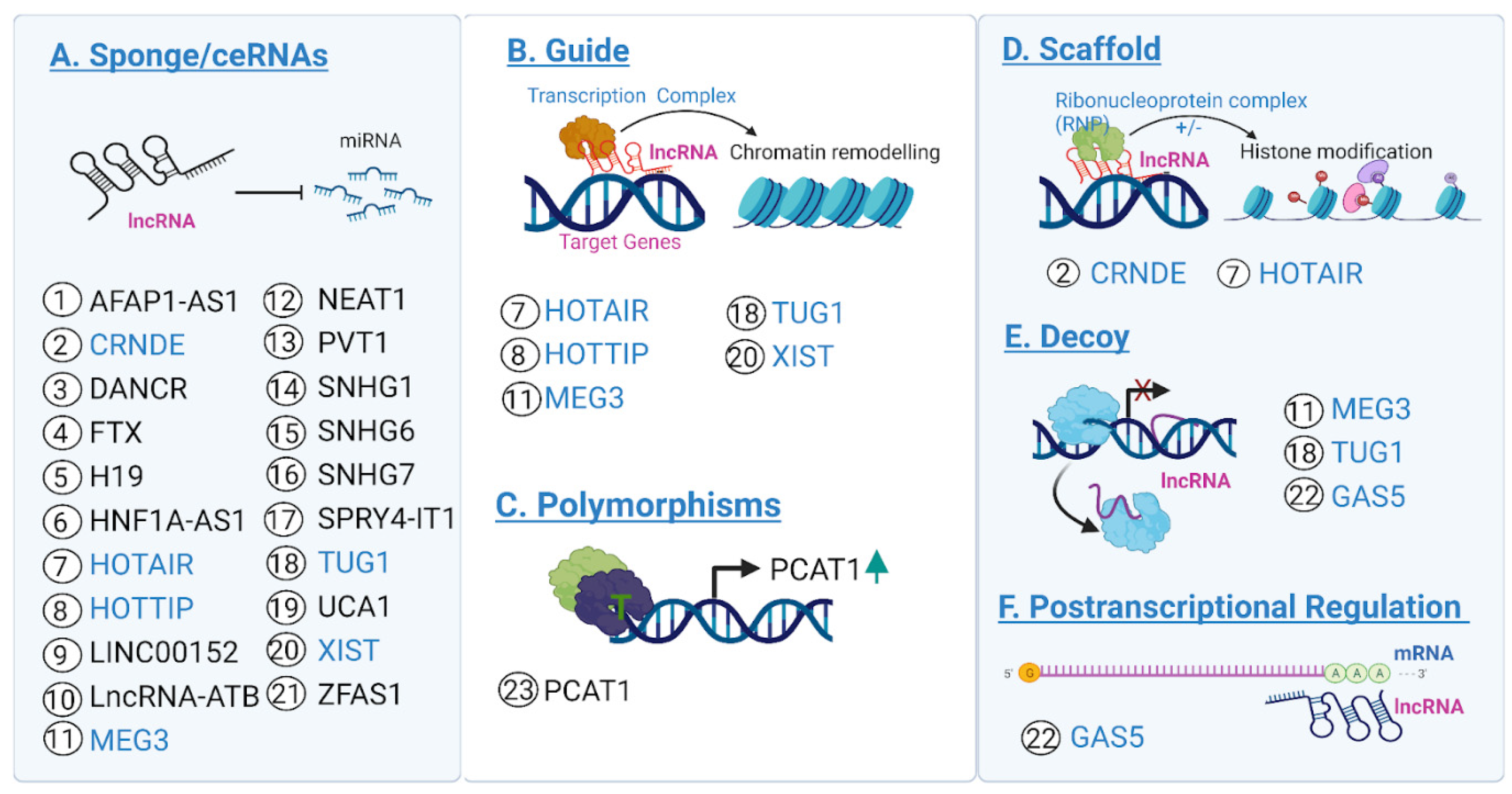
Figure 1. lncRNAs in CRC. Schematic description of the most frequent mechanisms of action (from AF) reported for the list of the candidate lncRNAs biomarkers (indicated by numbered circles) listed in Table 2, with strong supporting evidence in CRC. Highlighted in blue are the names of the lncRNAs with more than one mechanism reported. Created using Biorender.com.
Cancers 14 03866 g001
Figure 2. Methods for detecting and validating lncRNA biomarkers in CRC. After detection of candidate lncRNAs via the methods shown above (see text for full descriptions), validation techniques are used to characterize and assess which candidate lncRNAs are the most suitable for future studies. Regulatory assays may be carried out in cell lines, organoids, or in vivo models. Well characterized lncRNAs can be used as diagnostic/prognostic tools, as targets in future therapies, and as subjects of mechanistic studies. Created with Biorender.com.
Cancers 14 03866 g002
Figure 3. Evaluating clinical and prognostic significance of lncRNAs in CRC. Both insignificant (left, in red) and significant (right, in green) example graphics for Kaplan–Meier survival analysis, ROC curve analysis, and Cox regression analysis are shown. Created using Biorender.com.
Cancers 14 03866 g003
Table 1. Methods for determining lncRNA-dependent phenotypes of CRC.
MethodologyApplicationModelBiological
Process Tested
Dysregulation of lncRNA of Interest RequiredRefs. (PubMed ID) 1
Apoptotic assayIn vitro/in vivoCell lines, organoids, animal assaysCell proliferationYes 32144238
Transwell assayIn vitroCell lines, organoidsCell invasionYes 33328585
CCK-8 assayIn vitroCell lines, organoidsCell proliferationYes 34224294
MTT assayIn vitroCell lines, organoidsCell proliferationYes 33277833
Wound healing assayIn vitroCell lines, organoidsCell migrationYes 33570445
Colony formation assayIn vitroCell linesCell formationYes 34371180
Flow cytometryIn vitroCell linesCell cycle/apoptosisYes 33099922
Bioinformatic programs (RPIseq, lncPRO, lncBASE, Capsule-LPI)In silicoN/ACoexpression networkingNo 35034547
RNA sequencingIn vitroCell lines, organoidsCoexpression networkingYes 35039060
Western blotIn vitro/in vivoCell lines, organoids, animal assaysProtein expressionYes 34498706
Dual luciferase assayIn vitroCell linesInteractionsYes 35066433
RNA immunoprecipitationIn vivoCell lines, organoids, animal assaysInteractionsYes 35110535
RNA pull-downIn vitroCell lines, organoidsInteractionsYes 35107754
Tumor formation assayIn vivoOrganoids, animal assaysTumor formationYes 34477476
1 PMID = PubMed ID number [78].
Table 2. LncRNA candidates with strong supporting evidence for a regulatory role in CRC.
lncRNAMechanism of ActionRefs. (Pubmed ID) 1
AFAP1-AS1Proliferation, migration, invasion through the miR-195-5p/WISP1 axis. Tumor growth and metastasis34335760, 27578191
CRNDERegulation of apoptosis, proliferation, drug sensitivity via the Akt/mTORC1 pathway. Epigenetic transcriptional regulation of DUSP5 and CDNK1A35069879, 28796262
DANCRSuppression of apoptosis via RNA stabilization of MALAT1
Enhanced growth and metastasis via the DANCR/miR-518a-3p/MDM2 ceRNA network
33414433, 32423468
FTXProliferation, migration, invasion through the FTX-miR-214-5p-JAG1 regulatory axis. Enhanced growth and progression via the miR-192-5p/EIF5A2 axis34733921, 32280242
GAS5Inhibition of proliferation and migration, induction of apoptosis via the GAS5/miR-10b axis. Suppression of macroautophagy, induction of apoptosis
via the mTOR/SIRT1 pathway
35103069, 33416133
H19Migration, invasion, induction of EMT, metastasis via activation of Raf-ERK signaling
Proliferation, invasion, metastasis via the H19/miR-29b-3p/PGRN/Wnt axis
32698890, 29754471
HNF1A-AS1Migration, invasion, glycolysis via miR-124/MYO6. Angiogenesis via the PBX3/OTX1/ERK-MAPK pathway32110048, 32325080
HOTAIRMigration, invasion, EMT, cell viability via SNAIL/HNF4α transcriptional regulation. Suppression of miR-218 via the EZH2-targeting miR-218-2 promoter regulatory axis33588137, 28918035
HOTTIPProliferation, migration, invasion
Enhanced susceptibility via rs3807598, rs2067087, and rs17427960 SNPs
31945724, 30940774
LINC00152Proliferation and metastasis via promoter hypomethylation and the YAP1/LINC00152/miR-632/miR-185-3p/FSCN1 axis32307642, 32042551
lncRNA-ATBProliferation, migration, invasion via sponging miR-141-3p, metastasis
Developmental flexibility via transcriptional regulation of β-catenin
33199986, 32256798
MEG3Inhibited proliferation through targeting SOCS3/STAT3 signaling via miR-708
Inhibited proliferation and migration via the miR-376/PRDK1 signal axis
34934045, 31632544
NEAT1Proliferation, invasion, apoptotic suppression via the miR-138/SLC38A1 axis
Proliferation via the KDM5A/Cul4A/Wnt axis
32700988, 34109988
PCAT1Proliferation, migration, invasion, drug resistance. Proliferation, migration, invasion, apoptotic suppression via miR-149-5p regulation33277833, 31646561
PVT1 Proliferation, apoptotic regulation via the miR-761/MAPK1 axis. Epigenetic regulation of MYC, regulation of TGFβ/SMAD and Wnt/β-Catenin pathways 34515320, 33148262
SNHG1EMT regulation via miR-497-5p/miR-195-5p modulation. Proliferation, migration, invasion via Wnt/β-catenin signaling31276207, 29749530
SPRY4-IT1Cell growth and glycolysis via PDK1. Proliferation, migration, invasion, EMT regulation via miR-101-3p modulation33029299, 28720069
TUG1Proliferation, invasion, migration, apoptotic suppression, tumor growth via the miR-542-3p/TRIB2 axis and Wnt/β-catenin pathway. Proliferation, migration, cell viability via the TUG1/miR-145-5p/TRPC6 regulatory axis34030715, 32985219
TUSC7Inhibition of proliferation, invasion, EMT, enhanced apoptosis via the TUSC7/miR-23b/PDE7A axis33370523, 31002365
UCA1Proliferation, migration, invasion, EMT, drug resistance via the UCA1/miR-495-SP1/SP3 axis. Proliferation and drug resistance via UCA1/miR-495-HGF/c-MET33961855, 34976187
XISTProliferation, EMT, drug resistance via the XIST/miR-125b-2-3p/WEE1 axis. Proliferation, migration, invasion, apoptotic suppression via the miR-338-3p/PAX5 axis33666372, 32826710
ZEB1-AS1Proliferation via miR-141-3p regulation. Cell viability and apoptotic suppression via the MiR-205/YAP1 axis32669962, 32190742
ZFAS1Tumor size, metastasis, lipogenesis via PABP2/SREBP1. Proliferation, migration, invasion, metastasis via miR-34b/SOX435036050, 33725330
SNHG6Proliferation, apoptotic suppression via JAK2/SNHG6 regulation. Proliferation and invasion via miR-101-3p regulation and the Wnt/β-catenin signaling pathway32840014, 31533634
CCAT2Proliferation, apoptotic suppression. Proliferation, migration, invasion via TAF15/RAB14/AKT/GSK3β axis, tumor growth and metastasis33099922, 34868956
SNHG7Proliferation, migration, invasion, cell viability, and metastasis via miR-216b regulation and GALNT1 expression29915311, 33685194
FOXD2-AS1Proliferation, cell cycle regulation via miR-4306 regulation. Proliferation, migration, invasion via the miR-25-3p/Sema4C axis34396433, 31908535
LINC00460Metastasis via miR-149-5p and biglycan regulation. Proliferation, migration, invasion, apoptotic suppression via the miR-613/SphK1 axis33472555, 32821121
MIR4435-2HGProliferation, migration, invasion, metastasis via the miR-206/YAP1 axis. Proliferation, apoptotic suppression32154166, 32141545
ELFN1-AS1Proliferation, migration, invasion, apoptotic suppression via the miR-1205/MTA1 axis. Proliferation, migration, apoptotic suppression via the miR-4644/TRIM44 axis34337713, 31929141
LINC00858Suppression of apoptosis, senescence, autophagy. Tumor growth via WNK2 regulation. Proliferation, invasion, migration via the miR-4766-5p/PAK2 axis32768499, 31902050
CCAT1Proliferation, migration, invasion via the hsa-miR-4679/GNG10 axis. Migration, invasion, cell viability via the CCAT1/VEGF/miR-218 axis35005034, 32256733
1 PMID = PubMed ID number [78].
Publisher’s Note: MDPI stays neutral with regard to jurisdictional claims in published maps and institutional affiliations.