Review

Insights into the Possible Molecular Mechanisms of Resistance to PARP Inhibitors

Genetic Oncology Unit, Department of Oncology and Haematology, University Hospital of Modena, 41125 Modena, Italy
*
Author to whom correspondence should be addressed.
Cancers 2022, 14(11), 2804; https://doi.org/10.3390/cancers14112804
Submission received: 16 May 2022 / Revised: 2 June 2022 / Accepted: 3 June 2022 / Published: 5 June 2022
(This article belongs to the Special Issue A Deeper Dive into Signaling Pathways in Cancers)

Abstract

:

Simple Summary

The increasingly wide use of PARP inhibitors in breast, ovarian, pancreatic, and prostate cancers harbouring a pathogenic variant in BRCA1 or BRCA2 has highlighted the problem of resistance to therapy. This review summarises the complex interactions between PARP1, cell cycle regulation, response to stress replication, homologous recombination, and other DNA damage repair pathways in the setting of BRCA1/2 mutated cancers that could explain the development of primary or secondary resistance to PARP inhibitors.

Abstract

PARP1 enzyme plays an important role in DNA damage recognition and signalling. PARP inhibitors are approved in breast, ovarian, pancreatic, and prostate cancers harbouring a pathogenic variant in BRCA1 or BRCA2, where PARP1 inhibition results mainly in synthetic lethality in cells with impaired homologous recombination. However, the increasingly wide use of PARP inhibitors in clinical practice has highlighted the problem of resistance to therapy. Several different mechanisms of resistance have been proposed, although only the acquisition of secondary mutations in BRCA1/2 has been clinically proved. The aim of this review is to outline the key molecular findings that could explain the development of primary or secondary resistance to PARP inhibitors, analysing the complex interactions between PARP1, cell cycle regulation, PI3K/AKT signalling, response to stress replication, homologous recombination, and other DNA damage repair pathways in the setting of BRCA1/2 mutated cancers.

1. Introduction

Poly-(ADP-ribose) polymerase (PARP) enzyme PARP1 plays an important role in DNA damage recognition and signalling, as it binds single-stranded DNA breaks (SSBs) and then organizes their repair by synthesising PAR chains on target proteins (the so-called PARylation) [1]. In detail PARP1, once bound to SSBs via N-terminal zinc-finger DNA-binding domain, catalyses the polymerization of ADP-ribose moieties from NAD+ on target proteins, mainly PARP1 itself and histones. This process leads to chromatin relaxation and recruitment of other DNA repair enzymes such as XRCC1 [2,3,4,5]. The scaffolding protein XRCC1 stimulates the DNA kinase and DNA phosphatase activities of polynucleotide kinase at SSBs, accelerating the base excision repair reaction [6]. It is also reported that PARP1 promotes recruiting of MRE11, ATM and BRCA1, which are involved in double-stranded DNA break (DSB) repair by homologous recombination (HR) [7,8,9]. While PARP1 DNA binding is independent of its catalytic activity, dissociation of PARP1 from DNA requires PARylation presumably through a steric mechanism due to highly negatively charged PAR chains [10].
PARP inhibitors (PARPi) act mainly in a double way. The first proposed mechanism is the inhibition of the catalytic activity of PARP1, which results in synthetic lethality in cells with impaired HR [11,12,13]. In fact, inhibition of PARP1 promotes SSBs, which, if unrepaired, consequently lead to DSBs by collapse of the stalled replication fork during DNA replication [14]. In eukaryotic cells, DSBs are mainly resolved by the error-free mechanism of HR, which uses the intact sister chromatid as a template. HR deficiency induces activation of the more error-prone template-independent non-homologous end-joining (NHEJ) pathway, therefore, together with PARPi causing genomic instability followed by cell death [15,16]. Subsequent studies have revealed that most PARPi cause cytotoxicity by trapping PARP1 at sites of DNA damage [17,18,19]. According to the hypothesis proposed by Murai et al. [18], PARPi binding to the catalytic domain of PARP1 allosterically modifies interactions between DNA and the N-terminal DNA-binding domain of the protein, to the point that PARP1 becomes trapped on DNA. More recently, a third mechanism of PARPi sensitivity has been identified [20,21]. In cells with HR deficiency, aside from the NHEJ pathway, DSBs can be repaired by the microhomology-mediated end joining (MMEJ or Alt-EJ) pathway. Similarly to NHEJ, MMEJ is intrinsically error-prone, as the use of regions of microhomology inside DNA leads to deletions of nucleotides from the strand being repaired and to chromosomal translocations. In this pathway, the efficient recruitment of the DNA polymerase POLQ to the DSB requires PARP1. A PARPi will, therefore, block the MMEJ pathway and cause HR-deficient cell death.
PARP inhibitors are actually approved in breast, ovarian, pancreatic and prostate cancers harbouring a pathogenic or likely pathogenic variant in BRCA1 or BRCA2 (BRCA1/2) [22,23,24]. BRCA1/2 mutation prevalence varied widely from 1.8% in sporadic breast cancer to 36.9% in estrogen receptor/progesterone receptor low HER2 negative breast cancer [25]. Germline mutations in BRCA1/2 have been identified in 13–15% of women diagnosed with ovarian cancer, and somatic mutations are found in an additional 7% [26,27,28]. Germline BRCA1/2 mutations can be found in up to 8% of patients with sporadic pancreatic cancer [29]. In a sample of 692 patients with metastatic prostate cancer, unselected for family history or age at diagnosis, 5.3% carried a BRCA2 mutation, and 0.9% carried a BRCA1 mutation [30]. The increasingly wide use of PARPi in clinical practice is highlighting the problem of resistance to therapy. Considering the complex interaction between PARP1, HR and other DNA damage repair pathways in the setting of BRCA1/2 mutated cancers, several different mechanisms of resistance have been proposed, although some of them have been only described preclinically. The aim of this review is to outline the key molecular findings that could explain the mechanisms of primary or secondary resistance to PARPi (summarised in Table 1).

2. Primary Resistance

Primary resistance can be defined by a complete lack of response to the therapy. It can be mediated by co-occurring mutations in genes other than BRCA1/2 involved in cell cycle regulation and DNA damage repair, which could overcome HR deficiency, thus making cancer cells resistant to PARPi. Several mechanisms have been suggested but only in the preclinical setting.

2.1. PTEN Deficiency and PI3K/AKT Pathway Activation

Phosphatase and tensin homolog (PTEN) is a dual protein/lipid phosphatase that acts as a tumour suppressor gene inhibiting the PI3K/AKT pathway. In detail, PTEN converts phosphatidylinositol 3,4,5-triphosphate (PIP3) into phosphatidylinositol 4,5-bisphosphate (PIP2), antagonising PI3K action and preventing AKT activation and consequent cell growth and cell proliferation [62,63]. PTEN is often inactivated in different cancers [64]. Although AKT activation promotes BRCA1 expression through phosphorylation [65], it has been shown that BRCA1 can downregulate AKT activation in different ways: by acting on upstream kinases of AKT [31,66], by ubiquitin-mediated proteasomal degradation or by activating a protein serine/threonine phosphatase PP2A in breast cancer cells (Figure 1) [67]. Therefore, negative mutations and/or reduced expression of BRCA1 may activate the PI3K/AKT pathway [67]. Significantly, PTEN loss is highly associated with BRCA1-defective breast cancers, probably due to genomic instability resulting from deficient DSB repair [68], and the resulting PI3K/AKT activation stimulates the growth of those cancers [69].
Apart from its role in regulating the PI3K/AKT pathway, PTEN loss of function causes genetic instability, as PTEN-null cells lack competent HR DNA repair. In detail, PTEN has been suggested to contribute to RAD51 expression [70,71,72]. Dedes et al. [32] demonstrated that endometrioid endometrial cancer cell lines lacking PTEN function are unable to elicit competent HR DNA repair and are relatively sensitive to PARPi, and, as a consequence, PTEN silencing significantly increases the sensitivity to PARPi reducing RAD51 foci formation. Re-expression of PTEN in PTEN-mutant endometrioid endometrial cancer cells leads to relative resistance to PARPi [32].
Together, these observations corroborate the hypothesis that PTEN loss, which is highly associated with BRCA1-defective breast cancer, contributes to PARPi sensitivity. On the other hand, wild-type PTEN tumours could demonstrate relative resistance to PARPi. Considering that the PI3K/AKT pathway is constitutively active in BRCA1-defective human cancer cells [73], the combination of PTEN-related PI3K/AKT pathway inhibitors such as perifosine with DNA topoisomerase I (TOP1) or PARPi results in enhanced cancer cell killing in these tumours [74,75], suggesting a new possibly targetable pathway in case of PARPi resistance. A phase I trial of the PARPi Olaparib with the AKT inhibitor capivasertib, involving patients with advanced solid tumours carrying a germline BRCA1/2 mutation or somatic DNA damage response or PI3K/AKT pathway alterations, demonstrated the safety, tolerability and pharmacokinetic-pharmacodynamic activity of this combination [76].

2.2. ATM Roles and Loss of NHEJ Pathway

The choice between NHEJ and HR to repair DSBs is determined by several mechanisms, including activation of HR by cyclin-dependent kinase (CDK) activity (while NHEJ operates throughout interphase, HR is restricted to the S and G2 phase of the cell cycle, when a homologous sister chromatid is available as template) [16], or direct competition between HR and NHEJ stimulating factors at DSB sites [77]. During G2/S, HR is activated by binding of the MRE11–RAD50–NBS1 (MRN) complex to DSB ends. The MRN complex initiates DNA end resection, leading to the formation of single-strand DNA (ssDNA) at the extremity of the DSB. The ssDNA is protected from degradation by the loading of replication protein A (RPA) [78]. CtIP phosphorylated by CDK binds MRN complex to facilitate end resection [79]. The MRN complex recruits and activates the protein kinase ATM [80], while the sensor protein RPA finally drives ATR activation [81]. ATM and ATR phosphorylate several proteins involved in the HR [82]. The tumour suppressor complex BRCA1-BARD1 phosphorylated by ATM facilitates DNA end resection and interacts with PALB2, which in turn promotes the recruitment of BRCA2 [54]. PALB2 and BRCA2 remove RPA and facilitate the assembly of the RAD51 recombinase nucleoprotein filament. RAD51 nucleoprotein filament mediates the invasion of ssDNA into the intact sister chromatid, searching for a homologous template for DNA synthesis and repair [83,84,85]. During G1/2, 5′-3′ end resection is suppressed, and HR is inhibited due to the lack of sister chromatid [86]. ATM phosphorylates 53BP1 in multiple residues [87]. Once phosphorylated, 53BP1 binds and recruits RIF1 and PTIP that, together with the downstream effectors REV7 and Sheldin, inhibit 5′-3′ end resection and promote NHEJ [88]. Classical NHEJ starts when Ku70/80 heterodimer binds DSBs and recruits the DNA-dependent protein kinase catalytic subunit (DNA-PKcs) to form the DNA–PK complex. The latter engages XRCC4 and DNA ligase IV (LIG4) to align and ligates DNA ends regardless of sequence homology [89]. BRCA1 antagonizes 53BP1 by limiting its interaction with the chromatin in the S phase and stopping the translocation of RIF1 to DSBs in the G2/S phase, promoting HR [90,91]. Additionally, the different recruitment kinetics of the MRN and Ku complexes, which activate HR and NHEJ repair, respectively [92], as well as MRN/CtIP-dependent removal of Ku complex from DSBs [93], influence the choice between the two pathways (Figure 2).
Balmus et al. [33] demonstrated that ATM orchestrates the response to DSBs, promoting HR and preventing NHEJ that would result in chromosomal aberrations. They performed genome-wide CRISPR-Cas9 screens in wild-type and Atm-null mouse embryonic stem cells treated with the TOP1 inhibitor topotecan. They established that, in the absence of ATM, NHEJ-mediated repair of DSBs induced by TOP1 or PARPi leads to aberrant chromatid fusions and cell death. This could be avoided by impairing NHEJ through inactivation of the terminal components of the NHEJ machinery or promoting HR by inactivation of the BRCA1-A complex (BRCC45, ABRAXAS, MERIT40, RAP80, and BRCC36). In detail, the toxicity is mainly determined by the LIG4-mediated ligation step of NHEJ at DSBs and not by defective Ku removal, as previously suggested [93]. ATM phosphorylates hundreds of substrates [82], so it may operate at multiple levels to prevent NHEJ and ensure HR. The authors finally suggested that loss or reduced expression of the genes for the BRCA1-A complex and of the NHEJ components Ku70/80, XRCC4, XLF, and LIG4 in ATM-deficient tumours could explain resistance to PARPi in some cases [33].
The need for competent NHEJ to ensure synthetic lethality by PARPi in HR defective cells has been also proved by other studies. Patel et al. [34] disabled NHEJ by using genetic or pharmacologic approaches and rescued the lethality of PARP inhibition or down-regulation in cell lines lacking BRCA2, BRCA1, or ATM. McCormick and colleagues [35] assessed NHEJ in a panel of ovarian cancer cell lines and primary ascitic-derived ovarian cancer cultures, finding that NHEJ-defective cultures were resistant to the PARPi Rucaparib and only NHEJ-competent/HR-defective cultures were sensitive.

2.3. ALC1 Overexpression

Amplified in liver cancer 1 (ALC1) is a PAR-dependent chromatin remodeler that directly binds PAR chains, promoting PARP1 activation [94,95]. In a genome-wide CRISPR knockout screen with Olaparib, Juhász and colleagues [36] identified ALC1 as a key modulator of sensitivity to the PARPi Olaparib. ALC1 deficiency stimulates trapping of inhibited PARP1, which then impairs the binding of both the HR and NHEJ repair factors to DNA lesions. Moreover, they established that ALC1 overexpression, which is a common trait of many solid cancers, often associated with poor prognosis [96], reduces the sensitivity of BRCA1/2-deficient cells to PARPi. Analysis of the ALC1 expression levels before the use of PARPi could predict a mechanism of primary resistance to the therapy.

3. Secondary Resistance

Secondary or acquired resistance can be defined as the onset of the lack of response to treatment despite being successfully used before. Under therapeutic selective pressure, resistance to cytotoxic or target agents can emerge due to the expansion of pre-existing subclonal populations or from the evolution of drug-tolerant cells [97]. Several mechanisms of acquired resistance to PARPi have been suggested, although only reversion mutations in BRCA1/2 have been proved in the clinical setting.

3.1. Upregulation of ABC Transporters

Increased expression of the membrane-bound ATP-dependent efflux pump P-glicoprotein, encoded by ABCB1 (MDR1), is one of the most well-characterised mechanisms of multidrug resistance [98], in particular for doxorubicin, paclitaxel and related taxane drugs [99,100,101]. A chromosomal amplification event at 7q11.2-21 has been correlated to increased ABCB1 copy number and consequent P-glycoprotein expression in paclitaxel-resistant cancer cells [102].
Using a mouse model of BRCA2-deficient sarcomatoid breast cancer, Jaspers et al. [37] found that multidrug resistance was strongly associated with high expression of the ABCB1, known to be efflux transporter of Olaparib [103], docetaxel [104], and doxorubicin [105]. In novel A2780-derived ovarian cancer cell lines, paclitaxel-resistant cells were cross-resistant to the PARPi Olaparib and Rucaparib but not to Veliparib. This resistance correlated with increased ABCB1 expression and was reversible following treatment with ABCB1 inhibitors [38]. These findings, although cell-line based, could help in PARPi choice in paclitaxel-resistant patients, especially in second-line or maintenance therapy.

3.2. Decreased PARP1 Trapping

Through a genome-wide and high-density CRISPR-Cas9 mutagenesis screen to identify mouse embryonic stem cell mutants resistant to the PARPi Talazoparib, Pettitt and colleagues [39] identified point mutations in the N-terminal zinc-finger domain of PARP1 that, abolishing DNA binding, cause PARP1 resistance and alter PARP1 trapping. In support of this finding, they also observed a PARP1 mutation that abolished trapping in a patient with de novo resistance to Olaparib. In this experimental model, also mutations in amino-acid residues involved in hydrogen-bonding interactions that bridge the DNA-binding domain and the catalytic domain can cause PARPi resistance, likely by impairing PARP1 trapping; this reinforces the observations that inter-domain interactions are critical for PARP1 binding and activation [2,3,4].
PAR glycohydrolase (PARG) is the main enzyme responsible for degrading PAR chains, counteracting PARylation catalysed by PARP1 [106]. By combining genetic screens with multi-omics analysis of matched PARPi sensitive and resistant BRCA2-mutated mouse mammary tumours, Gogola et al. [40] identified loss of PARG as a major resistance mechanism. In a panel of 34 PARPi resistant tumours, they observed decreased expression of Parg in 17 tumours and acquired copy-number loss of the Parg locus in 22 tumours. Moreover, they demonstrated that PARG depletion does not increase PARP1 trapping per se but prevents excessive PARP1 binding to chromatin, thus reducing PARPi-dependent accumulation of toxic PARP1–DNA complexes. As a confirmation of this, PARG depletion also resulted in resistance to Talazoparib, a highly potent PARP1–DNA trapping agent. Finally, they found PARG-negative cell clones in a subset of human serous ovarian and triple-negative breast cancers (TNBCs), suggesting that this mechanism can contribute to PARPi resistance in vivo.

3.3. Restoration of HR

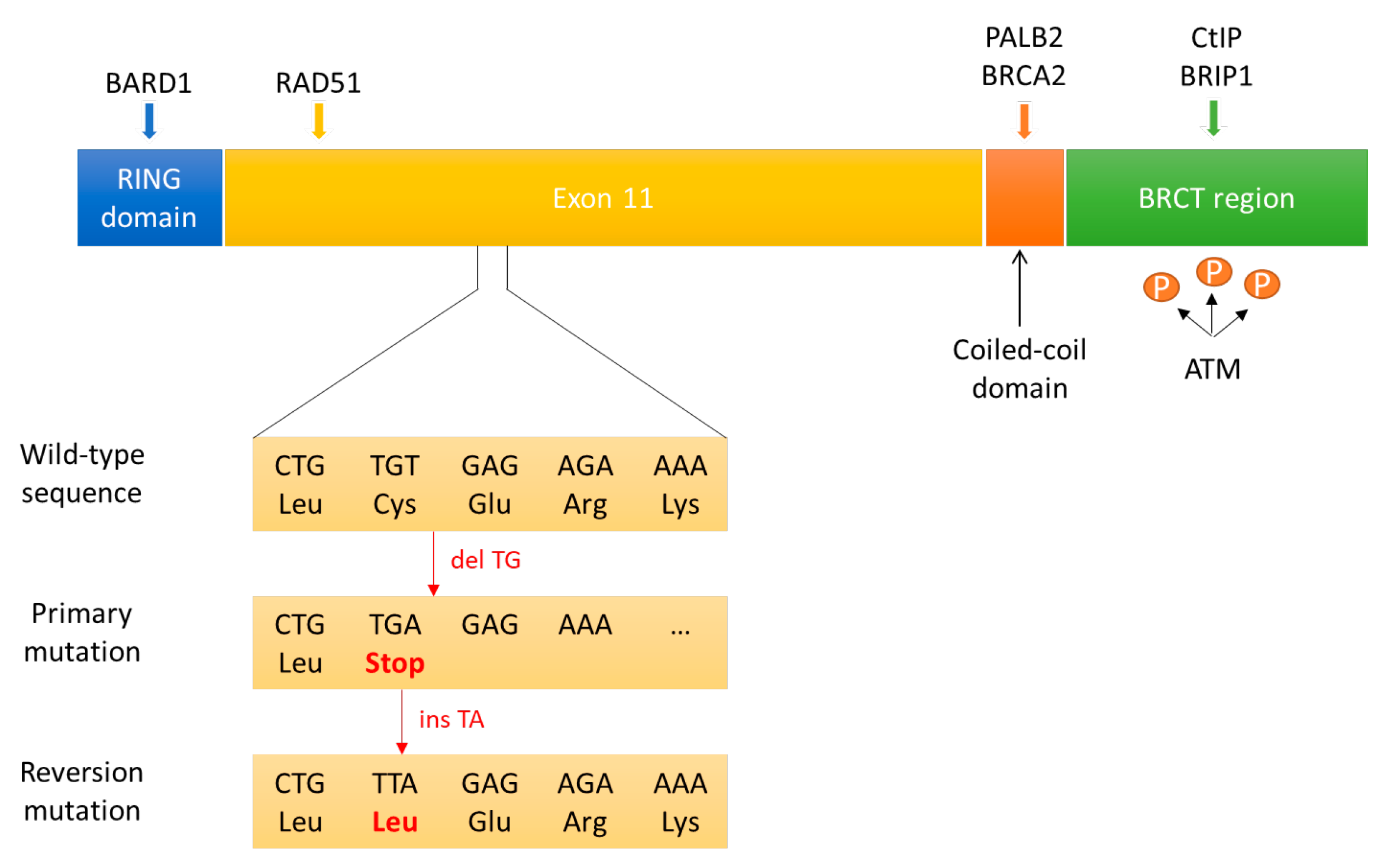
3.3.1. Reversion Mutations in BRCA1/2

The only mechanism of secondary resistance to PARPi currently validated in clinical practice is the acquisition of secondary mutations in BRCA1/2 restoring the open reading frame and detected upon treatment progression (Figure 3). Tobalina and colleagues [41] analysed sequencing data available in the literature from tumour or circulating tumour DNA (ctDNA) of 327 patients with tumours harbouring mutations in BRCA1/2 (234 patients with ovarian cancer, 27 with breast cancer, 13 with pancreatic cancer, 11 with prostate cancer and 42 with a cancer of unknown origin) on progression after platinum or PARPi treatment. Secondary mutations leading to the correction of the original mutation or to restoration of the BRCA1/2 open reading frame and functional proteins that were disrupted by it, the so-called reversion mutations, have been identified in 86 patients (26%). A comprehensive analysis of the reversion mutations points out that most amino acid sequences encoded by exon 11 in BRCA1 and BRCA2 are essential with regard to the protein function and consequentially to generate resistance to platinum or PARPi. Deletions accounted for the majority of secondary mutations, probably due to the upregulated use of the error-prone mechanisms of NHEJ or MMEJ to repair DNA DSBs in HR-deficient cells. As proof of this, researchers observed an increase in the length of micro-homologies (that can be considered MMEJ signatures) surrounding breakpoints in secondary deletion when compared with primary mutation and, in the case of BRCA2 deletions (regardless of being primary or secondary), compared with those in BRCA1, suggesting that several variables such as chromosomal location and/or chromatin landscape around BRCA1 and BRCA2 could determine the choice of the repair pathway when these genes are altered [107].

3.3.2. Hypomorphic BRCA1 Allele

It is unclear whether all pathogenic BRCA1 mutations have comparable effects on the response to therapy. C-terminal truncated BRCA1 proteins can be semifunctional, retaining the protein domains necessary to mediate interactions with PALB2-BRCA2 (Figure 3). Under PARPi selection pressure, the HSP90 protein can interact with and stabilize these mutant BRCA1 proteins, and the partial BRCA1 function can promote RAD51 loading onto DNA following DNA damage [108,109,110,111,112,113]. Even some N-terminal BRCA1 mutations may retain some residual activity in DNA damage response (Figure 3). Comparing two different BRCA1 mutations on N-terminal domain (185delAG and 5382insC) in genetically engineered mouse models, BRCA1 (185delAG) tumours responded significantly worse to PARPi than the BRCA1 (5382insC) tumours. The hypothesis suggested is that BRCA1 (185delAG) tumour cells produce a RING-less BRCA1 protein; this structure may lead to PARPi resistance through its residual activity by activating RAD51 [42].
Additionally, BRCA1 mRNA isoforms originated by alternative splicing and lacking exon 11 can produce truncated but hypomorphic proteins that have residual BRCA1 function (Figure 3). Ovarian cancer cells with BRCA1 splice isoforms lacking exon 11 may be clonally selected under PARPi treatment. Analysis of clinical ovarian cancer samples showed that exon 11 mutation carriers had worse overall survival when compared with non-exon 11 mutation carriers, probably due to less sensitivity to platinum-based chemotherapy by the cells with residual BRCA1 function of the hypomorphic protein [43]. The ability of BRCA1 hypomorphic protein to confer resistance to drug treatment has also been described in patient-derived xenograft (PDX) models [44,45,46].

3.3.3. Epigenetic Reversion of BRCA1 Promoter Hypermethylation

Epigenetic inactivation of BRCA1 through promoter hypermethylation occurs in approximately 15% of TNBCs and 11% to 15% of serous ovarian cancers [114,115]. In PDX models of BRCA1-deficient breast cancer, responses of the BRCA1-methylated PDX tumours to alkylating agents and Olaparib were comparable with those of the BRCA1-mutated model. However, several BRCA1-methylated PDX tumours acquired therapy resistance via re-expression of BRCA1 thanks to the loss of the BRCA1 promoter methylation [47]. This is consistent with previous cell line studies [48,49] and to what was shown in a patient with ovarian carcinoma [115]. Interestingly, in other BRCA1-methylated PDX tumours, BRCA1 re-expression was determined by de novo intrachromosomal genomic rearrangements through which BRCA1 transcription is placed under the control of a heterologous promoter [47].

3.3.4. Loss of End Resection Regulation

The tumour suppressor 53BP1 plays an important role in DSB repair pathway choice [82,83,116]. RIF1 is the most proximal effector of 53BP1 in its anti-resection function of DNA 5′ ends [117]; REV7 is a downstream factor in 53BP1-/RIF1-dependent NHEJ promotion [118]. Genetic depletion of 53BP1, RIF1, or REV7 provides synthetic viability to BRCA1-deficient cells and provides resistance to PARPi, restoring HR. Belotserkovskaya et al. [50] demonstrated that depleting 53BP1 in BRCA1-null cell lines reversed synthetic lethality mediated by PARPi, reactivating HR in a PALB2-dependent manner. Loss of REV7 re-establishes CtIP-dependent end resection of DSBs in BRCA1-deficient cells, leading to HR restoration and PARPi resistance [51]. Additionally, inactivating mutations in Sheldin, a 53BP1 effector complex, causes resistance to PARPi in BRCA1-deficient cells and cancers due to restoration of HR [52,53]. Interestingly, in all of these experiments, inhibition of NHEJ selectively reverts the BRCA1-depleted cell lines but not the BRCA2 mutant lines, reflecting the different roles of BRCA1 and BRCA2 in the DNA-damage repair pathways. As mentioned before, BRCA1 antagonizes 53BP1 and RIF1, avoiding NHEJ, so HR can still function in the absence of BRCA1, but cells will mainly proceed down the NHEJ pathway. Conversely, BRCA2 and PALB2 roles are within the HR pathway itself, so even inhibiting NHEJ, HR cannot work, leading to cell death.
Dynein light chain 1 (DYNLL1), like 53BP1, inhibits DNA end resection, promoting NHEJ. Although the exact relationship between DYNLL1 and the 53BP1 axis is still unclear [119,120], loss of DYNLL1 was associated with PARPi resistance in a panel of patient-derived BRCA1-mutant high-grade serous ovarian cancer lines [54].

3.3.5. Overexpression of RAD51 and RAD51 Paralogs

Sequencing core HR pathway genes in 12 pairs of pre-treatment and post-progression tumour biopsy samples collected from ovarian cancer patients treated with rucaparib, Kondrashova and colleagues [55] identified a truncation mutation in BRCA1, RAD51C, or RAD51D in 6 of 12 pre-treatment biopsies and one or more secondary mutations that restored the open reading frame in 5 of 6 paired post-progression biopsies; four distinct secondary mutations were observed for RAD51C. These mutations, as well as mutations in RAD51 itself identified in BRCA1-mutant TNBCs [56], are thought to restore RAD51 ability to mediate homologous sequence invasion during HR, cancelling the synthetic lethal effects of PARPi treatment.
Similarly, downregulation of EMI1 is associated with RAD51 stabilization. In detail, EMI1 assembles a ubiquitin ligase complex that constitutively targets RAD51 for degradation. A subset of BRCA1-mutant TNBC cells have proven to develop resistance to PARPi by downregulating EMI1 and restoring RAD51-dependent HR [57].

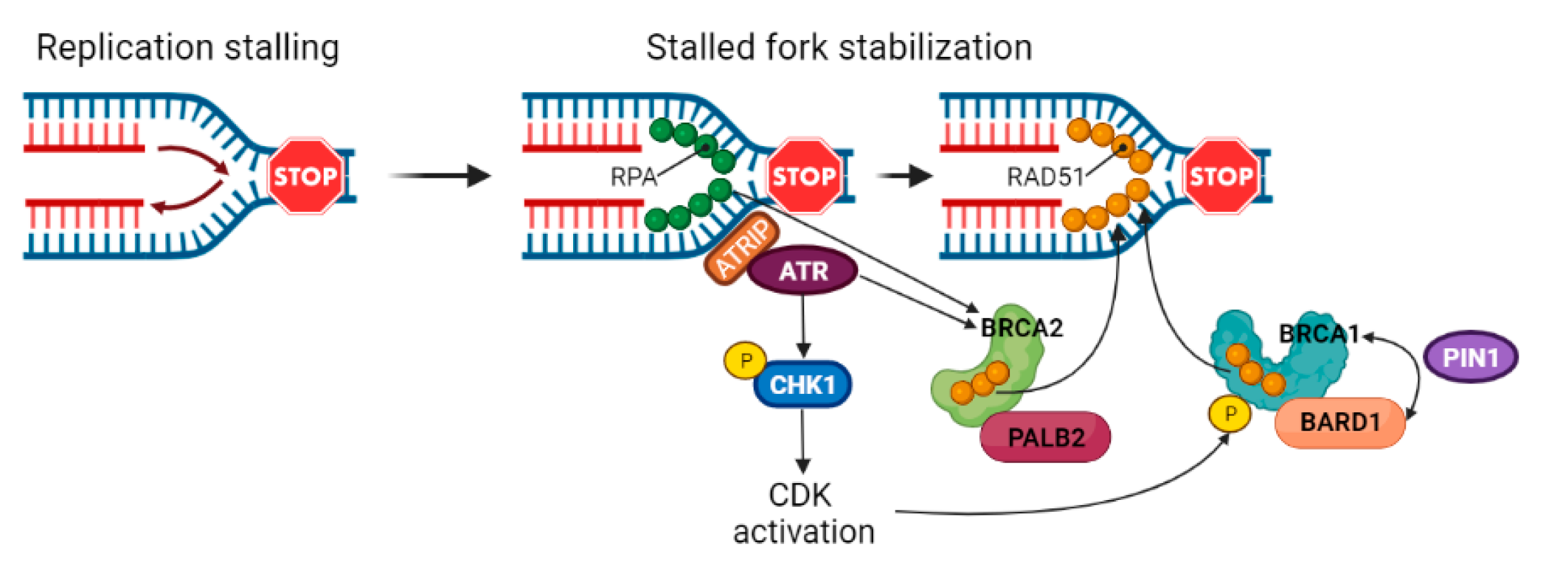
3.4. Stabilization of Stalled Forks

The HR pathway is not only involved in DNA DSB repair, but also in replication fork repair and restart [121]. During DNA duplication, replication forks often encounter obstacles that can lead to genotoxic fork stalling [122]. Cells have acquired several processes to maintain genome integrity by stabilizing, repairing and restarting stalled forks, thus preventing a possible mechanism of tumorigenesis [123,124]. Stalled forks are characterized by exposed DNA ends in the form of single- or double-strand DNA, which can be degraded by various cellular nucleases, including MRE11 and CtIP [125]. Under replication stress, extensive ssDNA is readily protected and stabilised by RPA; RPA phosphorylation by ATR and DNA-PKcs promotes PALB2 and BRCA2 translocation to stalled replication forks [126,127,128]. The RPA–ssDNA complex is then displaced by the RAD51 protein [129]. BRCA2 and BRCA1–BARD1 complex enhance the formation of stable RAD51 nucleoprotein filaments, which mediates replication fork reversal. The phosphorylation-directed prolyl isomerase PIN1 enhances BRCA1–BARD1 interaction with RAD51 (Figure 4). This mechanism is fundamental to regulate fork degradation mediated by the MRE11 nuclease [130,131,132,133,134]. In fact, in BRCA1/2-proficient cells, reversed forks are resected in a controlled manner, promoting HR-dependent fork repair and restart; in BRCA1/2-deficient cells, regressed forks are processed more extensively by MRE11 or converted into DSBs and can only be rescued by break-induce replication, a highly mutagenic process [121,125,132,135,136]. As proof that HR and replication fork protection are distinctive processes, the RAD51-binding region in the TR2 domain of BRCA2 or isomerization of the RING domain of BRCA1 are required for their function in DNA replication fork protection, but not in HR [130,134].
Like the BRCA2–RAD51 axis, also the Fanconi Anemia (FA) components FANCB and FANCD2 suppress MRE11-mediated fork degradation in a way linked to RAD51 protein [58,133,137]. FANCD2 is significantly overexpressed in BRCA1/2-mutated breast or ovarian cancers, highlighting its importance in limiting replication stress [58,138].
PARP1 activation at stalled replication forks contributes to MRE11 recruitment [136]; in addition, together with DNA-PKcs, PARP1 collaborates to engage XRCC1 for fork repair and restart, which suggests an involvement of the NHEJ machinery in stalled fork protection [139]. Since MRE11 recruitment by PARP1 is responsible for extensive fork degradation and genome instability in cells lacking BRCA1/2 [59,140], this genome maintenance pathway seems to be detrimental in BRCA1/2-deficient cells, suggesting an additional mechanism of synthetic lethality induced by PARPi. Remarkably, PARP1 depletion before BRCA1/2 loss restores stalled fork stability and even allows synthetic viability in mouse embryonic stem cells [60,140].
Multiple studies showed that restoring fork stabilization alone without restoring HR can drive the instauration of resistance to PARPi [59,60,61]. Interestingly, resistance to PARPi is associated with tolerance to replication stress-inducing chemotherapeutics such as cisplatin and topotecan [59]. Tobalina et al. [41] detected two reversion mutations in the isomerization region of BRCA1 that probably confer PARPi resistance through restoration of fork stabilization. Depletion of RADX, a RAD51 antagonist, restores stalled fork stability and induces chemoresistance in BRCA2-defective cells, probably due to enhanced association of RAD51 with stalled forks [61]. Inactivation of the remodelling protein SMARCAL1 confers chemoresistance, suppressing fork reversal and thus avoiding MRE11-dependent fork degradation [60]. Finally, clinical data revealed that low expression of RADX and SMARCAL1 correlates with poorer prognosis of BRCA1/2-mutated cancer patients, confirming the important role of fork stabilization in modulating chemosensitivities [59,60,61].

4. Future Perspectives

The discovery of multiple interactions between PARP1, DNA-damage repair pathways, and response to replication stress reinforces the increasing evidence that PARPi action is not limited to the inhibition of PARP1 catalytic activity. In this scenario, combination therapies could increase the sensitivity or overcome the resistance of certain tumours to PARPi, targeting pathways other than HR in which PARP1 is involved, both directly and indirectly. Considering that wild-type PTEN tumours could demonstrate relative resistance to PARPi and that the PI3K/AKT pathway is constitutively active in BRCA1-defective human cancer cells [73], the combination of PTEN-related PI3K/AKT pathway inhibitors with PARPi could be therapeutically beneficial. PI3K inhibitors in combination with PARPi have been shown to have synergistic therapeutic effects in BRCA1-deficient breast cancer in PDX models [141]. Targeting the replication checkpoint kinase ATR could represent another strategy. Combination of ATR inhibitors (ATRi) with PARPi in ATM-deficient BRCA1/2-mutated tumours achieved a therapeutic response at lower concentrations than monotherapy with either drug in vitro [138]. In addition, inhibition of ATR has the potential to reverse PARPi resistance provoked by reversions in BRCA1/2, as it limits HR acting on PALB2 [142]. More than a hundred ongoing clinical trials are investigating the combination of PARPi with immunotherapies and other agents, such as inhibitors of the replication stress, particularly ATRi [143] (Table S1). On the other hand, clinical trials involving MDR1 inhibitors have provided poor results, possibly due to the important endogenous function of MDR1 in cell-mediated immune responses as recently revealed [144]. Since FANC2 is upregulated in BRCA1/2-mutated cancers, therapeutic targeting of the FA pathway might be required to potentiate the PARPi or platinum-based treatment for BRCA1/2-mutated tumours [14].
Several different mechanisms of resistance to PARPi have been proposed, although only reversion mutations in BRCA1 and BRCA2 have been proved in the clinical setting. More studies are needed to investigate the clinical impact of the other suggested mechanisms of resistance, up to now highlighted only in cell lines or PDX models. The implementation of liquid biopsy, in particular the analysis of the ctDNA at the diagnosis and during the treatment, could help to identify subclones of tumour cells primarily resistant to PARPi and to monitor the onset of secondary resistance before the clinical progression. The development of combination therapies probably will be necessary to overcome PARPi resistance and would hopefully identify additional settings for the use of PARPi.

Supplementary Materials

The following supporting information can be downloaded at: https://www.mdpi.com/article/10.3390/cancers14112804/s1, Table S1: Ongoing (Recruiting and Active not recruiting) clinical trials investigating the combination of PARPi with immunotherapies and other agents (all accessed on 2 June 2022).

Author Contributions

C.P. and L.C. contributed equally to conception, data collection, analysis and writing of this paper. All authors have read and agreed to the published version of the manuscript.

Funding

The APC was funded by Associazione Angela Serra per la Ricerca sul Cancro, Modena (Angela Serra Association for Cancer Research).

Acknowledgments

We would like to thank and express our gratitude to Associazione Angela Serra per la Ricerca sul Cancro, Modena (Angela Serra Association for Cancer Research) for its support.

Conflicts of Interest

The authors declare no conflict of interest.

References

  1. Mortusewicz, O.; Amé, J.C.; Schreiber, V.; Leonhardt, H. Feedback-regulated poly(ADP-ribosyl)ation by PARP-1 is required for rapid response to DNA damage in living cells. Nucleic Acids Res. 2007, 35, 7665–7675. [Google Scholar] [CrossRef] [PubMed] [Green Version]
  2. Dawicki-McKenna, J.M.; Langelier, M.F.; DeNizio, J.E.; Riccio, A.A.; Cao, C.D.; Karch, K.R.; McCauley, M.; Steffen, J.D.; Black, B.E.; Pascal, J.M. PARP-1 Activation Requires Local Unfolding of an Autoinhibitory Domain. Mol. Cell 2015, 60, 755–768. [Google Scholar] [CrossRef] [PubMed] [Green Version]
  3. Eustermann, S.; Wu, W.F.; Langelier, M.F.; Yang, J.C.; Easton, L.E.; Riccio, A.A.; Pascal, J.M.; Neuhaus, D. Structural Basis of Detection and Signaling of DNA Single-Strand Breaks by Human PARP-1. Mol. Cell 2015, 60, 742–754. [Google Scholar] [CrossRef] [PubMed] [Green Version]
  4. Langelier, M.F.; Planck, J.L.; Roy, S.; Pascal, J.M. Structural basis for DNA damage-dependent poly(ADP-ribosyl)ation by human PARP-1. Science 2012, 336, 728–732. [Google Scholar] [CrossRef] [Green Version]
  5. El Khamisy, S.F.; Masutani, M.; Suzuki, H.; Caldecott, K.W. A requirement for PARP-1 for the assembly or stability of XRCC1 nuclear foci at sites of oxidative DNA damage. Nucleic Acids Res. 2003, 31, 5526–5533. [Google Scholar] [CrossRef] [Green Version]
  6. Whitehouse, C.J.; Taylor, R.M.; Thistlethwaite, A.; Zhang, H.; Karimi-Busheri, F.; Lasko, D.D.; Weinfeld, M.; Caldecott, K.W. XRCC1 stimulates human polynucleotide kinase activity at damaged DNA termini and accelerates DNA single-strand break repair. Cell 2001, 104, 107–117. [Google Scholar] [CrossRef] [Green Version]
  7. Haince, J.F.; McDonald, D.; Rodrigue, A.; Dery, U.; Masson, J.Y.; Hendzel, M.J.; Poirier, G.G. PARP1-dependent kinetics of recruitment of MRE11 and NBS1 proteins to multiple DNA damage sites. J. Biol. Chem. 2008, 283, 1197–1208. [Google Scholar] [CrossRef] [Green Version]
  8. Haince, J.F.; Kozlov, S.; Dawson, V.L.; Dawson, T.M.; Hendzel, M.J.; Lavin, M.F.; Poirier, G.G. Ataxia telangiectasia mutated (ATM) signaling network is modulated by a novel poly(ADP-ribose)-dependent pathway in the early response to DNA-damaging agents. J. Biol. Chem. 2007, 282, 16441–16453. [Google Scholar] [CrossRef] [Green Version]
  9. Li, M.; Yu, X. Function of BRCA1 in the DNA damage response is mediated by ADP-ribosylation. Cancer Cell 2013, 23, 693–704. [Google Scholar] [CrossRef] [Green Version]
  10. Zahradka, P.; Ebisuzaki, K. A shuttle mechanism for DNA-protein interactions. The regulation of poly(ADP-ribose) polymerase. Eur. J. Biochem. 1982, 127, 579–585. [Google Scholar] [CrossRef]
  11. Farmer, H.; McCabe, N.; Lord, C.J.; Tutt, A.N.; Johnson, D.A.; Richardson, T.B.; Santarosa, M.; Dillon, K.J.; Hickson, I.; Knights, C.; et al. Targeting the DNA repair defect in BRCA mutant cells as a therapeutic strategy. Nature 2005, 434, 917–921. [Google Scholar] [CrossRef] [PubMed]
  12. Bryant, H.E.; Schultz, N.; Thomas, H.D.; Parker, K.M.; Flower, D.; Lopez, E.; Kyle, S.; Meuth, M.; Curtin, N.J.; Helleday, T. Specific killing of BRCA2-deficient tumours with inhibitors of poly(ADP-ribose) polymerase. Nature 2005, 434, 913–917. [Google Scholar] [CrossRef] [PubMed]
  13. Lord, C.J.; Ashworth, A. PARP inhibitors: Synthetic lethality in the clinic. Science 2017, 355, 1152–1158. [Google Scholar] [CrossRef] [PubMed]
  14. Liao, H.; Ji, F.; Helleday, T.; Ying, S. Mechanisms for stalled replication fork stabilization: New targets for synthetic lethality strategies in cancer treatments. EMBO Rep. 2018, 19, e46263. [Google Scholar] [CrossRef] [PubMed]
  15. Lord, C.J.; Ashworth, A. The DNA damage response and cancer therapy. Nature 2012, 481, 287–294. [Google Scholar] [CrossRef] [PubMed]
  16. Chapman, J.R.; Taylor, M.R.; Boulton, S.J. Playing the end game: DNA double-strand break repair pathway choice. Mol. Cell 2012, 47, 497–510. [Google Scholar] [CrossRef] [PubMed] [Green Version]
  17. Murai, J.; Huang, S.Y.; Renaud, A.; Zhang, Y.; Ji, J.; Takeda, S.; Morris, J.; Teicher, B.; Doroshow, J.H.; Pommier, Y. Stereospecific PARP trapping by BMN 673 and comparison with laparib and rucaparib. Mol. Cancer Ther. 2014, 13, 433–443. [Google Scholar] [CrossRef] [Green Version]
  18. Murai, J.; Huang, S.Y.; Das, B.B.; Renaud, A.; Zhang, Y.; Doroshow, J.H.; Ji, J.; Takeda, S.; Pommier, Y. Trapping of PARP1 and PARP2 by clinical PARP inhibitors. Cancer Res. 2012, 72, 5588–5599. [Google Scholar] [CrossRef] [Green Version]
  19. Pettitt, S.J.; Rehman, F.L.; Bajrami, I.; Brough, R.; Wallberg, F.; Kozarewa, I.; Fenwick, K.; Assiotis, I.; Chen, L.; Campbell, J.; et al. A genetic screen using the PiggyBac transposon in haploid cells identifies Parp1 as a mediator of laparib toxicity. PLoS ONE 2013, 8, e61520. [Google Scholar] [CrossRef] [Green Version]
  20. Ceccaldi, R.; Liu, J.C.; Amunugama, R.; Hajdu, I.; Primack, B.; Petalcorin, M.I.; O’Connor, K.W.; Konstantinopoulos, P.A.; Elledge, S.J.; Boulton, S.J.; et al. Homologous recombination-deficient tumours are dependent on Poltheta-mediated repair. Nature 2015, 518, 258–262. [Google Scholar] [CrossRef] [Green Version]
  21. Mateos-Gomez, P.A.; Gong, F.; Nair, N.; Miller, K.M.; Lazzerini-Denchi, E.; Sfeir, A. Mammalian polymerase theta promotes alternative NHEJ and suppresses recombination. Nature 2015, 518, 254–257. [Google Scholar] [CrossRef] [PubMed] [Green Version]
  22. Golan, T.; Hammel, P.; Reni, M.; Van Cutsem, E.; Macarulla, T.; Hall, M.J.; Park, J.O.; Hochhauser, D.; Arnold, D.; Oh, D.Y.; et al. Maintenance Olaparib for Germline BRCA-Mutated Metastatic Pancreatic Cancer. N. Engl. J. Med. 2019, 381, 317–327. [Google Scholar] [CrossRef] [PubMed]
  23. De Bono, J.; Mateo, J.; Fizazi, K.; Saad, F.; Shore, N.; Sandhu, S.; Chi, K.N.; Sartor, O.; Agarwal, N.; Olmos, D.; et al. Olaparib for Metastatic Castration-Resistant Prostate Cancer. N. Engl. J. Med. 2020, 382, 2091–2102. [Google Scholar] [CrossRef] [PubMed]
  24. Mehta, P.; Bothra, S.J. PARP inhibitors in hereditary breast and ovarian cancer and other cancers: A review. Adv. Genet. 2021, 108, 35–80. [Google Scholar] [CrossRef] [PubMed]
  25. Armstrong, N.; Ryder, S.; Forbes, C.; Ross, J.; Quek, R.G. A systematic review of the international prevalence of BRCA mutation in breast cancer. Clin. Epidemiol. 2019, 11, 543–561. [Google Scholar] [CrossRef] [PubMed] [Green Version]
  26. Alsop, K.; Fereday, S.; Meldrum, C.; DeFazio, A.; Emmanuel, C.; George, J.; Dobrovic, A.; Birrer, M.J.; Webb, P.M.; Stewart, C.; et al. BRCA mutation frequency and patterns of treatment response in BRCA mutation-positive women with ovarian cancer: A report from the Australian Ovarian Cancer Study Group. J. Clin. Oncol. 2012, 30, 2654–2663. [Google Scholar] [CrossRef] [Green Version]
  27. Pennington, K.P.; Walsh, T.; Harrell, M.I.; Lee, M.K.; Pennil, C.C.; Rendi, M.H.; Thornton, A.; Norquist, B.M.; Casadei, S.; Nord, A.S.; et al. Germline and somatic mutations in homologous recombination genes predict platinum response and survival in ovarian, fallopian tube, and peritoneal carcinomas. Clin. Cancer Res. 2014, 20, 764–775. [Google Scholar] [CrossRef] [Green Version]
  28. Zhang, S.; Royer, R.; Li, S.; McLaughlin, J.R.; Rosen, B.; Risch, H.A.; Fan, I.; Bradley, L.; Shaw, P.A.; Narod, S.A. Frequencies of BRCA1 and BRCA2 mutations among 1342 unselected patients with invasive ovarian cancer. Gynecol. Oncol. 2011, 121, 353–357. [Google Scholar] [CrossRef]
  29. Rosen, M.N.; Goodwin, R.A.; Vickers, M.M. BRCA mutated pancreatic cancer: A change is coming. World J. Gastroenterol. 2021, 27, 1943–1958. [Google Scholar] [CrossRef]
  30. Pritchard, C.C.; Mateo, J.; Walsh, M.F.; De Sarkar, N.; Abida, W.; Beltran, H.; Garofalo, A.; Gulati, R.; Carreira, S.; Eeles, R.; et al. Inherited DNA-Repair Gene Mutations in Men with Metastatic Prostate Cancer. N. Engl. J. Med. 2016, 375, 443–453. [Google Scholar] [CrossRef]
  31. Yi, Y.W.; Kang, H.J.; Kim, H.J.; Hwang, J.S.; Wang, A.; Bae, I. Inhibition of constitutively activated phosphoinositide 3-kinase/AKT pathway enhances anti- tumor activity of chemotherapeutic agents in breast cancer susceptibility gene 1-defective breast cancer cells. Mol. Carcinog. 2013, 52, 667–675. [Google Scholar] [CrossRef] [PubMed] [Green Version]
  32. Dedes, K.J.; Wetterskog, D.; Mendes-Pereira, A.M.; Natrajan, R.; Lambros, M.B.; Geyer, F.C.; Vatcheva, R.; Savage, K.; Mackay, A.; Lord, C.J.; et al. PTEN Deficiency in Endometrioid Endometrial Adenocarcinomas Predicts Sensitivity to PARP Inhibitors. Sci. Transl. Med. 2010, 2, 53ra75. [Google Scholar] [CrossRef] [PubMed]
  33. Balmus, G.; Pilger, D.; Coates, J.; Demir, M.; Sczaniecka-Clift, M.; Barros, A.C.; Woods, M.; Fu, B.; Yang, F.; Chen, E.; et al. ATM orchestrates the DNA-damage response to counter toxic non-homologous end-joining at broken replication forks. Nat. Commun. 2019, 10, 87. [Google Scholar] [CrossRef] [PubMed] [Green Version]
  34. Patel, A.G.; Sarkaria, J.N.; Kaufmann, S.H. Nonhomologous end joining drives poly(ADP-ribose) polymerase (PARP) inhibitor lethality in homologous recombination-deficient cells. Proc. Natl. Acad. Sci. USA 2011, 108, 3406–3411. [Google Scholar] [CrossRef] [Green Version]
  35. McCormick, A.; Donoghue, P.; Dixon, M.; O’Sullivan, R.; O’Donnell, R.L.; Murray, J.; Kaufmann, A.; Curtin, N.J.; Edmondson, R.J. Ovarian Cancers Harbor Defects in Nonhomologous End Joining Resulting in Resistance to Rucaparib. Clin. Cancer Res. 2017, 23, 2050–2060. [Google Scholar] [CrossRef] [Green Version]
  36. Juhász, S.; Smith, R.; Schauer, T.; Spekhardt, D.; Mamar, H.; Zentout, S.; Chapuis, C.; Huet, S.; Timinszky, G. The chromatin remodeler ALC1 underlies resistance to PARP inhibitor treatment. Sci. Adv. 2020, 6, eabb8626. [Google Scholar] [CrossRef]
  37. Jaspers, J.E.; Sol, W.; Kersbergen, A.; Schlicker, A.; Guyader, C.; Xu, G.; Wessels, L.; Borst, P.; Jonkers, J.; Rottenberg, S. BRCA2-deficient sarcomatoid mammary tumors exhibit multidrug resistance. Cancer Res. 2015, 75, 732–741. [Google Scholar] [CrossRef] [Green Version]
  38. Vaidyanathan, A.; Sawers, L.; Gannon, A.L.; Chakravarty, P.; Scott, A.L.; Bray, S.E.; Ferguson, M.J.; Smith, G. ABCB1 (MDR1) induction defines a common resistance mechanism in paclitaxel- and olaparib-resistant ovarian cancer cells. Br. J. Cancer 2016, 115, 431–441. [Google Scholar] [CrossRef] [Green Version]
  39. Pettitt, S.J.; Krastev, D.B.; Brandsma, I.; Dréan, A.; Song, F.; Aleksandrov, R.; Harrell, M.I.; Menon, M.; Brough, R.; Campbell, J.; et al. Genome-wide and high-density CRISPR-Cas9 screens identify point mutations in PARP1 causing PARP inhibitor resistance. Nat. Commun. 2018, 9, 1849. [Google Scholar] [CrossRef]
  40. Gogola, E.; Duarte, A.A.; de Ruiter, J.R.; Wiegant, W.W.; Schmid, J.A.; de Bruijn, R.; James, D.I.; Guerrero Llobet, S.; Vis, D.J.; Annunziato, S.; et al. Selective Loss of PARG Restores PARylation and Counteracts PARP Inhibitor-Mediated Synthetic Lethality. Cancer Cell 2018, 33, 1078–1093.e12. [Google Scholar] [CrossRef] [Green Version]
  41. Tobalina, L.; Armenia, J.; Irving, E.; O’Connor, M.J.; Forment, J.V. A meta-analysis of reversion mutations in BRCA genes identifies signatures of DNA end-joining repair mechanisms driving therapy resistance. Ann. Oncol. 2021, 32, 103–112. [Google Scholar] [CrossRef] [PubMed]
  42. Drost, R.; Dhillon, K.K.; van der Gulden, H.; van der Heijden, I.; Brandsma, I.; Cruz, C.; Chondronasiou, D.; Castroviejo-Bermejo, M.; Boon, U.; Schut, E.; et al. BRCA1185delAG tumors may acquire therapy resistance through expression of RING-less BRCA1. J. Clin. Investig. 2016, 126, 2903–2918. [Google Scholar] [CrossRef] [PubMed]
  43. Wang, Y.; Bernhardy, A.J.; Cruz, C.; Krais, J.J.; Nacson, J.; Nicolas, E.; Peri, S.; van der Gulden, H.; van der Heijden, I.; O’Brien, S.W.; et al. The BRCA1-Δ11q Alternative Splice Isoform Bypasses Germline Mutations and Promotes Therapeutic Resistance to PARP Inhibition and Cisplatin. Cancer Res. 2016, 76, 2778–2790. [Google Scholar] [CrossRef] [PubMed] [Green Version]
  44. Cruz, C.; Castroviejo-Bermejo, M.; Gutiérrez-Enríquez, S.; Llop-Guevara, A.; Ibrahim, Y.H.; Gris-Oliver, A.; Bonache, S.; Morancho, B.; Bruna, A.; Rueda, O.M.; et al. RAD51 foci as a functional biomarker of homologous recombination repair and PARP inhibitor resistance in germline BRCA-mutated breast cancer. Ann. Oncol. 2018, 29, 1203–1210. [Google Scholar] [CrossRef] [PubMed]
  45. Wang, Y.; Bernhardy, A.J.; Nacson, J.; Krais, J.J.; Tan, Y.F.; Nicolas, E.; Radke, M.R.; Handorf, E.; Llop-Guevara, A.; Balmaña, J.; et al. BRCA1 intronic Alu elements drive gene rearrangements and PARP inhibitor resistance. Nat. Commun. 2019, 10, 5661. [Google Scholar] [CrossRef] [PubMed] [Green Version]
  46. Castroviejo-Bermejo, M.; Cruz, C.; Llop-Guevara, A.; Gutiérrez-Enríquez, S.; Ducy, M.; Ibrahim, Y.H.; Gris-Oliver, A.; Pellegrino, B.; Bruna, A.; Guzmán, M.; et al. A RAD51 assay feasible in routine tumor samples calls PARP inhibitor response beyond BRCA mutation. EMBO Mol. Med. 2018, 10, e9172. [Google Scholar] [CrossRef] [PubMed]
  47. Ter Brugge, P.; Kristel, P.; van der Burg, E.; Boon, U.; de Maaker, M.; Lips, E.; Mulder, L.; de Ruiter, J.; Moutinho, C.; Gevensleben, H.; et al. Mechanisms of Therapy Resistance in Patient-Derived Xenograft Models of BRCA1-Deficient Breast Cancer. J. Natl. Cancer Inst. 2016, 108, djw148, Erratum in J. Natl. Cancer Inst. 2020, 112, 1075. [Google Scholar] [CrossRef]
  48. Veeck, J.; Ropero, S.; Setien, F.; Gonzalez-Suarez, E.; Osorio, A.; Benitez, J.; Herman, J.G.; Esteller, M. BRCA1 CpG island hypermethylation predicts sensitivity to poly(adenosine diphosphate)-ribose polymerase inhibitors. J. Clin. Oncol. 2010, 28, e563–e564, author reply e565-6. [Google Scholar] [CrossRef]
  49. Wang, Y.Q.; Zhang, J.R.; Li, S.D.; He, Y.Y.; Yang, Y.X.; Liu, X.L.; Wan, X.P. Aberrant methylation of breast and ovarian cancer susceptibility gene 1 in chemosensitive human ovarian cancer cells does not involve the phosphatidylinositol 30-kinase-Akt pathway. Cancer Sci. 2010, 101, 1618–1623. [Google Scholar] [CrossRef]
  50. Belotserkovskaya, R.; Raga Gil, E.; Lawrence, N.; Butler, R.; Clifford, G.; Wilson, M.D.; Jackson, S.P. PALB2 chromatin recruitment restores homologous recombination in BRCA1-deficient cells depleted of 53BP1. Nat. Commun. 2020, 11, 819. [Google Scholar] [CrossRef] [Green Version]
  51. Xu, G.; Chapman, J.R.; Brandsma, I.; Yuan, J.; Mistrik, M.; Bouwman, P.; Bartkova, J.; Gogola, E.; Warmerdam, D.; Barazas, M.; et al. REV7 counteracts DNA double-strand break resection and affects PARP inhibition. Nature 2015, 521, 541–544. [Google Scholar] [CrossRef] [PubMed] [Green Version]
  52. Noordermeer, S.M.; Adam, S.; Setiaputra, D.; Barazas, M.; Pettitt, S.J.; Ling, A.K.; Olivieri, M.; Álvarez-Quilón, A.; Moatti, N.; Zimmermann, M.; et al. The shieldin complex mediates 53BP1-dependent DNA repair. Nature 2018, 560, 117–121. [Google Scholar] [CrossRef] [PubMed]
  53. Gupta, R.; Somyajit, K.; Narita, T.; Maskey, E.; Stanlie, A.; Kremer, M.; Typas, D.; Lammers, M.; Mailand, N.; Nussenzweig, A.; et al. DNA Repair Network Analysis Reveals Shieldin as a Key Regulator of NHEJ and PARP Inhibitor Sensitivity. Cell 2018, 173, 972–988.e23. [Google Scholar] [CrossRef] [PubMed] [Green Version]
  54. He, Y.J.; Meghani, K.; Caron, M.C.; Yang, C.; Ronato, D.A.; Bian, J.; Sharma, A.; Moore, J.; Niraj, J.; Detappe, A.; et al. DYNLL1 binds to MRE11 to limit DNA end resection in BRCA1-deficient cells. Nature 2018, 563, 522–526. [Google Scholar] [CrossRef] [PubMed]
  55. Kondrashova, O.; Nguyen, M.; Shield-Artin, K.; Tinker, A.V.; Teng, N.; Harrell, M.I.; Kuiper, M.J.; Ho, G.Y.; Barker, H.; Jasin, M.; et al. Secondary Somatic Mutations Restoring RAD51C and RAD51D Associated with Acquired Resistance to the PARP Inhibitor Rucaparib in High-Grade Ovarian Carcinoma. Cancer Discov. 2017, 7, 984–998. [Google Scholar] [CrossRef] [PubMed] [Green Version]
  56. Liu, Y.; Burness, M.L.; Martin-Trevino, R.; Guy, J.; Bai, S.; Harouaka, R.; Brooks, M.D.; Shang, L.; Fox, A.; Luther, T.K.; et al. RAD51 Mediates Resistance of Cancer Stem Cells to PARP Inhibition in Triple-Negative Breast Cancer. Clin. Cancer Res. 2017, 23, 514–522. [Google Scholar] [CrossRef] [PubMed] [Green Version]
  57. Marzio, A.; Puccini, J.; Kwon, Y.; Maverakis, N.K.; Arbini, A.; Sung, P.; Bar-Sagi, D.; Pagano, M. The F-Box Domain-Dependent Activity of EMI1 Regulates PARPi Sensitivity in Triple-Negative Breast Cancers. Mol. Cell 2019, 73, 224–237.e6. [Google Scholar] [CrossRef] [Green Version]
  58. Michl, J.; Zimmer, J.; Buffa, F.M.; McDermott, U.; Tarsounas, M. FANCD2 limits replication stress and genome instability in cells lacking BRCA2. Nat. Struct. Mol. Biol. 2016, 23, 755–757. [Google Scholar] [CrossRef]
  59. Chaudhuri, A.R.; Callen, E.; Ding, X.; Gogola, E.; Duarte, A.A.; Lee, J.E.; Wong, N.; Lafarga, V.; Calvo, J.A.; Panzarino, N.J.; et al. Replication fork stability confers chemoresistance in BRCA-deficient cells. Nature 2016, 535, 382–387. [Google Scholar] [CrossRef]
  60. Taglialatela, A.; Alvarez, S.; Leuzzi, G.; Sannino, V.; Ranjha, L.; Huang, J.W.; Madubata, C.; Anand, R.; Levy, B.; Rabadan, R.; et al. Restoration of replication fork stability in BRCA1-and BRCA2-deficient cells by inactivation of SNF2-family fork remodelers. Mol. Cell 2017, 68, 414–430. [Google Scholar] [CrossRef] [Green Version]
  61. Dungrawala, H.; Bhat, K.P.; Le Meur, R.; Chazin, W.J.; Ding, X.; Sharan, S.K.; Wessel, S.R.; Sathe, A.A.; Zhao, R.; Cortez, D. RADX promotes genome stability and modulates chemosensitivity by regulating RAD51 at replication forks. Mol. Cell 2017, 67, 374–386.e5. [Google Scholar] [CrossRef] [PubMed]
  62. Carnero, A.; Blanco-Aparicio, C.; Renner, O.; Link, W.; Leal, J.F. The PTEN/PI3K/AKT signalling pathway in cancer, therapeutic implications. Curr. Cancer Drug Targets 2008, 8, 187–198. [Google Scholar] [CrossRef] [PubMed]
  63. Carnero, A. The PKB/AKT pathway in cancer. Curr. Pharm. Des. 2010, 16, 34–44. [Google Scholar] [CrossRef] [PubMed] [Green Version]
  64. Hollander, M.C.; Blumenthal, G.M.; Dennis, P.A. PTEN loss in the continuum of common cancers, rare syndromes and mouse models. Nat. Rev. 2011, 11, 289–301. [Google Scholar] [CrossRef] [PubMed]
  65. Nelson, A.C.; Lyons, T.R.; Young, C.D.; Hansen, K.C.; Anderson, S.M.; Holt, J.T. AKT regulates BRCA1 stability in response to hormone signaling. Mol. Cell. Endocrinol. 2010, 319, 129–142. [Google Scholar] [CrossRef] [Green Version]
  66. Kang, H.J.; Yi, Y.W.; Kim, H.J.; Hong, Y.B.; Seong, Y.S.; Bae, I. BRCA1 negatively regulates IGF-1 expression through an estrogen-responsive element-like site. Cell Death Dis. 2012, 3, e336. [Google Scholar] [CrossRef] [Green Version]
  67. Xiang, T.; Ohashi, A.; Huang, Y.; Pandita, T.K.; Ludwig, T.; Powell, S.N.; Yang, Q. Negative Regulation of AKT Activation by BRCA1. Cancer Res. 2008, 68, 10040–10044. [Google Scholar] [CrossRef] [Green Version]
  68. Saal, L.H.; Gruvberger-Saal, S.K.; Persson, C.; Lövgren, K.; Jumppanen, M.; Staaf, J.; Jönsson, G.; Pires, M.M.; Maurer, M.; Holm, K.; et al. Recurrent gross mutations of the PTEN tumor suppressor gene in breast cancers with deficient DSB repair. Nat. Genet. 2008, 40, 102–107. [Google Scholar] [CrossRef]
  69. Foulkes, W.D. BRCA1—Sowing the seeds crooked in the furrow. Nat. Genet. 2008, 40, 8–9. [Google Scholar] [CrossRef]
  70. Shen, W.H.; Balajee, A.S.; Wang, J.; Wu, H.; Eng, C.; Pandolfi, P.P.; Yin, Y. Essential role for nuclear PTEN in maintaining chromosomal integrity. Cell 2007, 128, 157–170. [Google Scholar] [CrossRef] [Green Version]
  71. McEllin, B.; Camacho, C.V.; Mukherjee, B.; Hahm, B.; Tomimatsu, N.; Bachoo, R.M.; Burma, S. PTEN loss compromises homologous recombination repair in astrocytes: Implications for glioblastoma therapy with temozolomide or poly(ADP-ribose) polymerase inhibitors. Cancer Res. 2010, 70, 5457–5464. [Google Scholar] [CrossRef] [PubMed] [Green Version]
  72. Mendes-Pereira, A.M.; Martin, S.A.; Brough, R.; McCarthy, A.; Taylor, J.R.; Kim, J.S.; Waldman, T.; Lord, C.J.; Ashworth, A. Synthetic lethal targeting of PTEN mutant cells with PARP inhibitors. EMBO Mol. Med. 2009, 1, 315–322. [Google Scholar] [CrossRef] [PubMed]
  73. Minami, A.; Nakanishi, A.; Ogura, Y.; Kitagishi, Y.; Matsuda, S. Connection between Tumor Suppressor BRCA1 and PTEN in Damaged DNA Repair. Front. Oncol. 2014, 4, 318. [Google Scholar] [CrossRef] [PubMed] [Green Version]
  74. Chock, K.L.; Allison, J.M.; Shimizu, Y.; El Shamy, W.M. BRCA1-IRIS overexpression promotes cisplatin resistance in ovarian cancer cells. Cancer Res. 2010, 70, 8782–8791. [Google Scholar] [CrossRef] [Green Version]
  75. Burstein, H.J. Novel agents and future directions for refractory breast cancer. Semin. Oncol. 2011, 38, S17–S24. [Google Scholar] [CrossRef]
  76. Yap, T.A.; Kristeleit, R.; Michalarea, V.; Pettitt, S.J.; im, J.S.; Carreira, S.; Roda, D.; Miller, R.; Riisnaes, R.; Miranda, S.; et al. Phase I Trial of the PARP Inhibitor Olaparib and AKT Inhibitor Capivasertib in Patients with BRCA1/2- and Non-BRCA1/2-Mutant Cancers. Cancer Discov. 2020, 10, 1528–1543. [Google Scholar] [CrossRef]
  77. Ceccaldi, R.; Rondinelli, B.; D’Andrea, A.D. Repair pathway choices and consequences at the double-strand break. Trends Cell. Biol. 2016, 26, 52–64. [Google Scholar] [CrossRef] [Green Version]
  78. Myler, L.R.; Gallardo, I.F.; Soniat, M.M.; Deshpande, R.A.; Gonzalez, X.B.; Kim, Y.; Paull, T.T.; Finkelstein, I.J. Single-Molecule Imaging Reveals How Mre11-Rad50-Nbs1 Initiates DNA Break Repair. Mol. Cell 2017, 67, 891–898.e4. [Google Scholar] [CrossRef] [Green Version]
  79. Buis, J.; Stoneham, T.; Spehalski, E.; Ferguson, D.O. Mre11 regulates CtIP-dependent double-strand break repair by interaction with CDK2. Nat. Struct. Mol. Biol. 2012, 19, 246–252. [Google Scholar] [CrossRef]
  80. Lee, J.H.; Paull, T.T. Direct activation of the ATM protein kinase by the Mre11/Rad50/Nbs1 complex. Science 2004, 304, 93–96. [Google Scholar] [CrossRef]
  81. Kumagai, A.; Lee, J.; Yoo, H.Y.; Dunphy, W.G. TopBP1 activates the ATR-ATRIP complex. Cell 2006, 124, 943–955. [Google Scholar] [CrossRef] [PubMed] [Green Version]
  82. Matsuoka, S.; Ballif, B.A.; Smogorzewska, A.; McDonald, E.R.; Hurov, K.E.; Luo, J.; Bakalarski, C.E.; Zhao, Z.; Solimini, N.; Lerenthal, Y.; et al. ATM and ATR substrate analysis reveals extensive protein networks responsive to DNA damage. Science 2007, 316, 1160–1166. [Google Scholar] [CrossRef] [PubMed] [Green Version]
  83. Ducy, M.; Sesma-Sanz, L.; Guitton-Sert, L.; Lashgari, A.; Gao, Y.; Brahiti, N.; Rodrigue, A.; Margaillan, G.; Caron, M.C.; Côté, J.; et al. The Tumor Suppressor PALB2: Inside Out. Trends Biochem. Sci. 2019, 44, 226–240. [Google Scholar] [CrossRef] [PubMed]
  84. Sun, Y.; McCorvie, T.J.; Yates, L.A.; Zhang, X. Structural basis of homologous recombination. Cell. Mol. Life Sci. 2020, 77, 3–18. [Google Scholar] [CrossRef] [Green Version]
  85. Cortesi, L.; Piombino, C.; Toss, A. Germline Mutations in Other Homologous Recombination Repair-Related Genes Than BRCA1/2: Predictive or Prognostic Factors? J. Pers. Med. 2021, 11, 245. [Google Scholar] [CrossRef]
  86. Symington, L.S. Mechanism and regulation of DNA end resection in eukaryotes. Crit. Rev. Biochem. Mol. Biol. 2016, 51, 195–212. [Google Scholar] [CrossRef] [Green Version]
  87. Jowsey, P.; Morrice, N.A.; Hastie, C.J.; McLauchlan, H.; Toth, R.; Rouse, J. Characterisation of the sites of DNA damage-induced 53BP1 phosphorylation catalysed by ATM and ATR. DNA Repair 2007, 6, 1536–1544. [Google Scholar] [CrossRef]
  88. Zhang, F.; Gong, Z. Regulation of DNA double-strand break repair pathway choice: A new focus on 53BP1. J. Zhejiang Univ. Sci. B 2021, 22, 38–46. [Google Scholar] [CrossRef]
  89. Blackford, A.N.; Jackson, S.P. ATM, ATR, and DNA-PK: The trinity at the heart of the DNA damage response. Mol. Cell 2017, 66, 801–817. [Google Scholar] [CrossRef] [Green Version]
  90. Daley, J.M.; Sung, P. 53BP1, BRCA1 and the choice between recombination and end joining at DNA double-strand breaks. Mol. Cell. Biol. 2014, 34, 1380–1388. [Google Scholar] [CrossRef] [Green Version]
  91. Feng, L.; Li, N.; Li, Y.; Wang, J.; Gao, M.; Wang, W.; Chen, J. Cell cycle-dependent inhibition of 53BP1 signaling by BRCA1. Cell Discov. 2015, 1, 15019. [Google Scholar] [CrossRef] [PubMed]
  92. Hustedt, N.; Durocher, D. The control of DNA repair by the cell cycle. Nat. Cell Biol. 2017, 19, 1–9. [Google Scholar] [CrossRef] [PubMed]
  93. Chanut, P.; Britton, S.; Coates, J.; Jackson, S.P.; Calsou, P. Coordinated nuclease activities counteract Ku at single-ended DNA double-strand breaks. Nat. Commun. 2016, 7, 12889. [Google Scholar] [CrossRef] [PubMed] [Green Version]
  94. Ahel, D.; Hořejší, Z.; Wiechens, N.; Polo, S.E.; Garcia-Wilson, E.; Ahel, I.; Flynn, H.; Skehel, M.; West, S.C.; Jackson, S.P.; et al. Poly(ADP-ribose)-dependent regulation of DNA repair by the chromatin remodeling enzyme ALC1. Science 2009, 325, 1240–1243. [Google Scholar] [CrossRef] [Green Version]
  95. Gottschalk, A.J.; Timinszky, G.; Kong, S.E.; Jin, J.; Cai, Y.; Swanson, S.K.; Washburn, M.P.; Florens, L.; Ladurner, A.G.; Conaway, J.W.; et al. Poly(ADP-ribosyl)ation directs recruitment and activation of an ATP-dependent chromatin remodeler. Proc. Natl. Acad. Sci. USA 2009, 106, 13770–13774. [Google Scholar] [CrossRef] [Green Version]
  96. Ma, N.F.; Hu, L.; Fung, J.M.; Xie, D.; Zheng, B.J.; Chen, L.; Tang, D.J.; Fu, L.; Wu, Z.; Chen, M.; et al. Isolation and characterization of a novel oncogene, amplified in liver cancer 1, within a commonly amplified region at 1q21 in hepatocellular carcinoma. Hepatology 2008, 47, 503–510. [Google Scholar] [CrossRef]
  97. Dagogo-Jack, I.; Shaw, A. Tumour heterogeneity and resistance to cancer therapies. Nat. Rev. Clin. Oncol. 2018, 15, 81–94. [Google Scholar] [CrossRef]
  98. Kathawala, R.J.; Gupta., P.; Ashby, C.R., Jr.; Chen, Z.S. The modulation of ABC transporter-mediated multidrug resistance in cancer: A review of the past decade. Drug Resist. Updat. 2015, 18, 1–17. [Google Scholar] [CrossRef]
  99. Shen, D.W.; Fojo, A.; Chin, J.E.; Roninson, I.B.; Richert, N.; Pastan, I.; Gottesman, M.M. Human multidrug-resistant cell lines: Increased mdr1 expression can precede gene amplification. Science 1986, 232, 643–645. [Google Scholar] [CrossRef]
  100. Fojo, A.T.; Ueda, K.; Slamon, D.J.; Poplack, D.G.; Gottesman, M.M.; Pastan, I. Expression of a multidrug-resistance gene in human tumors and tissues. Proc. Natl. Acad. Sci. USA 1987, 84, 265–269. [Google Scholar] [CrossRef] [Green Version]
  101. Parekh, H.; Wiesen, K.; Simpkins, H. Acquisition of taxol resistance via P-glycoprotein- and non-P-glycoprotein-mediated mechanisms in human ovarian carcinoma cells. Biochem. Pharmacol. 1997, 53, 461–470. [Google Scholar] [CrossRef]
  102. Wang, Y.C.; Juric, D.; Francisco, B.; Yu, R.X.; Duran, G.E.; Chen, G.K.; Chen, X.; Sikic, B.I. Regional activation of chromosomal arm 7q with and without gene amplification in taxane-selected human ovarian cancer cell lines. Genes Chromosomes Cancer 2006, 45, 365–374. [Google Scholar] [CrossRef] [PubMed]
  103. Rottenberg, S.; Jaspers, J.E.; Kersbergen, A.; van der Burg, E.; Nygren, A.O.; Zander, S.A.; Derksen, P.W.; de Bruin, M.; Zevenhoven, J.; Lau, A.; et al. High sensitivity of BRCA1-deficient mammary tumors to the PARP inhibitor AZD2281 alone and in combination with platinum drugs. Proc. Natl. Acad. Sci. USA 2008, 105, 17079–17084. [Google Scholar] [CrossRef] [PubMed] [Green Version]
  104. Wils, P.; Phung-Ba, V.; Warnery, A.; Lechardeur, D.; Raeissi, S.; Hidalgo, I.J.; Scherman, D. Polarized transport of docetaxel and vinblastine mediated by P-glycoprotein in human intestinal epithelial cell monolayers. Biochem. Pharmacol. 1994, 48, 1528–1530. [Google Scholar] [CrossRef]
  105. Szakács, G.; Paterson, J.K.; Ludwig, J.A.; Booth-Genthe, C.; Gottesman, M.M. Targeting multidrug resistance in cancer. Nat. Rev. Drug Discov. 2006, 5, 219–234. [Google Scholar] [CrossRef]
  106. Pascal, J.M.; Ellenberger, T. The rise and fall of poly(ADP-ribose): An enzymatic perspective. DNA Repair 2015, 32, 10–16. [Google Scholar] [CrossRef] [Green Version]
  107. Clouaire, T.; Rocher, V.; Lashgari, A.; Arnould, C.; Aguirrebengoa, M.; Biernacka, A.; Skrzypczak, M.; Aymard, F.; Fongang, B.; Dojer, N.; et al. Comprehensive Mapping of Histone Modifications at DNA Double-Strand Breaks Deciphers Repair Pathway Chromatin Signatures. Mol. Cell 2018, 72, 250–262.e6. [Google Scholar] [CrossRef] [Green Version]
  108. Williams, R.S.; Glover, J.N. Structural consequences of a cancer-causing BRCA1-BRCT missense mutation. J. Biol. Chem. 2003, 278, 2630–2635. [Google Scholar] [CrossRef] [Green Version]
  109. Williams, R.S.; Chasman, D.I.; Hau, D.D.; Hui, B.; Lau, A.Y.; Glover, J.N. Detection of protein folding defects caused by BRCA1-BRCT truncation and missense mutations. J. Biol. Chem. 2003, 278, 53007–53016. [Google Scholar] [CrossRef] [Green Version]
  110. Lee, M.S.; Green, R.; Marsillac, S.M.; Coquelle, N.; Williams, R.S.; Yeung, T.; Foo, D.; Hau, D.D.; Hui, B.; Monteiro, A.N.; et al. Comprehensive analysis of missense variations in the BRCT domain of BRCA1 by structural and functional assays. Cancer Res. 2010, 70, 4880–4890. [Google Scholar] [CrossRef] [Green Version]
  111. Sy, S.M.; Huen, M.S.; Chen, J. PALB2 is an integral component of the BRCA complex required for homologous recombination repair. Proc. Natl. Acad. Sci. USA 2009, 106, 7155–7160. [Google Scholar] [CrossRef] [PubMed] [Green Version]
  112. Scully, R.; Chen, J.; Plug, A. Association of BRCA1 with Rad51 in mitotic and meiotic cells. Cell 1997, 88, 265–275. [Google Scholar] [CrossRef] [Green Version]
  113. Johnson, N.; Johnson, S.F.; Yao, W.; Li, Y.C.; Choi, Y.E.; Bernhardy, A.J.; Wang, Y.; Capelletti, M.; Sarosiek, K.A.; Moreau, L.A.; et al. Stabilization of mutant BRCA1 protein confers PARP inhibitor and platinum resistance. Proc. Natl. Acad. Sci. USA 2013, 110, 17041–17046. [Google Scholar] [CrossRef] [PubMed] [Green Version]
  114. Cancer Genome Atlas Research Network. Integrated genomic analyses of ovarian carcinoma. Nature 2011, 474, 609–615. [Google Scholar] [CrossRef]
  115. Patch, A.M.; Christie, E.L.; Etemadmoghadam, D.; Garsed, D.W.; George, J.; Fereday, S.; Nones, K.; Cowin, P.; Alsop, K.; Bailey, P.J.; et al. Whole-genome characterization of chemoresistant ovarian cancer. Nature 2015, 521, 489–494. [Google Scholar] [CrossRef]
  116. Prakash, R.; Zhang, Y.; Feng, W.; Jasin, M. Homologous recombination and human health: The roles of BRCA1, BRCA2, and associated proteins. Cold Spring Harb. Perspect. Biol. 2015, 7, a016600. [Google Scholar] [CrossRef] [Green Version]
  117. Mattarocci, S.; Hafner, L.; Lezaja, A.; Shyian, M.; Shore, D. Rif1: A Conserved Regulator of DNA Replication and Repair Hijacked by Telomeres in Yeasts. Front. Genet. 2016, 7, 45. [Google Scholar] [CrossRef] [Green Version]
  118. Boersma, V.; Moatti, N.; Segura-Bayona, S.; Peuscher, M.H.; van der Torre, J.; Wevers, B.A.; Orthwein, A.; Durocher, D.; Jacobs, J.J. MAD2L2 controls DNA repair at telomeres and DNA breaks by inhibiting 5′ end resection. Nature 2015, 521, 537–540. [Google Scholar] [CrossRef] [Green Version]
  119. Lo, K.W.; Kan, H.M.; Chan, L.N.; Xu, W.G.; Wang, K.P.; Wu, Z.; Sheng, M.; Zhang, M. The 8-kDa dynein light chain binds to p53-binding protein 1 and mediates DNA damage-induced p53 nuclear accumulation. J. Biol. Chem. 2005, 280, 8172–8179. [Google Scholar] [CrossRef] [Green Version]
  120. Becker, J.R.; Cuella-Martin, R.; Barazas, M.; Liu, R.; Oliveira, C.; Oliver, A.W.; Bilham, K.; Holt, A.B.; Blackford, A.N.; Heierhorst, J.; et al. The ASCIZ-DYNLL1 axis promotes 53BP1-dependent non-homologous end joining and PARP inhibitor sensitivity. Nat. Commun. 2018, 9, 5406. [Google Scholar] [CrossRef] [Green Version]
  121. Thangavel, S.; Berti, M.; Levikova, M.; Pinto, C.; Gomathinayagam, S.; Vujanovic, M.; Zellweger, R.; Moore, H.; Lee, E.H.; Hendrickson, E.A.; et al. DNA2 drives processing and restart of reversed replication forks in human cells. J. Cell Biol. 2015, 208, 545–562. [Google Scholar] [CrossRef] [PubMed] [Green Version]
  122. Cortez, D. Preventing replication fork collapse to maintain genome integrity. DNA Repair 2015, 32, 149–157. [Google Scholar] [CrossRef] [PubMed] [Green Version]
  123. McGrail, D.J.; Lin, C.C.; Dai, H.; Mo, W.; Li, Y.; Stephan, C.; Davies, P.; Lu, Z.; Mills, G.B.; Lee, J.S.; et al. Defective replication stress response is inherently linked to the cancer stem cell phenotype. Cell Rep. 2018, 23, 2095–2106. [Google Scholar] [CrossRef] [PubMed] [Green Version]
  124. Bartkova, J.; Rezaei, N.; Liontos, M.; Karakaidos, P.; Kletsas, D.; Issaeva, N.; Vassiliou, L.V.; Kolettas, E.; Niforou, K.; Zoumpourlis, V.C.; et al. Oncogene-induced senescence is part of the tumorigenesis barrier imposed by DNA damage checkpoints. Nature 2006, 444, 633–637. [Google Scholar] [CrossRef] [PubMed]
  125. Lemacon, D.; Jackson, J.; Quinet, A.; Brickner, J.R.; Li, S.; Yazinski, S.; You, Z.; Ira, G.; Zou, L.; Mosammaparast, N.; et al. MRE11 and EXO1 nucleases degrade reversed forks and elicit MUS81-dependent fork rescue in BRCA2-deficient cells. Nat. Commun. 2017, 8, 860. [Google Scholar] [CrossRef]
  126. Binz, S.K.; Sheehan, A.M.; Wold, M.S. Replication protein A phosphorylation and the cellular response to DNA damage. DNA Repair 2004, 3, 1015–1024. [Google Scholar] [CrossRef]
  127. Murphy, A.K.; Fitzgerald, M.; Ro, T.; Kim, J.H.; Rabinowitsch, A.I.; Chowdhury, D.; Schildkraut, C.L.; Borowiec, J.A. Phosphorylated RPA recruits PALB2 to stalled DNA replication forks to facilitate fork recovery. J. Cell Biol. 2014, 206, 493–507. [Google Scholar] [CrossRef] [Green Version]
  128. Liu, S.; Opiyo, S.O.; Manthey, K.; Glanzer, J.G.; Ashley, A.K.; Amerin, C.; Troksa, K.; Shrivastav, M.; Nickoloff, J.A.; Oakley, G.G. Distinct roles for DNA-PK, ATM and ATR in RPA phosphorylation and checkpoint activation in response to replication stress. Nucleic Acids Res. 2012, 40, 10780–10794. [Google Scholar] [CrossRef] [Green Version]
  129. Sugiyama, T.; Kowalczykowski, S.C. Rad52 protein associates with replication protein A (RPA)-single-stranded DNA to accelerate Rad51- mediated displacement of RPA and presynaptic complex formation. J. Biol. Chem. 2002, 277, 31663–31672. [Google Scholar] [CrossRef] [Green Version]
  130. Schlacher, K.; Christ, N.; Siaud, N.; Egashira, A.; Wu, H.; Jasin, M. Double-strand break repair-independent role for BRCA2 in blocking stalled replication fork degradation by MRE11. Cell 2011, 145, 529–542. [Google Scholar] [CrossRef] [Green Version]
  131. Ying, S.; Hamdy, F.C.; Helleday, T. Mre11-dependent degradation of stalled DNA replication forks is prevented by BRCA2 and PARP1. Cancer Res. 2012, 72, 2814–2821. [Google Scholar] [CrossRef] [PubMed] [Green Version]
  132. Mijic, S.; Zellweger, R.; Chappidi, N.; Berti, M.; Jacobs, K.; Mutreja, K.; Ursich, S.; Ray Chaudhuri, A.; Nussenzweig, A.; Janscak, P.; et al. Replication fork reversal triggers fork degradation in BRCA2-defective cells. Nat. Commun. 2017, 8, 859. [Google Scholar] [CrossRef] [PubMed]
  133. Schlacher, K.; Wu, H.; Jasin, M. A distinct replication fork protection pathway connects Fanconi anemia tumor suppressors to RAD51-BRCA1/2. Cancer Cell 2012, 22, 106–116. [Google Scholar] [CrossRef] [PubMed] [Green Version]
  134. Daza-Martin, M.; Starowicz, K.; Jamshad, M.; Tye, S.; Ronson, G.E.; MacKay, H.L.; Chauhan, A.S.; Walker, A.K.; Stone, H.R.; Beesley, J.F.; et al. Isomerization of BRCA1-BARD1 promotes replication fork protection. Nature 2019, 571, 521–527. [Google Scholar] [CrossRef]
  135. Kolinjivadi, A.M.; Sannino, V.; De Antoni, A.; Zadorozhny, K.; Kilkenny, M.; Techer, H.; Baldi, G.; Shen, R.; Ciccia, A.; Pellegrini, L.; et al. Smarcal1-Mediated Fork Reversal Triggers Mre11-Dependent Degradation of Nascent DNA in the Absence of Brca2 and Stable Rad51 Nucleofilaments. Mol. Cell 2017, 67, 867–881.e7. [Google Scholar] [CrossRef] [Green Version]
  136. Bryant, H.E.; Petermann, E.; Schultz, N.; Jemth, A.S.; Loseva, O.; Issaeva, N.; Johansson, F.; Fernandez, S.; McGlynn, P.; Helleday, T. PARP is activated at stalled forks to mediate Mre11-dependent replication restart and recombination. EMBO J. 2009, 28, 2601–2615. [Google Scholar] [CrossRef] [Green Version]
  137. Kim, T.M.; Son, M.Y.; Dodds, S.; Hu, L.; Luo, G.; Hasty, P. RECQL5 and BLM exhibit divergent functions in cells defective for the Fanconi anemia pathway. Nucleic Acids Res. 2015, 43, 893–903. [Google Scholar] [CrossRef]
  138. Kais, Z.; Rondinelli, B.; Holmes, A.; O’Leary, C.; Kozono, D.; D’Andrea, A.D.; Ceccaldi, R. FANCD2 maintains fork stability in BRCA1/2-deficient tumors and promotes alternative end-joining DNA repair. Cell Rep. 2016, 15, 2488–2499. [Google Scholar] [CrossRef] [Green Version]
  139. Ying, S.; Chen, Z.; Medhurst, A.L.; Neal, J.A.; Bao, Z.; Mortusewicz, O.; McGouran, J.; Song, X.; Shen, H.; Hamdy, F.C.; et al. DNA-PKcs and PARP1 Bind to Unresected Stalled DNA Replication Forks Where They Recruit XRCC1 to Mediate Repair. Cancer Res. 2016, 76, 1078–1088. [Google Scholar] [CrossRef] [Green Version]
  140. Ding, X.; Chaudhuri, A.R.; Callen, E.; Pang, Y.; Biswas, K.; Klarmann, K.D.; Martin, B.K.; Burkett, S.; Cleveland, L.; Stauffer, S.; et al. Synthetic viability by BRCA2 and PARP1/ARTD1 deficiencies. Nat. Commun. 2016, 7, 12425. [Google Scholar] [CrossRef]
  141. Juvekar, A.; Burga, L.N.; Hu, H.; Lunsford, E.P.; Ibrahim, Y.H.; Balmañà, J.; Rajendran, A.; Papa, A.; Spencer, K.; Lyssiotis, C.A.; et al. Combining a PI3K inhibitor with a PARP inhibitor provides an effective therapy for BRCA1-related breast cancer. Cancer Discov. 2012, 2, 1048–1063. [Google Scholar] [CrossRef] [PubMed] [Green Version]
  142. Lloyd, R.L.; Wijnhoven, P.; Ramos-Montoya, A.; Wilson, Z.; Illuzzi, G.; Falenta, K.; Jones, G.N.; James, N.; Chabbert, C.D.; Stott, J.; et al. Combined PARP and ATR inhibition potentiates genome instability and cell death in ATM-deficient cancer cells. Oncogene 2020, 39, 4869–4883. [Google Scholar] [CrossRef] [PubMed]
  143. Kim, H.; Xu, H.; George, E.; Hallberg, D.; Kumar, S.; Jagannathan, V.; Medvedev, S.; Kinose, Y.; Devins, K.; Verma, P.; et al. Combining PARP with ATR inhibition overcomes PARP inhibitor and platinum resistance in ovarian cancer models. Nat. Commun. 2020, 11, 3726. [Google Scholar] [CrossRef] [PubMed]
  144. Chen, M.L.; Sun, A.; Cao, W.; Eliason, A.; Mendez, K.M.; Getzler, A.J.; Tsuda, S.; Diao, H.; Mukori, C.; Bruno, N.E.; et al. Physiological expression and function of the MDR1 transporter in cytotoxic T lymphocytes. J. Exp. Med. 2020, 217, e20191388. [Google Scholar] [CrossRef]
Figure 1. PI3K/AKT pathway is an intracellular signal transduction pathway that promotes cell growth and proliferation in response to extracellular signals. The binding of the ligands such as growth factors to the receptor tyrosine kinases (RTKs) induces dimerization of two RTK monomers, which consequently leads to activation of the intracellular tyrosine kinase domain and auto-phosphorylation by each monomer. The phosphatidylinositol 3-kinase (PI3K), once activated through direct stimulation of the regulatory subunit bound to the activated receptor, converts phosphatidylinositol 4,5-bisphosphate (PIP2) into phosphatidylinositol 3,4,5-triphosphate (PIP3). PIP3 binds the 3-phosphoinositide-dependent protein kinase-1 (PDK1) at the plasma membrane. PDK1 in turn phosphorylates and activates AKT protein. Once activated, AKT via phosphorylation regulates activation or suppression of several proteins involved in cell growth and proliferation. Phosphatase and tensin homolog (PTEN) is the main downregulation protein that can convert PIP3 into PIP2. Although AKT activation promotes BRCA1 expression through phosphorylation, BRCA1 can downregulate AKT activation by different mechanisms, among which are the activation of protein phosphatase 2A (PP2A), which dephosphorylates AKT.
Cancers 14 02804 g001
Figure 2. PARP inhibitors and the interactions between homologous recombination and non-homologous end joining. PARPi act mainly in a double way: inhibiting the catalytic activity of PARP1 (the so-called PARylation) and trapping PARP1 at sites of single-stranded DNA breaks (SSBs). In both cases, unrepaired SSBs lead to double-stranded DNA breaks (DSBs), which can be resolved mainly by HR or NHEJ. The choice between NHEJ and HR to repair DSBs is determined by several mechanisms, including activation of HR by cyclin-dependent kinase (CDK) activity (HR is restricted to G2/S phase when a homologous sister chromatid is available as template), or direct competition between HR and NHEJ stimulating factors at DSB sites. During G2/S, HR is activated by the binding of the MRN complex to DSB ends; MRN complex initiates DNA 5′-3′ end resection, leading to the formation of single-strand DNA (ssDNA) at the extremity of the DSB. CDK phosphorylated CtIP binds MRN complex to facilitate end resection. The ssDNA is protected from degradation by the loading of replication protein A (RPA). The MRN complex recruits and activates the protein kinase ATM, while RPA finally drives ATR activation. ATM phosphorylated BRCA1–BARD1 complex interacts with the bridging protein PALB2, which in turn promotes the recruitment of BRCA2. PALB2 and BRCA2 remove RPA and facilitate the assembly of the RAD51 nucleoprotein filament. RAD51 nucleoprotein filament mediates the invasion of ssDNA into the intact sister chromatid, searching for a homologous template for DNA synthesis and faithful repair of DNA. During G1/2, 5′-3′ end resection is suppressed and HR is inhibited due to lack of a sister chromatid. ATM phosphorylated 53BP1 binds and recruits RIF1 and PTIP that, together with the downstream effectors REV7 and Sheldin, inhibit 5′-3′ end resection and promote NHEJ. Ku70/80 heterodimer binds DSBs and recruits the DNA-dependent protein kinase catalytic subunit (DNA-PKcs) to form the DNA–PK complex. The latter engages XRCC4, XLF, and DNA ligase IV (LIG4) to align and ligates DNA ends regardless of sequence homology. BRCA1 antagonizes 53BP1 by stopping the translocation of RIF1 to DSBs in the G2/S phase, promoting HR; also, MRN/CtIP-dependent removal of Ku complex from DSBs favours HR.
Cancers 14 02804 g002
Figure 3. BRCA1 structural domains and reversion mutations. BRCA1 is a multi-domain protein. The N-terminal RING domain mediates interactions between BRCA1 and BARD1. Some N-terminal BRCA1 mutations may result in RING-less BRCA1 protein that could retain some residual activity in DNA damage response, conferring resistance to PARPi. The coiled-coil domain associates with PALB2 and BRCA2. The C-terminal domain (BRCT) contains potential ATM phosphorylation sites and can bind CtIP and BRIP1. C-terminal truncated BRCA1 proteins can be semifunctional, retaining the protein domains necessary to mediate interactions with PALB2-BRCA2-RAD51. Most amino acid sequences encoded by exon 11 are essential with regard to the protein function and for binding important HR proteins including RAD51. BRCA1 mRNA isoforms originated by alternative splicing and lacking exon 11 can produce truncated but hypomorphic proteins that have residual BRCA1 function. Primary mutations are mostly deletions causing frameshifts, leading to premature STOP codons. Reversion mutations in these cases are deletions or insertions (as in the example above), leading to the restoration of the BRCA open reading frame and functional protein.
Cancers 14 02804 g003
Figure 4. Stalled fork stabilization. During DNA duplication, replication forks often encounter obstacles that can lead to genotoxic fork stalling. Stalled forks are characterized by exposed DNA ends, which can be degraded by various cellular nucleases. Under replication stress, extensive ssDNA is readily protected and stabilised by RPA. ATRIP binding to RPA–ssDNA promotes ATR localization. ATR phosphorylates RPA and CHK1, leading to CDK activation. Both ATR and RPA promote PALB2 and BRCA2 translocation to stalled replication forks, while CDK phosphorylates BRCA1. The RPA–ssDNA complex is displaced by the RAD51 protein. RAD51 loading and stabilization are enhanced by the BRCA2/PALB2 complex as well as by the BRCA1–BARD1 complex. The phosphorylation-directed prolyl isomerase PIN1 enhances BRCA1–BARD1 interaction with RAD51. Stable RAD51 nucleoprotein filaments then mediate replication fork reversal.
Cancers 14 02804 g004
Table 1. Proposed mechanisms of PARPi resistance.
Resistance MechanismEvidenceReferences
Primary resistance
PI3K/AKT pathway activationCell linesYi et al. [31]
Wild-type PTENCell linesDedes et al. [32]
Loss of NHEJCell linesBalmus et al. [33], Patel et al. [34], Mc Cormick et al. [35]
ALC1 overexpressionCell linesJuhász et al. [36]
Secondary resistance
Upregulation of ABC transportersMouse models,
cell lines
Jaspers et al. [37], Vaidyanathan et al. [38]
Decreased PARP1 trappingMouse models,
cell lines
Pettitt et al. [39], Gogola et al. [40]
Restoration of HR
-BRCA1/2 reversion mutations Tumour DNA and ctDNA from cancer patientsTobalina et al. [41]
-Hypomorphic BRCA1 alleleCell lines, mouse models, PDXsDrost et al. [42], Wang et al. [43], Cruz et al. [44], Wang et al. [45], Castroviejo-Bermejo et al. [46]
-Loss of BRCA1 promoter methylationCell lines, PDXsTer Brugge et al. [47], Veeck et al. [48], Wang et al. [49]
-Loss of end resection regulation (53BP1, RIF1, REV7, Sheldin complex or DYNLL1 depletion)Cell linesBelotserkovskaya et al. [50], Xu et al. [51], Noordermeer et al. [52], Gupta et al. [53], He et al. [54]
-RAD51 overexpressionOvarian cancer samples, cell linesKondrashova et al. [55], Liu et al. [56], Marzio et al. [57]
Stabilization of stalled fork (FANCD2 overexpression, RADX depletion, SMARCAL1 inactivation,)Cell linesMichl et al. [58], Chaudhuri et al. [59], Taglialatela et al. [60], Dungrawala et al. [61]
NHEJ: non-homologous end-joining. PDXs: patient-derived xenografts.
Publisher’s Note: MDPI stays neutral with regard to jurisdictional claims in published maps and institutional affiliations.

