Article

Primary High-Grade Myxoid Liposarcoma of the Extremities: Prognostic Factors and Metastatic Pattern

1
Unit of 3rd Orthopaedic and Traumatologic Clinic Prevalently Oncologic, IRCCS Istituto Ortopedico Rizzoli, Via Pupilli 1, 40136 Bologna, Italy
2
Department of Radiology, IRCCS Azienda Ospedaliero-Universitaria di Bologna, Via Albertoni 15, 40138 Bologna, Italy
*
Author to whom correspondence should be addressed.
Cancers 2022, 14(11), 2657; https://doi.org/10.3390/cancers14112657
Submission received: 27 March 2022 / Revised: 5 May 2022 / Accepted: 10 May 2022 / Published: 27 May 2022
(This article belongs to the Special Issue Metastatic Soft-Tissue Sarcomas)

Abstract

:

Simple Summary

Around 12–25% of patients with high-grade myxoid liposarcoma (MLS) develop local relapses and 30–60% develop pulmonary or extrapulmonary metastases over a 10-year follow-up period. The aim of this study was to evaluate factors that may influence overall survival (OS), local recurrence-free survival (LRFS), metastasis-free survival (MFS), post-relapse survival (PRS), and disease-free survival (DFS) in a select series of localized, deep-seated, high-grade myxoid liposarcomas of the extremities. The tumor sites and surgical margins were not found to be significant risk factors for OS, LRFS, MFS, PRS, and DFS in this study, confirming that patients with tumors in the extremities have a more favorable prognosis. Additionally, we concluded that tumor size is a significant risk factor for MFS and DFS.

Abstract

(1) Background: This retrospective study aimed to analyze the history and treatment outcomes of localized, high-grade MLS of the extremities. (2) Methods: We retrospectively reviewed 82 patients with primary high-grade MLS of the extremities. OS, LRFS, MFS, PRS, and DFS were analyzed. (3) Results: Five-year OS and LRS were 96% (95% CI: 86–98) and 94% (95% CI: 85–98), respectively. Statistical analysis indicated no risk factors for OS and LFRS. MFS was 77% (65–85) at 5-year follow-up. Size (p = 0.0337) was the only risk factor statistically significant for MFS (HR = 0.248, 95% CI: 0.07–0.84). Median PRS after distant metastasis was 34 months (range: 1–127 months). Five-year PRS was 79% (48–93). Overall, the 5-year DFS was 76% (65–85). (4) Conclusions: Patients with MLS were found to have a good prognosis. In high-grade deep-seated tumors, common risk factors for MLS do not correlate with survival. Tumor size appears to be the only predictor of long-term DSF and MSF.

1. Introduction

Liposarcoma—a malignant mesenchymal tumor originating in adipose tissue—is the second most common soft tissue sarcoma, accounting for between 15 and 20% of cases. Of the different variants, myxoid/round cell liposarcoma (MLS) is the second most common subtype (20–30% of cases), following well-differentiated liposarcoma/atypical lipomatous tumor [1].
A myxoid matrix with lipomatous differentiation characterizes the histopathology and is clearly distinguishable from the other variants due to the presence of the chimeric gene FUS-DDIT3 [2] or, less commonly, EWSR1-DDIT3 [3,4].
It is well known that myxoid liposarcomas and round cell liposarcomas rank in the same class of neoplasms, the round cell form appearing in a higher-grade and poorly differentiated form [4]. Additionally, previous studies have demonstrated that the presence and amount of a round cell variant greater than 5% results in an unfavorable prognosis and distinguishes low-grade from high-grade MLS [5,6,7].
High-grade MLS is highly sensitive to radiotherapy (RT) and partly sensitive to chemotherapy (Cht) [6,8], and treatment is usually a combination of surgery and RT associated with Cht, according to clinical presentation. The literature focusing on the characterization and management of high-grade MLS is currently limited. Most documented reports include either low-grade or high-grade MLS. MLS typically originates in the deeper soft tissue of the extremities, commonly in the leg. It does not show epidemiological differences between the two sexes, and the appearance of MLS peaks between the third and the fifth decade of life [5,9]. Local relapses occur in 12–25% and distant metastases in 30–60% of cases, even years after the initial diagnosis [10,11,12], in multiple interesting and atypical regions. The affected sites are usually the bones, soft tissues, lymph nodes, lungs, and abdomen [9,13].
According to the literature, there are many factors affecting prognosis in MLS, including tumor size and depth, surgical margins, tumor grade, and the use of neoadjuvant or adjuvant therapies [8,14,15]. This study aimed to evaluate factors that may influence overall survival (OS), local recurrence-free survival (LRFS), metastasis-free survival (MFS), post-relapse survival (PRS), and disease-free survival (DFS) in our department.

2. Patients and Methods

We retrospectively reviewed the histological and clinical records of 86 patients affected by myxoid liposarcoma treated between 2007 and 2021. From this first cohort, we selected 82 patients with primary, localized, high-grade, deep-seated tumors in their extremities. Follow-up data and patient status at the last follow-up were available and updated to their previous visit through a digital archive. Clinical data included patient characteristics (age, gender), tumor characteristics (site, size, depth, and histology), the diagnostic and therapeutic procedures used (type of surgery, surgical margins, neoadjuvant, and adjuvant therapy), and clinical outcomes (status, local recurrence, and distant metastasis after treatment).
Histological diagnosis was confirmed by open incisional biopsy or ultrasound needle biopsy. Round cell liposarcoma is recognized as a high-grade, cell-specific variant of MLS characterized by poor prognosis. Tumor size was assessed with a pre-operative MRI. Histologic slides were reviewed, and tumors were graded according to the WHO 2020 classifications [16,17].
The surgical approach was chosen according to the possibility of achieving the broadest oncological margins. In the event of adjacency to critical structures (blood vessels, nerves, or bones), planned marginal surgery was accepted. Surgical margins were assessed according to the Musculoskeletal Tumor Society as defined by Ennekin et al. [18]. Wide corresponds to the presence of normal tissue between tumor and margin. Marginal is defined when resection is along the pseudocapsule or reactive zone around the tumor; intralesional for a macroscopic or microscopic tumor at the margin.
RT was delivered with the most appropriate technique available, reaching a total dose of 50 Gy in 1.8–2 Gy fractions in the pre-operative setting. In the post-operative setting, doses up to 66 Gy were given, depending on the presentation, age, and resection margins. The administration of Cht in patients with MLS could be defined as a neoadjuvant for local control of primary MLS, an adjuvant in a post-operative setting, or for the treatment of metastatic MLS. Cht was applied in many cases due to our sarcoma center’s typical treatment regimen for soft tissue sarcomas. Combinations of doxorubicin and ifosfamide or epirubicin and ifosfamide were used as neoadjuvant and adjuvant treatments. For the treatment of metastatic MLS, trabectedin, eventually supplemented with neoadjuvant doxorubicin-based therapy, was the elected therapy. Surgery took place 4–8 weeks after the termination of the last cycle of Cht or the last fraction of RT [19].
At every visit, patients underwent clinical examination during follow-up: every 3 months for the first 2 years, every 4 months during the third year, every 6 months for the fourth to the fifth year, and annually from the sixth to the tenth year. An MRI with contrast enhancement of the primary tumor site and a chest CT scan were performed at every follow-up. An abdominal CT scan with contrast enhancement was performed every 6 months for the first 2 years, every 8 months during the third year, and annually during the rest of the follow-up until the tenth year. After relapse, patients started a new follow-up cycle from the beginning.

Statistical Methods

Patient characteristics were reported as medians and percentages for continuous and categorical variables, respectively. All variables were analyzed for their impact on OS, LRFS, MFS, PRS, and DFS, with a follow-up of 5 years. OS was calculated from diagnosis to death by any cause. LRFS was calculated from diagnosis to local recurrence onset or death due to the disease. MFS was calculated from diagnosis to metastasis onset to death due to the disease. PRFS was calculated from metastasis onset to death due to the disease. DFS was defined as the length of time, in months, from the date of surgery to the date of last follow-up or the local and distant recurrence.
In a univariate analysis of the OS estimates, LRFS, MFS, PRS, and DFS were calculated according to the Kaplan–Meier method. The log-rank test media performed the calculation and comparison of survival curves. The hazard ratios and confidence intervals (95%) were calculated using the Cox hazard test. Values of p ≤ 0.05 were considered statistically significant. The statistical analysis was performed with Stata software version 17.0.

3. Results

3.1. Clinicopathological Features

Our data included 82 primitive localized high-grade deep-seated myxoid liposarcomas (MLS). Table 1 summarizes characteristics of the patients and the tumors. In our study, we analyzed 53 men and 29 women. The mean age at diagnosis was 50 years (25–76). With regard to localization, the tumors were distributed as follows: 51 (62%) MLS were found in the thigh, 24 (29%) in the leg, 6 (7%) in the buttock, and 1 (1%) in the arm. The pre-operative MRI showed sizes over 10 cm in 50 (61%) patients, between 5 and 10 cm in 29 (35%) patients, and under 5 cm in 3 (4%) patients. All tumors were classified as high-grade (over 5% round cell).
The elective surgery treatment was excision (98% of cases). At the same time, only two patients were treated with amputation due to the tumor’s critical size and location (leg and thigh). Post-operative histology confirmed that 63 (77%) MLS were removed with wide margins, 16 (20%) with marginal margins, and 3 (4%) with intralesional margins. In the cases of amputation (2/86), surgical margins were radical (2%).
Pre-operative RT was performed in 57 MLS (40 cases over 10 cm in size, 14 cases between 5 and 10 cm, and 3 cases with size under 5 cm), while the post-operative RT was performed in 19 patients (7 over 10 cm, 12 between 5 and 10 cm, and 0 under 5 cm) (Table 1). Cht was administered to 60 (73%) MLS patients, while 45 (55%) patients underwent neoadjuvant Cht. Lastly, 13 (16%) patients had post-operative Cht and 2 (2%) patients had both pre- and post-operative treatment (Table 1).
Follow-up data were available for all patients. The median follow-up was 65 months (range: 9–146 months). At the end of the study, 95% of patients were alive, while 5% had died. In the cohort of surviving patients, 68 were alive without disease, and 10 patients were alive with metastases.

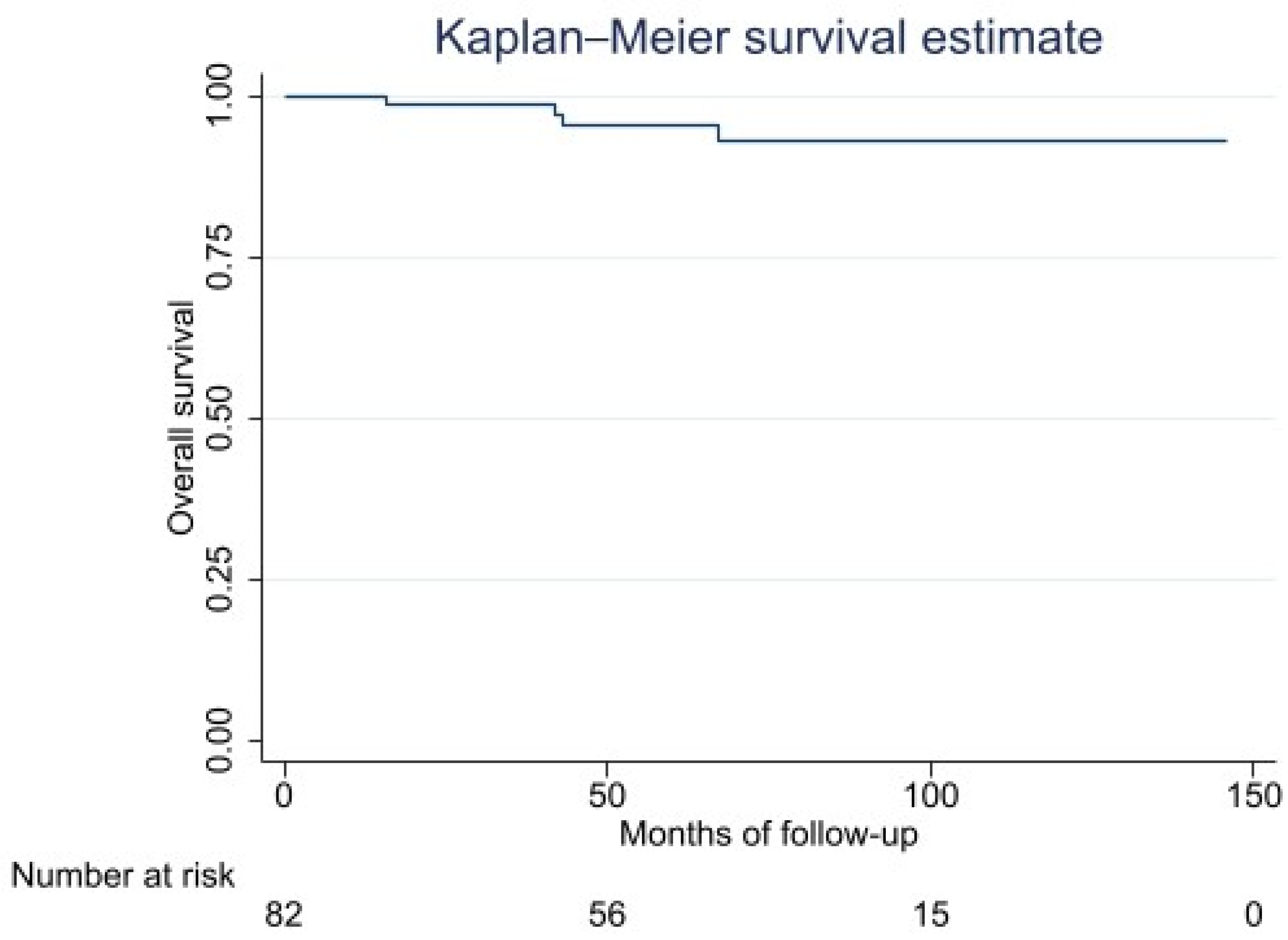
3.2. Overall Survival

Five-year OS was 96% (95% CI: 86–98) (Figure 1). Statistical analysis indicated that there were no risk factors for OS (Table 2).

3.3. Local Recurrence

Local recurrence was reported in four cases (5%), with a median free interval of 3 months (range: 0–32 months). Three out of four cases had a tumors over 10 cm, and one case exhibited a size ranging between 5 and 10 cm. Regarding treatment, two out of the four patients underwent pre-operative RT and all patients were treated with Cht. In all cases, the margin was wide/radical. Two patients were alive without evidence of disease at the last follow-up, one patient was alive with disease, and the last one died because of a distant progression. All local recurrences were re-excised. LRFS was 94% (95% CI: 85–98) at 5 years. Statistical analysis indicated that there were no risk factors for LRFS.

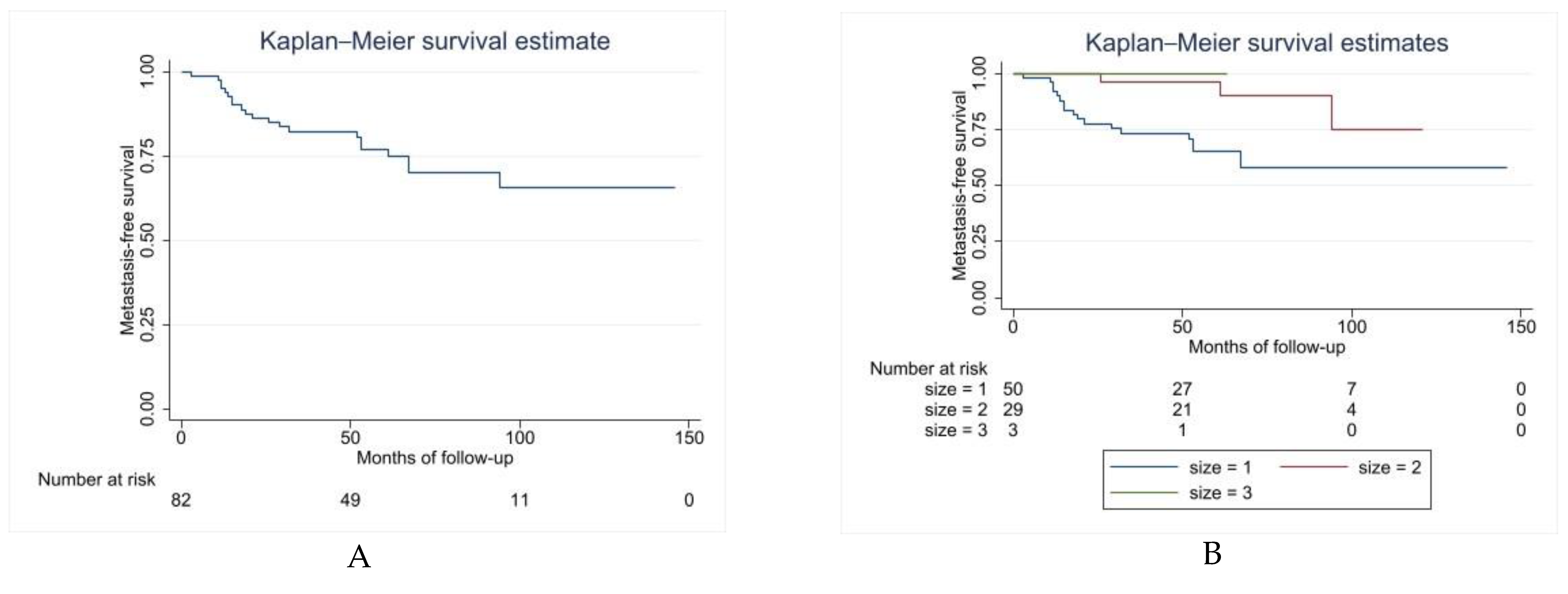
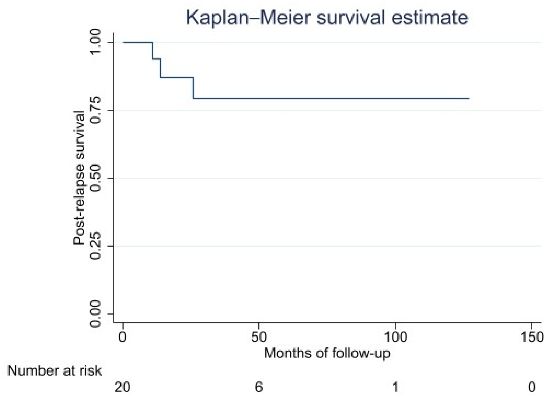
3.4. Distant Metastasis: Metastasis-Free Survival, Metastatic Pattern, and Post-Relapse Survival

Twenty MLS (24%) developed metastases, with a median free interval of 20 months (range: 3–94 months). MFS was 77% (65–85) at 5 years (Figure 2A). Size (p = 0.0337) was the only identified independent risk factor statistically significant for MFS (HR = 0.248, 95% CI: 0.07–0.84) (Figure 2B and Table 3).
The numbers and sites of metastases were one in the abdominal muscular wall (5%), five in bones (25%), two in lymph nodes (10%), three in lungs (15%), and the last nine patients had multiple locations (45%) (most frequently soft tissue, lungs, viscera, lymph nodes). Three out of twenty cases of metastatic MLS had tumors from 5–10 cm; all the others had tumors under 5 cm.
Among the metastatic patients, 35% showed no evidence of disease following treatment of metastasis, 50% were alive with the disease at the last follow-up and 15% had died. Median PRS was 34 months (range: 1–127 months). Five-year PRS was 79% (48–93 months) (Figure 3).
Metastasis treatment consisted of surgery alone (25%) or surgery combined with RT and Cht (55%). The preferred surgical procedure was excision (90%), though two patients were treated with amputation (one of them in combination with RT and the other with Cht). Four patients (20%) were treated with surgery and RT, and four patients (20%) had surgery plus Cht. The other three patients were treated with RT, surgery, and Cht. Among the 20 metastatic patients, only two were not treated with surgery (10%): one underwent Cht, and another was treated with RT. The last two patients did not undergo treatment (one patient underwent early palliative care, the other refused adjuvant therapies). No risk factors for PRS could be identified.

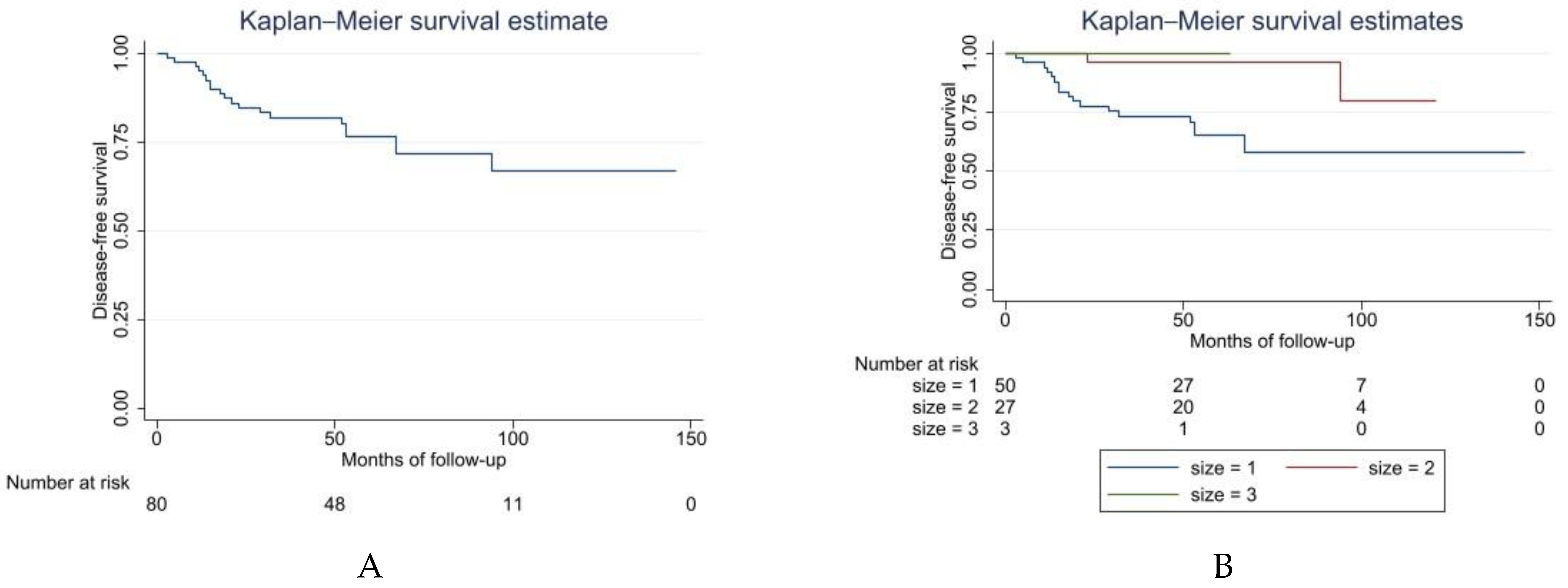
3.5. Disease-Free Survival

Altogether, 68 patients (83%) in this study showed no evidence of disease following treatment for local recurrence or metastases. Ten (12%) patients were alive with disease at the last follow-up, and 4 patients died (5%). Sixty-one patients (74%) were DF after primary surgery during the follow-up period. Overall, the 5-year DFS was 76% (65–85) (Figure 4A). We also recognized that size can be a risk factor for DFS (log-rank test p = 0.0199; COX proportional Hazard HR = 0.177; 95% CI: 0.04–0.76) (Figure 4B and Table 4).

4. Discussion

The study aimed to evaluate factors that may influence OS, LRFS, MFS, PRS, and DFS in high-grade MLS of the extremities. We also analyzed several factors that had a possible impact on the clinical outcomes of patients with MLS in a consecutive single-institution series. Histological grade is an established prognostic factor [7,10,11]. Muratori et al. reported that tumor grade was a significant risk factor affecting the OS of MLS [7]. Similarly, Whu et al. revealed that tumor grade was an independent prognostic factor of OS and cancer-specific survival [20]. In this study, we were not able to confirm these results and audit the effectiveness of histologic grade as a prognostic factor, since our series comprised only deep-seated high-grade tumors. While these limitations created a more homogeneous group for analysis and contributed novelty to the already published reports, they might have reduced the importance of other factors for prognosis.
In this work, the tumoral site and surgical margins were not significant risk factors for OS, LRFS, MFS, PRS, or DFS, confirming that tumors in the extremities have a good prognosis [11]. Different studies have analyzed MLS patients and identified trunk tumor location as being associated with a poorer outcome than an extremity location [10,21,22]. It is also noteworthy that the impact of tumor localization on the overall outcome is also related to the ability to perform resection with adequate surgical margins, as reported in other works on MLS [23,24].
Tumor size is a known prognostic factor for soft tissue sarcomas. According to the published data, the bigger the tumor, the worse the prognosis [25,26,27,28]. In our series, we showed that tumor size is a significant risk factor for MFS (log-rank test p = 0.0337, HR = 0.248. 95% CI 0.07–0.84) and DFS (log-rank test p = 0.0199, HR = 0.1773 95% CI: 0.04–0.76). These findings are congruent with those of Salduz et al. and Wu et al., who found that tumor size over 15 cm was significantly associated with increased overall mortality and demonstrated that tumor size under 10 cm was an independent prognostic factor of OS and cancer-specific survival [20,29]. In contrast, Nishida et al. and Oh et al. reported that tumor size was not associated with survival [21,22] but that size may influence higher-grade MLS.
Regarding treatment, the role of chemotherapy in patients with soft tissue sarcomas has been investigated extensively [23,30,31], as has the radio sensitivity behavior of MLS [31]. We did not succeed in showing whether chemotherapy or radiotherapy provided a clinical benefit for local recurrence, metastasis-free, post-relapse, or overall survival. However, it is essential to note that the selected employment of chemotherapy in the highest-risk patients confounded this analysis. Further investigation would be needed to help better determine the optimal use of chemotherapy in this group of patients.
Similarly, because most patients (97%) had received radiotherapy, we were not able to show the effect of radiation therapy in our series. There are contradictory opinions on the effects of radiotherapy on survival of extremity MLS patients. Some evidence supports the role of RT in improving survival [32,33]. Others found that RT had no impact on either the OS or DFS of MLS [21]. Additionally, it is well documented that this category of patients could achieve local control by only surgical treatment [11,21]. Further studies are warranted to investigate the effect of RT on the outcome and survival of extremity MLS.
Previous studies have shown that MLS patients display higher survival rates than other soft tissues sarcoma patients [34,35]. We observed a favorable estimated 5-year OS rate of 96%. Estimated OS rate was 96%. However, this is not confirmed in other datasets. We observed a higher rate in comparison to previous results reported by Guadagnolo et al. [30], and Saluz et al. [29] who described a 5-year OS corresponding to the 87% and 78.1% respectively. A possible explanation of this difference may be linked to the length of follow-up and to the number of cases analyzed.
LRFS was 94% (95% CI: 85–98) at 5 years. Despite previous results reported by other authors, our local recurrence rate was lower (5%), possibly due to the inclusion criteria included for our work. Selecting only patients with primary tumors treated for the first time at our institute made it possible to highlight the importance of accurate surgical planning and the involvement of specialized centers. This idea is highly accepted by other authors [10,15]. In addition, as confirmed in previous studies [7,30], this low rate of local relapse could be related to the positive effect of post-operative radiotherapy treatment and chemotherapy treatment, performed in 93% and 60% of the patients, respectively.
Finally, it should not be underestimated that the favorable localization of the tumor in the outer extremities made it possible to intervene with higher precision in resection and surgical margins.
We observed a metastatic rate of 24%; sites of distant metastasis included: bones (25%), lungs (15%), lymph nodes (10%), and more than one involved sites (soft tissue, lungs, viscera, lymph nodes) (45%).The crude rate of distant metastasis seems to be comparable with the published series reporting DM rates of 11–38% [4,5,21]. Moreover, these data confirmed previous results, which showed that MLS has a distinct non-pulmonary metastatic disease pattern [30]. Therefore, patients with high-risk extremity myxoid liposarcoma should undergo imaging studies of the chest, abdomen, spine, and pelvis as part of their staging and follow-up examinations, preferably with full-body MRI, or CT and MRI scans of the spine and pelvic region, for detection of suspected metastatic disease. Of the twenty metastatic patients, 35% had no evidence of disease following treatment of metastasis, 50% were alive with the disease at the last follow-up, and 15% died. Overall, 5-year PRS was 79% (95% CI: 48–93).
DFS is a direct measure of clinical benefit. Regarding our series, 74% of patients were DF after primary surgery at the end of the follow-up period, which indicates a generally favorable prognosis associated with this kind of soft tissue sarcoma. These results are similar (47–73%) to what has been reported in previously published reports [4,36]. Higher rates were reported by Nishida et al., who reviewed 53 patients with extremity and trunk MLS, with a 5-year DFS rate of 90% and a median follow-up time of 60 months [21]. This difference in outcomes between this study and previous reports may mainly reflect differences in tumor locations and the inclusion of the round cell subtype.
The homogeneity of the case series, the interest in choosing only high-grade tumors of the extremities, the absence of missing data, and the treatment of patients at a single institute are all strengths of our study. Moreover, considering that to date our cohort of 82 cases of primary high-grade MLS of the extremities, excluding other subtypes, is the biggest presented from a sole institute, makes an effort in growing the clinical record on this disease. However, some limitations must be acknowledged, the first of which is the relatively small number of patients and the follow-up period. Another limitation is the study’s retrospective design. An adequate control group could not be formed, and confounding variables were challenging to measure, making it hard to make simple statements.

5. Conclusions

Myxoid liposarcoma has a relatively good clinical outcome. Our analysis revealed that by selecting only high-grade, deep-seated tumors, the common risk factors for MLS do not correlate with survival. However, large tumors tend to be more aggressive, and tumor size is the most significant predictor of long-term DFS and MFS. In myxoid liposarcoma, it seems reasonable to expect an adequate local control rate after RT and Cht. Lastly, the distinctive pattern of metastatic relapse seen in MLS necessitates staging and surveillance imaging of the abdomen, pelvis, and the thorax because extrapulmonary soft tissue metastases are common sites for distant metastases.
Our findings have clinical implications for the treatment and surveillance of patients with localized myxoid liposarcoma and could be valuable in the daily clinical practice of oncologists and oncological orthopedic surgeons.

Author Contributions

Conceptualization, G.T. and G.B.; methodology, G.B.; software, R.L. and G.V.; validation, G.T., G.B. and R.L.; formal analysis, R.L. and G.V.; statistical analysis: R.L.; investigation, G.T.; resources, F.O. and E.B.; data curation, R.L.; writing—original draft preparation, R.L. and G.T.; writing—review and editing, R.L. and G.T.; supervision, G.B. All authors have read and agreed to the published version of the manuscript.

Funding

This research received no external funding.

Institutional Review Board Statement

The study was conducted according to the guidelines of the Declaration of Helsinki and approved by the Institutional Review Board (or Ethics Committee) of Rizzoli Orthopedic Institute of Bologna (protocol code: CE AVEC 58/2022/Oss/IOR; date of approval: 14 February 2022).

Informed Consent Statement

Due to the retrospective design of the study, informed consent was not requested.

Data Availability Statement

The data presented in this study are available on request from the corresponding author. The data are not publicly available due to privacy or ethical restrictions.

Acknowledgments

We thank our patients and their families. We would also like to show gratitude to Giulia Zoggia for her help with English revision.

Conflicts of Interest

The authors declare no conflict of interest.

References

  1. Dujardin, F.; Debled, M.; Guillemet, C.; Simonet, J.; Hamidou, H.; Cambon-Michot, C.; Dubray, B.; Vera, P. Diagnosis and treatment of soft-tissuesoft tissue tumors. Rev. Chir. Orthop. Reparatrice Appar. Mot. 2006, 92, 637–650. [Google Scholar] [CrossRef]
  2. Abaricia, S.; Hirbe, A.C. Diagnosis and Treatment of Myxoid Liposarcomas: Histology Matters. Curr. Treat. Options Oncol. 2018, 19, 64. [Google Scholar] [CrossRef] [PubMed]
  3. Pérez-Losada, J.; Sánchez-Martín, M.; Rodríguez-García, M.A.; Pérez-Mancera, P.A.; Pintado, B.; Flores, T.; Battaner, E.; Sánchez-Garćia, I. Liposarcoma initiated by FUS/TLS-CHOP: The FUS/TLS domain plays a critical role in the pathogenesis of liposarcoma. Oncogene 2000, 19, 6015–6022. [Google Scholar] [CrossRef] [PubMed] [Green Version]
  4. Smith, T.A.; Easley, K.A.; Goldblum, J.R. Myxoid/round cell liposarcoma of the extremities. A clinicopathologic study of 29 cases with particular attention to extent of round cell liposarcoma. Am. J. Surg. Pathol. 1996, 20, 171–180. [Google Scholar] [CrossRef]
  5. Dürr, H.R.; Rauh, J.; Baur-Melnyk, A.; Knösel, T.; Lindner, L.; Roeder, F.; Jansson, V.; Klein, A. Myxoid liposarcoma: Local relapse and metastatic pattern in 43 patients. BMC Cancer 2018, 18, 304. [Google Scholar] [CrossRef]
  6. Hoffman, A.; Ghadimi, M.P.H.; Demicco, E.G.; Creighton, C.J.; Torres, K.; Colombo, C.; Peng, T.; Lusby, K.; Ingram, D.; Hornick, J.L.; et al. Localized and Metastatic Myxoid/Round Cell Liposarcoma Clinical and Molecular Observations. Cancer 2013, 119, 1868–1877. [Google Scholar] [CrossRef]
  7. Muratori, F.; Bettini, L.; Frenos, F.; Mondanelli, N.; Greto, D.; Livi, L.; Franchi, A.; Roselli, G.; Scorianz, M.; Capanna, R.; et al. Myxoid Liposarcoma: Prognostic Factors and Metastatic Pattern in a Series of 148 Patients Treated at a Single Institution. Int. J. Surg. Oncol. 2018, 2018, 8928706. [Google Scholar] [CrossRef] [Green Version]
  8. Moreau, L.-C.; Turcotte, R.; Ferguson, P.; Wunder, J.; Clarkson, P.; Masri, B.; Isler, M.; Dion, N.; Werier, J.; Ghert, M.; et al. Myxoid\round cell liposarcoma (MRCLS) revisited: An analysis of 418 primarily managed cases. Ann. Surg. Oncol. 2012, 19, 1081–1088. [Google Scholar] [CrossRef]
  9. Schwab, J.H.; Boland, P.; Guo, T.; Brennan, M.F.; Singer, S.; Healey, J.H.; Antonescu, C.R. Skeletal metastases in myxoid liposarcoma: An unusual pattern of distant spread. Ann. Surg. Oncol. 2007, 14, 1507–1514. [Google Scholar] [CrossRef]
  10. Haniball, J.; Sumathi, V.P.; Kindblom, L.G.; Abudu, A.; Carter, S.R.; Tillman, R.M.; Jeys, L.; Spooner, D.; Peake, D.; Grimer, R.J. Prognostic factors and metastatic patterns in primary myxoid/round-cell liposarcoma. Sarcoma 2011, 2011, 538085. [Google Scholar] [CrossRef]
  11. Fiore, M.; Grosso, F.; Lo Vullo, S.; Pennacchioli, E.; Stacchiotti, S.; Ferrari, A.; Collini, P.; Lozza, L.; Mariani, L.; Casali, P.G.; et al. Myxoid/round cell and pleomorphic liposarcomas: Prognostic factors and survival in a series of patients treated at a single institution. Cancer 2007, 109, 2522–2531. [Google Scholar] [CrossRef] [PubMed]
  12. Spillane, A.J.; Fisher, C.; Thomas, J.M. Myxoid liposarcoma--the frequency and the natural history of nonpulmonary soft tissue metastases. Ann. Surg. Oncol. 1999, 6, 389–394. [Google Scholar] [CrossRef] [PubMed]
  13. Cheng, E.Y.; Springfield, D.S.; Mankin, H.J. Frequent incidence of extrapulmonary sites of initial metastasis in patients with liposarcoma. Cancer 1995, 75, 1120–1127. [Google Scholar] [CrossRef]
  14. Zheng, K.; Yu, X.-C.; Xu, M.; Yang, Y. Surgical Outcomes and Prognostic Factors of Myxoid Liposarcoma in Extremities: A Retrospective Study. Orthop. Surg. 2019, 11, 1020–1028. [Google Scholar] [CrossRef] [PubMed]
  15. Lemeur, M.; Mattei, J.-C.; Souteyrand, P.; Chagnaud, C.; Curvale, G.; Rochwerger, A. Prognostic factors for the recurrence of myxoid liposarcoma: 20 cases with up to 8 years follow-up. Orthop. Traumatol. Surg. Res. 2015, 101, 103–107. [Google Scholar] [CrossRef] [Green Version]
  16. Choi, J.H.; Ro, J.Y. The 2020 WHO Classification of Tumors of Bone: An Updated Review. Adv. Anat. Pathol. 2021, 28, 119–138. [Google Scholar] [CrossRef]
  17. Choi, J.H.; Ro, J.Y. The 2020 WHO Classification of Tumors of Soft Tissue: Selected Changes and New Entities. Adv. Anat. Pathol. 2021, 28, 44–58. [Google Scholar] [CrossRef]
  18. Enneking, W.F.; Spanier, S.S.; Goodman, M.A. A system for the surgical staging of musculoskeletal sarcoma. 1980. Clin. Orthop. Relat. Res. 2003, 415, 4–18. [Google Scholar] [CrossRef]
  19. Gronchi, A.; Miah, A.B.; Dei Tos, A.P.; Abecassis, N.; Bajpai, J.; Bauer, S.; Biagini, R.; Bielack, S.; Blay, J.Y.; Bolle, S.; et al. Soft tissue and visceral sarcomas: ESMO–EURACAN–GENTURIS Clinical Practice Guidelines for diagnosis, treatment and follow-up. Ann. Oncol. 2021, 32, 1348–1365. [Google Scholar] [CrossRef]
  20. Wu, J.; Qian, S.; Jin, L. Prognostic factors of patients with extremity myxoid liposarcomas after surgery. J. Orthop. Surg. Res. 2019, 14, 90. [Google Scholar] [CrossRef] [Green Version]
  21. Nishida, Y.; Tsukushi, S.; Nakashima, H.; Ishiguro, N. Clinicopathologic prognostic factors of pure myxoid liposarcoma of the extremities and trunk wall. Clin. Orthop. Relat. Res. 2010, 468, 3041–3046. [Google Scholar] [CrossRef] [PubMed] [Green Version]
  22. Oh, Y.J.; Yi, S.Y.; Kim, K.H.; Cho, Y.J.; Beum, S.H.; Lee, Y.H.; Suh, J.S.; Hur, H.; Kim, K.S.; Kim, S.H.; et al. Prognostic model to predict survival outcome for curatively resected liposarcoma: A multi-institutional experience. J. Cancer 2016, 7, 1174–1180. [Google Scholar] [CrossRef] [PubMed]
  23. Dalal, K.M.; Kattan, M.W.; Antonescu, C.R.; Brennan, M.F.; Singer, S. Subtype specific prognostic nomogram for patients with primary liposarcoma of the retroperitoneum, extremity, or trunk. Ann. Surg. 2006, 244, 381–389. [Google Scholar] [CrossRef] [PubMed]
  24. Singer, S.; Corson, J.M.; Demetri, G.D.; Healey, E.A.; Marcus, K.; Eberlein, T.J. Prognostic factors predictive of survival for truncal and retroperitoneal soft tissue sarcoma. Ann. Surg. 1995, 221, 185–195. [Google Scholar] [CrossRef] [PubMed]
  25. Zagars, G.K.; Ballo, M.T.; Pisters, P.W.T.; Pollock, R.E.; Patel, S.R.; Benjamin, R.S.; Evans, H.L. Prognostic Factors for Patients with Localized Soft tissue Sarcoma Treated with Conservation Surgery and Radiation Therapy An Analysis of 1225 Patients. 2003, 97, 2530–2543. Cancer Interdiscip. Int. J. Am. Cancer Soc. 2003, 97, 2530–2543. [Google Scholar] [CrossRef]
  26. Engstrim, K.; Bergh, P.; Gustafson, P.; Hultborn, R.; Johansson, H.; Lofvenberg, R.; Zaikova, O.; Trovik, C.; Wahlstrom, O.; Bauer, H.C.F.; et al. Liposarcoma Outcome Based on the Scandinavian Sarcoma Group Register. Cancer 2008, 113, 1649–1656. [Google Scholar] [CrossRef]
  27. Kilpatrick, S.E.; Doyon, J.; Choong, P.F.M.; Sim, F.H.; Nascimento, A.G. The Clinicopathologic Spectrum of Myxoid and Round Cell Liposarcoma A Study of 95 Cases. Int. J. Am. Cancer Soc. 1996, 77, 1450–1458. [Google Scholar] [CrossRef]
  28. Pitson, G.; Robinson, P.; Wilke, D.; Kandel, R.A.; White, L.; Griffin, A.M.; Bell, R.S.; Catton, C.N.; Wunder, J.S.; O’Sullivan, B. Radiation response: An additional unique signature of myxoid liposarcoma. Int. J. Radiat. Oncol. Biol. Phys. 2004, 60, 522–526. [Google Scholar] [CrossRef]
  29. Salduz, A.; Alpan, B.; Valiyev, N.; Özmen, E.; İribaş, A.; Ağaoğlu, F.; Bayram, A.; Bilgiç, B.; Özger, H. Neoadjuvant radiotherapy for myxoid liposarcomas: Oncologic outcomes and histopathologic correlations. Acta Orthop. Traumatol. Turc. 2017, 51, 355–361. [Google Scholar] [CrossRef]
  30. Guadagnolo, B.A.; Zagars, G.K.; Ballo, M.T.; Patel, S.R.; Lewis, V.O.; Benjamin, R.S.; Pollock, R.E. Excellent Local Control Rates and Distinctive Patterns of Failure in Myxoid Liposarcoma Treated With Conservation Surgery and Radiotherapy. Int. J. Radiat. Oncol. Biol. Phys. 2008, 70, 760–765. [Google Scholar] [CrossRef]
  31. De Vreeze, R.S.A.; de Jong, D.; Haas, R.L.; Stewart, F.; van Coevorden, F. Effectiveness of Radiotherapy in Myxoid Sarcomas Is Associated With a Dense Vascular Pattern. Int. J. Radiat. Oncol. Biol. Phys. 2008, 72, 1480–1487. [Google Scholar] [CrossRef] [PubMed]
  32. Kachare, S.D.; Brinkley, J.; Vohra, N.A.; Zervos, E.E.; Wong, J.H.; Fitzgerald, T.L. Radiotherapy Associated With Improved Survival for High-Grade Sarcoma of the Extremity. J. Surg. Oncol. 2015, 112, 338–343. [Google Scholar] [CrossRef] [PubMed]
  33. Zhao, R.P.; Yu, X.L.; Zhang, Z.; Jia, L.J.; Feng, Y.; Yang, Z.Z.; Chen, X.X.; Wang, J.; Ma, S.L.; Guo, X.M. The efficacy of postoperative radiotherapy in localized primary soft tissue sarcoma treated with conservative surgery. Radiat. Oncol. 2016, 11, 25. [Google Scholar] [CrossRef] [Green Version]
  34. Tirumani, S.H.; Tirumani, H.; Jagannathan, J.P.; Shinagare, A.B.; Hornick, J.L.; Ramaiya, N.H.; Wagner, A.J. Metastasis in dedifferentiated liposarcoma: Predictors and outcome in 148 patients. Eur. J. Surg. Oncol. 2015, 41, 899–904. [Google Scholar] [CrossRef] [PubMed]
  35. Hornick, J.L.; Bosenberg, M.W.; Mentzel, T.; McMenamin, M.E.; Oliveira, A.M.; Fletcher, C.D.M. Pleomorphic liposarcoma: Clinicopathologic analysis of 57 cases. Am. J. Surg. Pathol. 2004, 28, 1257–1267. [Google Scholar] [CrossRef] [PubMed]
  36. Ten Heuvel, S.E.; Hoekstra, H.J.; van Ginkel, R.J.; Bastiaannet, E.; Suurmeijer, A.J.H. Clinicopathologic prognostic factors in myxoid liposarcoma: A retrospective study of 49 patients with long-term follow-up. Ann. Surg. Oncol. 2007, 14, 222–229. [Google Scholar] [CrossRef]
Figure 1. Overall survival Kaplan–Meier curve.
Cancers 14 02657 g001
Figure 2. Metastasis-free survival (MFS) Kaplan–Meier curves. (A) Overall MFS. (B) Kaplan–Meier curve comparing local MFS by size of tumor: size 1 ≥ 10 cm, size 2 = 5–10 cm, size 3 ≤ 5 cm (p = 0.0337).
Cancers 14 02657 g002
Figure 3. Post-relapse survival estimates.
Cancers 14 02657 g003
Figure 4. Disease-free survival Kaplan–Meier curve. (A) Overall DSF. (B) Kaplan–Meier curve comparing DFS by size of tumor: size 1 ≥ 10 cm, size 2 = 5–10 cm, size 3 ≤ 5 cm (p = 0.0199).
Cancers 14 02657 g004
Table 1. Main clinicopathological features and therapeutic approaches.
FactorNumber of Patients%
Patients82100
Gender
Male5365
Female2935
Location
Thigh5162
Leg2429
Buttock67
Arm11
Tumor size
>10 cm5061
5–10 cm2935
<5 cm34
Surgery
Excision8098
Amputation22
Margin
Wide/radical6377
Marginal1620
Intralesional34
Radiotherapy
Pre-operative5768
Post-operative1923
None69
Chemotherapy
Pre-operative4555
Post-operative1316
Pre- and post-operative22
None2227
Table 2. Kaplan–Maier Overall Survival (OS) estimates from patient characteristics.
FactorLevelKaplan–Maier Estimates (95% CI) 5-Yearp > chi2
GenderF100% (ND)0.592
M93% (80–97)
Age>60100% (ND)0.787
<6094% (83–98)
Size>10 cm93% (79–97)0.2919
5–10 cm100%
<5 cm100%
LocationThigh93% (79–97)0.8938
Leg100%
Buttock100%
Other100%
MarginWide/Radical96% (85–99)0.0806
Marginal93% (59–99)
Intralesional100%
ChemotherapyYes94% (81–97)0.8497
No100%
RadiotherapyYes96% (87–99)0.1279
No83% (27–97)
95% CI: 95% Confidence Interval; F: Female; M: Male; ND: not determined.
Table 3. Kaplan–Maier estimates for MFS from patient characteristics. * Log-rank test. ** HRs unadjusted to each other in multivariable analysis. Results unchanged upon adjustment. (Size adjusted for location, RT and margin: HR = 0.24 (p = 0.029; 95% CI: 0.06–0.866)).
FactorLevelp > chi2HR (95%CI)
GenderF (ref)0.1270
M
Age>60 (ref)0.8263
<60
Size>10 cm (ref)* 0.0337** 0.248 (0.07–0.84)
p = 0.026
5–10 cm
<5 cm
LocationTight (ref)0.3708
Leg
Buttock
Other
MarginWide (ref)0.1099
Marginal
Intralesional
Radical
ChemotherapyYes (ref)0.0961
No
RadiotherapyYes (ref)0.1161
No
HR: Hazard Ratio; 95% CI: 95% Confidence Interval; F: Female; M: Male; ref: group of reference.
Table 4. Kaplan–Maier Disease-free survival (DFS) estimates from patient characteristics. ** HRs unadjusted to each other in multivariable analysis. Results did not change upon an adjustment in multivariate analysis.
FactorLevelp > chi2HR (95% CI)
GenderF (ref)0.1462
M
Age>60 (ref)0.7886
<60
Size>10 cm (ref)0.0199** 0.1773 p = 0.020 95% CI: 0.4–0.76
5–10 cm
<5 cm
LocationTight (ref)0.3236
Leg
Buttock
Arm
MarginWide/radical (ref)0.1097
Marginal
Intralesional
ChemotherapyYes (ref)0.1081
No
RadiotherapyYes (ref)0.2820
No
HR: Hazard Ratio; 95% CI: 95% Confidence Interval; F: Female; M: Male; ref: group of reference.
Publisher’s Note: MDPI stays neutral with regard to jurisdictional claims in published maps and institutional affiliations.

