
Prospective Comparison of 18-FDG PET/CT and Whole-Body MRI with Diffusion-Weighted Imaging in the Evaluation of Treatment Response of Multiple Myeloma Patients Eligible for Autologous Stem Cell Transplant

 ,
, 
Abstract
Simple Summary
Abstract
1. Introduction
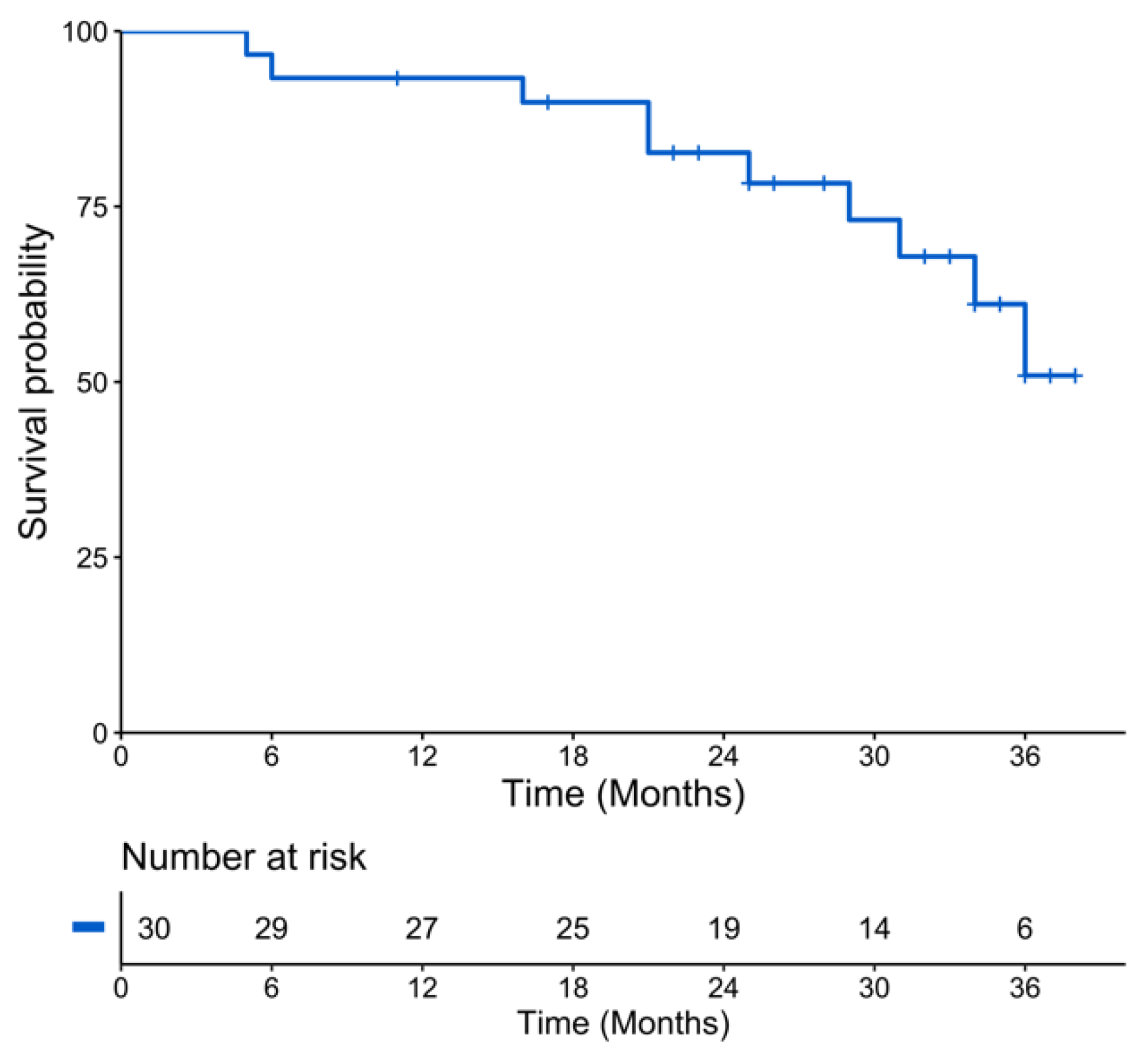
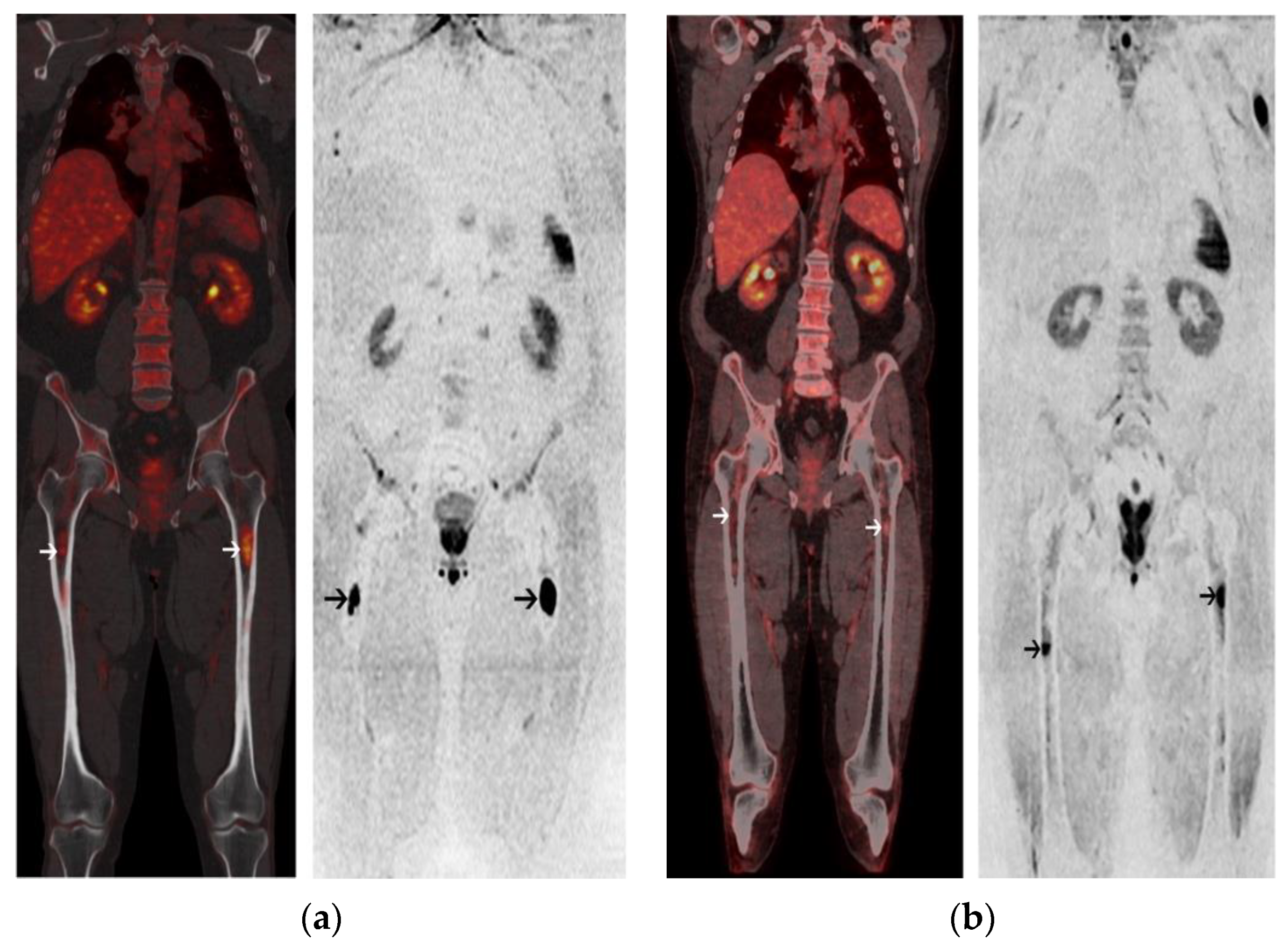
2. Results
2.1. Patients Characteristics
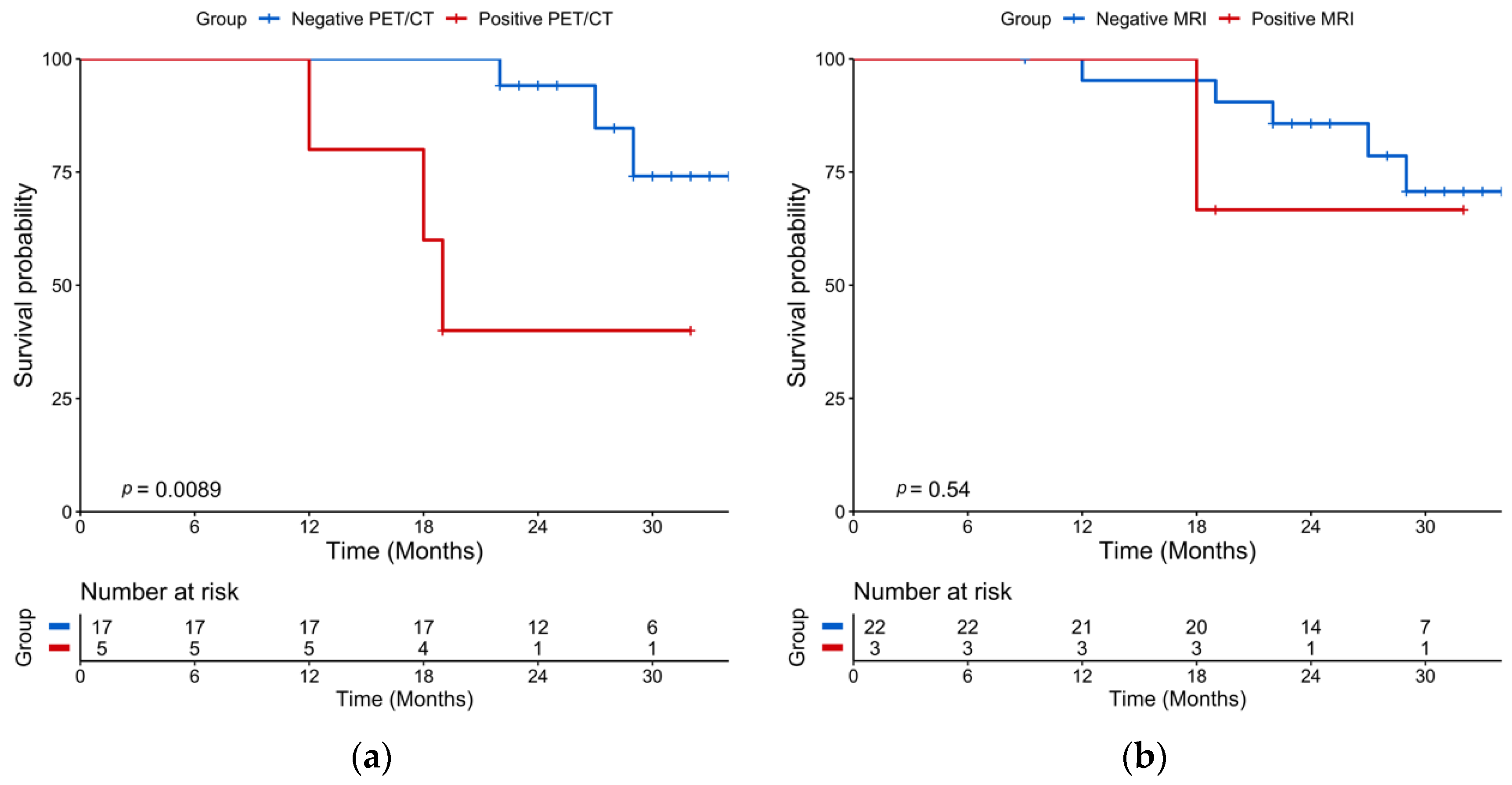
2.2. Post-Induction Chemotherapy Imaging
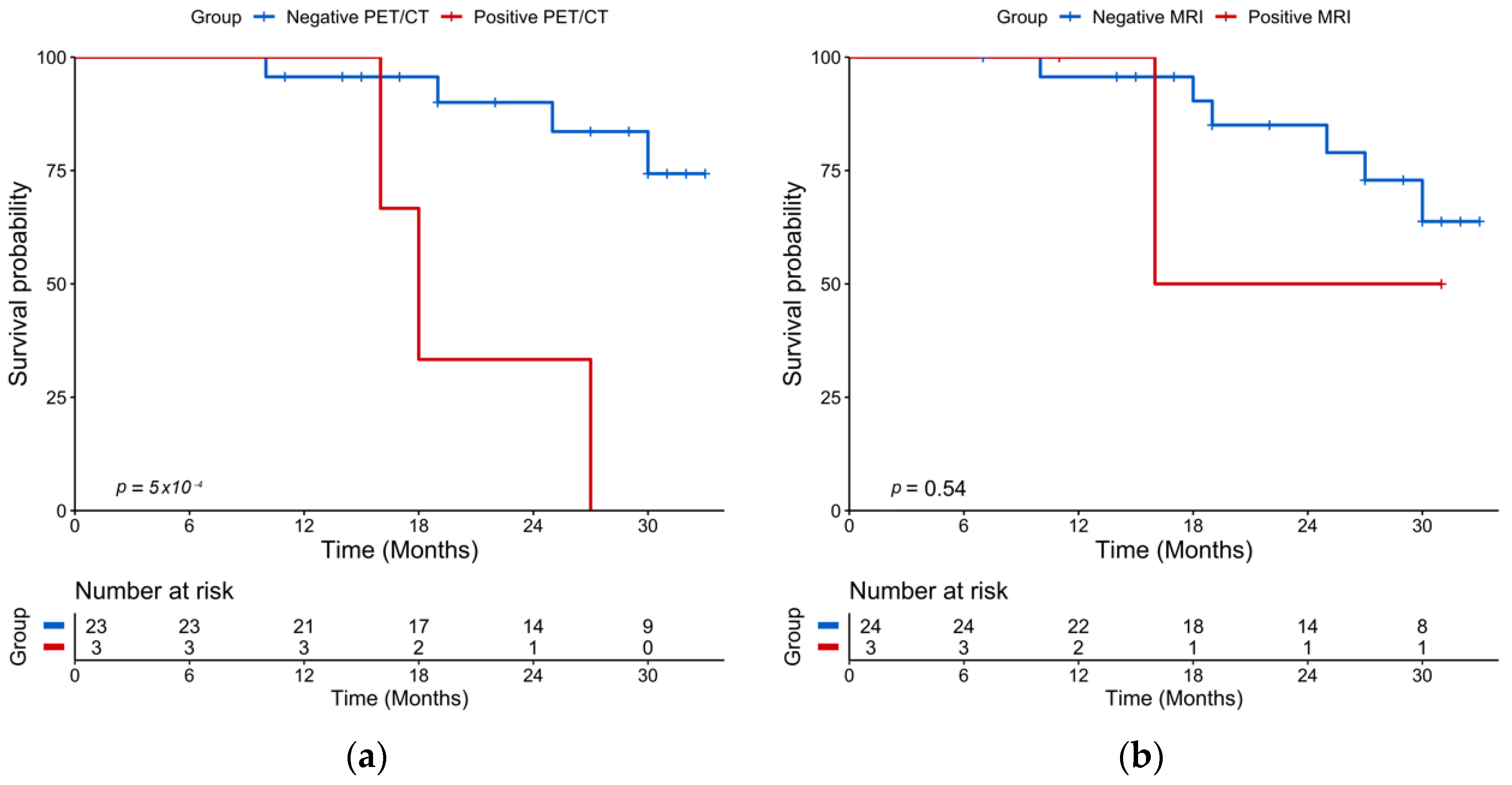
2.3. Post-ASCT Imaging
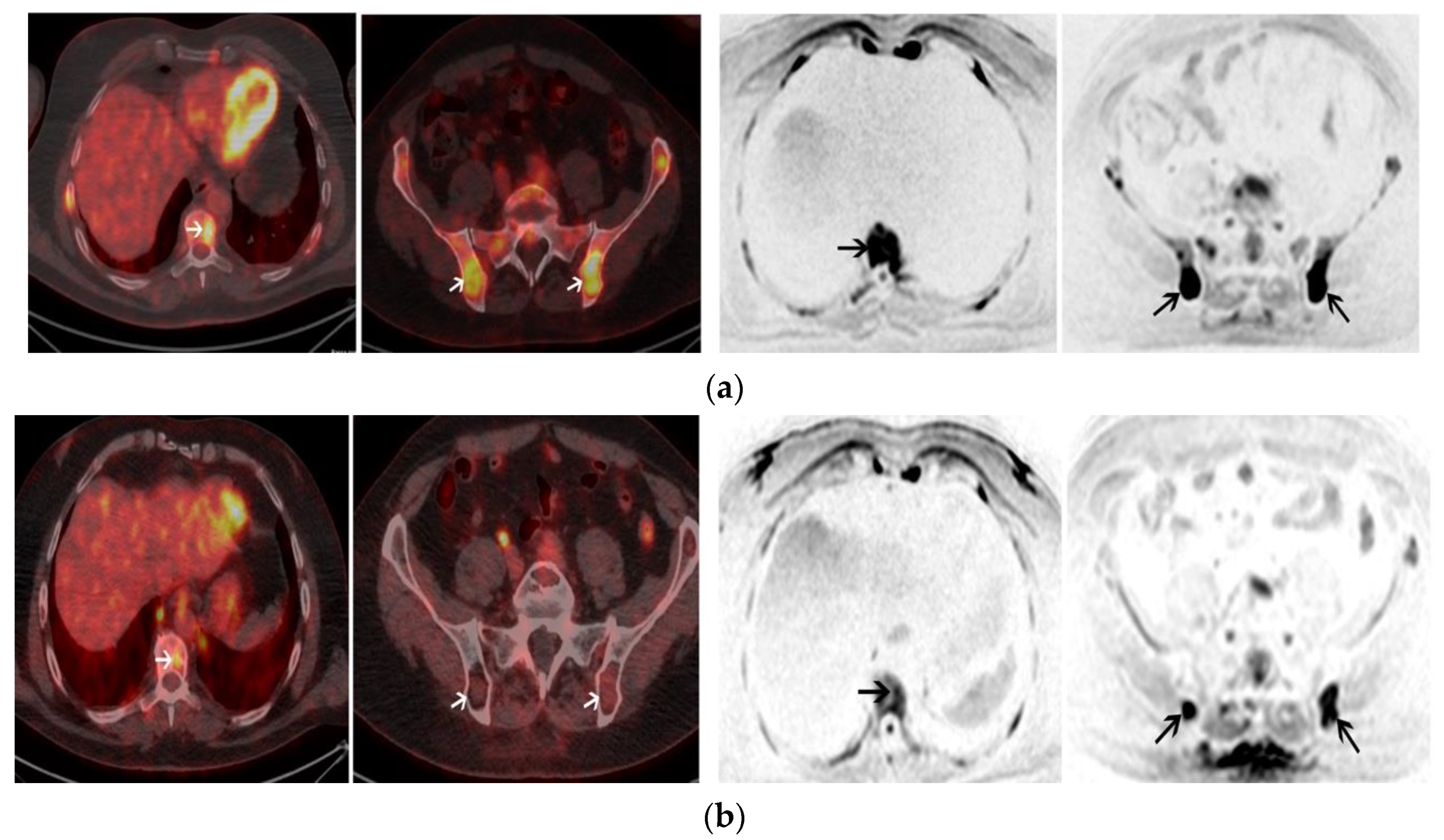
2.4. Baseline Imaging
3. Discussion
4. Materials and Methods
4.1. Patients
4.2. End Points
4.3. 18-FDG PET/CT
4.4. Whole-Body MRI with Diffusion-Weighted Imaging
4.5. Statistical Analysis
5. Conclusions
Author Contributions
Funding
Institutional Review Board Statement
Informed Consent Statement
Data Availability Statement
Conflicts of Interest
References
- Regelink, J.C.; Minnema, M.C.; Terpos, E.; Kamphuis, M.H.; Raijmakers, P.G.; Pieters-van den Bos, I.C.; Heggelman, B.G.; Nievelstein, R.J.; Otten, R.H.; van Lammeren-Venema, D.; et al. Comparison of modern and conventional imaging techniques in establishing multiple myeloma-related bone disease: A systematic review. Br. J. Hematol. 2013, 162, 50–61. [Google Scholar] [CrossRef]
- Hillengass, J.; Moulopoulos, L.A.; Delorme, S.; Koutoulidis, V.; Mosebach, J.; Hielscher, T.; Drake, M.; Rajkumar, S.V.; Oestergaard, B.; Abildgaard, N.; et al. Whole-body computed tomography versus conventional skeletal survey in patients with multiple myeloma: A study of the International Myeloma Working Group. Blood Cancer J. 2017, 7, e599. [Google Scholar] [CrossRef]
- Rajkumar, S.V.; Dimopoulos, M.A.; Palumbo, A.; Blade, J.; Merlini, G.; Mateos, M.V.; Kumar, S.; Hillengass, J.; Kastritis, E.; Richardson, P.; et al. International Myeloma Working Group updated criteria for the diagnosis of multiple myeloma. Lancet Oncol. 2014, 15, e538–e548. [Google Scholar] [CrossRef]
- Zamagni, E.; Patriarca, F.; Nanni, C.; Zannetti, B.; Englaro, E.; Pezzi, A.; Tacchetti, P.; Buttignol, S.; Perrone, G.; Brioli, A.; et al. Prognostic relevance of 18-F FDG PET/CT in newly diagnosed multiple myeloma patients treated with up-front autologous transplantation. Blood 2011, 118, 5989–5995. [Google Scholar] [CrossRef] [PubMed]
- Usmani, S.Z.; Mitchell, A.; Waheed, S.; Crowley, J.; Hoering, A.; Petty, N.; Brown, T.; Bartel, T.; Anaissie, E.; van Rhee, F.; et al. Prognostic implications of serial 18-fluoro-deoxyglucose emission tomography in multiple myeloma treated with total therapy 3. Blood 2013, 121, 1819–1823. [Google Scholar] [CrossRef] [PubMed]
- Moreau, P.; Attal, M.; Caillot, D.; Macro, M.; Karlin, L.; Garderet, L.; Facon, T.; Benboubker, L.; Escoffre-Barbe, M.; Stoppa, A.M.; et al. Prospective Evaluation of Magnetic Resonance Imaging and [(18)F] Fluorodeoxyglucose Positron Emission Tomography-Computed Tomography at Diagnosis and Before Maintenance Therapy in Symptomatic Patients with Multiple Myeloma Included in the IFM/DFCI 2009 Trial: Results of the IMAJEM Study. J. Clin. Oncol. 2017, 35, 2911–2918. [Google Scholar] [CrossRef]
- Spinnato, P.; Bazzocchi, A.; Brioli, A.; Nanni, C.; Zamagni, E.; Albisinni, U.; Cavo, M.; Fanti, S.; Battista, G.; Salizzoni, E. Contrast enhanced MRI and (1)(8)F-FDG PET-CT in the assessment of multiple myeloma: A comparison of results in different phases of the disease. Eur. J. Radiol. 2012, 81, 4013–4018. [Google Scholar] [CrossRef]
- Derlin, T.; Peldschus, K.; Munster, S.; Bannas, P.; Herrmann, J.; Stubig, T.; Habermann, C.R.; Adam, G.; Kroger, N.; Weber, C. Comparative diagnostic performance of (1)(8)F-FDG PET/CT versus whole-body MRI for determination of remission status in multiple myeloma after stem cell transplantation. Eur. Radiol. 2013, 23, 570–578. [Google Scholar] [CrossRef]
- Messiou, C.; Giles, S.; Collins, D.J.; West, S.; Davies, F.E.; Morgan, G.J.; Desouza, N.M. Assessing response of myeloma bone disease with diffusion-weighted MRI. Br. J. Radiol. 2012, 85, e1198–e1203. [Google Scholar] [CrossRef]
- Giles, S.L.; Messiou, C.; Collins, D.J.; Morgan, V.A.; Simpkin, C.J.; West, S.; Davies, F.E.; Morgan, G.J.; de Souza, N.M. Whole-body diffusion-weighted MR imaging for assessment of treatment response in myeloma. Radiology 2014, 271, 785–794. [Google Scholar] [CrossRef]
- Lacognata, C.; Crimi, F.; Guolo, A.; Varin, C.; De March, E.; Vio, S.; Ponzoni, A.; Barila, G.; Lico, A.; Branca, A.; et al. Diffusion-weighted whole-body MRI for evaluation of early response in multiple myeloma. Clin. Radiol. 2017, 72, 850–857. [Google Scholar] [CrossRef]
- Messiou, C.; Hillengass, J.; Delorme, S.; Lecouvet, F.E.; Moulopoulos, L.A.; Collins, D.J.; Blackledge, M.D.; Abildgaard, N.; Ostergaard, B.; Schlemmer, H.P.; et al. Guidelines for Acquisition, Interpretation, and Reporting of Whole-Body MRI in Myeloma: Myeloma Response Assessment and Diagnosis System (MY-RADS). Radiology 2019, 291, 5–13. [Google Scholar] [CrossRef]
- Rajkumar, S.V. Multiple myeloma: 2020 update on diagnosis, risk-stratification and management. Am. J. Hematol. 2020, 95, 548–567. [Google Scholar] [CrossRef]
- Caldarella, C.; Isgro, M.A.; Treglia, I.; Treglia, G. Is fluorine-18-fluorodeoxyglucose positron emission tomography useful in monitoring the response to treatment in patients with multiple myeloma? Int. J. Hematol. 2012, 96, 685–691. [Google Scholar] [CrossRef]
- Rasche, L.; Alapat, D.; Kumar, M.; Gershner, G.; McDonald, J.; Wardell, C.P.; Samant, R.; Van Hemert, R.; Epstein, J.; Williams, A.F.; et al. Combination of flow cytometry and functional imaging for monitoring of residual disease in myeloma. Leukemia 2019, 33, 1713–1722. [Google Scholar] [CrossRef] [PubMed]
- Zamagni, E.; Nanni, C.; Dozza, L.; Carlier, T.; Bailly, C.; Tacchetti, P.; Versari, A.; Chauvie, S.; Gallamini, A.; Gamberi, B.; et al. Standardization of (18)F-FDG-PET/CT According to Deauville Criteria for Metabolic Complete Response Definition in Newly Diagnosed Multiple Myeloma. J. Clin. Oncol. 2020, 39, 116–125. [Google Scholar] [CrossRef]
- Latifoltojar, A.; Hall-Craggs, M.; Bainbridge, A.; Rabin, N.; Popat, R.; Rismani, A.; D’Sa, S.; Dikaios, N.; Sokolska, M.; Antonelli, M.; et al. Whole-body MRI quantitative biomarkers are associated significantly with treatment response in patients with newly diagnosed symptomatic multiple myeloma following bortezomib induction. Eur. Radiol. 2017, 27, 5325–5336. [Google Scholar] [CrossRef]
- Zhang, Y.; Xiong, X.; Fu, Z.; Dai, H.; Yao, F.; Liu, D.; Deng, S.; Hu, C. Whole-body diffusion-weighted MRI for evaluation of response in multiple myeloma patients following bortezomib-based therapy: A large single-center cohort study. Eur. J. Radiol. 2019, 120, 108695. [Google Scholar] [CrossRef]
- Mesguich, C.; Zanotti-Fregonara, P.; Hindie, E. New Perspectives Offered by Nuclear Medicine for the Imaging and Therapy of Multiple Myeloma. Theranostics 2016, 6, 287–290. [Google Scholar] [CrossRef]
- Mesguich, C.; Hulin, C.; Lascaux, A.; Bordenave, L.; Marit, G.; Hindie, E. Choline PET/CT in Multiple Myeloma. Cancers 2020, 12, 1394. [Google Scholar] [CrossRef]
- Jamet, B.; Bailly, C.; Carlier, T.; Touzeau, C.; Nanni, C.; Zamagni, E.; Barre, L.; Michaud, A.V.; Cherel, M.; Moreau, P.; et al. Interest of Pet Imaging in Multiple Myeloma. Front. Med. 2019, 6, 69. [Google Scholar] [CrossRef]
- Usmani, S.Z.; Heuck, C.; Mitchell, A.; Szymonifka, J.; Nair, B.; Hoering, A.; Alsayed, Y.; Waheed, S.; Haider, S.; Restrepo, A.; et al. Extramedullary disease portends poor prognosis in multiple myeloma and is over-represented in high-risk disease even in the era of novel agents. Haematologica 2012, 97, 1761–1767. [Google Scholar] [CrossRef]
- Matsue, K.; Kobayashi, H.; Matsue, Y.; Abe, Y.; Narita, K.; Kitadate, A.; Takeuchi, M. Prognostic significance of bone marrow abnormalities in the appendicular skeleton of patients with multiple myeloma. Blood Adv. 2018, 2, 1032–1039. [Google Scholar] [CrossRef]
- Kumar, S.; Paiva, B.; Anderson, K.C.; Durie, B.; Landgren, O.; Moreau, P.; Munshi, N.; Lonial, S.; Blade, J.; Mateos, M.V.; et al. International Myeloma Working Group consensus criteria for response and minimal residual disease assessment in multiple myeloma. Lancet Oncol. 2016, 17, e328–e346. [Google Scholar] [CrossRef]
- Mesguich, C.; Hulin, C.; Latrabe, V.; Lascaux, A.; Bordenave, L.; Hindie, E.; Marit, G. Prospective comparison of 18-FDG PET/CT and whole-body diffusion-weighted MRI in the assessment of multiple myeloma. Ann. Hematol. 2020, 99, 2869–2880. [Google Scholar] [CrossRef]
- Zamagni, E.; Nanni, C.; Gay, F.; Pezzi, A.; Patriarca, F.; Bello, M.; Rambaldi, I.; Tacchetti, P.; Hillengass, J.; Gamberi, B.; et al. 18F-FDG PET/CT focal, but not osteolytic, lesions predict the progression of smoldering myeloma to active disease. Leukemia 2016, 30, 417–422. [Google Scholar] [CrossRef]
- Mesguich, C.; Fardanesh, R.; Tanenbaum, L.; Chari, A.; Jagannath, S.; Kostakoglu, L. State of the art imaging of multiple myeloma: Comparative review of FDG PET/CT imaging in various clinical settings. Eur. J. Radiol. 2014, 83, 2203–2223. [Google Scholar] [CrossRef] [PubMed]


| Characteristic | Value N = 30 | Range or % |
|---|---|---|
| Median age, years (range) | 56 | 34–66 |
| Male | 17 | 56 |
| Monoclonal protein isotype | ||
| IgG | 12 | 40 |
| IgA | 5 | 17 |
| IgD | 1 | 3 |
| Light chain | 12 | 40 |
| kappa | 10 | 33 |
| lambda | 2 | 7 |
| Low hemoglobin (≤10 g/dL) | 7 | 63 |
| Low platelets (<150 G/L) | 3 | 10 |
| High calcium (>12 mg/dL) | 4 | 13 |
| High LDH (>250 UI/L) | 13 | 43 |
| Altered renal function (creatinine ≤ 2 mg/dL) | 4 | 13 |
| Beta-2 microglobulin | ||
| 3.5–5.5 mg/L | 7 | 23 |
| >5.5 mg/L | 5 | 17 |
| Median bone marrow plasmocytosis, % | 28 | 0–87 |
| Median serum free light chain (mg/L) | 343 | 3.2–17,600 |
| High-risk cytogenetic: t (4;14) or del17p | 2 | 7 |
| ISS 1 | 17 | 57 |
| ISS 2 | 8 | 27 |
| ISS 3 | 5 | 17 |
| Induction chemotherapy | ||
| VRD | 12 | 40 |
| VTD | 6 | 20 |
| VTD-Dara | 6 | 20 |
| VRD-Dara | 4 | 13 |
| IRD | 1 | 3 |
| IRD-Dara | 1 | 3 |
| Variable | Hazard Ratio | ||
|---|---|---|---|
| Estimate | 95% CI | p | |
| Age | 0.99 | 0.92–1.09 | 0.96 |
| Male sex | 2.82 | 0.68–11.8 | 0.16 |
| Low hemoglobin (≤10 g/dL) | 2.37 | 0.61–9.21 | 0.21 |
| Low platelets (<150 G/L) | 1.55 | 0.15–13.2 | 0.59 |
| Altered renal function (creatinine ≤ 2mg/dL) | 4.10 | 0.74–22.6 | 0.11 |
| High calcium (>12 mg/dL) | 1.81 | 0.20–16.3 | 0.60 |
| High LDH (>250 UI/L) | 0.85 | 0.21–3.37 | 0.82 |
| Low albumin (<35 g/L) | 2.50 | 0.26–24.5 | 0.43 |
| High CRP (>5 mg/L) | 6.30 | 1.26–31,6 | 0.03 |
| Cytogenetic t (4;14); del17p | 12.84 | 2.06–80.1 | 0.006 |
| ISS 2 to 3 | 6.00 | 1.23–29.1 | 0.03 |
| FL SUVmax > 4.2 on baseline PET/CT | 2.47 | 0.31–19.6 | 0.39 |
| FL SUVmax > 6.1 on baseline PET/CT | 6.08 | 1.29–28.8 | 0.02 |
| FL ADC on baseline WB-DW-MRI | 1.00 | 0.99–1.00 | 0.58 |
| >3FL on baseline PET/CT | 2.95 | 0.33–0.63 | 0.17 |
| >11FL on baseline PET/CT | 5.06 | 1.28–20.0 | 0.02 |
| >7FL on baseline MRI | 4.38 | 0.23–0.87 | 0.07 |
| Inferior limb involvement on baseline PET/CT | 3.74 | 1.03–13.6 | 0.05 |
| >4 skeletal areas involved on baseline PET/CT | 5.46 | 1.49–20.0 | 0.01 |
| >4 skeletal areas involved on baseline MRI | 3.98 | 1.10–14.5 | 0.04 |
| Extramedullary disease on baseline PET/CT | 7.00 | 1.34–36.5 | 0.02 |
| Diffuse disease on baseline PET/CT | 1.16 | 0.33–4.12 | 0.82 |
| Diffuse disease on baseline MRI | 3.07 | 0.64–7.84 | 0.21 |
| Response ≥ VGPR after induction chemotherapy | 1.11 | 0.26–4.73 | 0.89 |
| Response ≥ VGPR after ASCT | 1.25 | 0.15–10.9 | 0.84 |
| Positive PET/CT after induction chemotherapy | 6.79 | 1.30–35.5 | 0.02 |
| FL SUVmax > 3.95 on post-induction PET/CT | 8.30 | 1.16–42.5 | 0.01 |
| Positive PET/CT after ASCT | 10.15 | 2.00–51.4 | 0.005 |
| FL SUVmax > 3.2 on post-ASCT PET/CT | 11.32 | 2.06–62.4 | 0.005 |
| Positive WB-DW-MRI after induction chemotherapy | 2.12 | 0.24–18.4 | 0.50 |
| FL ADC on post-induction WB-DW-MRI | 1.00 | 0.99–1.00 | 0.69 |
| Positive WB-DW-MRI after ASCT | 1.89 | 0.22–15.8 | 0.56 |
| FL ADC on post-ASCT WB-DW-MRI | 1.00 | 0.99–1.00 | 0.94 |
Publisher’s Note: MDPI stays neutral with regard to jurisdictional claims in published maps and institutional affiliations. |
© 2021 by the authors. Licensee MDPI, Basel, Switzerland. This article is an open access article distributed under the terms and conditions of the Creative Commons Attribution (CC BY) license (https://creativecommons.org/licenses/by/4.0/).
Share and Cite
Mesguich, C.; Latrabe, V.; Hulin, C.; Lascaux, A.; Bordenave, L.; Hindié, E.; Marit, G. Prospective Comparison of 18-FDG PET/CT and Whole-Body MRI with Diffusion-Weighted Imaging in the Evaluation of Treatment Response of Multiple Myeloma Patients Eligible for Autologous Stem Cell Transplant. Cancers 2021, 13, 1938. https://doi.org/10.3390/cancers13081938
Mesguich C, Latrabe V, Hulin C, Lascaux A, Bordenave L, Hindié E, Marit G. Prospective Comparison of 18-FDG PET/CT and Whole-Body MRI with Diffusion-Weighted Imaging in the Evaluation of Treatment Response of Multiple Myeloma Patients Eligible for Autologous Stem Cell Transplant. Cancers. 2021; 13(8):1938. https://doi.org/10.3390/cancers13081938
Chicago/Turabian StyleMesguich, Charles, Valérie Latrabe, Cyrille Hulin, Axelle Lascaux, Laurence Bordenave, Elif Hindié, and Gerald Marit. 2021. "Prospective Comparison of 18-FDG PET/CT and Whole-Body MRI with Diffusion-Weighted Imaging in the Evaluation of Treatment Response of Multiple Myeloma Patients Eligible for Autologous Stem Cell Transplant" Cancers 13, no. 8: 1938. https://doi.org/10.3390/cancers13081938
APA StyleMesguich, C., Latrabe, V., Hulin, C., Lascaux, A., Bordenave, L., Hindié, E., & Marit, G. (2021). Prospective Comparison of 18-FDG PET/CT and Whole-Body MRI with Diffusion-Weighted Imaging in the Evaluation of Treatment Response of Multiple Myeloma Patients Eligible for Autologous Stem Cell Transplant. Cancers, 13(8), 1938. https://doi.org/10.3390/cancers13081938